Article

The Strain Rates in the Brain, Brainstem, Dura, and Skull under Dynamic Loadings

by
Mohammad Hosseini-Farid
1,2,*,
MaryamSadat Amiri-Tehrani-Zadeh
3,
Mohammadreza Ramzanpour
1,
Mariusz Ziejewski
1 and
Ghodrat Karami
1
1
Department of Mechanical Engineering, North Dakota State University, Fargo, ND 58104, USA
2
Department of Orthopedic Surgery, Mayo Clinic, Rochester, MN 55905, USA
3
Department of Computer Science, North Dakota State University, Fargo, ND 58104, USA
*
Author to whom correspondence should be addressed.
Math. Comput. Appl. 2020, 25(2), 21; https://doi.org/10.3390/mca25020021
Submission received: 7 March 2020 / Revised: 5 April 2020 / Accepted: 5 April 2020 / Published: 7 April 2020
(This article belongs to the Special Issue Numerical Modelling and Simulation Applied to Head Trauma)

Abstract

:
Knowing the precise material properties of intracranial head organs is crucial for studying the biomechanics of head injury. It has been shown that these biological tissues are significantly rate-dependent; hence, their material properties should be determined with respect to the range of deformation rate they experience. In this paper, a validated finite element human head model is used to investigate the biomechanics of the head in impact and blast, leading to traumatic brain injuries (TBI). We simulate the head under various directions and velocities of impacts, as well as helmeted and unhelmeted head under blast shock waves. It is demonstrated that the strain rates for the brain are in the range of 36 to 241 s−1, approximately 1.9 and 0.86 times the resulting head acceleration under impacts and blast scenarios, respectively. The skull was found to experience a rate in the range of 14 to 182 s−1, approximately 0.7 and 0.43 times the head acceleration corresponding to impact and blast cases. The results of these incident simulations indicate that the strain rates for brainstem and dura mater are respectively in the range of 15 to 338 and 8 to 149 s−1. These findings provide a good insight into characterizing the brain tissue, cranial bone, brainstem and dura mater, and also selecting material properties in advance for computational dynamical studies of the human head.

Graphical Abstract

1. Introduction

Intracranial head organs have been known to be the most sensitive organs involved in life-threatening injuries caused by impact incidents and blast waves [1,2,3]. Due to the complex mechanical and physical reactions of the head and brain under impact and explosion waves, the mechanism of traumatic brain injuries (TBI) is not well understood [4]. Finite element (FE) simulations provide a convincing framework for determining biomechanical responses of the head exposed to those high rated loads [5]. These numerical models have been introduced to predict brain deformation for different applied loads and study intracranial organ behavior under TBI conditions [6,7,8,9,10,11,12,13]. The biofidelity of such computational simulations is strictly related to the accuracy of the material properties used to model these tissues.
Mechanical properties of brain tissue are a fundamental subject of biomechanics and have been extensively studied in the last few decades [3,14,15]. It has been shown that the mechanical behavior of the brain is dominated by the loading rate and varies nonlinearly with any change in strain, as well as with its strain rate [16,17,18,19,20]. Typically, soft biological materials such as brain and brainstem present complex mechanical responses characterized by large strains, load history, and rate sensitivity [21,22,23,24,25,26]. Since brain and brainstem tissues are rate-dependent materials, great care should be taken in selecting proper material properties from corresponding strain rates in different scenarios.
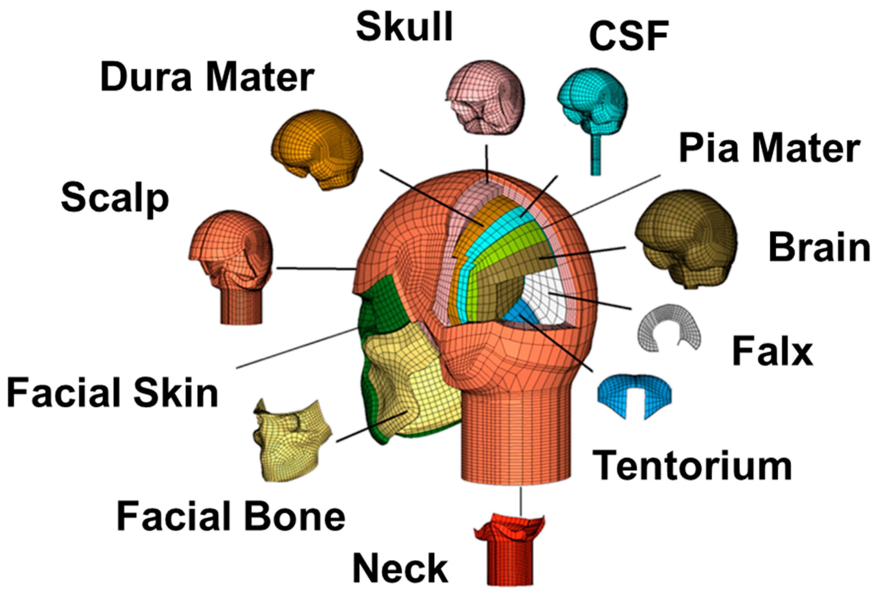
Dura mater (a surrounding membrane of brain) and skull, which hold and protect the brain, have significant roles in the analysis of the TBI incidents. Therefore, determining the material properties of the dura and cranial bone was the subject of earlier research in biomechanics [27,28,29]. Both theoretical and experimental studies have shown that the dura and cranial bone present rate-dependent material properties [30,31,32,33,34]. Although linear and hyperelastic models can approximate the behavior of dura and skull very well at each rate [31,35,36], they behave differently under various speeds of loading. For instance, Persson et al. [31] tested dura mater under uniaxial tension at three various strain rates of 0.01, 0.1, and 1.0 s−1. They postulated the mechanical responses of dura is rate-dependent; however, they fitted their results to the rate-independent hyperelastic Ogden model. Additionally, experimental tensile results indicated the Young’s modulus of the cranial bone varies from 8 to 19.5 GPa for quasistatic to dynamic rates [37]. Therefore, to have a correct result at each FE simulation, the right material properties corresponding to the rate of that application should be selected.
The majority of biomechanical characterizations for human head organs have been performed within experiments in relatively low strain rates. Recently, De Kegel et al. [35] conducted some experiments for human dura specimens within a strain rate of 1.0 s−1. They have obtained a highly nonlinear behavior for dura and characterized its mechanical response using three different hyperelastic material models. Franceschini et al. [38] have experimentally studied the mechanical behavior of human brain tissue at strain rates ranging between 0.0055 and 0.0093 s−1. Although their results are valid to be employed for studies in quasistatic loading, those material properties have been used in many computational simulations of TBI at dynamic loads [39,40]. It was confirmed [16,41] that, in addition to selecting an appropriate constitutive model, using the material constants derived from a mismatched strain rate may considerably affect the validity of the results. Farid et al. [16] showed that hyper-viscoelastic material models, which are optimized for various low strain rates, will result in considerable errors when predicting brain behavior at higher strain rates. Therefore, it is crucial to know the range of strain rates at dynamic loading conditions and characterize the material properties of head organs at those rates.
Finding the ranges of strain rate for brain, brainstem, dura, and skull in TBI through simulations can help in determining and using the material properties at the right rate. The strain rates of different incidents cannot be measured in vivo, and therefore, a computational finite element analysis of the human head was conducted to predict the strain rates for loading scenarios associated with brain injury [
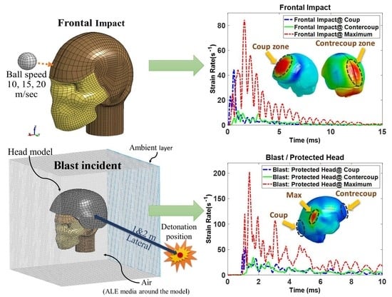
12]. In this paper, impact and blast assault simulations are conducted on a developed FE human head model to predict the range of strain rates. In this paper, it was assumed that the brain will experience minor injuries by blunt impact [1] and an effective (mild to severe) brain injury caused by blast [42]. This approach came from the fact that impact and blast load types are inherently different and cannot be directly compared to each other. The findings provide a ground basis for determining more relevant material properties for the strain rates corresponding to impact and blast cases.

2. Material and Methods

2.1. Finite Element of Head Model

Using MRI and CT scan techniques, the geometry of the 50th percentile human head–neck model was developed by Horgan and Gilchrest [43], and the detailed explanation of this head model has been provided in earlier studies [44,45]. As shown in Figure 1, the model contains all the major parts of the head and the neck, including the brain, falx, tentorium, cerebrospinal fluid (CSF), dura mater, pia mater, neck muscle, neck bone, facial bone, skull, and scalp. In this paper, linear elastic material behavior is used for all head components (Table 1) except the brain and brainstem, which are modeled as hyper-viscoelastic (Table 2) and viscoelastic (Table 3) material [1], respectively. The Mooney–Rivlin strain energy coupled with the linear viscoelastic function was used as the constitutive relation of the brain given by Equation (1). Furthermore, the linear viscoelastic material model expressed by Equation (2) was employed to represent the shear characteristics of the brainstem. A linear material model with 10 GPa of Young’s modulus was considered for the skull. The developed head model was earlier examined and validated under different blast scenarios [42,46,47]. Additionally, Horgan and Gilchrist [43,48] verified this head model against various mostly referenced cadaveric experiments of Nahum et al. [49], Hardy et al. [50], and Trosseille et al. [51].
W = 0 t { ( 1 k = 1 M g k ( 1 e ( t τ ) / τ k ) ) ( d d τ [ i + j = 1 N C i j ( I 1 3 ) i ( I 2 3 ) j ] ) } d τ
G ( t ) = G + ( G 0 G ) e β t

2.2. Impact Case

The injury caused by a golf ball impact is one of the most leading causes of head injury in sports-related impacts [53]. The possibility of being hit by an occasional golf ball is common for both the players and the people who watch the game [54]. This case of impact was simulated to evaluate the strain rate as an example of the general blunt impact applications. The simulations of a golf ball impact were done for a ball flying at speeds of 10, 15, and 20 m/s. As suggested by Lee et al. [52], these ball speeds were selected in such a way to avoid severe brain injury. Since directionality has notable influences [11], the simulation was performed for three directions of back, frontal, and lateral impact. Figure 2 presents the impact setup for two directions of frontal and lateral. Table 4 provides the summary of the simulated impact scenarios in this study. The neck is assumed to be fixed in the translational displacements [52].

2.3. Blast Case

High strength explosion waves are a possible source of TBI. In this paper, the same head model was also being used for blast simulations. Protecting the head with a helmet can reduce the severity of brain injury, but injury can still occur when the person is exposed to blast [55]. As illustrated in Figure 3, a three-dimensional model of an advanced combat helmet (ACH) is integrated with the head–neck model. The detailed explanation of helmet and its padding system modeling and their mechanical properties can be found in earlier study conducted by Salimi Jazi et al. [55]. In this study, the blast simulation was conducted for the protected and unprotected head in two back and lateral direction. The standoff distances are set to be one and two meters from the explosion site. In LS-Dyna (LS-Dyna Theory Manual, 2007), the standard trinitrotoluols (TNT) is selected as the high explosive (HE) material. Because of a very short effective time on the blast (less than 1 ms), a free boundary condition was applied to the end of neck. Rezaei et al. [56] have shown the blast waves of 95 g HE placed 750 mm far from the human head can cause a severe and fatal brain injury. In addition to this case (95 gr TNT), which is referred to as “heavy blast” in this paper, eight other blast scenarios with 75 gr of HE mass were chosen and are listed in Table 5.
Upon a burst in the air, a shock wave is generated moving away from the center of detonation mass. In this study, the multimaterial arbitrary Lagrangian–Eulerian (ALE) formulation method was used for simulating the blast scenarios in LS-Dyna. This method is established on explicit modelling of the air and exploding mass with multimaterial ALE equation for simulating the blast wave propagation. It solves the movement of the mesh using mesh smoothing algorithms [42]. To determine the behavior of the explosive gas, the equation of state (EOS) must be defined. This equation of state (EOS) explains pressure–volume–energy relationships of the gaseous form of detonation. The EOS regarding blast pressure wave generated by HE detonation can be expressed using the Jones–Wilkins–Lee (JWL) equation as [45]:
p = A ( 1 ω R 1 V ) e R 1 V + B ( 1 ω R 2 V ) e R 2 V + ω E V
where ω, A, B, R1, and R2 are constants that are defined by the user, E is the internal energy per initial volume, and V is the relative volume. The numerical data for these parameters are derived from the explosive handbook by Dobratz [57]. The discretized blast domain depicted in Figure 3 shows the location and direction of HE material. To model the blast wave–head interaction, a penalty-based fluid–structure interaction (FSI) algorithm was also used. FSI was employed through a coupling algorithm modeling the air as a moving fluid along with a moving mesh. For a more detailed explanation of blast modeling procedures, the readers are referred to earlier studies of Salimi Jazi et al. [55] and Rezaei et al. [42].

3. Results

FE simulations of the head impacted by a golf ball are performed for different impact directions and ball speeds as stated in Table 4. Moreover, the mentioned blast scenarios presented in Table 5 were implemented in the head model. In this study, the resultant linear acceleration of the skull was assumed to be the same as human head acceleration [58]. Therefore, this acceleration for three cases of impact and four scenarios of the blast are measured and shown in Figure 4a,b. Instead of measuring the acceleration at one point or element, the whole skull geometry is considered as one body, and the averaged acceleration of this part was evaluated by LS-Dyna.
Figure 5 presents the blast wave propagation and intracranial pressure (ICP) distribution in the brain which is necessary to predict brain behavior during the blast. As can be seen, for this case (backward heavy blast), the ICP in brain exceeds 300 kPa, and therefore, a severe TBI should be expected [1]. Furthermore, in comparison to Rezaei et al.’s study [56], it proves that the selection of HE masses and its distance from the head was adequate to create such a condition.
The strain rate distributions for all elements in the brain, brainstem, skull, and dura were examined. Similar to other mechanical parameters, such as ICP, strain or stress, the strain rate has a non-uniform distribution. For instance, in the brain, the maximum strain rate occurs only in a relatively small zone (some elements) which takes place in different locations for each case, and the rest of the brain observes a moderate regime of rates. Figure 6a,b illustrates the history of 1st principal strain rate in three different locations of the brain for the head impacted by a golf ball. For the case where the ball hit the front of the head with 15 m/s of speed, the maximum strain rate does not happen in coup nor contrecoup. However, it was found that for lateral impacts, the highest rate happens at the coup zone. This shows the location of the maximum rate can occur in every area of the brain and varies for several impact scenarios. Similarly, Figure 6c,d depicts the brain strain rates by the same measure (1st principal) for the protected and unprotected heads under heavy blast conditions. As shown in Figure 6c,d, for the heavy-blast scenario, the highest estimated strain rates of the protected and unprotected head are close to each other, although due to inherent high nonlinearity of blast analysis, it is difficult to find a rational explanation for this, and it seems the helmet in this scenario has a minor effect on the strain-rate measure. One probable reason for this can be the severe explosion happening in a very short distance from the head, which leads to the helmet not being effective in serving as a deep protective tool here. For blunt impact cases, the elements of the skull, which are located at the impact side, are observed to have the highest strain rates. Additionally, for all blast simulations (lateral and backside), the maximum rates occur at the temporal bone, which has the lowest cranial thickness.
In this study, the maximum shear strain rate was determined for brainstem since it has been shown to be the dominant cause of injury at brainstem occurring due to shear strain [55]. 
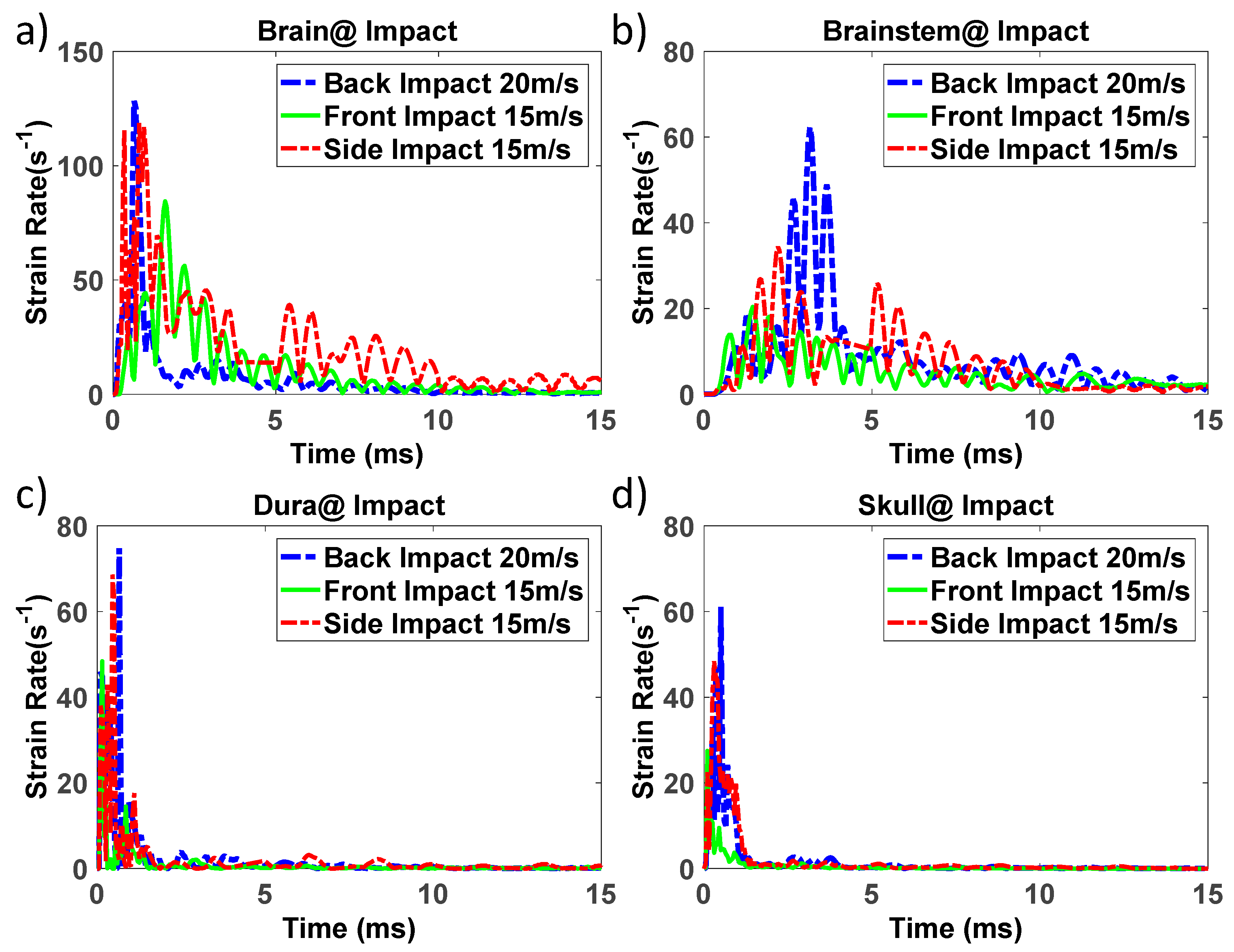
Figure 7a–d demonstrates the histories of the strain rate at regions with highest values of rate in brain, brainstem, skull, and dura under three directions of impact. In addition, the computed strain rates at points with maximum rates in these head parts under four scenarios of blast are plotted and shown in Figure 8a–d. Finally, the maximum linear acceleration of the head and highest calculated strain rate in the brain, brainstem, skull, and dura for all impact and blast cases are demonstrated in Table 6 and Table 7, respectively. Since the values of the acceleration in all impact scenarios are less than 90 g [1], the damage level can be considered minor or close to the mild TBI. Head accelerations in some blast cases exceed 90 g, so severe brain injuries are expected under those circumstances.

4. Discussion

Evaluating mechanical properties of the human head organs is fundamental in understanding intracranial brain deformation under different loading conditions. It is known that the brain has a rate-dependent behavior and gets stiffer by increasing the loading rate [16,59]. That is why, in literature, it is common to conduct in vitro tests with certain strain rates to capture the wider properties of the brain [18,19,60]. Similar approaches have been applied to identify the material parameters of brainstem, dura, and other rate-dependent biological tissue [31,61]. Cranial bone was found to behave differently at various rates [30]. It was reported that the bone tissue for the skull at high rates (150 s−1) can become up to 200% stiffer as skull in quasistatic rates [37]. Therefore, to simulate these rate-dependent materials in dynamical studies such as TBI cases, the validated properties over the desired range of strain rates must be employed. In this regard, the findings of this paper propose a measure for the range of the rates needed for conducting the in vitro test and also selecting material properties from the right strain rates.
The time duration of a general head impacts is in the order of milliseconds, while in the case of a high-speed load such as a blast wave less than one millisecond. Regardless of the strain value, it was found that brain injury caused by impact happens with a brain strain rate of 23 to 140 s−1 with an average of 84 s−1 [62,63]. Using a validated FE head model is a very feasible approach for determining the strain rates of the brain and skull under loading. Clinical studies of reconstructed head impact simulations which led to mild TBI for 58 football players proposed a range of 35–97 s−1 for the brain strain rate [64]. Viano et al. [5] analyzed 28 cases of NFL impacts involving 22 concussions by FEM, and they found the brain strain rates for those incidents were in the range of 19–162 s−1. To the best of the authors’ knowledge, there is no reported study in the literature pointing out the range of the strain rate from experimental measurement of an in vivo or cadaver test.
In this paper, some impact and blast scenarios were modeled on a validated head model. Results (Table 6 and Table 7) showed that under these dynamic loads, the response of brain and brainstem were in the range of 36 to 241 and 15 to 412 s−1, respectively. Meanwhile, the estimated strain rates of the dura and skull during such impact and blast loads were respectively in the range of 8 to 149 and 14 to 182 s−1. Significant correlations of strain rates with linear head acceleration for brain, brainstem, dura, and skull were found and are presented in Figure 9a–d. These linear regressions were independent of the effects of direction and speed of impactor, as well as the HE mass, distance, direction, and head condition (protection) under blast [65]. The proposed strain rates of brain, brainstem, dura, and skull are, respectively, 1.9 (R = 0.8, p < 0.01), 0.7 (R = 0.9, p < 0.01), 1.18 (R = 0.9, p < 0.001) and 0.7 (R = 0.6, p < 0.01) times of resulting head acceleration by impact. In addition, under blast incidents, the brain, brainstem, dura, and skull behaved with a strain rate of 0.86 (R = 0.96, p < 0.001), 1.25 (R = 0.9, p < 0.001), 0.52 (R = 0.95, p < 0.001) and 0.43 (R = 0.9, p < 0.001) times of acceleration, respectively. Figure 9 shows that for the equivalent linear acceleration of head, the strain rates induced by impact for the brain, dura, and skull are higher in comparison to the blast one.
As the first limitation of this study, the result was not based on a general impact or blast conditions. Numerous cases of other simulations for various scenarios of blunt impact and blast are required instead of the chosen limited representative cases. Therefore, it should be mentioned that the estimated range of rates in this study is more expected for a blunt impact causing mild TBI, and blast cases generating concussion or brain injury [1,63]. In this research, we did not study the effect of element size on the strain rate. We have used a validated head model that has been used in many computational studies of human head under similar dynamical loads. Therefore, we did not change the mesh size, and of course, once we had changed the element sizes, we needed to validate the new head model again. As another limitation, the FE simulations were done with one set of material properties, and we used a simple elastic material model for the cranial bone and dura mater. The most accurate material properties for this kind of simulation would be the nonhomogeneous anisotropic visco-hyperelastic materials. However, first, we could not find these properties for human head tissues in the current literature, and second, our head model was not developed for utilizing such material models. Among material properties available in the literature, those which were determined at the highest dynamic rates were selected. However, the findings of this study still suggest that there is a necessity to characterize those material properties at even higher ranges. Thus, the results of this paper can be considered as the first step in having the true estimation of selecting the right material properties in dynamical simulations.

5. Conclusions

Cranial bone, and particularly brain, brainstem, and dura are highly rate-dependent materials and will behave differently in various strain rates. In order to study the biomechanics of traumatic brain injury (TBI) through FE methods, it is necessary to implement correct properties associated with anticipated application rates. However, to calculate the material properties of the brain, brainstem, dura, and skull related to TBI cases, we need to know for which range of strain rates they are required to be examined. In this study, FE simulation was performed to estimate the strain rate ranges in impact and blast. It was found that the brain and skull experience strain rates in the range of 36 to 241 and 14 to 182 s−1 for impact and blast incidents, respectively. Additionally, in such dynamical simulations, the brainstem and dura behave at strain rates of 15 to 412 and 8 to 149 s−1, respectively. Knowing these ranges provides a good measurement for the more accurate characterization of these materials. Additionally, representing the rates based on head acceleration helps to get a better idea of selecting material properties in advance. It was shown in the impact incidents that the strain rates of the brain, brainstem, dura, and skull were approximately 1.9, 0.7, 1.18, and 0.7 times the resulting head acceleration. Furthermore, under blast loadings, the strain rates for those head organs were around 0.86, 1.25, 0.52, and 0.43 times head acceleration.

Author Contributions

Conceptualization, M.H.-F. and G.K.; methodology, M.H.-F.; software, M.H.-F. and M.A.-T.-Z.; validation, M.H.-F. and M.R.; formal analysis, M.H.-F.; investigation, M.H.-F.; resources, M.H.-F., M.Z., G.K.; data curation, M.H.-F. and M.A.-T.-Z.; writing—original draft preparation, M.H.-F.; writing—review and editing, M.H.-F., M.A.-T.-Z., M.R., M.Z., G.K.; visualization, M.H.-F.; supervision, M.Z. and G.K.; All authors have read and agreed to the published version of the manuscript.

Acknowledgments

The Authors would like to thank Mehdi Salimi Jazi, Hesam Sarvghad-Moghaddam, Asghar Rezaei and Ashkan Eslaminejad for their support and providing us with the validated FE head model.

Conflicts of Interest

The authors declare no conflict of interest.

References

  1. Tse, K.M.; Lim, S.P.; Tan, V.B.C.; Lee, H.P. A review of head injury and finite element head models. Am. J. Eng. Technol. Soc. 2014, 1, 28–52. [Google Scholar]
  2. Bass, C.R.; Panzer, M.B.; Rafaels, K.A.; Wood, G.; Shridharani, J.; Capehart, B. Brain injuries from blast. Ann. Biomed. Eng. 2012, 40, 185–202. [Google Scholar] [CrossRef] [PubMed]
  3. Saboori, P.; Walker, G. Brain injury and impact characteristics. Ann. Biomed. Eng. 2019, 47, 1982–1992. [Google Scholar] [CrossRef] [PubMed]
  4. Meaney, D.F.; Morrison, B.; Bass, C.D. The mechanics of traumatic brain injury: A review of what we know and what we need to know for reducing its societal burden. J. Biomed. Eng. 2014, 136, 021008. [Google Scholar] [CrossRef] [PubMed]
  5. Viano, D.C.; Casson, I.R.; Pellman, E.J.; Zhang, L.; King, A.I.; Yang, K.H. Concussion in professional football: Brain responses by finite element analysis: Part 9. Neurosurgery 2005, 57, 891–916. [Google Scholar] [CrossRef]
  6. Rueda, M.F.; Gilchrist, M.D. Comparative multibody dynamics analysis of falls from playground climbing frames. Forensic Sci. Int. 2009, 191, 52–57. [Google Scholar] [CrossRef]
  7. Faul, M.; Xu, L.; Wald, M.M.; Coronado, V.G. Traumatic Brain Injury in the United States; Centers for Disease Control and Prevention, National Center for Injury Prevention and Control: Atlanta, GA, USA, 2010.
  8. Zhang, L.; Yang, K.H.; Dwarampudi, R.; Omori, K.; Li, T.; Chang, K.; Hardy, W.N.; Khalil, T.B.; King, A.I. Recent advances in brain injury research: A new human head model development and validation. Stapp Car Crash J. 2001, 45, 369–394. [Google Scholar]
  9. Raul, J.-S.; Baumgartner, D.; Willinger, R.; Ludes, B. Finite element modelling of human head injuries caused by a fall. Int. J. Leg. Med. 2006, 120, 212–218. [Google Scholar] [CrossRef]
  10. Giordano, C.; Cloots, R.; Van Dommelen, J.; Kleiven, S. The influence of anisotropy on brain injury prediction. J. Biomech. 2014, 47, 1052–1059. [Google Scholar] [CrossRef]
  11. Hosseini-Farid, M.; Ramzanpour, M.; Eslaminejad, A.; Ziejewski, M.; Karami, G. Computational Simulation of Brain Injury by Golf Ball Impacts in Adult and Children. Biomed. Sci. Instrum. 2018, 54, 369–376. [Google Scholar]
  12. Hosseini-Farid, M.; Ramzanpour, M.; Ziejewski, M.; Karami, G. Estimating the Brain Strain Rates During Traumatic Brain Injury. Biomed. Sci. Instrum. 2018, 54, 361–368. [Google Scholar]
  13. Fernandes, F.A.; Alves de Sousa, R.J.; Ptak, M.; Wilhelm, J. Certified Motorcycle Helmets: Computational Evaluation of the Efficacy of Standard Requirements with Finite Element Models. Math. Comput. Appl. 2020, 25, 12. [Google Scholar] [CrossRef] [Green Version]
  14. Holbourn, A. Mechanics of head injuries. Lancet 1943, 242, 438–441. [Google Scholar] [CrossRef]
  15. Hosseini-Farid, M.; Ramzanpour, M.; Ziejewski, M.; Karami, G. A compressible hyper-viscoelastic material constitutive model for human brain tissue and the identification of its parameters. Int. J. Non-Linear Mech. 2019, 116, 147–154. [Google Scholar] [CrossRef]
  16. Farid, M.H.; Eslaminejad, A.; Ziejewski, M.; Karami, G. A Study on the Effects of Strain Rates on Characteristics of Brain Tissue. In Proceedings of the ASME 2017 International Mechanical Engineering Congress and Exposition, Tampa, FL, USA, 3–9 November 2017. [Google Scholar]
  17. Hosseini-Farid, M.; Ramzanpour, M.; McLean, J.; Ziejewski, M.; Karami, G. A poro-hyper-viscoelastic rate-dependent constitutive modeling for the analysis of brain tissues. J. Mech. Behav. Biomed. Mater. 2020, 102, 103457. [Google Scholar] [CrossRef]
  18. Hosseini Farid, M.; Ramzanpour, M.; Ziejewski, M.; Karami, G. A Biphasic Viscoelastic Constitutive Model for Brain Tissue. In Proceedings of the ASME 2019 International Mechanical Engineering Congress and Exposition, Salt Lake City, UT, USA, 11–14 November 2019. [Google Scholar]
  19. Hosseini Farid, M.; Ramzanpour, M.; Ziejewski, M.; Karami, G. A Constitutive Material Model With Strain-Rate Dependency for Brain Tissue. In Proceedings of the ASME 2019 International Mechanical Engineering Congress and Exposition, Salt Lake City, UT, USA, 11–14 November 2019. [Google Scholar]
  20. Hosseini-Farid, M.; Ramzanpour, M.; McLean, J.; Ziejewski, M.; Karami, G. Rate-dependent constitutive modeling of brain tissue. Biomech. Model. Mechanobiol. 2020, 19, 621–632. [Google Scholar] [CrossRef]
  21. El Sayed, T.; Mota, A.; Fraternali, F.; Ortiz, M. A variational constitutive model for soft biological tissues. J. Biomech. 2008, 41, 1458–1466. [Google Scholar] [CrossRef] [Green Version]
  22. Meyers, M.A.; Chawla, K.K. Mechanical Behavior of Materials; Cambridge University Press: Cambridge, UK, 2009; Volume 2. [Google Scholar]
  23. Ning, X.; Zhu, Q.; Lanir, Y.; Margulies, S.S. A transversely isotropic viscoelastic constitutive equation for brainstem undergoing finite deformation. J. Biomech. Eng. 2006, 128, 925–933. [Google Scholar] [CrossRef]
  24. Arbogast, K.B.; Margulies, S.S. A fiber-reinforced composite model of the viscoelastic behavior of the brainstem in shear. J. Biomech. 1999, 32, 865–870. [Google Scholar] [CrossRef]
  25. Ramzanpour, M.; Hosseini-Farid, M.; Ziejewski, M.; Karami, G. Microstructural Hyperelastic Characterization of Brain White Matter in Tension. In Proceedings of the ASME 2019 International Mechanical Engineering Congress and Exposition, Salt Lake City, UT, USA, 11–14 November 2019. [Google Scholar]
  26. Ramzanpour, M.; Hosseini-Farid, M.; Ziejewski, M.; Karami, G. Particle Swarm Optimization Method for Hyperelastic Characterization of Soft Tissues. In Proceedings of the ASME 2019 International Mechanical Engineering Congress and Exposition, Salt Lake City, UT, USA, 11–14 November 2019. [Google Scholar]
  27. McElhaney, J.H.; Fogle, J.L.; Melvin, J.W.; Haynes, R.R.; Roberts, V.L.; Alem, N.M. Mechanical properties of cranial bone. J. Biomech. 1970, 3, 495–511. [Google Scholar] [CrossRef] [Green Version]
  28. Galford, J.E.; McElhaney, J.H. A viscoelastic study of scalp, brain, and dura. J. Biomech. 1970, 3, 211–221. [Google Scholar] [CrossRef]
  29. Zhai, X.; Nauman, E.A.; Moryl, D.; Lycke, R.; Chen, W.W. The effects of loading-direction and strain-rate on the mechanical behaviors of human frontal skull bone. J. Mech. Behav. Biomed. Mater. 2019, 103597. [Google Scholar] [CrossRef] [PubMed]
  30. Motherway, J.A.; Verschueren, P.; Van der Perre, G.; Vander Sloten, J.; Gilchrist, M.D. The mechanical properties of cranial bone: The effect of loading rate and cranial sampling position. J. Biomech. 2009, 42, 2129–2135. [Google Scholar] [CrossRef] [PubMed]
  31. Persson, C.; Evans, S.; Marsh, R.; Summers, J.L.; Hall, R.M. Poisson’s ratio and strain rate dependency of the constitutive behavior of spinal dura mater. Ann. Biomed. Eng. 2010, 38, 975–983. [Google Scholar] [CrossRef] [PubMed]
  32. Maikos, J.T.; Elias, R.A.; Shreiber, D.I. Mechanical properties of dura mater from the rat brain and spinal cord. J. Neurotrauma 2008, 25, 38–51. [Google Scholar] [CrossRef] [PubMed]
  33. Eslaminejad, A.; Hosseini-Farid, M.; Ziejewski, M.; Karami, G. Constitutive Properties Determination of Human Cranium by an Experimental-Computational Modal Analysis. J. Vib. Acoust. 2019, 142, 011013. [Google Scholar] [CrossRef]
  34. Farid, M.H.; Eslaminejad, A.; Ramzanpour, M.; Ziejewski, M.; Karami, G. The Strain Rates of the Brain and Skull Under Dynamic Loading. In Proceedings of the ASME 2018 International Mechanical Engineering Congress and Exposition, Pittsburgh, PA, USA, 9–15 November 2018. [Google Scholar]
  35. De Kegel, D.; Vastmans, J.; Fehervary, H.; Depreitere, B.; Vander Sloten, J.; Famaey, N. Biomechanical characterization of human dura mater. J. Mech. Behav. Biomed. Mater. 2018, 79, 122–134. [Google Scholar] [CrossRef]
  36. Eslaminejad, A.; Hosseini-Farid, M.; Ramzanpour, M.; Ziejewski, M.; Karami, G. Determination of Mechanical Properties of Human Skull With Modal Analysis. In Proceedings of the ASME 2018 International Mechanical Engineering Congress and Exposition, Pittsburgh, PA, USA, 9–15 November 2018. [Google Scholar]
  37. Wood, J.L. Dynamic response of human cranial bone. J. Biomech. 1971, 4, IN1–IN3. [Google Scholar] [CrossRef]
  38. Franceschini, G.; Bigoni, D.; Regitnig, P.; Holzapfel, G.A. Brain tissue deforms similarly to filled elastomers and follows consolidation theory. J. Mech. Phys. Solids 2006, 54, 2592–2620. [Google Scholar] [CrossRef]
  39. Ghajari, M.; Hellyer, P.J.; Sharp, D.J. Computational modelling of traumatic brain injury predicts the location of chronic traumatic encephalopathy pathology. Brain 2017, 140, 333–343. [Google Scholar] [CrossRef] [Green Version]
  40. Patton, D.A.; McIntosh, A.S.; Kleiven, S.; Frechede, B. Injury data from unhelmeted football head impacts evaluated against critical strain tolerance curves. Proc. Inst. Mech. Eng. P J. Sports Eng. Technol. 2012, 226, 177–184. [Google Scholar] [CrossRef]
  41. Eslaminejad, A.; Hosseini Farid, M.; Ziejewski, M.; Karami, G. Brain Tissue Constitutive Material Models and the Finite Element Analysis of Blast-Induced Traumatic Brain Injury. Sci. Iran. 2018. [Google Scholar] [CrossRef]
  42. Rezaei, A.; Salimi Jazi, M.; Karami, G.; Ziejewski, M. A computational study on brain tissue under blast: Primary and tertiary blast injuries. Int. J. Numer. Methods Biomed. Eng. 2014, 30, 781–795. [Google Scholar] [CrossRef] [PubMed]
  43. Horgan, T.; Gilchrist, M.D. The creation of three-dimensional finite element models for simulating head impact biomechanics. Int. J. Crashworthiness 2003, 8, 353–366. [Google Scholar] [CrossRef]
  44. Sarvghad-Moghaddam, H.; Rezaei, A.; Ziejewski, M.; Karami, G. Correlative analysis of head kinematics and brain’s tissue response: A computational approach toward understanding the mechanisms of blast TBI. Shock Waves 2017, 27, 919–927. [Google Scholar] [CrossRef]
  45. Salimi Jazi, M.; Rezaei, A.; Karami, G.; Azarmi, F.; Ziejewski, M. A computational study of influence of helmet padding materials on the human brain under ballistic impacts. Comput. Methods Biomech. Biomed. Eng. 2014, 17, 1368–1382. [Google Scholar] [CrossRef]
  46. Chafi, M.; Karami, G.; Ziejewski, M. Biomechanical assessment of brain dynamic responses due to blast pressure waves. Ann. Biomed. Eng. 2010, 38, 490–504. [Google Scholar] [CrossRef]
  47. Edri, I.; Savir, Z.; Feldgun, V.; Karinski, Y.; Yankelevsky, D. On blast pressure analysis due to a partially confined explosion: I. Experimental studies. Int. J. Prot. Struct. 2011, 2, 1–20. [Google Scholar] [CrossRef]
  48. Horgan, T.J.; Gilchrist, M.D. Influence of FE model variability in predicting brain motion and intracranial pressure changes in head impact simulations. Int. J. Crashworthiness 2004, 9, 401–418. [Google Scholar] [CrossRef]
  49. Nahum, A.M.; Smith, R.; Ward, C.C. Intracranial Pressure Dynamics during Head Impact. Available online: https://www.sae.org/publications/technical-papers/content/770922/ (accessed on 6 April 2020).
  50. Hardy, W.N.; Foster, C.D.; Mason, M.J.; Yang, K.H.; King, A.I.; Tashman, S. Investigation of head injury mechanisms using neutral density technology and high-speed biplanar X-ray. Stapp Car Crash J. 2001, 45, 337–368. [Google Scholar]
  51. Trosseille, X.; Tarriere, C.; Lavaste, F.; Guillon, F.; Domont, A. Development of a FEM of the human head according to a specific test protocol. In Proceedings of the 36th Stapp Car Crash Conference, Seattle, WA, USA, 2–4 November 1992. [Google Scholar]
  52. Lee, H.P.; Wang, F. Assessment of head injury of children due to golf ball impact. Comput. Methods Biomech. Biomed. Eng. 2010, 13, 523–535. [Google Scholar] [CrossRef] [PubMed]
  53. Ridenour, M.V. Golf clubs: Hidden home hazard for children. Percept. Mot. Ski. 1998, 86, 747–753. [Google Scholar] [CrossRef] [PubMed]
  54. Bait, M.E. Golfing injuries. Sports Med. 1993, 16, 64–71. [Google Scholar] [CrossRef] [PubMed]
  55. Salimi Jazi, M.; Rezaei, A.; Karami, G.; Azarmi, F. Biomechanical parameters of the brain under blast loads with and without helmets. Int. J. Exp. Comput. Biomech. 2014, 2, 223–244. [Google Scholar] [CrossRef]
  56. Rezaei, A.; Salimi Jazi, M.; Karami, G. Computational modeling of human head under blast in confined and open spaces: Primary blast injury. Int. J. Numer. Methods Biomed. Eng. 2014, 30, 69–82. [Google Scholar] [CrossRef] [PubMed]
  57. Dobratz, B.; Crawford, P. Properties of Chemical Explosives & Explosives Stimulants, LLNL Explosive Handbook; University of California Report UCRL-52997; Lawrence Livermore National Laboratory: Livermore, CA, USA, 1981.
  58. Ruan, J.; Khalil, T.; King, A. Human head dynamic response to side impact by finite element modeling. J. Biomech. Eng. 1991, 113, 276–283. [Google Scholar] [CrossRef]
  59. Farid, M.H. Mechanical Characterization and Constitutive Modeling of Rate-Dependent Viscoelastic Brain Tissue under High Rate Loadings. Available online: https://library.ndsu.edu/ir/handle/10365/29786 (accessed on 6 April 2020).
  60. Forte, A.E.; Gentleman, S.M.; Dini, D. On the characterization of the heterogeneous mechanical response of human brain tissue. Biomech. Model. Mechanobiol. 2017, 16, 907–920. [Google Scholar] [CrossRef] [Green Version]
  61. Zhao, H.; Yin, Z.; Li, K.; Liao, Z.; Xiang, H.; Zhu, F. Mechanical characterization of immature porcine brainstem in tension at dynamic strain rates. Med. Sci. Monit. Basic Res. 2016, 22, 6. [Google Scholar] [CrossRef] [Green Version]
  62. Viano, D.C.; Lovsund, P. Biomechanics of brain and spinal-cord injury: Analysis of neuropathologic and neurophysiology experiments. Traffic Inj. Prev. 1999, 1, 35–43. [Google Scholar] [CrossRef]
  63. Zhang, L.; Yang, K.H.; King, A.I. A proposed injury threshold for mild traumatic brain injury. Trans. Am. Soc. Mech. Eng. J. Biomech. Eng. 2004, 126, 226–236. [Google Scholar] [CrossRef]
  64. Kleiven, S. Predictors for traumatic brain injuries evaluated through accident reconstructions. Stapp Car Crash J. 2007, 51, 81. [Google Scholar] [PubMed]
  65. Jahani, B.; Salimi Jazi, M.; Azarmi, F.; Croll, A. Effect of volume fraction of reinforcement phase on mechanical behavior of ultra-high-temperature composite consisting of iron matrix and TiB2 particulates. J. Compos. Mater. 2018, 52, 609–620. [Google Scholar] [CrossRef]
Figure 1. The finite element (FE) head model used in this study includes the main components of the human head [44].
Mca 25 00021 g001
Figure 2. Setup of frontal and lateral impact by golf ball in the FE head model.
Mca 25 00021 g002
Figure 3. The position of the detonation at side and back of the protected head model.
Mca 25 00021 g003
Figure 4. Resultant linear acceleration for the head skull (head) at: (a) impact in three directions, and (b) four cases of blast.
Mca 25 00021 g004
Figure 5. Intracranial pressure (ICP) (kPa) contours in the brain for the protected head in heavy blast for whole brain and also cross-sectional view.
Mca 25 00021 g005
Figure 6. Histories of strain rate at three different regions of the brain under: (a) frontal and (b) lateral impact by golf ball, and heavy blast scenarios for (c) protected and (d) unprotected head.
Mca 25 00021 g006
Figure 7. History of highest calculated strain rate under three blunt impact cases for: (a) brain, (b) brainstem, (c) dura, and (d) skull.
Mca 25 00021 g007
Figure 8. History of highest calculated strain rate during four blast scenarios for: (a) brain, (b) brainstem, (c) dura, and (d) skull.
Mca 25 00021 g008
Figure 9. Linear regression of highest evaluated strain rate versus maximum acceleration of head under impact and blast cases for: (a) brain, (b) brainstem, (c) dura, and (d) skull.
Mca 25 00021 g009
Table 1. Head model component properties and mechanical properties [1,52].
Segment NameConstitutive ModelFE ModelDensity (kg/m3)Poisson’s RatioYoung’s Modulus (MPa)
TentoriumLinear ElasticShell Element11330.4531.5
Dura MaterLinear ElasticShell Element11330.4531.5
Pia MaterLinear ElasticShell Element11300.4511.5
FalxLinear ElasticShell Element11330.4531.5
SkullLinear ElasticSolid Element13000.2410000
Neck-BoneLinear ElasticSolid Element13000.2410000
Neck-FleshLinear ElasticSolid Element11300.450.1
Face-BoneLinear ElasticSolid Element21000.425540
Face-SkinLinear ElasticSolid Element12000.4216.7
ScalpLinear ElasticSolid Element12000.4216.7
Golf BallLinear ElasticSolid Element11000.49300
CSFLinear Elastic (Fluid Opt.)Solid Element1040Poisson’s RatioBulk ModulusViscosity Coefficient
0.4921900.2
Table 2. Mooney–Rivlin hyper-viscoelastic constants of the brain used in FE modeling [1].
Density (kg/m3) C 10   ( Pa ) C 01   ( Pa ) g 1   ( Pa ) g 2   ( Pa ) β1 (s−1)β2 (s−1)
104031354072331256.7
Table 3. Viscoelastic material parameters of the brainstem used in FE modeling [1].
Density (kg/m3)Bulk Modulus (GPa) G 0   ( kPa ) G   ( kPa ) β (s−1)
10602.1922.54.580
Table 4. The summary of impact cases by golf ball applied in FE modeling for estimating the ranges of strain rates.
Case CodeImpactorSpeed (m/s)Direction
Impact# 1Golf ball10Lateral
Impact# 2Golf ball15Lateral
Impact# 3Golf ball20Lateral
Impact# 4Golf ball10Back
Impact# 5Golf ball15Back
Impact# 6Golf ball20Back
Impact# 7Golf ball10Frontal
Impact# 8Golf ball15Frontal
Impact# 9Golf ball20Frontal
Table 5. The summary of blast scenarios implemented in FE simulations to predict the ranges of strain rate associated to traumatic brain injuries (TBI).
Case CodeHead ConditionExplosive Mass (gr)Distance (m)Direction
Blast# 1Protected751Back
Blast# 2Protected752Back
Blast# 3Protected751Lateral
Blast# 4Protected752Lateral
Blast# 5Protected950.75Back
Blast# 6Unprotected751Back
Blast# 7Unprotected752Back
Blast# 8Unprotected751Lateral
Blast# 9Unprotected752Lateral
Blast# 10Unprotected950.75Back
Table 6. The estimated maximum linear acceleration of skull, and strain rate of the brain, brainstem, skull, and dura at different impact scenarios.
Case CodeMax. Skull Acceleration (g)Max. Strain Rate in Brain (s−1)Max. Strain Rate in Brainstem (s−1)Max. Strain Rate in Dura (s−1)Max. Strain Rate in Skull (s−1)
Impact# 13385244935
Impact# 247105356845
Impact# 3821806810348
Impact# 42342213822
Impact# 56167436237
Impact# 668132607561
Impact# 73456154316
Impact# 85281245431
Impact# 970142357642
Table 7. The predicted maximum linear acceleration of skull, and strain rate of the brain, brainstem, skull, and dura at different blast scenarios.
Case CodeMax. Skull Acceleration (g)Max. Strain Rate in Brain (s−1)Max. Strain Rate in Brainstem (s−1)Max. Strain Rate in Dura (s−1)Max. Strain Rate in Skull (s−1)
Blast# 1931001653955
Blast# 2304070814
Blast# 31401202285551
Blast# 44136711326
Blast# 5226205338144122
Blast# 61801152146264
Blast# 73848621520
Blast# 820518015112266
Blast# 96373473228
Blast# 10432241412149182

