
Carnosic Acid and Carnosol: Analytical Methods for Their Determination in Plants, Foods and Biological Samples


Abstract
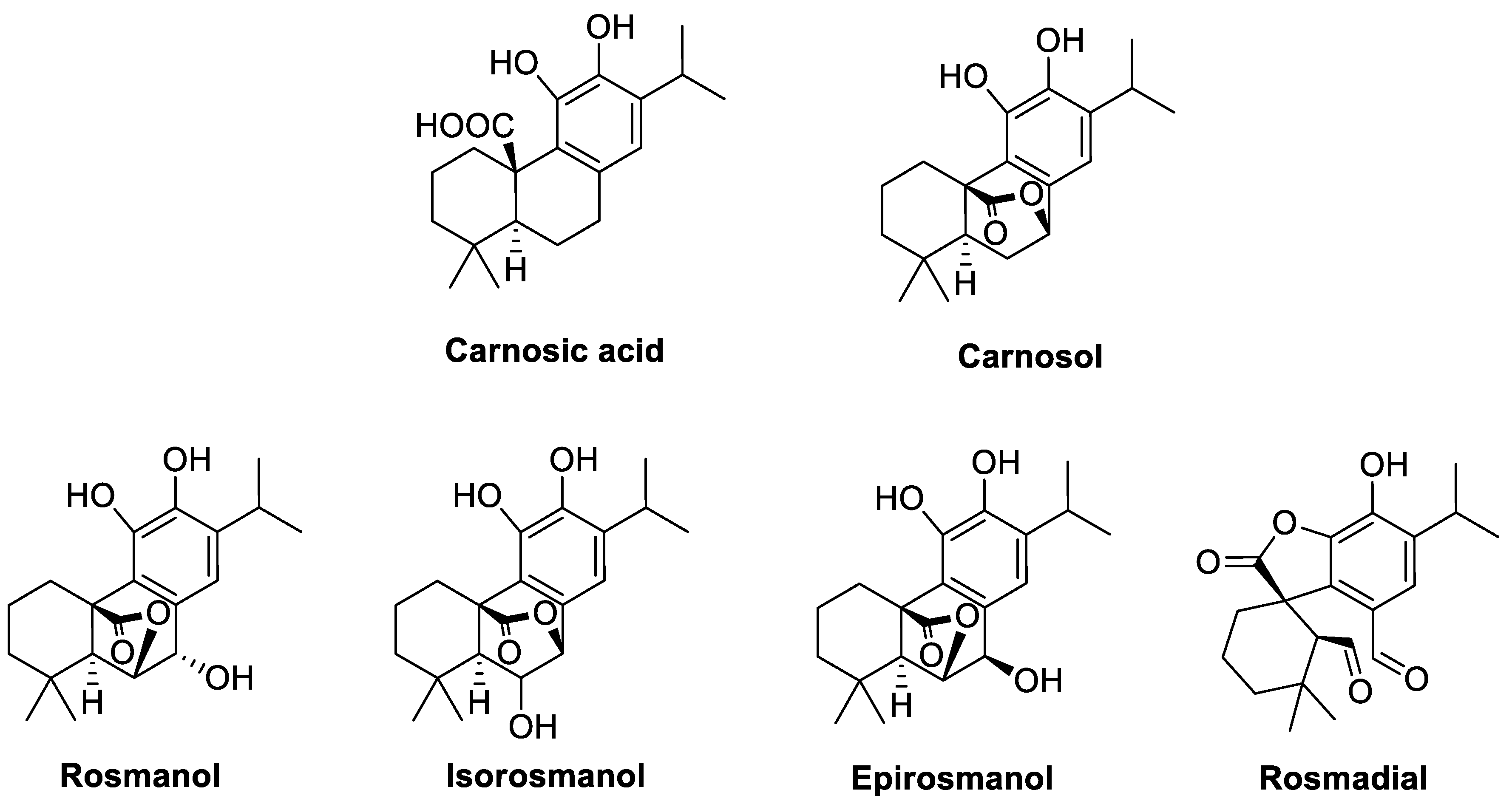
:1. Introduction
2. Extraction Methods
2.1. Sample Pretreatment
2.2. Conventional Extraction Methods
2.3. Ultrasound-Assisted Extraction (UAE)
2.4. Microwave-Assisted Extraction (MAE)
2.5. Supercritical Fluid Extraction (SFE)
2.6. Accelerated Solvent Extraction (ASE)
2.7. Green and Sustainable Solvents
3. Analysis of Carnosic Acid and Carnosol
3.1. High-Performance Liquid Chromatography–UV Detection (HPLC–UV)
3.2. Liquid Chromatography–Mass Spectrometry (LC–MS)
3.3. Capillary Electrophoresis (CE) and Other Techniques
4. Biological Activities of Carnosic Acid and Carnosol
4.1. Antioxidant Activity
4.2. Anticancer Activity
4.3. Anti-Inflammatory Activity
4.4. Neuroprotective Activity
5. Conclusions
Author Contributions
Funding
Institutional Review Board Statement
Informed Consent Statement
Data Availability Statement
Acknowledgments
Conflicts of Interest
References
- Jassbi, A.R.; Zare, S.; Firuzi, O.; Xiao, J. Bioactive phytochemicals from shoots and roots of Salvia species. Phytochem. Rev. 2016, 15, 829–867. [Google Scholar] [CrossRef]
- Carović-Stanko, K.; Petek, M.; Grdiša, M.; Pintar, J.; Bedeković, D.; Herak Ćustić, M.; Satovic, Z. Medicinal plants of the family Lamiaceae as functional foods—A review. Czech J. Food Sci. 2016, 34, 377–390. [Google Scholar] [CrossRef]
- Pizani, R.S.; Viganó, J.; De Souza Mesquita, L.M.; Contieri, L.S.; Sanches, V.L.; Chaves, J.O.; Souza, M.C.; Da Silva, L.C.; Rostagno, M.A. Beyond aroma: A review on advanced extraction processes from rosemary (Rosmarinus officinalis) and sage (Salvia officinalis) to produce phenolic acids and diterpenes. Trends Food Sci. Technol. 2022, 127, 245–262. [Google Scholar] [CrossRef]
- Pizzale, L.; Bortolomeazzi, R.; Vichi, S.; Überegger, E.; Conte, L.S. Antioxidant activity of sage (Salvia officinalis and S. fruticosa) and oregano (Origanum onites and O. indercedens) extracts related to their phenolic compound content: Antioxidant activity of sage and oregano extracts. J. Sci. Food Agric. 2002, 82, 1645–1651. [Google Scholar] [CrossRef]
- Schwarz, K.; Ternes, W. Antioxidative constituents of Rosmarinus officinalis and Salvia officinalis: II. Isolation of carnosic acid and formation of other phenolic diterpenes. Z. Lebensm. Unters. Forschung 1992, 195, 99–103. [Google Scholar] [CrossRef] [PubMed]
- Munné-Bosch, S.; Alegre, L. Subcellular compartmentation of the diterpene carnosic acid and its derivatives in the leaves of rosemary. Plant Physiol. 2001, 125, 1094–1102. [Google Scholar] [CrossRef]
- Luis, J.C.; Johnson, C.B. Seasonal variations of rosmarinic and carnosic acids in rosemary extracts. Analysis of their in vitro antiradical activity. Span. J. Agric. Res. 2005, 3, 106. [Google Scholar] [CrossRef]
- Brewer, M.S. Natural antioxidants: Sources, compounds, mechanisms of action, and potential applications. Compr. Rev. Food Sci. Food Saf. 2011, 10, 221–247. [Google Scholar] [CrossRef]
- Munné-Bosch, S.; Alegre, L. Drought-induced changes in the redox state of α-tocopherol, ascorbate, and the diterpene carnosic acid in chloroplasts of Labiatae species differing in carnosic acid contents. Plant Physiol. 2003, 131, 1816–1825. [Google Scholar] [CrossRef]
- Lefebvre, T.; Destandau, E.; Lesellier, E. Selective extraction of bioactive compounds from plants using recent extraction techniques: A Review. J. Chromatogr. A 2021, 1635, 461770. [Google Scholar] [CrossRef]
- Boscaiu, M.; Vicente, O.; Bautista, I.; Ranga, F.; Socaciu, C. HPLC-DAD-ESI+-MS phytochemical profiles of several Rosmarinus officinalis accessions from Spain as influenced by different environmental stress conditions. Stud. UBB Chem. 2019, 64, 163–180. [Google Scholar] [CrossRef]
- Nakatani, N.; Inatani, R. Two antioxidative diterpenes from rosemary (Rosmarinus officinalis L.) and a revised structure for rosmanol. Agric. Biol. Chem. 1984, 48, 2081–2085. [Google Scholar] [CrossRef]
- Cuvelier, M.-E.; Richard, H.; Berset, C. Antioxidative activity and phenolic composition of pilot-plant and commercial extracts of sage and rosemary. J. Am. Oil Chem. Soc. 1996, 73, 645–652. [Google Scholar] [CrossRef]
- Hossain, M.B.; Rai, D.K.; Brunton, N.P.; Martin-Diana, A.B.; Barry-Ryan, C. Characterization of phenolic composition in Lamiaceae spices by LC-ESI-MS/MS. J. Agric. Food Chem. 2010, 58, 10576–10581. [Google Scholar] [CrossRef]
- Vági, E.; Rapavi, E.; Hadolin, M.; Vásárhelyiné Perédi, K.; Balázs, A.; Blázovics, A.; Simándi, B. Phenolic and triterpenoid antioxidants from Origanum majorana L. Herb and Extracts Obtained with Different Solvents. J. Agric. Food Chem. 2005, 53, 17–21. [Google Scholar] [CrossRef]
- Herodež, Š.S.; Hadolin, M.; Škerget, M.; Knez, Ž. Solvent extraction study of antioxidants from balm (Melissa officinalis L.) leaves. Food Chem. 2003, 80, 275–282. [Google Scholar] [CrossRef]
- EFSA ANS Panel; Younes, M.; Aggett, P.; Aguilar, F.; Crebelli, R.; Dusemund, B.; Filipič, M.; Frutos, M.J.; Galtier, P.; Gott, D.; et al. Refined exposure assessment of extracts of rosemary (E 392) from its use as food Additive. EFS2 2018, 16, e05373. [Google Scholar] [CrossRef]
- Choi, S.-H.; Jang, G.-W.; Choi, S.-I.; Jung, T.-D.; Cho, B.-Y.; Sim, W.-S.; Han, X.; Lee, J.-S.; Kim, D.-Y.; Kim, D.-B.; et al. Development and validation of an analytical method for carnosol, carnosic acid and rosmarinic acid in food matrices and evaluation of the antioxidant activity of rosemary extract as a food additive. Antioxidants 2019, 8, 76. [Google Scholar] [CrossRef]
- Sharma, Y.; Velamuri, R.; Fagan, J.; Schaefer, J. Full-spectrum analysis of bioactive compounds in rosemary (Rosmarinus officinalis L.) as influenced by different extraction methods. Molecules 2020, 25, 4599. [Google Scholar] [CrossRef]
- Ali, A.; Chua, B.L.; Chow, Y.H. An insight into the extraction and fractionation technologies of the essential oils and bioactive compounds in Rosmarinus officinalis L.: Past, present and future. TrAC Trends Anal. Chem. 2019, 118, 338–351. [Google Scholar] [CrossRef]
- Conde-Hernández, L.A.; Espinosa-Victoria, J.R.; Trejo, A.; Guerrero-Beltrán, J.Á. CO2 -Supercritical extraction, hydrodistillation and steam distillation of essential oil of rosemary (Rosmarinus officinalis). J. Food Eng. 2017, 200, 81–86. [Google Scholar] [CrossRef]
- Fotovvat, M.; Radjabian, T.; Saboora, A. HPLC fingerprint of important phenolic compounds in some Salvia L. species from Iran. Rec. Nat. Prod. 2018, 13, 37–49. [Google Scholar] [CrossRef]
- Lemos, M.F.; Lemos, M.F.; Pacheco, H.P.; Endringer, D.C.; Scherer, R. Seasonality modifies rosemary’s composition and biological activity. Ind. Crops Prod. 2015, 70, 41–47. [Google Scholar] [CrossRef]
- Lesellier, E.; Lefebvre, T.; Destandau, E. Recent developments for the analysis and the extraction of bioactive compounds from Rosmarinus officinalis and medicinal plants of the Lamiaceae family. TrAC Trends Anal. Chem. 2021, 135, 116158. [Google Scholar] [CrossRef]
- Ben Farhat, M.; Jordán, M.J.; Chaouech-Hamada, R.; Landoulsi, A.; Sotomayor, J.A. Variations in essential oil, phenolic compounds, and antioxidant activity of Tunisian cultivated Salvia Officinalis L. J. Agric. Food Chem. 2009, 57, 10349–10356. [Google Scholar] [CrossRef]
- Jordán, M.J.; Lax, V.; Rota, M.C.; Lorán, S.; Sotomayor, J.A. Relevance of carnosic Acid, carnosol, and rosmarinic acid concentrations in the in vitro antioxidant and antimicrobial activities of Rosmarinus officinalis (L.) methanolic extracts. J. Agric. Food Chem. 2012, 60, 9603–9608. [Google Scholar] [CrossRef]
- Hcini, K.; Sotomayor, J.A.; Jordan, M.J.; Bouzid, S. Identification and quantification of phenolic compounds of Tunisian Rosmarinus officinalis L. Asian J. Chem. 2013, 25, 9299–9301. [Google Scholar] [CrossRef]
- Jordán, M.J.; Martínez, R.M.; Martínez, C.; Moñino, I.; Sotomayor, J.A. Polyphenolic extract and essential oil quality of Thymus zygis Ssp. gracilis shrubs cultivated under different watering levels. Ind. Crops Prod. 2009, 29, 145–153. [Google Scholar] [CrossRef]
- Kontogianni, V.G.; Tomic, G.; Nikolic, I.; Nerantzaki, A.A.; Sayyad, N.; Stosic-Grujicic, S.; Stojanovic, I.; Gerothanassis, I.P.; Tzakos, A.G. Phytochemical profile of Rosmarinus officinalis and Salvia officinalis extracts and correlation to their antioxidant and anti-proliferative activity. Food Chem. 2013, 136, 120–129. [Google Scholar] [CrossRef]
- Rodríguez-Solana, R.; Salgado, J.M.; Domínguez, J.M.; Cortés-Diéguez, S. Comparison of soxhlet, accelerated solvent and supercritical fluid extraction techniques for volatile (GC-MS and GC/FID) and phenolic compounds (HPLC-ESI/MS/MS) from Lamiaceae species: Lamiaceae essential oils. Phytochem. Anal. 2015, 26, 61–71. [Google Scholar] [CrossRef]
- Koutsoulas, A.; Čarnecká, M.; Slanina, J.; Tóth, J.; Slaninová, I. Characterization of phenolic compounds and antiproliferative effects of Salvia pomifera and Salvia fruticosa extracts. Molecules 2019, 24, 2921. [Google Scholar] [CrossRef] [PubMed]
- Birtić, S.; Dussort, P.; Pierre, F.-X.; Bily, A.C.; Roller, M. Carnosic acid. Phytochemistry 2015, 115, 9–19. [Google Scholar] [CrossRef] [PubMed]
- Schwarz, K.; Ternes, W. Antioxidative constituents of Rosmarinus officinalis and Salvia officinalis: I. Determination of phenolic diterpenes with antioxidative activity amongst tocochromanols using HPLC. Z. Lebensm. Unters. Forschung 1992, 195, 95–98. [Google Scholar] [CrossRef] [PubMed]
- Ozkan, G.; Sagdic, O.; Ekici, L.; Ozturk, I.; Ozcan, M.M. Phenolic compounds of Origanum sipyleum L. extract and its antioxidant and antibacterial activities. J. Food Lipids 2007, 14, 157–169. [Google Scholar] [CrossRef]
- Mei, X.; Tan, J.; Xiao, N.; Fang, X.; Gu, S.; Li, J.; Wang, J. Simultaneous qualitative and quantitative determination of 17 bioactive components in the fibrous roots of Salvia miltiorrhiza bunge by a combinatorial ultra performance liquid chromatography- ultraviolet characteristic spectra analysis. J. Appl. Res. Med. Aromat. Plants 2021, 20, 100291. [Google Scholar] [CrossRef]
- Xie, L.; Li, Z.; Li, H.; Sun, J.; Liu, X.; Tang, J.; Lin, X.; Xu, L.; Zhu, Y.; Liu, Z.; et al. Fast quantitative determination of principal phenolic anti-oxidants in rosemary using ultrasound-assisted extraction and chemometrics-enhanced HPLC–DAD Method. Food Anal. Methods 2023, 16, 386–400. [Google Scholar] [CrossRef]
- Zabot, G.L.; Moraes, M.N.; Rostagno, M.A.; Meireles, M.A.A. Fast analysis of phenolic terpenes by high-performance liquid chromatography using a fused-core column. Anal. Methods 2014, 6, 7457–7468. [Google Scholar] [CrossRef]
- Ge, X.; Jing, L.; Zhao, K.; Su, C.; Zhang, B.; Zhang, Q.; Han, L.; Yu, X.; Li, W. The phenolic compounds profile, quantitative analysis and antioxidant activity of four naked barley grains with different color. Food Chem. 2021, 335, 127655. [Google Scholar] [CrossRef]
- Duan, H.; Wang, W.; Li, Y.; Jilany Khan, G.; Chen, Y.; Shen, T.; Bao, N.; Hua, J.; Xue, Z.; Zhai, K.; et al. Identification of phytochemicals and antioxidant activity of Premna microphylla turcz. stem through UPLC-LTQ-Orbitrap-MS. Food Chem. 2022, 373, 131482. [Google Scholar] [CrossRef]
- Gkioni, M.D.; Zeliou, K.; Dimaki, V.D.; Trigas, P.; Lamari, F.N. GC-MS and LC-DAD-MS phytochemical profiling for characterization of three native Salvia taxa from eastern Mediterranean with antiglycation properties. Molecules 2022, 28, 93. [Google Scholar] [CrossRef]
- Li, P.; Liu, A.; Li, Y.; Yuan, B.; Xiao, W.; Liu, Z.; Zhang, S.; Lin, H. Development and validation of an analytical method based on HPLC-ELSD for the simultaneous determination of rosmarinic acid, carnosol, carnosic acid, oleanolic acid and ursolic acid in rosemary. Molecules 2019, 24, 323. [Google Scholar] [CrossRef] [PubMed]
- Albu, S.; Joyce, E.; Paniwnyk, L.; Lorimer, J.P.; Mason, T.J. Potential for the use of ultrasound in the extraction of antioxidants from Rosmarinus officinalis for the food and pharmaceutical industry. Ultrason. Sonochem. 2004, 11, 261–265. [Google Scholar] [CrossRef] [PubMed]
- Bernatoniene, J.; Cizauskaite, U.; Ivanauskas, L.; Jakstas, V.; Kalveniene, Z.; Kopustinskiene, D.M. Novel approaches to optimize extraction processes of ursolic, oleanolic and rosmarinic acids from Rosmarinus officinalis leaves. Ind. Crops Prod. 2016, 84, 72–79. [Google Scholar] [CrossRef]
- Paniwnyk, L.; Cai, H.; Albu, S.; Mason, T.J.; Cole, R. The enhancement and scale up of the extraction of anti-oxidants from Rosmarinus officinalis using ultrasound. Ultrason. Sonochem. 2009, 16, 287–292. [Google Scholar] [CrossRef]
- Borrás-Linares, I.; Stojanović, Z.; Quirantes-Piné, R.; Arráez-Román, D.; Švarc-Gajić, J.; Fernández-Gutiérrez, A.; Segura-Carretero, A. Rosmarinus officinalis leaves as a natural source of bioactive compounds. Int. J. Mol. Sci. 2014, 15, 20585–20606. [Google Scholar] [CrossRef]
- Ferreira, D.F.; Lucas, B.N.; Voss, M.; Santos, D.; Mello, P.A.; Wagner, R.; Cravotto, G.; Barin, J.S. Solvent-free simultaneous extraction of volatile and non-volatile antioxidants from rosemary (Rosmarinus officinalis L.) by microwave hydrodiffusion and gravity. Ind. Crops Prod. 2020, 145, 112094. [Google Scholar] [CrossRef]
- Jacotet-Navarro, M.; Rombaut, N.; Fabiano-Tixier, A.-S.; Danguien, M.; Bily, A.; Chemat, F. Ultrasound versus microwave as green processes for extraction of rosmarinic, carnosic and ursolic acids from rosemary. Ultrason. Sonochem. 2015, 27, 102–109. [Google Scholar] [CrossRef]
- Stashenko, E.E.; Martínez, J.R.; Cala, M.P.; Durán, D.C.; Caballero, D. Chromatographic and mass spectrometric characterization of essential oils and extracts from Lippia (Verbenaceae) aromatic plants: Gas chromatography. J. Sep. Sci. 2013, 36, 192–202. [Google Scholar] [CrossRef]
- Herrero, M.; Plaza, M.; Cifuentes, A.; Ibáñez, E. Green processes for the extraction of bioactives from rosemary: Chemical and functional characterization via ultra-performance liquid chromatography-tandem mass spectrometry and in-vitro assays. J. Chromatogr. A 2010, 1217, 2512–2520. [Google Scholar] [CrossRef]
- Borrás Linares, I.; Arráez-Román, D.; Herrero, M.; Ibáñez, E.; Segura-Carretero, A.; Fernández-Gutiérrez, A. Comparison of different extraction procedures for the comprehensive characterization of bioactive phenolic compounds in Rosmarinus officinalis by reversed-phase high-performance liquid chromatography with diode array detection coupled to electrospray time-of-flight mass spectrometry. J. Chromatogr. A 2011, 1218, 7682–7690. [Google Scholar] [CrossRef]
- Šulniūtė, V.; Pukalskas, A.; Venskutonis, P.R. Phytochemical composition of fractions isolated from ten Salvia species by supercritical carbon dioxide and pressurized liquid extraction Methods. Food Chem. 2017, 224, 37–47. [Google Scholar] [CrossRef]
- Ramírez, P.; García-Risco, M.R.; Santoyo, S.; Señoráns, F.J.; Ibáñez, E.; Reglero, G. Isolation of functional ingredients from rosemary by preparative-supercritical fluid chromatography (Prep-SFC). J. Pharm. Biomed. Anal. 2006, 41, 1606–1613. [Google Scholar] [CrossRef] [PubMed]
- Bicchi, C.; Binello, A.; Rubiolo, P. Determination of phenolic diterpene antioxidants in rosemary (Rosmarinus officinalis L.) with different methods of extraction and analysis. Phytochem. Anal. 2000, 11, 236–242. [Google Scholar] [CrossRef]
- Kompelly, A.; Kompelly, S.; Vasudha, B.; Narender, B. Rosmarinus officinalis L.: An update review of its phytochemistry and biological activity. J. Drug Deliv. Ther. 2019, 9, 323–330. [Google Scholar] [CrossRef]
- Sánchez-Camargo, A.D.P.; Valdés, A.; Sullini, G.; García-Cañas, V.; Cifuentes, A.; Ibáñez, E.; Herrero, M. Two-step sequential supercritical fluid extracts from rosemary with enhanced anti-proliferative activity. J. Funct. Foods 2014, 11, 293–303. [Google Scholar] [CrossRef]
- Vicente, G.; García-Risco, M.R.; Fornari, T.; Reglero, G. Isolation of carsonic acid from rosemary extracts using semi-preparative supercritical fluid chromatography. J. Chromatogr. A 2013, 1286, 208–215. [Google Scholar] [CrossRef]
- Sánchez-Camargo, A.; García-Cañas, V.; Herrero, M.; Cifuentes, A.; Ibáñez, E. Comparative study of green sub- and supercritical processes to obtain carnosic acid and carnosol-enriched rosemary extracts with in vitro anti-proliferative activity on colon cancer cells. Int. J. Mol. Sci. 2016, 17, 2046. [Google Scholar] [CrossRef]
- Herrero, M.; Arráez-Román, D.; Segura, A.; Kenndler, E.; Gius, B.; Raggi, M.A.; Ibáñez, E.; Cifuentes, A. Pressurized liquid extraction–capillary electrophoresis–mass spectrometry for the analysis of polar antioxidants in rosemary extracts. J. Chromatogr. A 2005, 1084, 54–62. [Google Scholar] [CrossRef]
- Ibañez, E.; Kubátová, A.; Señoráns, F.J.; Cavero, S.; Reglero, G.; Hawthorne, S.B. Subcritical water extraction of antioxidant compounds from rosemary plants. J. Agric. Food Chem. 2003, 51, 375–382. [Google Scholar] [CrossRef]
- Liu, T.; Sui, X.; Zhang, R.; Yang, L.; Zu, Y.; Zhang, L.; Zhang, Y.; Zhang, Z. Application of ionic liquids based microwave-assisted simultaneous extraction of carnosic acid, rosmarinic acid and essential oil from rosmarinus officinalis. J. Chromatogr. A 2011, 1218, 8480–8489. [Google Scholar] [CrossRef]
- Zu, G.; Zhang, R.; Yang, L.; Ma, C.; Zu, Y.; Wang, W.; Zhao, C. Ultrasound-assisted extraction of carnosic acid and rosmarinic acid using ionic liquid solution from rosmarinus officinalis. Int. J. Mol. Sci. 2012, 13, 11027–11043. [Google Scholar] [CrossRef] [PubMed]
- Hansen, B.B.; Spittle, S.; Chen, B.; Poe, D.; Zhang, Y.; Klein, J.M.; Horton, A.; Adhikari, L.; Zelovich, T.; Doherty, B.W.; et al. Deep eutectic solvents: A review of fundamentals and applications. Chem. Rev. 2021, 121, 1232–1285. [Google Scholar] [CrossRef]
- Liu, Y.; Friesen, J.B.; McAlpine, J.B.; Lankin, D.C.; Chen, S.-N.; Pauli, G.F. Natural deep eutectic solvents: Properties, applications, and perspectives. J. Nat. Prod. 2018, 81, 679–690. [Google Scholar] [CrossRef] [PubMed]
- Barbieri, J.B.; Goltz, C.; Batistão Cavalheiro, F.; Theodoro Toci, A.; Igarashi-Mafra, L.; Mafra, M.R. Deep eutectic solvents applied in the extraction and stabilization of rosemary (Rosmarinus officinalis L.) phenolic compounds. Ind. Crops Prod. 2020, 144, 112049. [Google Scholar] [CrossRef]
- Wojeicchowski, J.P.; Marques, C.; Igarashi-Mafra, L.; Coutinho, J.A.P.; Mafra, M.R. Extraction of phenolic compounds from rosemary using choline chloride–based deep eutectic solvents. Sep. Purif. Technol. 2021, 258, 117975. [Google Scholar] [CrossRef]
- Wang, T.; Wang, Q.; Guo, Q.; Li, P.; Yang, H. A hydrophobic deep eutectic solvents-based integrated method for efficient and green extraction and recovery of natural products from rosmarinus officinalis leaves, ginkgo biloba leaves and salvia miltiorrhiza roots. Food Chem. 2021, 363, 130282. [Google Scholar] [CrossRef]
- Wang, T.; Guo, Q.; Li, P.; Yang, H. Deep-eutectic solvents/ionic liquids/water mixture as a novel type of green thermo-switchable solvent system for selective extraction and separation of natural products from Rosmarinus officinalis leaves. Food Chem. 2022, 390, 133225. [Google Scholar] [CrossRef] [PubMed]
- Vieira, C.; Rebocho, S.; Craveiro, R.; Paiva, A.; Duarte, A.R.C. Selective extraction and stabilization of bioactive compounds from rosemary leaves using a biphasic NADES. Front. Chem. 2022, 10, 954835. [Google Scholar] [CrossRef]
- Mazaud, A.; Lebeuf, R.; Laguerre, M.; Nardello-Rataj, V. Hydrotropic extraction of carnosic acid from rosemary with short-chain alkyl polyethylene glycol ethers. ACS Sustain. Chem. Eng. 2020, 8, 15268–15277. [Google Scholar] [CrossRef]
- Zhu, C.; Fan, Y.; Bai, X. A Green and effective polyethylene glycols-based microwave-assisted extraction of carnosic and rosmarinic acids from Rosmarinus officinalis leaves. Foods 2023, 12, 1761. [Google Scholar] [CrossRef]
- Thorsen, M.A.; Hildebrandt, K.S. Quantitative determination of phenolic diterpenes in rosemary extracts. J. Chromatogr. A 2003, 995, 119–125. [Google Scholar] [CrossRef]
- Del Baño, M.J.; Lorente, J.; Castillo, J.; Benavente-García, O.; Del Río, J.A.; Ortuño, A.; Quirin, K.-W.; Gerard, D. Phenolic diterpenes, flavones, and rosmarinic acid distribution during the development of leaves, flowers, stems, and roots of Rosmarinus officinalis. Antioxidant activity. J. Agric. Food Chem. 2003, 51, 4247–4253. [Google Scholar] [CrossRef] [PubMed]
- Wellwood, C.R.L.; Cole, R.A. Relevance of carnosic acid concentrations to the selection of rosemary, Rosmarinus officinalis (L.), accessions for optimization of antioxidant yield. J. Agric. Food Chem. 2004, 52, 6101–6107. [Google Scholar] [CrossRef] [PubMed]
- Troncoso, N.; Sierra, H.; Carvajal, L.; Delpiano, P.; Günther, G. Fast high performance liquid chromatography and ultraviolet–visible quantification of principal phenolic antioxidants in fresh rosemary. J. Chromatogr. A 2005, 1100, 20–25. [Google Scholar] [CrossRef] [PubMed]
- Rau, O.; Wurglics, M.; Paulke, A.; Zitzkowski, J.; Meindl, N.; Bock, A.; Dingermann, T.; Abdel-Tawab, M.; Schubert-Zsilavecz, M. Carnosic acid and carnosol, phenolic diterpene compounds of the Labiate herbs rosemary and sage, are activators of the human peroxisome proliferator-activated receptor gamma. Planta Med. 2006, 72, 881–887. [Google Scholar] [CrossRef]
- Kamatou, G.P.P.; Viljoen, A.M.; Steenkamp, P. Antioxidant, antiinflammatory activities and HPLC analysis of South African Salvia species. Food Chem. 2010, 119, 684–688. [Google Scholar] [CrossRef]
- Erkan, N.; Akgonen, S.; Ovat, S.; Goksel, G.; Ayranci, E. Phenolic compounds profile and antioxidant activity of Dorystoechas hastata L. Boiss et Heldr. Food Res. Int. 2011, 44, 3013–3020. [Google Scholar] [CrossRef]
- Zhang, Y.; Smuts, J.P.; Dodbiba, E.; Rangarajan, R.; Lang, J.C.; Armstrong, D.W. Degradation study of carnosic acid, carnosol, rosmarinic acid, and rosemary extract (Rosmarinus officinalis L.) assessed using HPLC. J. Agric. Food Chem. 2012, 60, 9305–9314. [Google Scholar] [CrossRef]
- Meziane-Assami, D.; Tomao, V.; Ruiz, K.; Meklati, B.Y.; Chemat, F. Geographical differentiation of rosemary based on GC/MS and fast HPLC analyses. Food Anal. Methods 2013, 6, 282–288. [Google Scholar] [CrossRef]
- Mira-Sánchez, M.D.; Castillo-Sánchez, J.; Morillas-Ruiz, J.M. Comparative study of rosemary extracts and several synthetic and natural food antioxidants. Relevance of carnosic acid/carnosol ratio. Food Chem. 2020, 309, 125688. [Google Scholar] [CrossRef]
- Ceylan, B.; Tırıs, G.; Tekkeli, S.E.K. A New HPLC method with UV detection for the determination of carnosol in human plasma and application to a pharmacokinetic study. Chromatographia 2021, 84, 855–860. [Google Scholar] [CrossRef]
- Lešnik, S.; Furlan, V.; Bren, U. Rosemary (Rosmarinus officinalis L.): Extraction techniques, analytical methods and health-promoting biological effects. Phytochem. Rev. 2021, 20, 1273–1328. [Google Scholar] [CrossRef]
- Almela, L.; Sánchez-Muñoz, B.; Fernández-López, J.A.; Roca, M.J.; Rabe, V. Liquid chromatograpic–mass spectrometric analysis of phenolics and free radical scavenging activity of rosemary extract from different raw material. J. Chromatogr. A 2006, 1120, 221–229. [Google Scholar] [CrossRef] [PubMed]
- Mulinacci, N.; Innocenti, M.; Bellumori, M.; Giaccherini, C.; Martini, V.; Michelozzi, M. Storage method, drying processes and extraction procedures strongly affect the phenolic fraction of rosemary leaves: An HPLC/DAD/MS study. Talanta 2011, 85, 167–176. [Google Scholar] [CrossRef]
- Zimmermann, B.F.; Walch, S.G.; Tinzoh, L.N.; Stühlinger, W.; Lachenmeier, D.W. Rapid UHPLC determination of polyphenols in aqueous infusions of Salvia officinalis L. (sage tea). J. Chromatogr. B 2011, 879, 2459–2464. [Google Scholar] [CrossRef]
- Napoli, E.M.; Siracusa, L.; Saija, A.; Speciale, A.; Trombetta, D.; Tuttolomondo, T.; La Bella, S.; Licata, M.; Virga, G.; Leone, R.; et al. Wild Sicilian rosemary: Phytochemical and morphological screening and antioxidant activity evaluation of extracts and essential oils. Chem. Biodivers. 2015, 12, 1075–1094. [Google Scholar] [CrossRef] [PubMed]
- Rajauria, G.; Foley, B.; Abu-Ghannam, N. Identification and characterization of phenolic antioxidant compounds from brown Irish seaweed Himanthalia elongata using LC-DAD–ESI-MS/MS. Innov. Food Sci. Emerg. Technol. 2016, 37, 261–268. [Google Scholar] [CrossRef]
- Wang, L.; Gan, C.; Wang, Z.; Liu, L.; Gao, M.; Li, Q.; Yang, C. Determination and pharmacokinetic study of three diterpenes in rat plasma by UHPLC-ESI-MS/MS after oral administration of Rosmarinus officinalis L. extract. Molecules 2017, 22, 934. [Google Scholar] [CrossRef]
- Zhong, X.; Wang, X.; Zhou, N.; Li, J.; Liu, J.; Yue, J.; Hao, X.; Gan, M.; Lin, P.; Shang, X. Chemical characterization of the polar antibacterial fraction of the ethanol extract from Rosmarinus officinalis. Food Chem. 2021, 344, 128674. [Google Scholar] [CrossRef]
- Paloukopoulou, C.; Karioti, A. A validated method for the determination of carnosic acid and carnosol in the fresh foliage of Salvia rosmarinus and Salvia officinalis from Greece. Plants 2022, 11, 3106. [Google Scholar] [CrossRef]
- Santana-Méridas, O.; Polissiou, M.; Izquierdo-Melero, M.E.; Astraka, K.; Tarantilis, P.A.; Herraiz-Peñalver, D.; Sánchez-Vioque, R. Polyphenol composition, antioxidant and bioplaguicide activities of the solid residue from hydrodistillation of Rosmarinus officinalis L. Ind. Crops Prod. 2014, 59, 125–134. [Google Scholar] [CrossRef]
- Song, Y.; Yan, H.; Chen, J.; Wang, Y.; Jiang, Y.; Tu, P. Characterization of in vitro and in vivo metabolites of carnosic acid, a natural antioxidant, by high performance liquid chromatography coupled with tandem mass spectrometry. J. Pharm. Biomed. Anal. 2014, 89, 183–196. [Google Scholar] [CrossRef] [PubMed]
- Bonoli, M.; Pelillo, M.; Lercker, G. Fast separation and determination of carnosic acid and rosmarinic acid in different rosemary (Rosmarinus officinalis) extracts by capillary zone electrophoresis with ultra violet-diode array detection. Chromatographia 2003, 57, 505–512. [Google Scholar] [CrossRef]
- Baskan, S.; Oztekin, N.; Erim, F. Determination of carnosic acid and rosmarinic acid in sage by capillary electrophoresis. Food Chem. 2007, 101, 1748–1752. [Google Scholar] [CrossRef]
- Lu, P.; Ma, J.; Li, P.; Zhou, H.; Xu, X.; Guo, W. Determination of carnosic acid and carnosol in Rosmarinus officinalis L. by high-performance capillary electrophoresis. Instrum. Sci. Technol. 2017, 45, 268–275. [Google Scholar] [CrossRef]
- Adımcılar, V.; Kalaycıoğlu, Z.; Aydoğdu, N.; Dirmenci, T.; Kahraman, A.; Erim, F.B. Rosmarinic and carnosic acid contents and correlated antioxidant and antidiabetic activities of 14 salvia species from Anatolia. J. Pharm. Biomed. Anal. 2019, 175, 112763. [Google Scholar] [CrossRef]
- Pachura, N.; Zimmer, A.; Grzywna, K.; Figiel, A.; Szumny, A.; Łyczko, J. Chemical investigation on Salvia officinalis L. affected by multiple drying techniques—The comprehensive analytical approach (HS-SPME, GC–MS, LC-MS/MS, GC-O and NMR). Food Chem. 2022, 397, 133802. [Google Scholar] [CrossRef]
- Khaliullina, A.S.; Khaziev, R.S.; Salamatin, A.A. Quantitative determination of diterpene acids in garden sage leaves. J. Anal. Chem. 2017, 72, 810–814. [Google Scholar] [CrossRef]
- Yilmaz, Ü.T.; Calik, E.; Akdulum, B.; Yilmaz, H. Determination of carnosic acid in Rosmarinus officinalis L. using square wave voltammetry and electrochemical behavior. J. Food Drug Anal. 2018, 26, 300–308. [Google Scholar] [CrossRef]
- Satoh, T.; Trudler, D.; Oh, C.-K.; Lipton, S.A. Potential therapeutic use of the rosemary diterpene carnosic acid for Alzheimer’s disease, Parkinson’s disease, and long-COVID through NRF2 activation to counteract the NLRP3 inflammasome. Antioxidants 2022, 11, 124. [Google Scholar] [CrossRef]
- Petiwala, S.M.; Johnson, J.J. Diterpenes from rosemary (Rosmarinus officinalis): Defining their potential for anti-cancer activity. Cancer Lett. 2015, 367, 93–102. [Google Scholar] [CrossRef] [PubMed]
- Islam, M.T. Diterpenes and their derivatives as potential anticancer agents: Diterpenes in cancer. Phytother. Res. 2017, 31, 691–712. [Google Scholar] [CrossRef] [PubMed]
- Allegra, A.; Tonacci, A.; Pioggia, G.; Musolino, C.; Gangemi, S. Anticancer activity of Rosmarinus officinalis L.: Mechanisms of action and therapeutic potentials. Nutrients 2020, 12, 1739. [Google Scholar] [CrossRef] [PubMed]
- Chan, E.W.C.; Wong, S.K.; Chan, H.T. An overview of the chemistry and anticancer properties of rosemary extract and its diterpenes. J. Herbmed Pharmacol. 2021, 11, 10–19. [Google Scholar] [CrossRef]
- Yesil-Celiktas, O.; Sevimli, C.; Bedir, E.; Vardar-Sukan, F. Inhibitory effects of rosemary extracts, carnosic acid and rosmarinic acid on the growth of various human cancer cell lines. Plant Foods Hum. Nutr. 2010, 65, 158–163. [Google Scholar] [CrossRef]
- Kakouri, E.; Nikola, O.; Kanakis, C.; Hatziagapiou, K.; Lambrou, G.I.; Trigas, P.; Kanaka-Gantenbein, C.; Tarantilis, P.A. Cytotoxic effect of Rosmarinus officinalis extract on glioblastoma and rhabdomyosarcoma cell lines. Molecules 2022, 27, 6348. [Google Scholar] [CrossRef] [PubMed]
- López-Jiménez, A.; García-Caballero, M.; Medina, M.Á.; Quesada, A.R. Anti-angiogenic properties of carnosol and carnosic acid, two major dietary compounds from rosemary. Eur. J. Nutr. 2013, 52, 85–95. [Google Scholar] [CrossRef]
- O’Neill, E.J.; Den Hartogh, D.J.; Azizi, K.; Tsiani, E. Anticancer properties of carnosol: A summary of in vitro and in vivo evidence. Antioxidants 2020, 9, 961. [Google Scholar] [CrossRef]
- Jiang, S.; Qiu, Y.; Wang, Z.; Ji, Y.; Zhang, X.; Yan, X.; Zhan, Z. Carnosic acid induces antiproliferation and anti-metastatic property of esophageal cancer cells via MAPK signaling pathways. J. Oncol. 2021, 2021, 4451533. [Google Scholar] [CrossRef]
- Zhao, L.; Zhang, J.; Fan, Y.; Li, Y. Antiproliferative activity of carnosic acid is mediated via inhibition of cell migration and invasion, and suppression of phosphatidylinositol 3-kinases (PI3K)/AKT/mammalian target of rapamycin (mTOR) signaling pathway. Med. Sci. Monit. 2019, 25, 7864–7871. [Google Scholar] [CrossRef]
- D’Alesio, C.; Bellese, G.; Gagliani, M.C.; Aiello, C.; Grasselli, E.; Marcocci, G.; Bisio, A.; Tavella, S.; Daniele, T.; Cortese, K.; et al. Cooperative antitumor activities of carnosic acid and trastuzumab in ERBB2+ breast cancer cells. J. Exp. Clin. Cancer Res. 2017, 36, 154. [Google Scholar] [CrossRef]
- Shao, N.; Mao, J.; Xue, L.; Wang, R.; Zhi, F.; Lan, Q. Carnosic acid potentiates the anticancer effect of temozolomide by inducing apoptosis and autophagy in glioma. J. Neurooncol. 2019, 141, 277–288. [Google Scholar] [CrossRef] [PubMed]
- El-Huneidi, W.; Bajbouj, K.; Muhammad, J.S.; Vinod, A.; Shafarin, J.; Khoder, G.; Saleh, M.A.; Taneera, J.; Abu-Gharbieh, E. Carnosic acid induces apoptosis and inhibits Akt/mTOR signaling in human gastric cancer cell lines. Pharmaceuticals 2021, 14, 230. [Google Scholar] [CrossRef]
- Min, F.; Liu, X.; Li, Y.; Dong, M.; Qu, Y.; Liu, W. Carnosic acid suppresses the development of oral squamous cell carcinoma via mitochondrial-mediated apoptosis. Front. Oncol. 2021, 11, 760861. [Google Scholar] [CrossRef] [PubMed]
- Johnson, J.J.; Syed, D.N.; Suh, Y.; Heren, C.R.; Saleem, M.; Siddiqui, I.A.; Mukhtar, H. Disruption of androgen and estrogen receptor activity in prostate cancer by a novel dietary diterpene carnosol: Implications for chemoprevention. Cancer Prev. Res. 2010, 3, 1112–1123. [Google Scholar] [CrossRef] [PubMed]
- Alsamri, H.; El Hasasna, H.; Al Dhaheri, Y.; Eid, A.H.; Attoub, S.; Iratni, R. Carnosol, a natural polyphenol, inhibits migration, metastasis, and tumor growth of breast cancer via a ROS-dependent proteasome degradation of STAT3. Front. Oncol. 2019, 9, 743. [Google Scholar] [CrossRef] [PubMed]
- Habtemariam, S. Anti-inflammatory therapeutic mechanisms of natural products: Insight from rosemary diterpenes, carnosic acid and carnosol. Biomedicines 2023, 11, 545. [Google Scholar] [CrossRef]
- Maione, F.; Cantone, V.; Pace, S.; Chini, M.G.; Bisio, A.; Romussi, G.; Pieretti, S.; Werz, O.; Koeberle, A.; Mascolo, N.; et al. Anti-inflammatory and analgesic activity of carnosol and carnosic acid in vivo and in vitro and in silico analysis of their target interactions: Anti-inflammatory response of CA and CS. Br. J. Pharmacol. 2017, 174, 1497–1508. [Google Scholar] [CrossRef]
- Xia, G.; Wang, X.; Sun, H.; Qin, Y.; Fu, M. Carnosic acid (CA) attenuates collagen-induced arthritis in db/db mice via inflammation suppression by regulating ROS-dependent P38 pathway. Free Radic. Biol. Med. 2017, 108, 418–432. [Google Scholar] [CrossRef]
- Li, L.; Pan, Z.; Ning, D.; Fu, Y. Rosmanol and carnosol synergistically alleviate rheumatoid arthritis through inhibiting TLR4/NF-κB/MAPK pathway. Molecules 2021, 27, 78. [Google Scholar] [CrossRef]
- Ishitobi, H.; Sanada, Y.; Kato, Y.; Ikuta, Y.; Shibata, S.; Yamasaki, S.; Lotz, M.K.; Matsubara, K.; Miyaki, S.; Adachi, N. Carnosic acid attenuates cartilage degeneration through induction of heme oxygenase-1 in human articular chondrocytes. Eur. J. Pharmacol. 2018, 830, 1–8. [Google Scholar] [CrossRef] [PubMed]
- Kalantar, H.; Sadeghi, E.; Abolnezhadian, F.; Goudarzi, M.; Hemmati, A.A.; Basir, Z.; Kalantar, M. Carnosol attenuates bleomycin-induced lung damage via suppressing fibrosis, oxidative stress and inflammation in rats. Life Sci. 2021, 287, 120059. [Google Scholar] [CrossRef] [PubMed]
- Mirza, F.J.; Zahid, S.; Holsinger, R.M.D. Neuroprotective effects of carnosic acid: Insight into its mechanisms of action. Molecules 2023, 28, 2306. [Google Scholar] [CrossRef] [PubMed]
- Wu, C.-R.; Tsai, C.-W.; Chang, S.-W.; Lin, C.-Y.; Huang, L.-C.; Tsai, C.-W. Carnosic acid protects against 6-hydroxydopamine-induced neurotoxicity in in vivo and in vitro model of Parkinson’s disease: Involvement of antioxidative enzymes induction. Chem.-Biol. Interact. 2015, 225, 40–46. [Google Scholar] [CrossRef]


| Lamiaceae Plants | Carnosic Acid | Carnosol | Rosmanol | Epi-, iso- Rosmanol | Rosmadial | Methyl Carnosate |
|---|---|---|---|---|---|---|
| Rosemary | X | X | X | X | X | X |
| Sage | X | X | X | X | X | X |
| Oregano | X | X | X | |||
| Thyme | X | X | ||||
| Lemon balm | X | |||||
| Marjoram | X | X |
| Analyte | Origin | Analytical Technique | Instrumental Analysis | Column/Mobile Phase | Sample Preparation— Solvent Extraction | Ref. |
|---|---|---|---|---|---|---|
| Carnosic acid + carnosol | Salvia rosmarinus L. | HPLC–UV | HPLC pump type: 64 (Knauer, Bad Homburg, FRG) | ODS Hypersil column (250 mm × 4 mm, 5 μm, Knauer, Berlin, Germany). The mobile phase consisted of (A) acetonitrile/distilled water/2 M citric acid (51:49:0.83) and (B) acetonitrile/water/2 M citric acid (97:3:0.5); flow rate 0.6 mL/min; temperature 25 °C. | Extraction with methanol containing citric and ascorbic acid (50 ppm); ultrasonication (Sonicator Bandelin sonoplus HD 200 Berlin, Germany, equipped with an MS 73 probe) | [5,33] |
| Carnosic acid + carnosol | Salvia officinalis and S. fruticosa, Origanum onites and Origanum indercedens | HPLC–UV | Varian 9010 HPLC pump, connected to a Varian 9050 UV-vis detector (Mulgrave Victoria, Australia) | Spherisorb ODS 2 (C18) (250 mm × 4.6 mm, 5 μm, Alltech, Deer Field, IL, USA). The mobile phase consisted of (A) 5% (v/v) acetic acid/acetonitrile 85:15 and (B) methanolic 5% (v/v) acetic acid; flow rate 1.0 mL/min; temperature 25 °C. | Extraction with methanol | [4] |
| Carnosic acid + carnosol | Salvia rosmarinus L. | HPLC–DAD | 1100 Series (Agilent Technologies, Waldbronn, Germany) | Zorbax SB-C18 (150 mm × 3 mm, 3.5 μm, Zorbax, Agilent Technologies, Palo Alto, CA, USA). The mobile phase consisted of (A) 400 mL water 600 mL acetonitrile, 1.5 mL trifluoroacetic acid and (B) 1000 mL MeOH, 1.5 mL TFA; flow rate 0.42 mL/min; temperature 45 °C. | Commercially available rosemary extract (powder) | [48] |
| Carnosic acid + carnosol | Salvia rosmarinus L. | HPLC–DAD | Hewlett-Packard HP 1100 equipped with a diode array detector (Agilent, Palo Alto, CA, USA) | C18 LiChrospher 100 analytical column (250 mm × 4 mm, 5 μm, Merck, Darmstadt, Germany). The mobile phase consisted of (A) water with 1% acetic acid and (B) methanol; flow rate 1.0 mL/min; temperature 30 °C. | Extraction with DMSO | [72] |
| Carnosic acid + carnosol | Salvia rosmarinus L. | HPLC–UV | HPLC equipped with a Gilson Holochrome UV detector (Gilson Incorporated, Middleton, WI, USA) | C18 Hypersil-ODS column (250 mm × 4.6 mm, 5 μm, Supelco, Dorset, England). The mobile phase consisted of (A) deionized wateR, acetic acid and acetonitrile and (B) methanol; flow rate 1.5 mL/min; temperature 25 °C. | Extraction with dichloromethane and ethanol 75:25 v/v) | [73] |
| Carnosic acid + carnosol | Salvia rosmarinus L. | HPLC–DAD | Merck-HITACHI LaChrom system combined with a L-7100 pump and a Merck-HITACHI photodiode array detector DAD L-7450 (Tokyo, Japan) | Merck Chromolith Performance RP-18e, (100 mm × 4.6 mm, 5 μm). The mobile phase consisted of (A) acetate buffer pH 3.5 and (B) methanol; flow rate 1.5 mL/min; temperature 25 °C. | Extraction with methanol | [74] |
| Carnosic acid + carnosol | Salvia rosmarinus L. and Salvia officinalis L. | HPLC–UV | Waters 600 Controller, 2487 Dual λ Absorbance Detector, 717plus Autosampler (Milford, MA, USA) | Nucleodur column 100-5 C18ec, (125 mm × 2 mm, 5 μm, Macherey-Nagel, Duren, Germany). The mobile phase consisted of (A) acetonitrile–water–phosphoric acid (65.1%:34.9%:0.02%) and (B) acetonitrile–water–phosphoric acid (22%:78%:0.25%); flow rate 0.5 mL/min; temperature 25 °C. | Extraction with 80% ethanol | [75] |
| Carnosic acid + carnosol | Salvia officinalis L. and Salvia rosmarinus L. | HPLC–DAD | Hewlett-Packard system with a G1311A quaternary pump and G1315A photodiode array UV-vis detector (Palo Alto, CA, USA) | Zorbax SB-C18 column (250 mm × 4.6 mm, 5 μm, Hewlett-Packard). The mobile phase consisted of (A) acetonitrile and (B) acidified water containing 5% formic acid%); flow rate 1.0 mL/min; temperature 25 °C. | Soxhlet extraction with methanol (B-811, Buchi, Flawil, Switzerland) | [25,26,27,28] |
| Carnosic acid + carnosol | 16 South African Salvia species | HPLC–DAD | Waters 2695 HPLC system (Waters Corporation, Milford, MA, USA) with a 2996 photodiode array detector | Phenomenex Aqua C18 column (250 mm × 2.1 mm, 5 μm, Phenomenex, Torrance, CA, USA). The mobile phase consisted of (A) 10% acetonitrile and (B) 90% water containing 10 mM formic acid; flow rate 0.2 mL/min; temperature 40 °C. | Extraction with methanol:chloroform (1:1) | [76] |
| Carnosic acid + carnosol | Dorystoechas hastata L. | HPLC–DAD | Agilent 1100 series HPLC instrument equipped with an autosampler and a diode array detector (Agilent, Palo Alto, CA, USA) | Hypersil ODS C18 type (250 mm × 4.6 mm, 5 μm). The mobile phase consisted of (A) 5% acetic acid in water and (B) methanol; flow rate 0.9 mL/min; temperature 28 °C. | Extraction under reflux with methanol, water, acetone, ethyl acetate or petroleum ether | [77] |
| Carnosic acid + carnosol | Salvia rosmarinus L. | HPLC–DAD | Agilent 1200 series autosampler, pump, diode array detector (Agilent, Palo Alto, CA, USA) | Cyclobond I 2000 RSP column (250 mm× 4.6 mm, 5 μm).The mobile phase consisted of (A) 70% water, 30% acetonitrile, 0.1% formic acid and (B) 40% water, 60% acetonitrile and 0.1% formic acid. | Commercially available | [78] |
| Carnosic acid | Lippia alba, Lippia origanoides, Lippia micromera, Lippia americana, Lippia graveolens and Lippia citriodora | HPLC–DAD | Agilent Technologies 1200 LC, with a quaternary pump (AT G1353A), a manual injector (G1328B), and a UV-Vis DAD (G1315B) (Agilent, Palo Alto, CA, USA) | ZORBAX Eclipse XDB-C18 (150 mm × 4.6 mm, 5 μm). The mobile phase consisted of (A) o-phosphoric acid aqueous solution (0.1%) and (B) methanol; flow rate 1.0 mL/min; temperature 35 °C. | Extraction with aqueous methanol and supercritical fluid extraction (Thar SFE-2000–2-FMC50, Thar Instruments, Pittsburgh, PA, USA) | [48] |
| Carnosic acid + carnosol | Salvia rosmarinus L. | HPLC–DAD | Waters LC (Milford, MA, USA) with a Model 600 pump and a Model 600 gradient controller, connected to a Model 717 autosampler and a Model 996 photodiode-array detector. | Chromolith Performance RP-18e (100 mm × 4.6 mm I.D, VWR International, Radnor, PA, USA).The mobile phase consisted of (A) water/formic acid (99.5/0.5) and (B) acetonitrile; flow rate 4.0 mL/min. | Extraction with methanol/water (80:20, v/v) | [79] |
| Carnosic acid | Origanum sipyleum L. | HPLC–DAD | SCL-10 Avp System controller (Shimadzu Scientific Instruments, Columbia, MD), SIL–10AD vp Autosampler (Shimadzu), LC–10AD vp pump, DGU-14a degasser (Shimadzu), CTO-10 Avp column heater (Shimadzu) and a diode array detector | Zorbax Agilent Eclipse XDB (250 mm × 4.6 mm, 5 μm, Agilent Part No. 990967-902, Palo Alto, CA). The mobile phase consisted of (A) acetic–water (2:98, v/v) and (B) methanol; flow rate 0.8 mL/min; temperature 30 °C. | Extraction with methanol. Ultrasonication (Super RK 255 H, Bandelin Electronic, Berlin, Germany) | [34] |
| Carnosic acid | 41 populations from 27 Iranian Salvia species | HPLC–UV | Smartline HPLC (Kenuer, Germany) with a quaternary pump and a UV-VIS detector (D-14163 model) | C18 Eurospher-100 (125 mm × 4.6 mm, 5 μm). The mobile phase consisted of (A) 0.2% (v/v) glacial acetic acid in water and (B) acetonitrile; flow rate 1.0 mL/min. | Maceration in methanol | [22] |
| Carnosic acid + carnosol | Salvia rosmarinus L. | HPLC–DAD | Hewlett-Packard HP 1100 with a diode array detector (Palo Alto, CA, USA) | C18 LiChrospher 100 analytical column (250 mm × 4.6 mm, 5 μm, Merck, Darmstadt, Germany). The mobile phase consisted of (A) acetonitrile and (B) water, phosphoric acid (0.2%); flow rate 0.75 mL/min; temperature 30 °C. | Commercial extracts | [80] |
| Carnosic acid | Salvia miltiorrhiza Bunge | HPLC–DAD | Waters Acquity UPLC H-class with a quaternary solvent manager and a photodiode array detector (Waters Co., Milford, MA, USA) | Waters Acquity UPLC BEH Shield RP18 Column (150 mm × 3 mm, 1.7 μm, Waters, USA). The mobile phase consisted of (A) acetonitrile and (B) water with 0.01% (v/v) formic acid%; flow rate 0.5 mL/min. | Extraction with 80% aqueous methanol. Ultrasonication (Scientz SB-5200DTD ultrasonic instrument, Ningbo, China) | [35] |
| Carnosic acid + carnosol | Salvia rosmarinus L. | HPLC–DAD | Agilent Technologies 1260 series Infinity System LC (Santa Clara, CA, USA), with a photodiode-array detector, a 1260 quaternary pump VL, and an AT-330 thermostatted column compartment | Agilent Poroshell 120 EC-C18 column (150 mm × 4.6 mm, 4 μm, Wilmington, NC, USA). The mobile phase consisted of (A) acetonitrile and (B) 0.1% aqueous phosphoric acid (v/v); flow rate 1.0 mL/min; temperature 30 °C. | Extraction with ethanol Ultrasonication (KQ3200DE, Kunshan Ultrasonic Instrument Co., Ltd., Jiangsu, China) | [36] |
| Carnosic acid | Thymus zygis ssp. gracilis | HPLC–DAD | Hewlett Packard system (Germany) with a G1311A quaternary pump and G1315A photodiode array UV-vis detector | ZORBAX SB-C18 column (250 mm × 4.6 mm, 5 μm, Hewlett Packard, Palo Alto, CA, USA). The mobile phase consisted of (A) acetonitrile and (B) acidified water containing 5% formic acid; flow rate 1.0 mL/min. | Soxhlet extraction with methanol (B-811) (Buchi, Flawil, Switzerland) | [28] |
| Carnosic acid + carnosol | Food samples | HPLC–DAD | Waters 2695 separation module HPLC system (Waters Co., Milford, MA, USA) with a pump, an autosampler, a column oven, and a 996 photodiode array detector | Shiseido Capcell Pak C18 UG120 (4.6 mm × 250 mm, 5.0 μm, Shiseido, Tokyo, Japan). The mobile phase consisted of (A) methanol and (B) 1% acetic acid in water; flow rate 1.0 mL/min; temperature 30 °C. | Extraction with n-hexane-saturated acetonitrile | [18] |
| Carnosol | Human plasma | HPLC–DAD | Shimadzu (Tokyo, Japan) LC 20 with a LC20AT pump, SIL AH-HT autosampler part, a SPD-20A HT UV detector and CTO 10 AC column oven | C18 column (150 mm × 4.6 mm, 5 μm, ShimPack, Shimadzu Corporations, Tokyo, Japan). The mobile phase consisted of (A) methanol and (B) 2% aqueous o-phosphoric acid (v/v); flow rate 1.2 mL/min; temperature 25 °C. | Extraction with n-hexane | [81] |
| Analyte | Origin | Analytical Technique | Instrumental Analysis | Column/Mobile Phase | Sample Preparation— Solvent Extraction | Ref. |
|---|---|---|---|---|---|---|
| Carnosic acid + carnosol | Salvia rosmarinus L. | HPLC–ESI-MS (positive + negative mode) | Agilent 1100 series LC/MSD ion trap (Agilent Technologies, Waldbronn, Germany). | Ultrabase C-18 (250 mm × 4.6 mm, 5 μm, Scharlau, Barcelona, Spain). The mobile phase consisted of (A) acetonitrile and (B) acidified water containing 0.1% formic acid. | Extraction with methanol | [83] |
| Carnosic acid + carnosol | Salvia rosmarinus L. | UPLC–MS/MS (ESI negative mode) | Accela liquid chromatograph (Thermo Scientific, San Jose, CA, USA) equipped with a DAD and an autosampler coupled to a TSQ Quantum triple quadrupole analyzer (Thermo Scientific, San Jose, CA, USA) | Hypersil Gold column (50 mm × 2.1 mm, 1.9 μm, Thermo Scientific). The mobile phase consisted of (A) acetonitrile and (B) acidified water containing 0.1% formic acid; flow rate 0.4 mL/min. | Supercritical fluid extraction (Thar Technology, Pittsburgh, PA, USA, model SF2000); accelerated solvent extraction (ASE 200, Dionex, Sunnyvale, CA, USA); water extraction and particle formation on-line (WEPO) | [49] |
| Carnosic acid + carnosol | Salvia rosmarinus L. | HPLC–DAD-ESI-TOF-MS (positive + negative mode) | Agilent 1200 Series Rapid Resolution LC system (Agilent Technologies, Palo Alto, CA, USA) coupled to a microTOFTM (Bruker Daltonik, Bremen, Germany), an orthogonal accelerated TOF mass spectrometer (oaTOFMS), with an ESI interface (model G1607A from Agilent Technologies, Palo Alto, CA, USA) | Zorbax Eclipse Plus C18 (150 mm × 4.6 mm, 1.8 μm, Agilent Technologies, Palo Alto, CA, USA). The mobile phase consisted of (A) acetonitrile and (B) acidified water containing 0.1% formic acid; flow rate 0.8 mL/min. | Supercritical fluid extraction (Suprex Prep Master, Suprex Corporation, Pittsburg, PA, USA). Accelerated solvent extraction (ASE 200, Dionex, Sunnyvale, CA, USA) | [50] |
| Carnosic acid + carnosol | Salvia rosmarinus L. | HPLC/DAD/MS (positive + negative mode) | HP 1100L LC with a DAD detector and a HP 1100 MSD mass spectrometer with an API/electrospray interface (Agilent Technologies, Palo Alto, CA, USA) | Fusion, RP18 column (150 mm × 3.9 mm, 4 μm, Phenomenex, USA). The mobile phase consisted of (A) acetonitrile and (B) acidified water containing 0.1% formic acid; flow rate 0.8 mL/min; temperature 26 °C. | Extraction with ethanol | [84] |
| Carnosic acid + carnosol | Salvia officinalis L. (commercially available sage tea) | UHPLC–UV-MS (negative mode) | Acquity UPLC (Waters, Milford, MA, USA) and a triple quadrupole mass spectrometer (Acquity TQD) with electrospray interface | Acquity BEH Shield RP18 column (150 mm × 2.1 mm, 1.7 μm, Waters). The mobile phase consisted of (A) acetonitrile and (B) water both acidified with 0.1% (v/v) formic acid; flow rate 0.4 mL/min. | Infused with boiling water | [85] |
| Carnosic acid + carnosol | Salvia rosmarinus L. and Salvia officinalis L. | LC/DAD/ESI-MSn (negative mode) | MSD trap SL quadrupole ion trap mass analyzer (Agilent Technologies) and a 1100 binary HPLC with a degasser, autosampler, diode array detector and ESI source (Agilent Technologies, Karlsruhe, Germany) | Altima C18 analytical column (250 mm × 4.6 mm, 5 μm, Alltech, Deerfield, USA). The mobile phase consisted of (A) acetonitrile and (B) (water/acetic acid, 99.9: 0.1 v/v); flow rate 0.6 mL/min. | Soxhlet extraction with ethyl acetate | [29] |
| Carnosic acid | M. piperita, O. vulgare, S. rosmarinus L. and T. vulgaris L. | HPLC–ESI/MS/MS (negative mode) | HPLC Thermo Finnigan Spectra System UV 6000 LP coupled to a quadrupole MS: Finnigan TSQ Quantum Discovery equipped with an electrospray ionisation interface Thermo Scientific (Olten, Switzerland) | Kinetex™ XB-C18 100 Å, LC Column (100 mm × 4.6 mm, 2.6 μm, Phenomenex, Macclesfield, UK). The mobile phase consisted of (A) methanol and (B) acidified water containing 0.1% formic acid; flow rate 1.0 mL/min. | Soxhlet extraction (Behrotest®, Düsseldorf, Germany); accelerated solvent extraction (Dionex ASE 350 from Vertex Technics, Barcelona, Spain); supercritical fluid extraction (Thar process, Pittsburgh, PA, USA) | [30] |
| Carnosic acid + carnosol | Salvia rosmarinus L. | HPLC–ESI-QTOF-MS (negative mode) | UPLC Acquity (Waters, Millford, MA, USA) with a microTOF-Q II mass spectrometer (Bruker Daltoniks, Bremen, Germany) and an ESI interface (Bruker Daltoniks, Bremen, Germany) | Zorbax Eclipse Plus C18 (4.6 mm × 150 mm, 1.8 μm). The mobile phase consisted of (A) acetonitrile and (B) acidified water containing 0.1% formic acid; flow rate 0.8 mL/min; temperature 4 °C. | Microwave-assisted extraction with methanol-water 70:30 (v/v) (microwave oven, LG Electronics, Seoul, Republic of Korea) | [45] |
| Carnosic acid + carnosol | Salvia officinalis L. Salvia rosmarinus L. mixture of herbs (sage, oregano, thyme and rosemary), mixture for chimichurri sauce and Origanum vulgare L. | UPLC–MS (negative mode) | UPLC–MS (Waters, Milford, MA, USA) with a single quadrupole mass detector | Acquity, UPLC BEH C18, (50 mm × 2.1 mm, 1.7 μm, Waters, Milford, USA). The mobile phase consisted of (A) acetonitrile with 0.1% (v/v) of acetic acid and (B) acidified water with 0.1% (v/v) of acetic acid; flow rate 0.8 mL/min; temperature 55 °C. | Supercritical fluid extraction and extraction with ethanol assisted by ultrasonication (Unique, Indaiatuba Brazil) | [37] |
| Carnosic acid | Salvia rosmarinus L. | LC–ESI-MS/MS (negative mode) | Agilent 1200 series HPLC tandem triple quadrupole API 3200 mass spectrometer (Applied Biosystems, Foster City, CA, USA) | Agilent Eclipse PlusC18column (150 mm × 4.6 mm, 5 μm) The mobile phase consisted of (A), formic acid in water (0.1%, v/v), and (B), formic acid in methanol (0.1%, v/v); flow rate 0.8 mL/min; temperature 35 °C. | Maceration with ethanol | [23] |
| Carnosic acid + carnosol | Salvia rosmarinus L. | HPLC–PDA/ESI-MS (positive and negative mode) | 1525 binary HPLC pump, PDA 996 photodiode array detector and Micromass ZQ mass analyzer with a ESI Z-spray source (Waters Italia S.p.A., Milan, Italy) | Luna C18 RP column (250 mm × 4.6 mm, 5 μm, Phenomenex Italia). The mobile phase consisted of (A) acetonitrile and (B) acidified water containing 2.5% formic acid; flow rate 1.0 mL/min; temperature 25 °C. | Extraction with ethyl acetate and ethanol | [86] |
| Carnosic acid | Himanthalia elongata (Irish seaweed) | LC–DAD–ESI-MS/MS (negative mode) | Agilent Technologies 6410 Triple Quadrupole LC/MS with Agilent 1200 series LC, G1315B DAD (Agilent Technologies, Santa Clara, CA, USA) | Atlantis C-18 (250 mm × 4.6 mm, 5 μm, Waters, Milford, MA, USA). The mobile phase consisted of (A) acetonitrile/water and (B) 0.25% aqueous acetic acid; flow rate 1.0 mL/min; temperature 25 °C. | Extraction with 60% (v/v) methanol in water | [87] |
| Carnosic acid + carnosol | Salvia rosmarinus L.. in rat plasma samples | UHPLC–ESI-MS/MS (negative mode) | UHPLC–MS 1290 series (Agilent Technologies, Santa Clara, CA, USA) | C18 column ACQUITY UPLC HSS T3 (100 mm × 2.1 mm, 1.8 μm) The mobile phase consisted of (A) acetonitrile and (B) acidified water containing 0.1% formic acid; flow rate 0.3 mL/min. | Extraction with ethanol water 80:20 (v/v) | [88] |
| Carnosic acid + carnosol | Salvia spp. plants | UPLC–MS (negative mode) | Waters Acquity UPLCTM H-Class (Waters, Milford, MA) with Xevo TQ-S tandem quadrupole mass spectrometer (Waters, Milford, MA, USA) | Acquity BEH C18 column (100 mm × 2.1 mm, 1.7 μm, Waters, Milford, MA, USA). The mobile phase consisted of (A) acetonitrile and (B) acidified water containing 0.1% formic acid; flow rate 0.4 mL/min; temperature 40 °C. | Supercritical fluid extraction (Helix extraction system, Applied Separation, PA, USA). Ethanol/water accelerate solvent extraction (Dionex ASE 350, Sunnyvale, CA, USA) | [51] |
| Carnosic acid + carnosol | Salvia rosmarinus L. | HPLC–DAD-ESI-MS (positive mode) | Agilent 1200 HPLC Series system (Agilent Technologies, Santa Clara, CA, USA) with a diode array (DADG1315D) and a single quadrupole mass spectrometer (Agilent Technologies 6110 system, Santa Clara, USA) | Zorbax Eclipse XDBC18 column (4.6 mm × 150 mm, 5 μm). The mobile phase consisted of (A) water:acetonitrile:acetic acid (99:0.9:0.1, v/v/v) and (B) acetonitrile:acetic acid (99.9:0.1, v/v); flow rate 0.5 mL/min; temperature 25 °C. | Extraction with aq. methanol 95% containing 1% HCl (aq.), Ultrasonication (XUB5 model) | [36] |
| Carnosic acid + carnosol | Salvia pomifera L. Salvia fruticosa Mill. | LCz-Q/TOF-MS/MS (positive and negative mode) | Dionex Ultimate 3000RS HPLC (ThermoScientific, California, CA, USA) with a Bruker MicrOTOF-Q II mass spectrometer (Bruker Daltonics, Bremen, Germany) | Dionex Acclaim 120 C8 column (2.1 mm × 150 mm, 5 μm, ThermoScientific, California, CA, USA). The mobile phase consisted of (A) acetonitrile and (B) acidified water containing 0.1% acetic acid; flow rate 0.3 mL/min; temperature 23 °C. | Soxhlet extraction with methanol | [31] |
| Carnosic acid + carnosol | Naked barley flours | LC–MS-IT-TOF (positive and negative mode) | LC–MS-IT-TOF (ABT4700, USA) | Shimadzu Shim-pack VP-ODS column (150 mm × 2.0 mm, 5 μm). The mobile phase consisted of (A) acetonitrile and (B) acidified water containing 0.1% formic acid; flow rate 1.0 mL/min. | Extraction with aq. methanol (80%) Ultrasonication (KQ-500DE, Kunshan, Jiangsu, China) | [38] |
| Carnosic acid + carnosol | Salvia rosmarinus L. | UPLC–Orbitrap-MS/MS (positive and negative mode) | Thermo Vanquish Flex Binary RSLC platform (Thermo Fisher Scientific, Waltham, MA, USA) Q-Exactive Plus Orbitrap mass spectrometer (Thermo Scientific, Waltham, MA, USA) | Hypersil GOLD aQ C18 column (100 mm × 2.1 mm, 1.9 μm, Thermo Fisher, Waltham, MA, USA) The mobile phase consisted of (A) methanol and (B) acidified water containing 0.1% formic acid; flow rate 0.3 mL/min; temperature 40 °C. | Extraction with aq. ethanol | [89] |
| Carnosic acid + carnosol | Salvia rosmarinus L. Salvia officinalis L. | HPLC–PDA-MS (positive and negative mode) | LC-PDA-MS Thermo Finnigan (LC Pump Plus, Autosampler, Surveyor PDA Plus Detector) with an ESI MSQ Plus (Thermo Finnigan, MA, USA) | SB-Aq RP-C18 column (150 mm × 3 mm, 5μm, Agilent Agilent Technologies, Palo Alto, CA, USA). The mobile phase consisted of (A) acetonitrile and (B) water-0.05% formic acid; flow rate 0.4 mL/min. | Extraction with methanol | [90] |
| Carnosic acid | Premna microphylla Turcz. | UPLC–LTQ-Orbitrap-MS (positive and negative mode) | Hypersil GOLD Thermo Scientific HPLC system (Thermo Fisher Scientifc, Waltham, MA, USA) coupled to a LTQ mass Spectrometer | Thermo Hypersil Gold Column (100 mm × 2.1 mm, 1.9 μm). The mobile phase consisted of (A) acetonitrile and (B) acidified water containing 0.075% formic acid; flow rate 0.2 mL/min. | Extraction with methanol. Ultrasonication (AnonKia Shenzhen Guan Yijia Technology Co., Ltd., Guangdong, China) | [39] |
| Carnosic acid + carnosol | Salvia fruticosa and S. pomifera subsp. Calycina S. pomifera subsp. pomifera | UHPLC–DAD-ESI-MS (positive and negative mode) | Dionex UltiMate 3000 LC (Thermo Fisher Scientific, Waltham, MA, USA) with a quadrupole ion-trap Bruker amaZon SL MS (Bruker Daltonics, Billerica, MA, USA) | Acclaim 120 C18 (2.1 mm × 100 mm, 3 μm, Thermo Fisher Scientific, Waltham, MA, USA). The mobile phase consisted of (A) 0.2% (v/v) formic acid in water and (B) 0.2% (v/v) formic acid in acetonitrile; flow rate 0.3 mL/min; temperature 35 °C. | Extraction with methanol and ultrasonication | [40] |
| Carnosic acid + carnosol | Salvia rosmarinus L. | LC–DAD-ESI-MS (negative mode) | Shimadzu LC/MS-2010A with a LC–10ADvp binary pump, a DGU-14A degasser, a SIL-10ADvp autosampler, and a SPD-M10Avp Photo Diode Array Detector (Shimadzu, Tokyo, Japan) | Discovery HS-C18 column (4.6 mm × 250 mm, 5 μm, Supelco, Bellefonte, PA, USA) The mobile phase consisted of (A) 0.1% (v/v) formic acid in water at pH 2.5 and (B) methanol; flow rate 0.4 mL/min; temperature 25 °C. | Clevenger hydrodistillation and UAE with ethanol (Sonorex Super RK 255H type, Bandelin Electronic, Berlin, Germany) | [91] |
| Carnosic acid | Subcellular fractions from human and rats, C. elegans culture, Rat urine and feces | LC–UV-MS/MS (positive and negative mode) | Agilent series 1200SL (Agilent Technologies, USA) with a vacuum degasser, a binary pump, an autosampler and a diode array detector coupled to an ABSciex API 4500 Q-Trap mass spectrometer (Foster City, CA, USA) with a Turbo VTM ion source | Zorbox Extend-C18 column (100 mm × 2.1 mm, 3.5 μm, Agilent, USA). The mobile phase consisted of (A) 0.1% (v/v) formic acid in water and (B) 0.1% (v/v) formic acid in acetonitrile; flow rate 0.4 mL/min; temperature 30 °C. | Extraction with acetonitrile (in vitro samples) Agilent Accubond II ODS-C18 solid phase extraction cartridges (Agilent Technologies, USA) water/methanol (in vivo samples) | [92] |
| Analyte | Origin | Analytical Technique | Instrumental Analysis | Column/Mobile Phase | Sample Preparation— Solvent Extraction | Ref. |
|---|---|---|---|---|---|---|
| Carnosic acid | Salvia rosmarinus L. | HPCE–UV-DAD | Beckman CE instrument P/ACE 5500 (Beckman Instruments, Inc., Fullerton, CA, USA) with a diode array detector | 50 μm ID × 375 μm OD (Beckman); 47 cm total capillary length; running buffer 20 mM sodium tetraborate (pH 9.0); temperature 35 °C. | Extraction with methanol, chloroform, isopropyl alcohol and 1:1 methanol: chloroform | [93] |
| Carnosic acid | Salvia rosmarinus L. | CE–ESI–MS (negative mode) | CE apparatus (P/ACE5500, Beckman Instruments, Fullerton, CA, USA) with a UV-vis detector and coupled to an ion-trap mass spectrometer (Esquire 2000, Bruker Daltonik, Bremen, Germany) with an orthogonal ESI (model G1607A, Agilent Technologies, Palo Alto, CA, USA) | Fused-silica capillary μm ID (Composite Metal Services, Worcester, UK). Length to the UV detector was 20 cm; running buffer 40 mM ammonium acetate/ammonium hydroxide (pH 9.0). | ASE with water (200 system, Dionex, Sunnyvale, CA, USA) | [58] |
| Carnosic acid + carnosol | Salvia rosmarinus L. | SFC-UV | Supercritical fluid chromatography pilot plant (Thar Designs, USA) coupled to a UV/vis detector UV 1000 model (San Jose, CA, USA) | 25 cm × 10 mm ID Supelco SIL LC–Diol packed column, 5 μm (Supelco, Bellefonte, PA, USA); CO2 flow rate 20 g/min. | Supercritical fluid extraction (Iberfluid, Spain) | [52] |
| Carnosic acid | Salvia officinalis L. (Commercially available tea bags) | CE-DAD | Agilent CE system with a diode-array detector (Agilent, Palo Alto, CA, USA) | Capillary 50 μm ID (Polymicro Technologies, Phoenix, AZ, USA); 53 cm total capillary length; running buffer sodium tetraborate (pH 9.6). | Infused with boiling water | [94] |
| Carnosic acid + carnosol | Salvia rosmarinus L. | HPCE-DAD | P/ACE MDQ capillary electrophoresis instrument (Beckman, CA, USA) equipped with a diode array detector | Fused silica capillary (75 μm × 57 cm, Yongnian Fibre Factory, China). Effective length 50 cm; running buffer 12 mmol/L borax and 20% methanol (pH 9.9). | Extraction with methanol | [95] |
| Carnosic acid | 14 Salvia species from Anatolia | CE | Agilent 1600 capillary electrophoresis system (Waldbronn, Germany) | Silica capillaries (50 μm_m i.d., Polymicro Technology, Phoenix, AZ, USA). Capillary length 67 cm; running buffer 20 mM borate (pH 9.6). | Extraction with methanol | [96] |
| Carnosic acid + carnosol | Salvia rosmarinus L. | HPLC–ELSD (evaporative light scattering detection) | SCL-10Avp HPLC (Shimadzu, Kyoto, Japan), LC–10ATvp pumps (Shimadzu), CTO-10ASvp column oven (Shimadzu) coupled to a SofTA Model 400 ELSD (SofTA Corporation, Boulder, CO, USA) | Zorbax SB-C18 column (4.6 mm × 250 mm, 5 μm, Agilent, Palo Alto, CA, USA). The mobile phase consisted of (A) methanol and (B) 0.6% acetic acid in water; flow rate 1.0 mL/min; temperature 30 °C. | Extraction with methanol. Ultrasonication (KQ-5200DE, Kun Shan Ultrasonic Instruments Co., Ltd., Jiangsu, China) | [41] |
| Carnosic acid + carnosol | Salvia officinalis L. | GC-MS | GC coupled to a mass spectrometer (Shimadzu GCMS QP 2020, Shimadzu, Kyoto, Japan) | Zebron ZB-5 MSi capillary column (30 m × 0.25 mm, 0.25 μm; Phenomenex, Torrance, CA, USA). The carrier gas was helium at a flow rate of 1.02 mL/min. | Extraction with 2 M sodium hydroxide and ethyl acetate. Derivatization with N,O-bis-(trimethylsilyl)trifluoroacetamide (BSTFA) | [97] |
| Diterpene acids | Salvia officinalis L. | UV | LAMDA 25 spectrophotometer (Perkin Elmer, United States) | - | Extraction with petroleum ether 40/70 | [98] |
| Carnosic acid | Salvia rosmarinus L. | SWV | IviumStat electrochemical analyzer (Potentiostat/Galvanostat, The Netherlands) | - | Maceration with ethanol | [99] |
Disclaimer/Publisher’s Note: The statements, opinions and data contained in all publications are solely those of the individual author(s) and contributor(s) and not of MDPI and/or the editor(s). MDPI and/or the editor(s) disclaim responsibility for any injury to people or property resulting from any ideas, methods, instructions or products referred to in the content. |
© 2023 by the authors. Licensee MDPI, Basel, Switzerland. This article is an open access article distributed under the terms and conditions of the Creative Commons Attribution (CC BY) license (https://creativecommons.org/licenses/by/4.0/).
Share and Cite
Mantzourani, C.; Tarantilis, P.A.; Kokotou, M.G. Carnosic Acid and Carnosol: Analytical Methods for Their Determination in Plants, Foods and Biological Samples. Separations 2023, 10, 481. https://doi.org/10.3390/separations10090481
Mantzourani C, Tarantilis PA, Kokotou MG. Carnosic Acid and Carnosol: Analytical Methods for Their Determination in Plants, Foods and Biological Samples. Separations. 2023; 10(9):481. https://doi.org/10.3390/separations10090481
Chicago/Turabian StyleMantzourani, Christiana, Petros A. Tarantilis, and Maroula G. Kokotou. 2023. "Carnosic Acid and Carnosol: Analytical Methods for Their Determination in Plants, Foods and Biological Samples" Separations 10, no. 9: 481. https://doi.org/10.3390/separations10090481