Next Issue
Volume 9, March
Previous Issue
Volume 8, September


Dermatopathology, Volume 8, Issue 4 (December 2021) – 11 articles

Cover Story (view full-size image): 
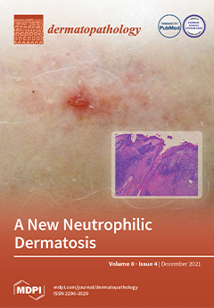
Erosive pustular dermatosis of the scalp (EPDS) is an enigmatic condition, and its diagnosis depends mainly on the recognition of the evolving clinical features, supported by histopathology. Moreover, the histopathologic changes depend on the examined lesion, the disease stage, and the biopsy site. In this retrospective study, we gain more insight into the disease through the review of the epidemiological, clinical, histopathological, therapeutic, and follow-up data of 50 EPDS cases. A continuing, intraepidermal/intrafollicular spongiform pustule formation with a chronically impaired, wound healing process gradually leads to scarring alopecia, as clearly evidenced in the histopathological sections. The clinicopathologic similarities with other neutrophilic dermatoses suggest this condition should be included in this spectrum, where pathergy plays a pathogenetic role.View this paper
  • Issues are regarded as officially published after their release is announced to the table of contents alert mailing list.
  • You may sign up for e-mail alerts to receive table of contents of newly released issues.
  • PDF is the official format for papers published in both, html and pdf forms. To view the papers in pdf format, click on the "PDF Full-text" link, and use the free Adobe Reader to open them.
Order results
Result details
Section
Select all
Export citation of selected articles as:
13 pages, 2176 KiB  
Article
The “Virtual Biopsy” of Cancerous Lesions in 3D: Non-Invasive Differentiation between Melanoma and Other Lesions Using Vibrational Optical Coherence Tomography
by Frederick H. Silver, Tanmay Deshmukh, Nikita Kelkar, Kelly Ritter, Nicole Ryan and Hari Nadiminti
Dermatopathology 2021, 8(4), 539-551; https://doi.org/10.3390/dermatopathology8040058 - 13 Dec 2021
Cited by 3 | Viewed by 3060
Abstract
Early detection of skin cancer is of critical importance to provide five year survival rates that approach 99%. By 2050, one out of five Americans by age 70 will develop some form of skin cancer. This will result in a projected rate of [...] Read more.
Early detection of skin cancer is of critical importance to provide five year survival rates that approach 99%. By 2050, one out of five Americans by age 70 will develop some form of skin cancer. This will result in a projected rate of 50 million skin biopsies per year given the current rate of escalation. In addition, the ability to differentiate between pigmented lesions and melanomas has proven a diagnostic challenge. While dermoscopy and visual analysis are useful in identifying many skin lesions, additional non-invasive techniques are needed to assist in the analysis of difficult to diagnose skin tumors. To augment dermoscopy data, we have developed 3D maps based on physical biomarker characteristics of benign and cancerous lesions using vibrational optical coherence tomography (VOCT). 3D images based on quantitative physical data involving changes in cellular and fibrous tissue stiffness along with changes in vascular quality are used to map and evaluate different types of cancers. 3D tumor maps constructed using quantitative VOCT data and OCT images have been used to characterize the differences between melanoma and other lesions. These characteristics can be used to plan the excision of difficult lesions where extensive surgery may be needed to remove the entire tumor in one step. In addition, it is now possible to use dermoscopy and VOCT to non-invasively differentiate between different cancerous lesion types using measurements of the resonant frequency of new cellular and vascular peaks. Quantitative VOCT information along with dermoscopic findings can be collected and analyzed remotely using artificial intelligence to improve cancerous tissue diagnosis. Full article
Show Figures

Figure 1

4 pages, 956 KiB  
Case Report
An Ecchymosis with Fulminant Evolution
by Andrea Michelerio, Stefania Barruscotti, Nathalie Rizzo and Carlo Tomasini
Dermatopathology 2021, 8(4), 535-538; https://doi.org/10.3390/dermatopathology8040057 - 11 Dec 2021
Viewed by 2613
Abstract
We present the case of an 83-year-old woman who presented with an advanced cutaneous angiosarcoma of the head. The tumor had first appeared as a small ecchymosis on her forehead 3 months before admission. There was an extremely rapid and dramatic evolution, as [...] Read more.
We present the case of an 83-year-old woman who presented with an advanced cutaneous angiosarcoma of the head. The tumor had first appeared as a small ecchymosis on her forehead 3 months before admission. There was an extremely rapid and dramatic evolution, as evidenced by photographic documentation by her relatives. Unfortunately, the delay in access to the healthcare system due to the COVID-19 pandemic lockdown and the fulminant growth were the main determinants for our patient outcome. Full article
(This article belongs to the Section Clinico-Pathological Correlation in Dermatopathology)
Show Figures

Figure 1

4 pages, 196 KiB  
Editorial
New Insights in Paediatric Dermatopathology
by Sylvie Fraitag
Dermatopathology 2021, 8(4), 531-534; https://doi.org/10.3390/dermatopathology8040056 - 07 Dec 2021
Viewed by 2408
Abstract
Paediatric dermatology is an expanding subspeciality [...] Full article
(This article belongs to the Special Issue New Insights in Pediatric Dermatopathology)
6 pages, 5640 KiB  
Article
Spitz Nevus with Features of Clark Nevus, So-Called SPARK Nevus: Case Series Presentation with Emphasis on Cytological and Histological Features
by Antonietta Cimmino, Gerardo Cazzato, Anna Colagrande, Eugenio Maiorano, Lucia Lospalluti, Giuseppe Ingravallo and Leonardo Resta
Dermatopathology 2021, 8(4), 525-530; https://doi.org/10.3390/dermatopathology8040055 - 01 Dec 2021
Cited by 5 | Viewed by 5654
Abstract
Background: SPARK nevus represents a little-known and characterized entity, with few case series available in the literature. Methods and results: we present a case series of 12 patients (6 F and 6 M) between January 2005 and December 2020 and conduct [...] Read more.
Background: SPARK nevus represents a little-known and characterized entity, with few case series available in the literature. Methods and results: we present a case series of 12 patients (6 F and 6 M) between January 2005 and December 2020 and conduct a review of the current literature. Ten articles were selected on the basis of the adopted inclusion criteria and the PRISMA guidelines. Conclusions: The definition of histopathological and dermoscopic criteria are important to allow for an agreement to be reached among dermopathologists, and for the development of a consensus on higher case studies. To our knowledge, there are not many case series in the literature, and ours is part of the attempt to increase the knowledge of an entity that remains little-known and characterized. Full article
(This article belongs to the Special Issue In Memory of Raffaele Gianotti)
Show Figures

Figure 1

10 pages, 3689 KiB  
Article
Primary Cutaneous Gamma-Delta T Cell Lymphomas: A Case Series and Overview of the Literature
by Silvia Alberti-Violetti, Carlo Alberto Maronese, Luigia Venegoni, Valentina Merlo and Emilio Berti
Dermatopathology 2021, 8(4), 515-524; https://doi.org/10.3390/dermatopathology8040054 - 17 Nov 2021
Cited by 9 | Viewed by 5008
Abstract
Primary cutaneous gamma-delta T cell lymphomas (PCGDTCLs) are rare and aggressive cutaneous malignancies that have been diagnostically challenging for dermopathologists and clinicians since their first published descriptions in 1991. Since then, the availability of immunostaining for T cell receptors γ and δ in [...] Read more.
Primary cutaneous gamma-delta T cell lymphomas (PCGDTCLs) are rare and aggressive cutaneous malignancies that have been diagnostically challenging for dermopathologists and clinicians since their first published descriptions in 1991. Since then, the availability of immunostaining for T cell receptors γ and δ in formalin-fixed paraffin-embedded samples has greatly increased our knowledge of the gamma-delta phenotype by showing that it may also be present in the context of indolent entities, such as mycosis fungoides (MFs) and lymphomatoid papulosis, and this has raised questions concerning its diagnostic and prognostic implications. We here describe the histological and clinical differences between the dermo-epidermal and subcutaneous sub-groups of PCGDTCL observed in a cohort of 20 patients attending a single experienced centre, with particular focus on cases with an MF-like presentation, which are still less well defined than those of classic MF. Full article
(This article belongs to the Special Issue In Memory of Raffaele Gianotti)
Show Figures

Figure 1

6 pages, 22025 KiB  
Case Report
Skin Mycetoma in an 11-Year-Old African Boy: Case Presentation with Emphasis on Histopathological Features and Differential Diagnosis
by Gerardo Cazzato, Anna Colagrande, Antonietta Cimmino, Lucia Lospalluti, Aurora Demarco, Caterina Foti, Paolo Romita, Francesca Arezzo, Vera Loizzi, Paola Parente, Leonardo Resta and Giuseppe Ingravallo
Dermatopathology 2021, 8(4), 509-514; https://doi.org/10.3390/dermatopathology8040053 - 26 Oct 2021
Cited by 1 | Viewed by 4119
Abstract
Mycetoma is an uncommon, chronic infective disease of the skin and subcutaneous tissues, characterized by the triad of tumefaction, draining sinuses, and the presence in the exudate of colonial grains. In cases of long-term disease, the presence of colonial grains together with the [...] Read more.
Mycetoma is an uncommon, chronic infective disease of the skin and subcutaneous tissues, characterized by the triad of tumefaction, draining sinuses, and the presence in the exudate of colonial grains. In cases of long-term disease, the presence of colonial grains together with the host’s derivative material can lead to the formation of real sinuses. Histological analysis is of fundamental importance to allow an accurate etiological diagnosis and to understand if the basic pathogen is an actinomycete (bacterium) or a real fungus (eumycetic mycetomas) and is also fundamental for therapy, which is quite different. Here, we present a case of Mycetoma in an 11-year-old patient who emigrated from Djibouti, Somalia, and showed the essential histopathological features of this rare and forgotten nosographic entity in the industrialized world and briefly discuss the major and most important differential diagnoses. Full article
Show Figures

Figure 1

7 pages, 7663 KiB  
Case Report
A Rare Case of Transient Acantholytic Dermatosis (AKA. Grover’s Disease) with Concomitant Pediculosis Pubis: An Atypical Presentation and First Documented Case Report
by Gehan A. Pendlebury, Peter Oro, Drew Merideth and Eric Rudnick
Dermatopathology 2021, 8(4), 502-508; https://doi.org/10.3390/dermatopathology8040052 - 22 Oct 2021
Cited by 1 | Viewed by 5296
Abstract
A 66-year-old male presented with a one-month history of persistent pruritic eruptions distributed mainly on the trunk. A punch biopsy from the left upper abdomen revealed focal acantholytic dyskeratosis with mixed inflammatory infiltrate in the dermis composed of numerous eosinophils. Grover’s disease was [...] Read more.
A 66-year-old male presented with a one-month history of persistent pruritic eruptions distributed mainly on the trunk. A punch biopsy from the left upper abdomen revealed focal acantholytic dyskeratosis with mixed inflammatory infiltrate in the dermis composed of numerous eosinophils. Grover’s disease was diagnosed based on the clinical and histopathological findings. Appropriate treatment was initiated but failed to relieve symptoms of itchiness. A further investigation of the hair follicles under mineral oil preparation revealed an infestation of pediculosis pubis. Subsequent treatment with Ivermectin and permethrin cream led to the complete resolution of his symptoms. This case report highlights an unusual and first documented case of Grover’s disease with a concomitant infestation of pediculosis pubis. To date, no reported cases in the literature have associated Grover’s disease with pubic lice infestation. However, there are three reported cases of concurrent scabies and Grover’s disease in the literature. This rare case underscores the clinical value in further investigating treatable underlying conditions in patients with suspected transient acantholytic dermatosis. Full article
Show Figures

Figure 1

8 pages, 4827 KiB  
Review
Dedifferentiated Melanoma: A Diagnostic Histological Pitfall—Review of the Literature with Case Presentation
by Gerardo Cazzato, Lucia Lospalluti, Anna Colagrande, Antonietta Cimmino, Paolo Romita, Caterina Foti, Aurora Demarco, Francesca Arezzo, Vera Loizzi, Gennaro Cormio, Sara Sablone, Leonardo Resta, Roberta Rossi and Giuseppe Ingravallo
Dermatopathology 2021, 8(4), 494-501; https://doi.org/10.3390/dermatopathology8040051 - 15 Oct 2021
Cited by 8 | Viewed by 2925
Abstract
Dedifferentiated melanoma is a particular form of malignant melanoma with a progressive worsening of the patient’s clinical outcome. It is well known that melanoma can assume different histo-morphological patterns, to which specific genetic signatures correspond, sometimes but not always. In this review we [...] Read more.
Dedifferentiated melanoma is a particular form of malignant melanoma with a progressive worsening of the patient’s clinical outcome. It is well known that melanoma can assume different histo-morphological patterns, to which specific genetic signatures correspond, sometimes but not always. In this review we address the diagnostic difficulties in correctly recognizing this entity, discuss the major differential diagnoses of interest to the dermatopathologist, and conduct a review of the literature with particular attention and emphasis on the latest molecular discoveries regarding the dedifferentiation/undifferentiation mechanism and more advanced therapeutic approaches. Full article
(This article belongs to the Section Clinico-Pathological Correlation in Dermatopathology)
Show Figures

Figure 1

17 pages, 10393 KiB  
Review
Venous Malformations in Childhood: Clinical, Histopathological and Genetics Update
by Isabel Colmenero and Nicole Knöpfel
Dermatopathology 2021, 8(4), 477-493; https://doi.org/10.3390/dermatopathology8040050 - 15 Oct 2021
Cited by 10 | Viewed by 8059
Abstract
Our knowledge in vascular anomalies has grown tremendously in the past decade with the identification of key molecular pathways and genetic mutations that drive the development of vascular tumors and vascular malformations. This has led us to better understand the pathogenesis of vascular [...] Read more.
Our knowledge in vascular anomalies has grown tremendously in the past decade with the identification of key molecular pathways and genetic mutations that drive the development of vascular tumors and vascular malformations. This has led us to better understand the pathogenesis of vascular lesions, refine their diagnosis and update their classification while also exploring the opportunity for a targeted molecular treatment. This paper aims to provide an overview of venous malformations (VM) in childhood. Specific entities include common VMs, cutaneo-mucosal VM, blue rubber bleb nevus syndrome or Bean syndrome, glomuvenous malformation, cerebral cavernous malformation, familial intraosseous vascular malformation and verrucous venous malformation. The clinicopathological features and the molecular basis of each entity are reviewed. Full article
(This article belongs to the Special Issue New Insights in Pediatric Dermatopathology)
Show Figures

Figure 1

14 pages, 93415 KiB  
Article
Single-Center Clinico-Pathological Case Study of 19 Patients with Cutaneous Adverse Reactions Following COVID-19 Vaccines
by Dennis Niebel, Joerg Wenzel, Dagmar Wilsmann-Theis, Jana Ziob, Jasmin Wilhelmi and Christine Braegelmann
Dermatopathology 2021, 8(4), 463-476; https://doi.org/10.3390/dermatopathology8040049 - 27 Sep 2021
Cited by 23 | Viewed by 5083
Abstract
(1) Background: Coronavirus disease 2019 (COVID-19) vaccines are currently employed on a population-wide scale in most countries worldwide. Data about unusual cutaneous adverse drug reactions (ADR) are scant, though. (2) Methods: We retrospectively analyzed moderate to severe vaccine-related ADR in the Department of [...] Read more.
(1) Background: Coronavirus disease 2019 (COVID-19) vaccines are currently employed on a population-wide scale in most countries worldwide. Data about unusual cutaneous adverse drug reactions (ADR) are scant, though. (2) Methods: We retrospectively analyzed moderate to severe vaccine-related ADR in the Department of Dermatology and Allergy of the University Hospital Bonn between May to June 2021 and analyzed related skin biopsies. (3) Results: As a specialized dermatological academic center, we encountered a total of n = 19 clinically and pathologically heterogeneous cutaneous ADR with a female predominance. Delayed cutaneous ADR occurred as late as 30 days after vaccination. The majority of ADR were mild, though a few patients required systemic treatment (antihistamines, glucocorticosteroids). (4) Conclusions: The clinico-pathological spectrum of cutaneous side effects with COVID-19 vaccines is wide; however, the benefits outweigh the risks by far. More dermatopathological studies on cutaneous ADR not limited to COVID-19 vaccines are desirable to enable a better understanding of underlying pathophysiological mechanisms. Full article
(This article belongs to the Special Issue Dermatopathology and COVID-19)
Show Figures

Figure 1

13 pages, 8580 KiB  
Article
Erosive Pustular Dermatosis of the Scalp: A Clinicopathologic Study of Fifty Cases
by Andrea Michelerio, Camilla Vassallo, Giacomo Fiandrino and Carlo Francesco Tomasini
Dermatopathology 2021, 8(4), 450-462; https://doi.org/10.3390/dermatopathology8040048 - 23 Sep 2021
Cited by 4 | Viewed by 7041
Abstract
Erosive pustular dermatosis of the scalp (EPDS) is an uncommon, pustular, idiopathic disorder typically occurring on the scalp of the elderly, whose diagnosis requires close clinicopathologic correlations. Recently, the primary histopathologic characteristic of EPDS has been identified in some biopsies from hair-bearing scalp [...] Read more.
Erosive pustular dermatosis of the scalp (EPDS) is an uncommon, pustular, idiopathic disorder typically occurring on the scalp of the elderly, whose diagnosis requires close clinicopathologic correlations. Recently, the primary histopathologic characteristic of EPDS has been identified in some biopsies from hair-bearing scalp lesions as a sterile, vesiculo-pustule involving the infundibulum of hair follicles. To further delineate the clinicopathologic spectrum of the disease, we led a retrospective study of 50 patients (36 males and 14 females) with a diagnosis of EPDS between 2011 and 2021, reviewing clinical and histopathological data. Androgenetic alopecia was present in 32 patients. Triggering factors were present in 21 patients. The vertex was the most common location; one patient also had leg involvement. Two cases were familial. Disease presentation varied markedly from tiny, erosive, scaly lesions to crusted and hemorrhagic plaques, mimicking pustular pyoderma gangrenosum (PPG). Biopsies of patients with severe androgenetic or total baldness produced specimens showing nonspecific pathologic changes (39/50), while in 11 patients with a hair-bearing scalp histopathologic examination, changes were specific. The clinicopathologic similarities between EPDS and PPG suggest that EPDS should be included in the spectrum of autoinflammatory dermatoses. Clinicians could consider the possibility of associated disorders rather than managing EPDS as a sui generis skin disorder. Full article
(This article belongs to the Special Issue In Memory of Raffaele Gianotti)
Show Figures

Figure 1

Previous Issue
Next Issue
Back to TopTop