Next Issue
Volume 8, December
Previous Issue
Volume 8, June


Dermatopathology, Volume 8, Issue 3 (September 2021) – 20 articles

Cover Story (view full-size image): 
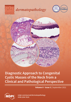
This review presents the clinical, pathological, and radiological features of the most common congenital cystic lesions of the neck, emphasizing their embryologic origin, the differential diagnosis, and the possible association with chromosomal defects. These lesions vary in prevalence from common (thyroglossal duct cysts, branchial cleft cysts, and lymphangioma) to very rare (thymic cysts and cervical bronchogenic cysts).The age of the child (infant or child) and the localization of the mass (median, lateral, or parotid) are extremely important elements of orientation. Most of the time, clinical examination and US are sufficient for clear identification and correct treatment of the cervical cyst. Large predominance of benign cystic lesions does not rule out the possibility of any rare malignancies associated with a cystic presentation. View this paper.
  • Issues are regarded as officially published after their release is announced to the table of contents alert mailing list.
  • You may sign up for e-mail alerts to receive table of contents of newly released issues.
  • PDF is the official format for papers published in both, html and pdf forms. To view the papers in pdf format, click on the "PDF Full-text" link, and use the free Adobe Reader to open them.
Order results
Result details
Section
Select all
Export citation of selected articles as:
4 pages, 1795 KiB  
Case Report
Eccrine Nevus in the Forearm of a 16-Year-Old Presenting as Unilateral Hyperhidrosis: A Clinicopathological Correlation Paradigm
by Alejandro Martin-Gorgojo, Ignacio Sanchez-Carpintero, Ricardo Ruiz-Rodriguez and Ana-Belen Enguita-Valls
Dermatopathology 2021, 8(3), 446-449; https://doi.org/10.3390/dermatopathology8030047 - 18 Sep 2021
Cited by 2 | Viewed by 4395
Abstract
A case of a purely eccrine nevus in an adolescent patient presenting with focal hyperhidrosis on an area comprising the left forearm and the dorsal aspect of the left hand is described. No clinically evident lesions were identifiable. Dermatopathologic findings were subtle, showing [...] Read more.
A case of a purely eccrine nevus in an adolescent patient presenting with focal hyperhidrosis on an area comprising the left forearm and the dorsal aspect of the left hand is described. No clinically evident lesions were identifiable. Dermatopathologic findings were subtle, showing only a slight increase in the number of eccrine glands. Clinicopathological correlation was paramount to achieve the diagnosis. Full article
(This article belongs to the Section Clinico-Pathological Correlation in Dermatopathology)
Show Figures

Figure 1

4 pages, 527 KiB  
Case Report
A New Case of Hybrid Epidermoid and Apocrine Cyst
by Fulvia Serra and Gürkan Kaya
Dermatopathology 2021, 8(3), 442-445; https://doi.org/10.3390/dermatopathology8030046 - 01 Sep 2021
Cited by 1 | Viewed by 2714
Abstract
We described a new case of a hybrid epidermoid and apocrine cyst, known to be a rare histopathological entity. The cyst was located in the axillary region and completely excised, without complication. The diagnosis was made at the histological analysis, where we found [...] Read more.
We described a new case of a hybrid epidermoid and apocrine cyst, known to be a rare histopathological entity. The cyst was located in the axillary region and completely excised, without complication. The diagnosis was made at the histological analysis, where we found a cystic lesion in the dermis, lined with both epidermoid and apocrine epithelium. Full article
Show Figures

Figure 1

16 pages, 3195 KiB  
Article
Immunohistopathological Analysis of Immunoglobulin E-Positive Epidermal Dendritic Cells with House Dust Mite Antigens in Naturally Occurring Skin Lesions of Adult and Elderly Patients with Atopic Dermatitis
by Ryoji Tanei and Yasuko Hasegawa
Dermatopathology 2021, 8(3), 426-441; https://doi.org/10.3390/dermatopathology8030045 - 01 Sep 2021
Cited by 3 | Viewed by 3236
Abstract
The immunopathogenic role of house dust mite (HDM) allergens in the development of skin lesions in atopic dermatitis (AD) has not yet been precisely clarified. We immunohistopathologically evaluated the localization of immunoglobulin E (IgE)-positive epidermal dendritic cells with HDM antigens in the skin [...] Read more.
The immunopathogenic role of house dust mite (HDM) allergens in the development of skin lesions in atopic dermatitis (AD) has not yet been precisely clarified. We immunohistopathologically evaluated the localization of immunoglobulin E (IgE)-positive epidermal dendritic cells with HDM antigens in the skin lesions of patients with IgE-allergic AD. Using double-immunofluorescence and single-immunochemical staining methods, we analyzed biopsy specimens from the skin lesions of six patients with IgE-allergic AD and HDM allergy and 11 control subjects with inflammatory skin disorders. Inflammatory dendritic epidermal cells (IDECs; CD11c+ and CD206+ cells) were markedly observed in the central area of the spongiotic epidermis of skin lesions in all AD patients. Furthermore, IgE-positive IDECs with HDM antigens in the central areas of the spongiosis were found in four of the six (66.7%) AD patients. Langerhans cells (LCs; CD207+ cells) with HDM antigens were also observed in the peripheral areas of the spongiosis. Infiltration of CD4+ and CD8+ T cells in association with IgE-positive IDECs and LCs with HDM antigens was seen in the spongiotic epidermis. An IgE-mediated delayed-type hypersensitivity reaction, in combination with IgE-bearing dendritic cells, specific T cells, keratinocytes, and HDM antigens, may lead to spongiotic tissue formation in eczematous dermatitis in AD. Full article
Show Figures

Figure 1

8 pages, 1172 KiB  
Review
Artificial Intelligence in Dermatopathology: New Insights and Perspectives
by Gerardo Cazzato, Anna Colagrande, Antonietta Cimmino, Francesca Arezzo, Vera Loizzi, Concetta Caporusso, Marco Marangio, Caterina Foti, Paolo Romita, Lucia Lospalluti, Francesco Mazzotta, Sebastiano Cicco, Gennaro Cormio, Teresa Lettini, Leonardo Resta, Angelo Vacca and Giuseppe Ingravallo
Dermatopathology 2021, 8(3), 418-425; https://doi.org/10.3390/dermatopathology8030044 - 01 Sep 2021
Cited by 18 | Viewed by 4148
Abstract
In recent years, an increasing enthusiasm has been observed towards artificial intelligence and machine learning, involving different areas of medicine. Among these, although still in the embryonic stage, the dermatopathological field has also been partially involved, with the attempt to develop and train [...] Read more.
In recent years, an increasing enthusiasm has been observed towards artificial intelligence and machine learning, involving different areas of medicine. Among these, although still in the embryonic stage, the dermatopathological field has also been partially involved, with the attempt to develop and train algorithms that could assist the pathologist in the differential diagnosis of complex melanocytic lesions. In this article, we face this new challenge of the modern era, carry out a review of the literature regarding the state of the art and try to determine promising future perspectives. Full article
(This article belongs to the Section Artificial Intelligence in Dermatopathology)
Show Figures

Figure 1

28 pages, 66904 KiB  
Article
What to Look Out for in a Newborn with Multiple Papulonodular Skin Lesions at Birth
by Sylvie Fraitag and Olivia Boccara
Dermatopathology 2021, 8(3), 390-417; https://doi.org/10.3390/dermatopathology8030043 - 17 Aug 2021
Cited by 4 | Viewed by 6922
Abstract
Multiple papulonodular skin lesions at birth can indicate the presence of various benign and malignant disorders. Although the lesions’ clinical aspect (color and consistency, in particular) may steer the clinician towards one disorder or another (infantile myofibromatosis, xanthogranuloma, or metastatic neuroblastoma), the diagnosis [...] Read more.
Multiple papulonodular skin lesions at birth can indicate the presence of various benign and malignant disorders. Although the lesions’ clinical aspect (color and consistency, in particular) may steer the clinician towards one disorder or another (infantile myofibromatosis, xanthogranuloma, or metastatic neuroblastoma), the diagnosis can only be confirmed by the histopathologic assessment of a biopsy. In neonates, a rapid but accurate diagnosis is critical because skin lesions may be the first manifestation of a malignant disorder like leukemia cutis or metastatic neuroblastoma. Here, we review the various disorders that may manifest themselves as multiple skin lesions at birth. Full article
(This article belongs to the Special Issue New Insights in Pediatric Dermatopathology)
Show Figures

Figure 1

14 pages, 6906 KiB  
Review
Pseudomalignancies in Children: Histological Clues, and Pitfalls to Be Avoided
by Sébastien Menzinger and Sylvie Fraitag
Dermatopathology 2021, 8(3), 376-389; https://doi.org/10.3390/dermatopathology8030042 - 14 Aug 2021
Cited by 1 | Viewed by 3913
Abstract
The term “pseudomalignancy” covers a large, heterogenous group of diseases characterized by a benign cellular proliferation, hyperplasia, or infiltrate that resembles a true malignancy clinically or histologically. Here, we (i) provide a non-exhaustive review of several inflammatory skin diseases and benign skin proliferations [...] Read more.
The term “pseudomalignancy” covers a large, heterogenous group of diseases characterized by a benign cellular proliferation, hyperplasia, or infiltrate that resembles a true malignancy clinically or histologically. Here, we (i) provide a non-exhaustive review of several inflammatory skin diseases and benign skin proliferations that can mimic a malignant neoplasm in children, (ii) give pathologists some helpful clues to guide their diagnosis, and (iii) highlight pitfalls to be avoided. The observation of clinical–pathological correlations is often important in this situation and can sometimes be the only means (along with careful monitoring of the disease’s clinical course) of reaching a firm diagnosis. Full article
(This article belongs to the Special Issue New Insights in Pediatric Dermatopathology)
Show Figures

Figure 1

5 pages, 888 KiB  
Communication
Granular Cell Dermatofibroma: When Morphology Still Matters
by Gerardo Cazzato, Anna Colagrande, Antonietta Cimmino, Maricla Marrone, Alessandra Stellacci, Francesca Arezzo, Teresa Lettini, Leonardo Resta and Giuseppe Ingravallo
Dermatopathology 2021, 8(3), 371-375; https://doi.org/10.3390/dermatopathology8030041 - 13 Aug 2021
Cited by 8 | Viewed by 3687
Abstract
Dermatofibroma, also known as “fibrous histiocytoma”, is one of the most common cutaneous soft-tissue tumors. Many variants of dermatofibromas have been described, and knowledge of these variations is important to avoid misdiagnosis of a possibly more aggressive tumor. Histological features of different variants [...] Read more.
Dermatofibroma, also known as “fibrous histiocytoma”, is one of the most common cutaneous soft-tissue tumors. Many variants of dermatofibromas have been described, and knowledge of these variations is important to avoid misdiagnosis of a possibly more aggressive tumor. Histological features of different variants can coexist in the same lesion, but typical common fibrous histiocytoma features are generally found, at least focally, in all cases. However, when cellular changes make up the majority of the lesion, the histopathological diagnosis can become more complex and requires immunohistochemical investigations for correct nosographic classification. We present the case of a cutaneous fibrous histiocytoma, “granular cell” variant, found on the left leg of a 74-year-old woman. Full article
Show Figures

Figure 1

12 pages, 7084 KiB  
Review
Aberrant Expression of Immunohistochemical Markers in Malignant Melanoma: A Review
by Elie Saliba and Jag Bhawan
Dermatopathology 2021, 8(3), 359-370; https://doi.org/10.3390/dermatopathology8030040 - 03 Aug 2021
Cited by 19 | Viewed by 8464
Abstract
Immunohistochemical stains are increasingly used to aid in the diagnosis of malignant melanoma, especially when the differentiation of the tumor is unclear based on examination with hematoxylin and eosin. However, aberrant expression of non-melanocytic markers has been reported in melanomas, which can sometimes [...] Read more.
Immunohistochemical stains are increasingly used to aid in the diagnosis of malignant melanoma, especially when the differentiation of the tumor is unclear based on examination with hematoxylin and eosin. However, aberrant expression of non-melanocytic markers has been reported in melanomas, which can sometimes be further complicated by the loss of conventional melanocytic markers. This review aims to summarize available data regarding unusual staining patterns in primary and metastatic malignant melanoma. It also raises awareness of the potential pitfalls and highlights the importance of appropriate use and interpretation of broad immunohistochemical markers in the context of clinical and histopathologic findings to facilitate the diagnosis of atypical cases of malignant melanoma. Full article
Show Figures

Figure 1

17 pages, 5447 KiB  
Review
Diagnostic Approach to Congenital Cystic Masses of the Neck from a Clinical and Pathological Perspective
by Amanda Fanous, Guillaume Morcrette, Monique Fabre, Vincent Couloigner and Louise Galmiche-Rolland
Dermatopathology 2021, 8(3), 342-358; https://doi.org/10.3390/dermatopathology8030039 - 01 Aug 2021
Cited by 12 | Viewed by 8587
Abstract
Background: neck cysts are frequently encountered in pediatric medicine and can present a diagnostic dilemma for clinicians and pathologists. Several clinical items enable to subclassify neck cyst as age at presentation, anatomical location, including compartments and fascia of the neck, and radiological presentation. [...] Read more.
Background: neck cysts are frequently encountered in pediatric medicine and can present a diagnostic dilemma for clinicians and pathologists. Several clinical items enable to subclassify neck cyst as age at presentation, anatomical location, including compartments and fascia of the neck, and radiological presentation. Summary: this review will briefly describe the clinical, imaging, pathological and management features of (I) congenital and developmental pathologies, including thyroglossal duct cyst, branchial cleft cysts, dermoid cyst, thymic cyst, and ectopic thymus; (II) vascular malformations, including lymphangioma. Key Messages: pathologists should be familiar with the diagnostic features and clinicopathologic entities of these neck lesions in order to correctly diagnose them and to provide proper clinical management. Full article
(This article belongs to the Special Issue New Insights in Pediatric Dermatopathology)
Show Figures

Figure 1

5 pages, 2859 KiB  
Case Report
Plaque-Like Dermatofibroma: Case Report of a Rare Entity
by Sara Moradi, Laila Mnayer, Jonathan Earle, Alex C. Cech and Torsten Ehrig
Dermatopathology 2021, 8(3), 337-341; https://doi.org/10.3390/dermatopathology8030038 - 01 Aug 2021
Cited by 2 | Viewed by 3095
Abstract
A case of a well-demarcated plaque measuring 11 cm without satellites of several years’ duration is presented. It showed typical histologic findings of dermatofibroma, prompting a diagnosis of plaque-like dermatofibroma. The relationship to multiple clustered dermatofibromas and plaque-like myofibroblastic tumor is discussed. Full article
Show Figures

Figure 1

22 pages, 4989 KiB  
Review
Panniculitis in Children
by Isabelle Moulonguet and Sylvie Fraitag
Dermatopathology 2021, 8(3), 315-336; https://doi.org/10.3390/dermatopathology8030037 - 01 Aug 2021
Cited by 8 | Viewed by 7297
Abstract
Panniculitides form a heterogenous group of inflammatory diseases that involve the subcutaneous adipose tissue. These disorders are rare in children and have many aetiologies. As in adults, the panniculitis can be the primary process in a systemic disorder or a secondary process that [...] Read more.
Panniculitides form a heterogenous group of inflammatory diseases that involve the subcutaneous adipose tissue. These disorders are rare in children and have many aetiologies. As in adults, the panniculitis can be the primary process in a systemic disorder or a secondary process that results from infection, trauma or exposure to medication. Some types of panniculitis are seen more commonly or exclusively in children, and several new entities have been described in recent years. Most types of panniculitis have the same clinical presentation (regardless of the aetiology), with tender, erythematous subcutaneous nodules. Although the patient’s age and the lesion site provide information, a histopathological assessment is sometimes required for a definitive diagnosis and classification of the disorder. In children, most panniculitides are lobular. At present, autoimmune inflammatory diseases and primary immunodeficiencies have been better characterised; panniculitis can be the presenting symptom in some of these settings. Unexplained panniculitis in a young child should prompt a detailed screen for monogenic immune disorders because the latter usually manifest themselves early in life. Here, we review forms of panniculitis that occur primarily in children, with a focus on newly described entities. Full article
(This article belongs to the Special Issue New Insights in Pediatric Dermatopathology)
Show Figures

Figure 1

14 pages, 5599 KiB  
Review
Cutaneous Melanomas Arising during Childhood: An Overview of the Main Entities
by Arnaud de la Fouchardière, Felix Boivin, Heather C. Etchevers and Nicolas Macagno
Dermatopathology 2021, 8(3), 301-314; https://doi.org/10.3390/dermatopathology8030036 - 01 Aug 2021
Cited by 2 | Viewed by 2951
Abstract
Cutaneous melanomas are exceptional in children and represent a variety of clinical situations, each with a different prognosis. In congenital nevi, the risk of transformation is correlated with the size of the nevus. The most frequent type is lateral transformation, extremely rare before [...] Read more.
Cutaneous melanomas are exceptional in children and represent a variety of clinical situations, each with a different prognosis. In congenital nevi, the risk of transformation is correlated with the size of the nevus. The most frequent type is lateral transformation, extremely rare before puberty, reminiscent of a superficial spreading melanoma (SSM) ex-nevus. Deep nodular transformation is much rarer, can occur before puberty, and must be distinguished from benign proliferative nodules. Superficial spreading melanoma can also arise within small nevi, which were not visible at birth, usually after puberty, and can reveal a cancer predisposition syndrome (CDKN2A or CDK4 germline mutations). Prognosis is correlated with classical histoprognostic features (mainly Breslow thickness). Spitz tumors are frequent in adolescents and encompass benign (Spitz nevus), intermediate (atypical Spitz tumor), and malignant forms (malignant Spitz tumor). The whole spectrum is characterized by specific morphology with spindled and epithelioid cells, genetic features, and an overall favorable outcome even if a regional lymph node is involved. Nevoid melanomas are rare and difficult to diagnose clinically and histologically. They can arise in late adolescence. Their prognosis is currently not very well ascertained. A small group of melanomas remains unclassified after histological and molecular assessment. Full article
(This article belongs to the Special Issue New Insights in Pediatric Dermatopathology)
Show Figures

Figure 1

16 pages, 152907 KiB  
Review
Update on Superficial Spindle Cell Mesenchymal Tumors in Children
by Philippe Drabent and Sylvie Fraitag
Dermatopathology 2021, 8(3), 285-300; https://doi.org/10.3390/dermatopathology8030035 - 21 Jul 2021
Cited by 5 | Viewed by 7446
Abstract
The diagnosis of cutaneous and subcutaneous spindle cell neoplasms in children is often challenging and has potential therapeutic and prognostic implications. Although correctly diagnosing dermatofibrosarcoma protuberans and infantile fibrosarcoma is paramount, pathologists should not ignore a number of diagnostic pitfalls linked to mostly [...] Read more.
The diagnosis of cutaneous and subcutaneous spindle cell neoplasms in children is often challenging and has potential therapeutic and prognostic implications. Although correctly diagnosing dermatofibrosarcoma protuberans and infantile fibrosarcoma is paramount, pathologists should not ignore a number of diagnostic pitfalls linked to mostly rare tumors with completely different clinical outcomes. In the last decade, a spectrum of novel entities has been described; information from molecular biology has helped to shape this new landscape for spindle cell tumors. Here, we review the most noteworthy neoplasms in this spectrum, with a focus on their histological similarities: fibroblastic connective tissue nevus, medallion-like dermal dendrocyte hamartoma, or plaque-like CD34-positive dermal fibroma, which share features with fibrous hamartoma of infancy; lipofibromatosis and lipofibromatosis-like neural tumor; and plexiform myofibroblastoma, a recently described neoplasm that should be distinguished from plexiform fibrohistiocytic tumor. These tumors also have genetic similarities, particularly gene rearrangements involving NTRK3 or NTRK1. These genetic features are not only essential for the differential diagnosis of infantile fibrosarcoma but are also of diagnostic value for lipofibromatosis-like neural tumors. The more recently described RET, RAF1, and BRAF gene fusions are also discussed. Full article
(This article belongs to the Special Issue New Insights in Pediatric Dermatopathology)
Show Figures

Figure 1

8 pages, 1719 KiB  
Article
Evaluation of Melanocyte Loss in Mycosis Fungoides Using SOX10 Immunohistochemistry
by Cynthia Reyes Barron and Bruce R. Smoller
Dermatopathology 2021, 8(3), 277-284; https://doi.org/10.3390/dermatopathology8030034 - 08 Jul 2021
Cited by 1 | Viewed by 3120
Abstract
Mycosis fungoides (MF) is a subtype of primary cutaneous T-cell lymphoma (CTCL) with an indolent course that rarely progresses. Histologically, the lesions display a superficial lymphocytic infiltrate with epidermotropism of neoplastic T-cells. Hypopigmented MF is a rare variant that presents with hypopigmented lesions [...] Read more.
Mycosis fungoides (MF) is a subtype of primary cutaneous T-cell lymphoma (CTCL) with an indolent course that rarely progresses. Histologically, the lesions display a superficial lymphocytic infiltrate with epidermotropism of neoplastic T-cells. Hypopigmented MF is a rare variant that presents with hypopigmented lesions and is more likely to affect young patients. The etiology of the hypopigmentation is unclear. The aim of this study was to assess melanocyte loss in MF through immunohistochemistry (IHC) with SOX10. Twenty cases were evaluated, including seven of the hypopigmented subtype. The neoplastic epidermotropic infiltrate consisted predominantly of CD4+ T-cells in 65% of cases; CD8+ T-cells were present in moderate to abundant numbers in most cases. SOX10 IHC showed a decrease or focal complete loss of melanocytes in 50% of the cases. The predominant neoplastic cell type (CD4+/CD8+), age, race, gender, histologic features, and reported clinical pigmentation of the lesions were not predictive of melanocyte loss. A significant loss of melanocytes was observed in 43% of hypopigmented cases and 54% of conventional cases. Additional studies will increase our understanding of the relationship between observed pigmentation and the loss of melanocytes in MF. Full article
Show Figures

Figure 1

6 pages, 1950 KiB  
Review
“Animal-Type Melanoma/Pigmented Epithelioid Melanocytoma”: History and Features of a Controversial Entity
by Gerardo Cazzato, Francesca Arezzo, Anna Colagrande, Antonietta Cimmino, Teresa Lettini, Sara Sablone, Leonardo Resta and Giuseppe Ingravallo
Dermatopathology 2021, 8(3), 271-276; https://doi.org/10.3390/dermatopathology8030033 - 05 Jul 2021
Cited by 6 | Viewed by 2738
Abstract
Animal-type melanoma (ATM) was first described in the literature by Levene in 1979 in relation to a patient with a characteristic clinical presentation, and only later, rare and anecdotal case series have tried to shed light on an entity that has undergone several [...] Read more.
Animal-type melanoma (ATM) was first described in the literature by Levene in 1979 in relation to a patient with a characteristic clinical presentation, and only later, rare and anecdotal case series have tried to shed light on an entity that has undergone several nosographic classification changes, and which, since 2018, is classified under the term “pigmented epithelioid melanocytoma”. Here, we conduct a brief review of the current literature on ATM and present a new clinical case with histopathological, immunophenotypic, and molecular investigations. Full article
Show Figures

Figure 1

6 pages, 1386 KiB  
Review
The Multiple Faces of Nodular Trichoblastoma: Review of the Literature with Case Presentation
by Gerardo Cazzato, Antonietta Cimmino, Anna Colagrande, Francesca Arezzo, Lucia Lospalluti, Sara Sablone, Teresa Lettini, Leonardo Resta and Giuseppe Ingravallo
Dermatopathology 2021, 8(3), 265-270; https://doi.org/10.3390/dermatopathology8030032 - 05 Jul 2021
Cited by 5 | Viewed by 5143
Abstract
Trichoblastoma (TB) is a rare biphasic benign adnexal neoplasm originating from follicular germ cells but clinically, it can simulate basal cell carcinoma (BCC), making the diagnosis more difficult. There are several variants of Trichoblastoma and a good knowledge of these is essential for [...] Read more.
Trichoblastoma (TB) is a rare biphasic benign adnexal neoplasm originating from follicular germ cells but clinically, it can simulate basal cell carcinoma (BCC), making the diagnosis more difficult. There are several variants of Trichoblastoma and a good knowledge of these is essential for correct diagnosis and management. We report two new cases observed in the last year at our Pathological Anatomy Operative Unit, and conduct a careful review of the literature, from the first description of this lesion by Headington in 1970 to the most recent classifications. Full article
Show Figures

Figure 1

7 pages, 3243 KiB  
Article
GLUT1 Expression in Cutaneous Sebaceous Lesions Determined by Immunohistochemical Staining Patterns
by Cynthia Reyes Barron and Bruce R. Smoller
Dermatopathology 2021, 8(3), 258-264; https://doi.org/10.3390/dermatopathology8030031 - 05 Jul 2021
Viewed by 2547
Abstract
GLUT1 is a membrane associated carrier protein that functions in the physiologic transport of glucose across cell membranes. Multiple studies have shown an increased GLUT1 expression in various tumor types and a role in cancer prognosis. The aim of this study was to [...] Read more.
GLUT1 is a membrane associated carrier protein that functions in the physiologic transport of glucose across cell membranes. Multiple studies have shown an increased GLUT1 expression in various tumor types and a role in cancer prognosis. The aim of this study was to determine whether cutaneous sebaceous lesions have a differential expression of GLUT1 by immunohistochemistry (IHC). GLUT1 IHC was performed on excision specimens of ten cases of sebaceous carcinoma, nine of sebaceoma, ten of sebaceous adenoma, and ten of sebaceous hyperplasia. Intense, diffuse cytoplasmic staining was observed in sebaceous carcinoma. The pattern of GLUT1 staining in sebaceomas and sebaceous adenomas consisted of a gradient of intense cytoplasmic staining in the basaloid cells with a decreased intensity to membranous staining only and absent staining in mature sebaceous cells. In lesions of sebaceous hyperplasia, GLUT1 staining outlined the basal layer of each gland; cytoplasmic staining was minimal to absent. Increased cytoplasmic staining of GLUT1 may correlate with cellular metabolic and proliferative activity. GLUT1 has potential utility in differentiating sebaceous lesions. Full article
Show Figures

Figure 1

5 pages, 10532 KiB  
Case Report
A Wolf in Sheep’s Clothing: Collision of Melanoma and Keratoacanthoma
by Matthias Walther, Sandra Falkvoll and Sebastian Leibl
Dermatopathology 2021, 8(3), 253-257; https://doi.org/10.3390/dermatopathology8030030 - 04 Jul 2021
Viewed by 4387
Abstract
Collision tumors consisting of melanoma and squamous cell carcinoma are very rare. We present the case of a deceptive hyperkeratotic nodule on the forearm of a 72-year-old woman, which clinically appeared to be a squamous cell carcinoma, keratoacanthoma type. Histological examination surprisingly revealed [...] Read more.
Collision tumors consisting of melanoma and squamous cell carcinoma are very rare. We present the case of a deceptive hyperkeratotic nodule on the forearm of a 72-year-old woman, which clinically appeared to be a squamous cell carcinoma, keratoacanthoma type. Histological examination surprisingly revealed a coexisting epithelioid melanoma. Thus, this case report shows the importance of an early histopathological and immunohistochemical workup to prevent unnecessary diagnostic and therapeutic delay with negative effects on prognosis. Full article
Show Figures

Figure 1

17 pages, 10399 KiB  
Review
Histological Patterns of Skin Lesions in Tuberous Sclerosis Complex: A Panorama
by Marine Cascarino and Stéphanie Leclerc-Mercier
Dermatopathology 2021, 8(3), 236-252; https://doi.org/10.3390/dermatopathology8030029 - 04 Jul 2021
Cited by 8 | Viewed by 9279
Abstract
Tuberous Sclerosis Complex (TSC) is a multisystem genetic disease characterized by cutaneous and extracutaneous hamartomas. The diagnosis is based on the association of major and minor criteria, defined by a consensus conference updated in 2012. The clinical examination of the skin is crucial [...] Read more.
Tuberous Sclerosis Complex (TSC) is a multisystem genetic disease characterized by cutaneous and extracutaneous hamartomas. The diagnosis is based on the association of major and minor criteria, defined by a consensus conference updated in 2012. The clinical examination of the skin is crucial because seven diagnostic criteria are dermatological: four major (hypomelanotic macules, angiofibroma or fibrous cephalic plaques, ungual fibromas, shagreen patches) and three minor criteria (confetti skin lesions, dental enamel pits, intraoral fibromas). Skin biopsy is commonly performed to assert the diagnosis of TSC when the clinical aspect is atypical. Histopathology of TSC cutaneous lesions have been poorly reported until now. In this article, we review the histologic features described in the literature and share our experience of TSC skin biopsies in our pediatric hospital specialized in genetic disorders. Both hypomelanotic lesions and cutaneous hamartomas (angiofibroma/fibrous cephalic plaques, ungual fibromas, shagreen patches) are discussed, including the recent entity called folliculocystic and collagen hamartoma, with a special emphasis on helpful clues for TSC in such lesions. Full article
(This article belongs to the Special Issue New Insights in Pediatric Dermatopathology)
Show Figures

Figure 1

7 pages, 2211 KiB  
Case Report
Primary Localized Cutaneous Nodular Amyloidosis and Limited Cutaneous Systemic Sclerosis: Additional Cases with Dermatoscopic and Histopathological Correlation of Amyloid Deposition
by Laura Atzori, Caterina Ferreli, Caterina Matucci-Cerinic, Luca Pilloni and Franco Rongioletti
Dermatopathology 2021, 8(3), 229-235; https://doi.org/10.3390/dermatopathology8030028 - 02 Jul 2021
Cited by 5 | Viewed by 4960
Abstract
Primary localized cutaneous nodular amyloidosis (PLCNA) is a rare condition due to the plasma cell proliferation and skin deposition of immunoglobulin light chains, without systemic amyloidosis or hematological dyscrasias. The association with autoimmune connective tissue diseases has been reported, especially with Sjogren’s syndrome, [...] Read more.
Primary localized cutaneous nodular amyloidosis (PLCNA) is a rare condition due to the plasma cell proliferation and skin deposition of immunoglobulin light chains, without systemic amyloidosis or hematological dyscrasias. The association with autoimmune connective tissue diseases has been reported, especially with Sjogren’s syndrome, and in a few cases with systemic sclerosis. Herein, we describe three cases of PLCNA occurring in women with a diagnosis of limited cutaneous systemic sclerosis and review the literature on the topic to highlight a stereotypical presentation. Moreover, we support the usefulness of dermoscopy, characterized by a yellow–orange waxy pattern surrounded by telangiectasias, for a rapid and non-invasive diagnostic assessment. Thus, when asymptomatic nodules occur on lower limbs of women affected with limited systemic sclerosis, and dermoscopy identifies yellow–orange blotches, a diagnosis of PLCNA can be considered and further confirmed by histopathology. Monitoring for systemic amyloidosis development is advisable, although the risk of progression is considered very low. Full article
(This article belongs to the Special Issue In Memory of Raffaele Gianotti)
Show Figures

Figure 1

Previous Issue
Next Issue
Back to TopTop