Article

Combined Biocidal Effect of Gaseous Ozone and Citric Acid on Acinetobacter baumannii Biofilm Formed on Ceramic Tiles and Polystyrene as a Novel Approach for Infection Prevention and Control

1
Department of Microbiology and Parasitology, Faculty of Medicine, University of Rijeka, 51000 Rijeka, Croatia
2
Department of Social Medicine and Epidemiology, Faculty of Medicine, University of Rijeka, 51000 Rijeka, Croatia
3
Toxicology Unit, Institute for Medical Research and Occupational Health, 10000 Zagreb, Croatia
4
Department of Basic Medical Sciences, Faculty of Health Studies, University of Rijeka, 51000 Rijeka, Croatia
5
Department of Wood Science and Technology, Biotechnical Faculty, University of Ljubljana, 1000 Ljubljana, Slovenia
6
Faculty of Health Studies, University of Mostar, 88000 Mostar, Bosnia and Herzegovina
7
Department of Sanitary Engineering, Faculty of Health Sciences, University of Ljubljana, 1000 Ljubljana, Slovenia
*
Author to whom correspondence should be addressed.
Processes 2022, 10(9), 1788; https://doi.org/10.3390/pr10091788
Submission received: 12 August 2022 / Revised: 26 August 2022 / Accepted: 1 September 2022 / Published: 5 September 2022
(This article belongs to the Special Issue Sanitary and Environmental Engineering: Relevance and Concerns)

Abstract

:
Acinetobacter baumannii is a prominent emerging pathogen responsible for a variety of hospital-acquired infections. It can contaminate inanimate surfaces and survive in harsh environmental conditions for prolonged periods of time in the form of biofilm. Biofilm is difficult to remove with only one method of disinfection, so combined disinfection methods and biocidal active substances are needed for biofilm eradication. Additionally, having in mind ecological demands, legislators are more prone using fewer toxic substances for disinfection that produce less solid waste and hazardous disinfection byproducts. Gaseous ozone and citric acid are natural biocidal compounds, and the purpose of this study was to determine their combined biocidal effects on A. baumannii biofilm formed on ceramics and polystyrene. Twenty-four-hour A. baumannii biofilm formed on ceramic tiles and polystyrene was exposed to different combinations of disinfection protocols with 25 ppm of gaseous ozone for 1 h exposure time and 15% citric acid for 10 min exposure. The total number of bacteria was counted afterwards and expressed as CFU/cm2. The determined disinfection protocols of A. baumannii biofilm with combined citric acid and gaseous ozone caused reduction of 2.8 to 5.89 log10 CFU (99.99% inhibition rate) of total viable bacteria for each method, with the citric acid–ozone–citric acid disinfection protocol being most successful in eradication of viable bacteria on both ceramics and polystyrene. In conclusion, gaseous ozone and citric acid showed good combined biocidal effects on A. baumannii biofilm and successfully reduced early A. baumannii biofilm from ceramic and polystyrene surfaces. The given combination of active substances can be a good option for eco-friendly disinfection of hospital inanimate surfaces from A. baumannii biofilm contamination with prior mechanical cleaning.

1. Introduction

Amongst the many virulence factors that make Acinetobacter baumannii interesting for hospital infection control and prevention is its ability to form biofilm on abiotic and biotic surfaces [1,2,3,4]. This Gram-negative opportunistic pathogen, during its evolution, acquired characteristics such as flagella and pili, adhesins on bacterial surface) and certain genes that allow it to successfully adhere to surfaces and then form biofilm [3,4,5,6]. Because of biofilm formation, A. baumannii can survive long stretches of time in harsh environments such as hospital inanimate surfaces [4,5,7,8,9]. Biofilm represents bacterial congregation embedded in a self-produced extracellular polymeric substance (EPS) that serves as a shield and protects bacteria within from environmental influences [5,10,11,12,13]. A. baumannii in biofilm form can withstand prolonged and frequent use of antibiotics and prolonged and frequent disinfection and expresses a high survival rate in environments without nutrients or water availability [2,3,4,5,9,14]. It is hypothesized that capability of biofilm formation of A. baumannii is one of the factors responsible for the remarkable ability of A. baumannii to acquire multidrug resistance [15]. A. baumannii is one of the nosocomial pathogens that has rapidly developed resistance to multiple available antimicrobial agents and poses great challenge for therapy and outcome of hospital acquired infections [6,16,17]. Even though A. baumannii is considered to be a low-virulent bacteria, its ability to rapidly form resistance factors has made it endemic to many hospitals, especially intensive care units, despite implemented infection control strategies [15,18]. The main sources of A. baumannii in hospital environment are infected patients and staff, contaminated surfaces, and previous room occupancy by colonized patients [19,20]. Key strategies to stop the spreading of A. baumannii contamination through hospital environment are staff hand hygiene and frequent mechanical cleaning and surface disinfection [15,21,22,23]. These infection control measures can only be achieved by frequent use of antiseptics and disinfectants [15,23], but sometimes frequent or incorrect use of the same available disinfectants can result in development of bacterial resistance towards it [8,24,25,26,27,28]. Additionally, the ability of A. baumannii to form biofilm on abiotic hospital surfaces can be related to greater resistance to available disinfectants [9,29]. Numerous studies highlight the fact that bacteria in biofilm are more resistant to disinfectants, potentially due to EPS production that limits disinfectant penetration into biofilm [30,31,32,33,34,35]. Some authors reported that in Gram-negative bacteria, concomitant antibiotic and antiseptic/disinfectant resistance can occur, and that high multidrug resistance can also result in higher resistance towards disinfectants [8,19,36]. Usually, the introduction of novel disinfection methods to hospital environments can be a potential solution to this challenge [30,37,38], so the occurrence of resistant A. baumannii towards standardly used disinfectants in hospitals can possibly be mitigated with the introduction of innovative technologies to fight A. baumannii biofilm in hospitals. Such innovative strategies can involve application of gaseous disinfectants, hot vapor, and combinations of the effects of two biocidal active substances trying to evade A. baumannii resistance mechanism [22,23,30,39]. Ozone gas is the strongest known oxidizing agent and has proven antimicrobial properties against planktonic bacterial forms and certain biofilms [40,41]. Even though ozone is toxic to humans, it rapidly dissociates into oxygen and is considered environmentally friendly [40]. It achieves its oxidizing action through molecular ozone or free oxygen radicals. An antimicrobial effect is expressed by the oxidation of glycolipids and glycoproteins, and by oxidizing sulfhydryl groups and amino acids of enzymes and oxidizing peptides and proteins. It is also toxic to nucleic acids [42,43,44,45]. Due to modern legislators’ strategic goals of uses of efficient but less environmentally toxic biocidal substances producing less solid waste, ozone can be considered as a potential good novel disinfectant in A. baumannii infection prevention and control [46]. Citric acid is a weak organic acid and a natural compound widely distributed in plant and animal material. It is considered safe to humans and the environment with no toxic residues and has good antimicrobial properties [47,48]. Citric acid achieves its antimicrobial effect by lowering intracellular pH and subsequently damaging proteins, DNA and membranes, leading to cell death [13,49]. It also exhibits chelating activity sequestering metal ions such as calcium, iron and magnesium ions which are (e.g., Ca2+, Mg2+, Fe3+) essential for bacterial homeostasis [50,51]. The antibiofilm action of citric acid has been studied against, Gram negative bacteria like Pseudomonas aeruginosa, Klebsiella pneumoniae, Escherichia coli and Gram positive bacteria such as Staphylococcus aureus, mostly in food processing industry [35,49,51,52], but less frequently in hospital acquired infection prevention and control or wound treatments [47,48,53,54]. The combination of two different biocidal substances, as compared with a single biocidal substance, may result in more efficient antibacterial activity that can allow the use of lower doses of each biocidal substance [13,49]. The combined effect of gaseous ozone and citric acid on A. baumannii biofilm has not been previously described, so the main objective of this study was to investigate the combined biocidal effect of gaseous ozone and citric acid on A. baumannii biofilm formed on ceramics and polystyrene, which are relatively frequently used materials in healthcare settings, as a potential novel environmentally friendly strategy for infection prevention and control.

2. Materials and Methods

2.1. Bacterial Strains and Biofilm Formation on Ceramic Tile and Polystyrene

Two standard strains of A. baumannii obtained from culture collection of the Department of Microbiology, University of Rijeka used in this study were A. baumannii ATCC BAA-1605 and A. baumannii ATCC 19606. Materials used for biofilm formation were upper smooth surface mosaic ceramic tile (2.5 cm × 2.5 cm) and standard 96-well microtiter plates for polystyrene (Pierce™ 96-well polystyrene plates, Thermofisher Scientific, Whaltmann, MA, USA). Prior to the biofilm formation on ceramic tiles, tiles were washed and sterilized, and then a modified method previously described by Ivanković et al. [29] was used. Briefly, 2% agar was melted in autoclave and when cooled, poured aside three mosaic tiles in petri dish, leaving upper surface of the tile’s agar free. Then, around 105 CFU/mL of diluted overnight bacterial suspension was poured over upper ceramic tiles side and incubated for 24 h at 25 ± 2 °C. Ninety-six-well microtiter plates were used for biofilm formation on polystyrene. In sterile microplates, around 105 CFU/mL of diluted overnight bacterial suspension was poured and incubated for 24 h at 25 ± 2 °C.

2.2. Ozone Treatment Test Protocol (Protocol A)

A sealed off experimental chamber with volume of 125 L was used for ozone treatment [55]. Ozone was generated using a mobile ozone generator, model Mozon GPF 8008 (Mozon d.o.o., Sisak, Croatia), and insufflated into the chamber via 6 mm silicone tube. Before the ozone treatment, both ceramic tiles and microtiters plates were rinsed off with saline and dried out for 1 min in a sterile chamber before the ozonation. 24-hour old biofilm on ceramic tiles and microtiters plates re e exposed to ozone action with following parameters: 25 ppm ozone, 1 h exposure time, relative humidity 55–57% and room temperature of 23 ± 5 °C. During the experiment, ozone concentration was monitored with mobile detector model Keernuo GT901 (Shanghai, China), relative humidity and temperature was monitored with an Auriol 4-LD5531 (Berlin, Germany). After the treatment, ceramic tiles were aseptically removed from agar, rinsed off with saline and placed in 50 mL tube with 10 mL saline (1 tile per 1 tube). Tubes containing tiles were then sonicated at 40 kHz/1 min to detach leftover biofilm from the tiles. Afterwards, tiles were once again homogenized using vortex and ten-fold serial dilutions were prepared. Process was similar with microtiter plates. After ozone treatment, saline was added in microplates, then microplates were sonicated at 40 kHz/1 min. After homogenization, bottom of the microplates was scraped off with sterile pipette tip to enhance biofilm detachment. Afterwards, ten-fold serial dilutions were made. When samples were prepared, the number of bacteria was determined, and results were expressed as CFU/cm2. Non treated biofilm on ceramic tiles and microtiter plates within the same environmental parameters (temperature and relative humidity) served as control. The experiment were done in triplicate.

2.3. Citric Acid Treatment Test Protocol (Protocol B)

Twenty-four-old biofilm of two standard A. baumannii strains (ATCC BAA-1605 and ATCC 19606) formed on three ceramic tiles and polystyrene microtiter plates were treated with previously prepared 15% citric acid solution for 10 min exposure time, according to the manufacturer’s instructions regarding the usual concentration and exposure time that exhibits adverse biocidal effect. Ceramic tiles were then further processed according to Section 2.2.

2.4. Combined Ozone–Citric Acid Test Protocols (Protocols C–E)

Different combinations of ozone -citric acid disinfection protocols were performed to determine biocidal effect of both active substances. Twenty-four-hour-old biofilms of A. baumannii ATCC BAA-1605 and A. baumannii ATCC 19606 formed on ceramics and polystyrene were exposed to the following treatments: citric acid–ozone (Protocol C), ozone–citric acid (Protocol D), and citric acid–ozone–citric acid (Protocol E). For Protocol C, biofilm was firstly exposed to 15% of citric acid as described in Section 2.3. After the treatment, ceramic tiles and polystyrene were rinsed off with sterile saline, dried off for 1 min in a sterile environment, and exposed to gaseous ozone as described in Section 2.2. Afterwards, ten-fold serial dilutions were made and inoculated on MH agar and then incubated for 24–48 h at 35 ± 2 °C. For Protocol D treatment, 24 h-old biofilm of A. baumannii formed on ceramic tiles and polystyrene was first exposed to gaseous ozone action as described in Section 2.2. Ozone treated biofilm on both materials was removed from the chamber and then exposed to citric acid treatment as described in Section 2.3. In Protocol E, biofilm of A. baumannii was first exposed to citric acid as described in Section 2.3, then to gaseous ozone as described in Section 2.2, and then again to citric acid. After all combinations used in this study and after the incubation period, the number of cultivable bacteria was expressed as CFU/cm2.

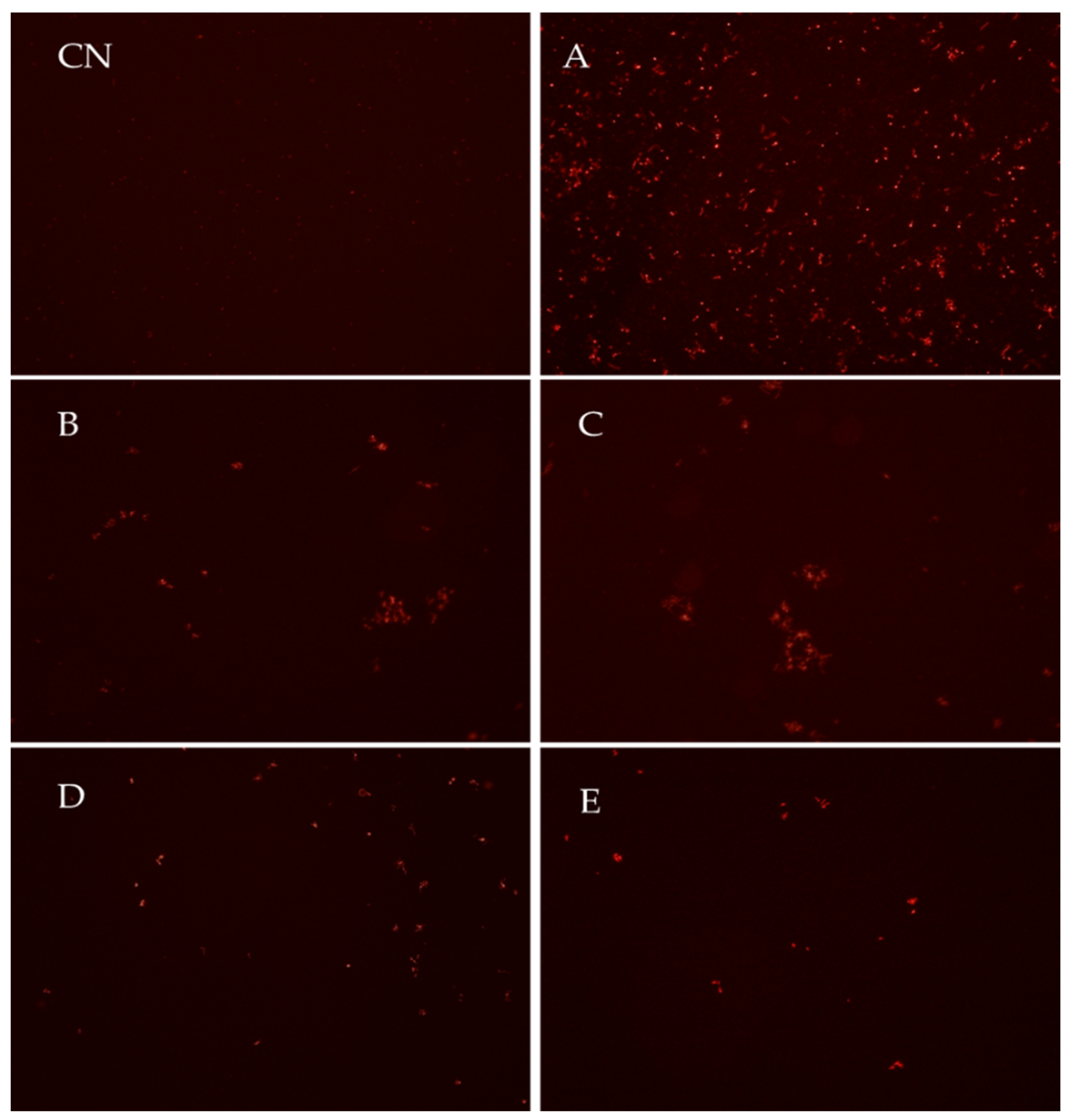
2.5. Cell Viability Using Propidium Iodide Dye

Propidium iodide (PI) dye (Molecular Probes, Eugene, OR, USA) was used to determine cell viability and to detect dead bacterial cells. Briefly, following the treatment with ozone gas and citric acid, ceramic tiles and microtiter plates were rinsed off with sterile saline solution and fluorescent dye PI was applied to biofilm on both materials and incubated in the dark for 15 min. After the incubation, to remove the excess dye, both materials were rinsed off with sterile saline. For fluorescence observation, an inverted microscope at 20× magnification (Olympus IX51, Tokyo, Japan) was used. The obtained images were analyzed in ImageJ 1.47 (National Institute for Health, Bethesda, MD, USA). Untreated biofilm represented control samples.

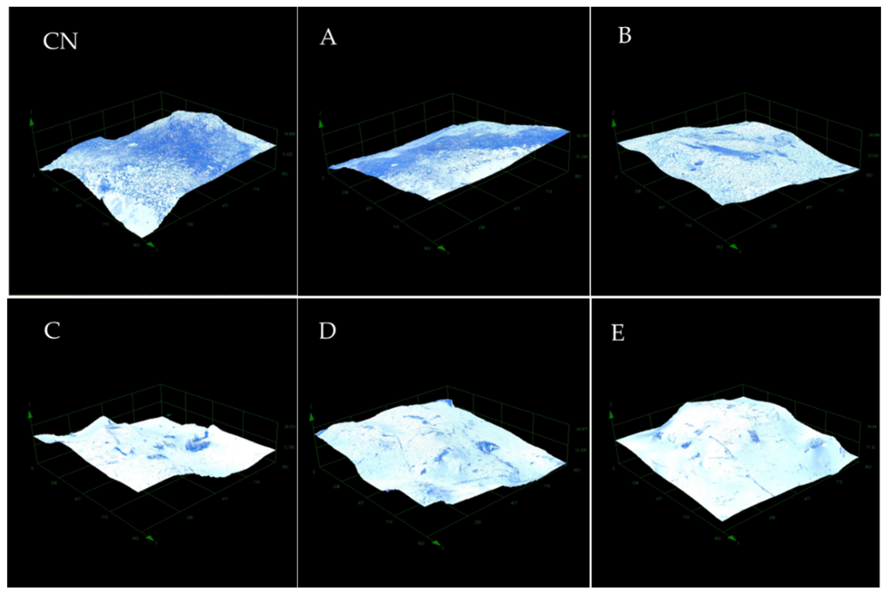
2.6. Crystal Violet Staining and Digital Microscopy

A. baumannii biofilm formed on polystyrene and ceramic tiles was stained with crystal-violet (CV) dye to perform digital microscope images. Treated biofilm from ceramic tiles was rinsed off with sterile saline and then fixated in a dry heat sterilizer (ST-01/02, Instrumentaria, Zagreb, Croatia) at 80 °C for 30 min, while on polystyrene, a blow dryer was used for fixation. Following fixation, biofilm was stained with 0.1% CV for 30 min.
Ceramic surfaces were microscopically examined with a DSX 1000 digital microscope (Olympus, Tokyo, Japan) at 20× magnification with 2-dimensional and 3-dimensional images with the digital microscope. Polystyrene surfaces were examined with an inverted microscope (Olympus, Tokyo, Japan) at magnification 20×.

2.7. Statistical Analyses and Graphing

For evaluation of both combined biocidal effects and solitary effects of gaseous ozone and citric acid, data were analyzed using Wilcoxon signed rank tests (p < 0.05), while Microsoft Excel, version 11.00 (Microsoft Home Office, 2021) was used for graphic images. For evaluation of differences between the used material and methods between one strain, Mann-Whitney test was performed (p < 0.05), as well as for determination of differences between tested strains and used methods. For determination of differences between the tested methods, Kruskal-Wallis multiple comparison test were performed (p < 0.05).

3. Results

To determine the biocidal effects of gaseous ozone and citric acid as solitary disinfectants, and to determine the combined biocidal effects of both disinfectants, a series of experiments with different disinfection protocols with both disinfectants was done, all resulting in significant reductions of viable bacteria in biofilm in comparison to the control (Wilcoxon signed rank test, p < 0.05).

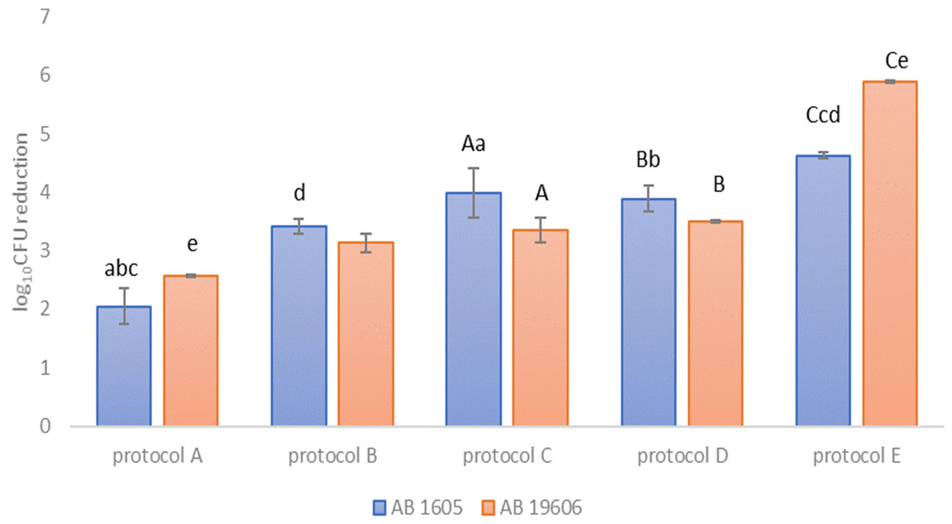
3.1. Viable Bacteria Reduction with Gaseous Ozone–Citric Acid Solitary Effect and Combinations on Ceramic Tiles

Tested strains of both A. baumannii ATCC BAA-1605 and A. baumannii ATCC 19606 showed biofilm reduction on ceramic tiles after the disinfection protocols. The combined biocidal effects of gaseous ozone and citric acid are shown in Figure 1 with the highest log10 CFU reductions of both strains observed with protocol E (combination of pretreatment with citric acid, main treatment with gaseous ozone, and then posttreatment with citric acid). Both citric acid and gaseous ozone in combination, regardless of disinfection protocol used, showed greater biofilm reduction in comparison to solitary gaseous ozone and citric acid treatments. There was a significant difference observed between the two bacterial strains of A. baumannii on ceramics and the used disinfection protocols (Mann–Whitney, p < 0.05), as well as significant difference between different disinfection protocols on each bacterial strain used on ceramics (Kruskal–Wallis multiple comparison test was performed, p < 0.05).

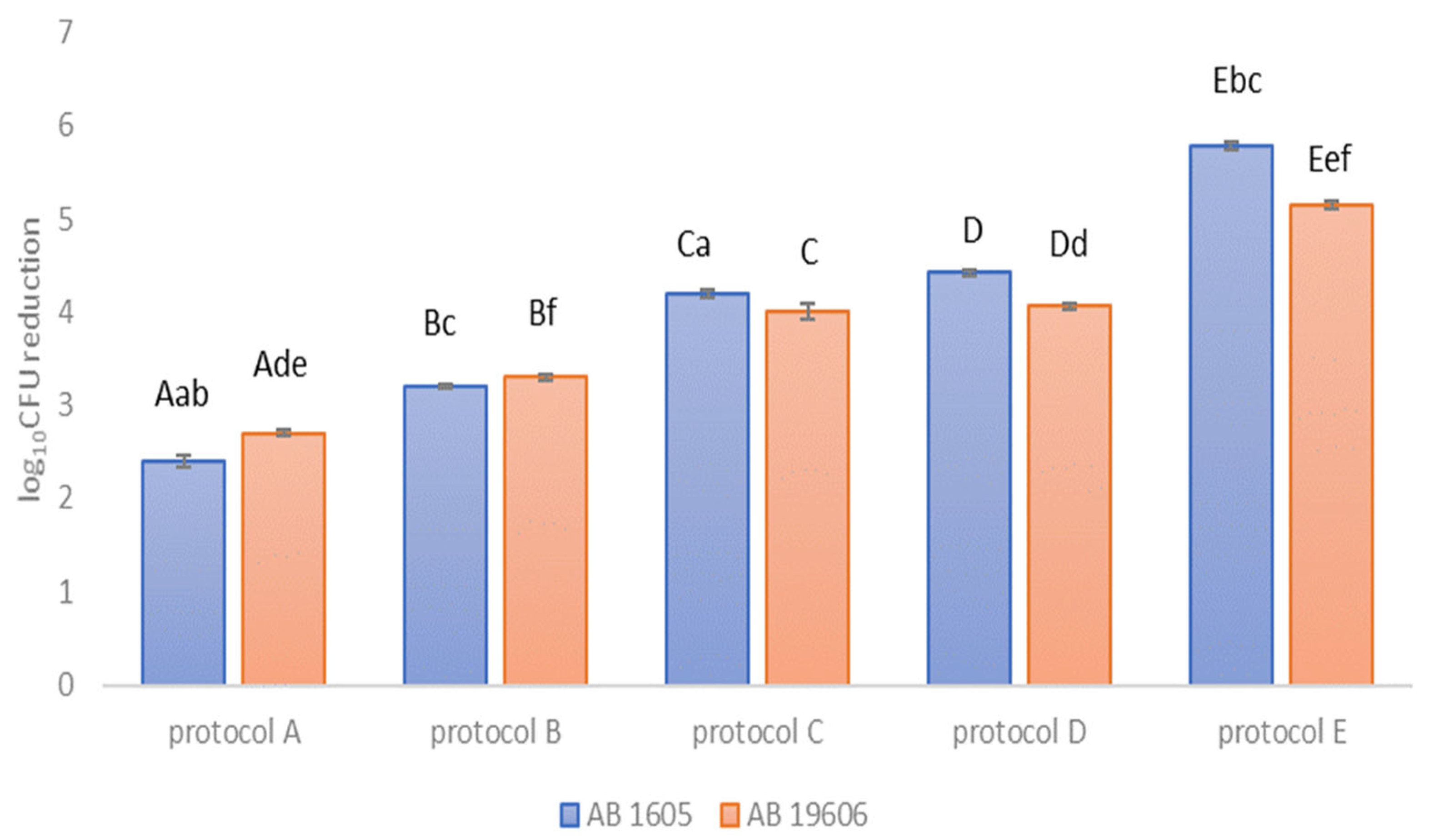
3.2. Viable Bacteria Reduction with Gaseous Ozone–Citric Acid Solitary and Combination Effects on Polystyrene

The reduction of viable bacteria using different disinfection protocols on biofilm of A. baumannii ATCC BAA-1605 and A. baumannii ATCC 19606 formed on polystyrene is shown in Figure 2. Protocol E (combination of pretreatment with citric acid, main treatment with gaseous ozone, and then posttreatment with citric acid) caused a reduction greater than 5 log10CFU for both strains. Both strains showed slightly higher bacterial reductions of combined disinfection protocols on polystyrene in comparison to ceramics. There were statistically significant differences between A. baumannii ATCC BAA-1605 and A. baumannii ATCC 19606 for all protocols (Kruskal–Wallis multiple comparison test was performed (p < 0.05). Additionally, there was no statistically significant difference between the used materials and the efficacy of protocols for strain A. baumannii ATCC BAA-1605, while for strain A. baumannii ATCC 19606, there was a significant difference between the used materials and the efficacy of the tested methods (not shown).

3.3. Different Disinfection Protocols with Gaseous Ozone and Citric Acid Caused Different Inhibition Rates on Polystyrene and Ceramic Tiles

Gaseous ozone and citric acid used in different combinations of disinfection protocols on A. baumannii ATCC BAA-1605 and A. baumannii ATCC 19606 biofilms formed on polystyrene and ceramic tiles caused different inhibition rates (Table 1). The inhibition rates varied from 61.63% up to 99.99%, depending on solitary or combined use of biocidal active substances. Biofilm formed on polystyrene showed slightly higher inhibition rates for both strains than biofilm formed on ceramic tiles.

3.4. Combined Biocidal Effect of Gaseous Ozone and Citric Acid on Cell Viability

A representative strain of A. baumannii ATCC 19606 treated with different disinfection protocols (Protocol A (solitary ozone), Protocol B (solitary citric acid), Protocol C (citric acid–ozone), Protocol D (ozone–citric acid), and Protocol E (citric acid–ozone–citric acid)) is shown in Figure 3. Biofilm not exposed to disinfection with ozone gas and citric acid did not show dead bacterial cells (Figure 3CN), while clustered dead bacterial cells (red) could be seen in the treated biofilm (Figure 3A–E). The absence of dead bacterial cells marked the destruction of bacterial cells and biofilm detachment from the material.

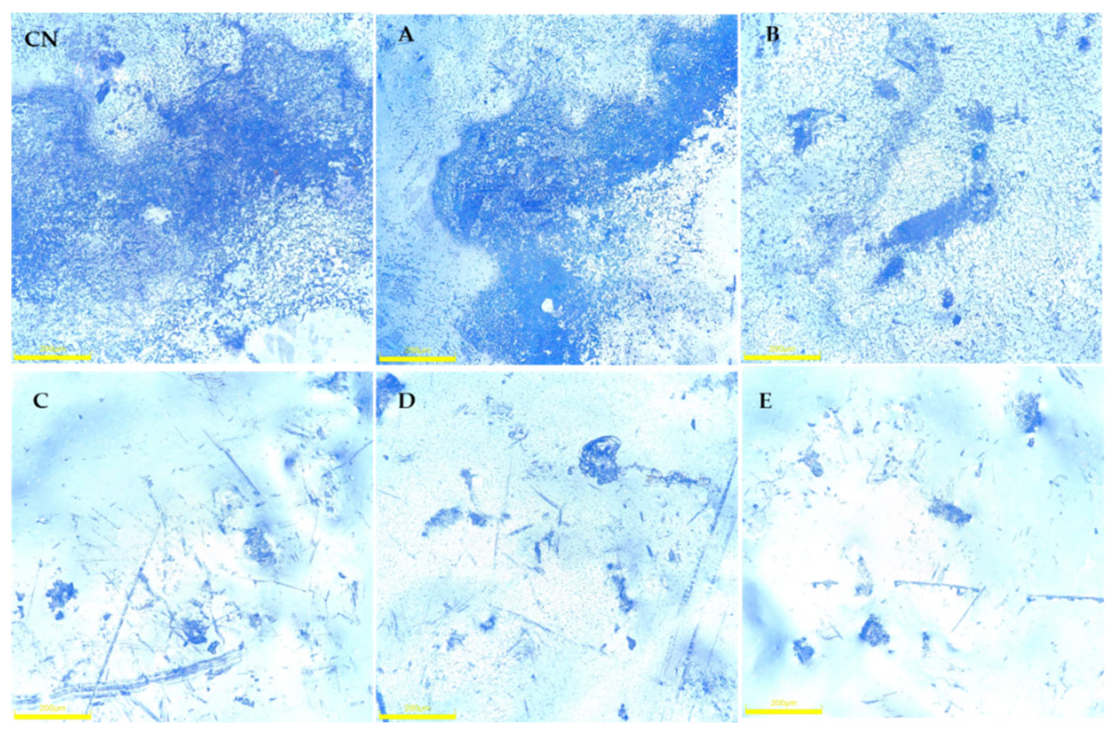
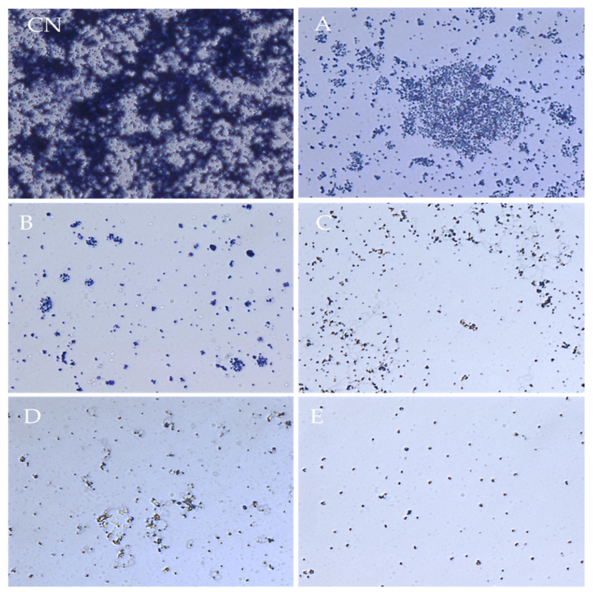
3.5. Crystal Violet Microscopy
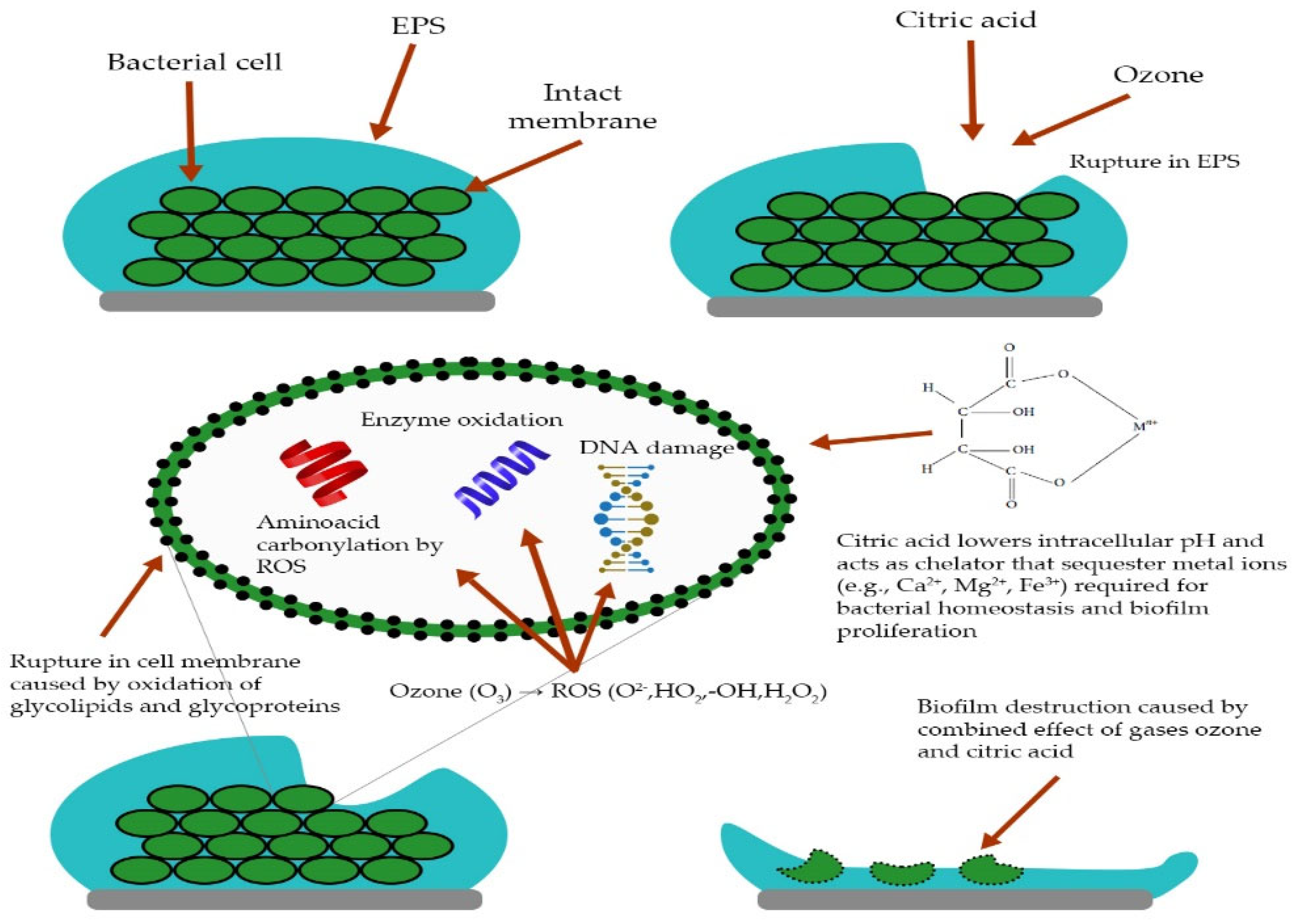
To determine changes in biofilm morphology after disinfection protocols (Protocol A—solitary gaseous ozone, Protocol B—solitary citric acid, Protocol C—citric acid–ozone, Protocol D—ozone–citric acid, and Protocol E–citric acid–gaseous ozone–citric acid), light microscopy was performed. Morphological changes in biofilm of representative strain A. baumannii ATCC 19606 formed on ceramics were found, where 3-dimensional images of Protocols A–D and E can be seen in Figure 4A–E. The distribution of crystal violet dye marked bacterial cell presence. The highest 3-dimensional peaks of A. baumannii biofilm were the first exposed to biocidal action and the first to deteriorate after the disinfection. Combined biocidal effects can be clearly shown in Figure 4E, where the absence of crystal violet dye and cells marked the disinfection effects of combined biocides and biofilm destruction and detachment from ceramics and polystyrene.
Figure 5 shows 2-dimensional images of representative A. baumannii ATCC 19606 biofilm on ceramics after disinfection protocols (Protocol A—solitary ozone, Protocol B—solitary citric acid, Protocol C—citric acid–ozone, Protocol D—ozone–citric acid, and Protocol E—citric acid–gaseous ozone–citric acid). The presented images show that the material surface of tile if rough or uneven can influence the distribution of bacterial cells. Cells were growing more easily in depressed areas or small cavities (crystal violet-stained parts).
Different combinations and application orders of gaseous ozone and citric acid on representative A. baumannii ATCC 19606 biofilm on polystyrene caused morphological changes in biofilm density (Figure 6). The best antibiofilm effect for both materials was observed for Protocol E.

4. Discussion

Contamination of hospital environments with A. baumannii biofilm is a potential source of infections, especially in hospital wards with immunocompromised patients, so one way of infection prevention and control is keeping the wards free of biofilm [3,11,14,18]. Once formed on surfaces, A. baumannii biofilm is challenging to remove because of biofilm characteristics such as resistance to disinfectants, desiccation, long survival in nutrient free environments, and resistance to antibiotics [3,4,14]. Additionally, the survival of A. baumannii in hospital wards where high levels of conventional disinfectants are used on a daily basis can lead to acquired resistance of A. baumannii towards them, especially if disinfectants are used contrary to manufacturer’s instructions [1,15,16,29]. Thus, one of the main goals of infection prevention teams in hospitals is to maintain high levels of hand and environmental hygiene, choosing the correct disinfectant, and use the optimal concentration to fight biofilm contamination [23,38,56,57]. One of the novel trends in surface disinfection of both environment and foods is the use of environmentally friendly disinfectants such as ozone gas and organic acids such as citric acid [37,40,47,48,52,58,59,60]. When used in combination, two biocidal active substances can achieve stronger antimicrobial effects with smaller dosages of both disinfectants, which can also have better effects on chemical waste management, less toxicity for staff and the environment, and lower costs of disinfection [35,61]. The aim of this study was to investigate antibiofilm effects of combined disinfection with gaseous ozone and citric acid on early A. baumannii biofilm formed on ceramics and polystyrene, two relatively frequent materials in hospitals.
In trying to determine the optimal disinfection protocol for A. baumannii biofilm removal, different ozone gas–citric acid combinations were investigated (pretreatment with citric acid then treatment with gaseous ozone, pretreatment with gaseous ozone then citric acid, pretreatment with citric acid then gaseous ozone and posttreatment with citric acid, solitary citric acid treatment and solitary gaseous ozone treatment) using gaseous ozone in effective concentrations determined in a previous study done by authors and citric acid in concentrations according to the manufacturer’s instructions. Both ozone gas and citric acid when used in combination, showed good antibiofilm effects and caused significant reduction in viable bacteria count. Additionally, the combination of ozone gas and citric acid resulted in greater reduction of viable bacteria when used in combination in comparison to their effect as solitary disinfectants. These results were similar to the results of Britton et al., Vankerckhoven et al., Cho et al. and Jung et al., where greater antibiofilm effect were achieved when two biocidal active substances were used in combination [37,61,62,63]. Interestingly, there was no significant difference for both strains and used materials between Protocol C and Protocol D combined disinfection on the viable bacteria count, indicating that in achieving its antimicrobial effect, there was no difference in order of application of these disinfectants. Only the combination of citric acid–gaseous ozone–citric acid showed the highest reduction rates in elimination above 5 log10 CFU reduction of both materials, so only this disinfection protocol can be regarded as efficient according to the European Chemical Agency Guidance on the Biocidal product Regulation, Volume II—efficacy [64]. Additionally, this combined disinfection protocol showed a statistically significant difference in comparison to other protocols on both tested strains and materials.
Regarding differences between combined disinfection protocols and the tested materials for each tested bacterial strain (results not shown), there were no statistically significant differences between polystyrene and ceramics on A. baumannii ATCC BAA-1605 biofilm, while there were significant differences between the used combined disinfection protocols and A. baumannii ATCC 19606 biofilm grown on polystyrene and ceramic tiles, indicating that numerous factors, such as strain sensitivity, material selection, and sanitation protocol, can influence the success of sanitation and disinfection on formed biofilm, as previously emphasized by Gaddy et al., Qi et al., and Rodriguez-Bano et al. [2,3,65].
There is a lack of data on the combined effect of gaseous ozone and citric acid on A. baumannii formed on polystyrene and ceramic tiles. Some studies, such as Jung et. al. [61], explored the synergistic effects of sequential or combined uses of ozone and UV radiation on B. subtilis spores, and studies such as Cho et al. [63] explored the synergistic effects of citric acid and xenon light on the inactivation of food pathogens. Other studies such as Ha et al. [66] explored the synergistic effects of combined disinfection using sodium hypochlorite and ethanol with UV light to reduce Staphylococcus aureus, while Vankerckhoven et al. [61] studied the potential synergistic effects of chemical disinfectants and UV light on biofilm [62]. All these studies have similar conclusions, namely, when used in combination, two biocidal active substances yield better antimicrobial effects than when used alone.
Ozone gas achieves its antimicrobial action through oxidation, while citric acid achieves its action by lowering the intracellular pH and acting as a metal chelator, leading to membrane instability and cell death [35,43,47,48], as shown in Figure 7. Both ozone gas and citric acid achieve their biocidal effects with different mechanisms of actions, so it can be expected that this combination can have good antimicrobial effects on A. baumannii biofilm. Additionally, another element of good combined action of ozone and citric acid is emphasized in studies such as Hirahara et al. [67], where the authors underlined the fact that when used in aqueous form, citric acid enables ozone to have a longer half-life in water, which certainly enhances its antimicrobial action.
To determine the effect of ozone and citric acid combination on cell membrane permeability, propidium iodide staining was performed. The combined effect of ozone gas and citric acid led to a greater number of dead bacterial cells as a result of disturbed membrane permeability, which was also previously confirmed by studies such as Piletić et al. and Nagayoshi et al. [67,68]. Additionally, this combination of biocides, in combination citric acid-ozone gas-citric acid, caused total destruction and biofilm detachment from the material, which indicated good practical potential for eradication of biofilm from inanimate surfaces.
To determine how the combined biocidal effect of gaseous ozone and citric acid influenced A. baumannii biofilm morphology, light microscopy of all sanitation protocols was performed. All treated biofilms showed reductions in numbers of viable bacteria, clearly visible as blank areas in biofilm surfaces, and the combination of citric acid-ozone gas-citric acid caused total biofilm destruction and detachment from the surface. Additionally, using 3D imagery of treated biofilm, it was clearly shown that both ozone gas and citric acid attacked the topologically highest peaks of biofilm, starting to degrade it from the top, possibly because the highest parts are the first contact points for both biocides. These findings are in line with a previous study done by the authors on gaseous ozone efficacy on K. pneumoniae biofilm formed on ceramics, where most morphological changes were observed at the highest topological points of 3D biofilm structures [55].
Regarding the cost effectiveness of using this combination for disinfection protocols in hospitals or other healthcare facilities, there are some issues to address. Ozone gas is relatively cheap to produce with an ozone generator, and it rapidly dissociates to oxygen, therefore leaving no solid waste. In gaseous form it can reach all surfaces in hospital wards, and it is considered to be environmentally friendly [40,58,59]. Additionally, it can be used for air disinfection, which is especially important during hospital-acquired infection outbreaks or COVID-19 outbreaks [69]. Negative side effects are that ozone gas is toxic and must be administered with qualified personnel and can leave an odor, but this can be mitigated with ozone quench gas [55]. Citric acid has numerous advantages; it is not toxic to humans and can be used without the usual precautions, and it is also considered to be environmentally friendly [48]. Both biocides can be favorable according to new policies regarding the usage of chemicals and chemical waste of the European Commission Chemicals strategy for sustainability, which is part of the European Union’s zero pollution ambition [46].
Although ozone gas is a strong oxidative agent, and citric acid is known to be a good food disinfectant and to successfully reduce the number of viable bacteria, they failed to completely eradicate early A. baumannii biofilm from ceramic tiles and polystyrene in the designed concentrations and exposure times for all tested protocol. This issue can potentially be overcome with thorough mechanical cleaning and prior disinfection using stronger detergents, as surfactants can make changes in membrane permeability and make them more susceptible to biocidal effects.

5. Conclusions

Gaseous ozone and citric acid show good antibiofilm effects when used in sequential, combined disinfection and in comparison, to when used as solitary disinfectants.
The best results, achieving almost eradication of A. baumannii biofilm, were observed while using the sequential combination protocol of citric acid–gaseous ozone–citric acid, causing almost full destruction and detachment of biofilm from the surfaces. This combination of biocides has the most implications for practical applications of both biocides, especially for infection prevention and control in healthcare settings.
All tested protocols of combined and solitary used gaseous ozone and citric acid caused biofilm reduction and morphological changes in biofilm structure and made changes in the membrane permeability.
Gaseous ozone and citric acid, when used in the proposed combinations, have good potential to be used for combined disinfection as eco-friendly disinfectants to fight A. baumannii biofilm formed on polystyrene and ceramics.

Author Contributions

Conceptualization, M.O. and I.G.; methodology, K.P. and I.G.; validation, B.K.; investigation, K.P., B.K., M.P., V.V. and I.B.K.; resources, I.G. and M.O.; writing—original draft preparation, K.P., B.K. and M.P.; writing—review and editing, I.G., M.O. and I.B.K.; visualization, J.Ž. and I.G.; supervision, I.G. and M.O.; project administration, M.O. and I.G. All authors have read and agreed to the published version of the manuscript.

Funding

This research was funded by the University of Rijeka, grant number UNIRI-biomed 18-171.

Data Availability Statement

Not applicable.

Acknowledgments

The authors express gratitude to the company Mozone d.o.o., Sisak, Croatia, for donation of the ozone generator and other equipment for the experiment.

Conflicts of Interest

The authors declare no conflict of interest.

References

  1. Liu, W.L.; Liang, H.W.; Lee, M.F.; Lin, H.L.; Lin, Y.H.; Chen, C.C.; Chang, P.C.; Lai, C.C.; Chuang, Y.C.; Tang, H.J. The impact of inadequate terminal disinfection on an outbreak of imipenem-resistant Acinetobacter Baumanniiin an intensive care unit. PLoS ONE 2014, 9, e107975. [Google Scholar] [CrossRef]
  2. Qi, L.; Li, H.; Zhang, C.; Liang, B.; Li, J.; Wang, L.; Du, X.; Liu, X.; Qiu, S.; Song, H. Relationship between antibiotic resistance, biofilm formation, and biofilm-specific resistance in Acinetobacter baumannii. Front. Microbiol. 2016, 7, 483. [Google Scholar] [CrossRef] [PubMed]
  3. Gaddy, A.; Actis, J. A Regulation of Acinetobacter baumannii biofilm formation. Mol. Cell. Biochem. 2012, 23, 273–278. [Google Scholar] [CrossRef] [PubMed]
  4. Espinal, P.; Martí, S.; Vila, J. Effect of biofilm formation on the survival of Acinetobacter baumannii on dry surfaces. J. Hosp. Infect. 2012, 80, 56–60. [Google Scholar] [CrossRef]
  5. Tomaras, A.P.; Dorsey, C.W.; Edelmann, R.E.; Actis, L.A. Attachment to and biofilm formation on abiotic surfaces by Acinetobacter baumannii: Involvement of a novel chaperone-usher pili assembly system. Microbiology 2003, 149, 3473–3484. [Google Scholar] [CrossRef]
  6. Bergogne-Bé, E.; Zin, R.É.; Towner, K.J. Acinetobacter ssp. as nosocomial Pathogens. Clin. Microbiol. Rev. 1996, 9, 148–165. [Google Scholar] [CrossRef]
  7. Nor A’shimi, M.H.; Alattraqchi, A.G.; Rani, F.M.; Rahman, N.I.A.; Ismail, S.; Abdullah, F.H.; Othman, N.; Cleary, D.W.; Clarke, S.C.; Yeo, C.C. Biocide susceptibilities and biofilm-forming capacities of Acinetobacter baumannii clinical isolates from Malaysia. J. Infect. Dev. Ctries. 2019, 13, 626–633. [Google Scholar] [CrossRef]
  8. Wisplinghoff, H.; Schmitt, R.; Wöhrmann, A.; Stefanik, D.; Seifert, H. Resistance to disinfectants in epidemiologically defined clinical isolates of Acinetobacter baumannii. J. Hosp. Infect. 2007, 66, 174–181. [Google Scholar] [CrossRef]
  9. Rajamohan, G.; Srinivasan, V.B.; Gebreyes, W.A. Biocide-tolerant multidrug-resistant Acinetobacter baumannii clinical strains are associated with higher biofilm formation. J. Hosp. Infect. 2009, 73, 287–289. [Google Scholar] [CrossRef]
  10. Donlan, M. Rodney Biofilms: Microbial life on surfaces. An. Real Acad. Nac. Farm. 2016, 82, 108–126. [Google Scholar]
  11. Vickery, K.; Deva, A.; Jacombs, A.; Allan, J.; Valente, P.; Gosbell, I.B. Presence of biofilm containing viable multiresistant organisms despite terminal cleaning on clinical surfaces in an intensive care unit. J. Hosp. Infect. 2012, 80, 52–55. [Google Scholar] [CrossRef] [PubMed]
  12. Vickery, K. Special Issue: Microbial biofilms in healthcare: Formation, prevention and treatment. Materials 2019, 12, 2001. [Google Scholar] [CrossRef] [PubMed]
  13. Kim, H.W.; Lee, N.Y.; Park, S.M.; Rhee, M.S. A fast and effective alternative to a high-ethanol disinfectant: Low concentrations of fermented ethanol, caprylic acid, and citric acid synergistically eradicate biofilm-embedded methicillin-resistant Staphylococcus aureus. Int. J. Hyg. Environ. Health 2020, 229, 113586. [Google Scholar] [CrossRef] [PubMed]
  14. Narayanan, A.; Nair, M.S.; Karumathil, D.P.; Baskaran, S.A. Inactivation of Acinetobacter baumannii Biofilms on Polystyrene, Stainless Steel, and Urinary Catheters by Octenidine Dihydrochloride. Front. Microbiol. 2016, 7, 847. [Google Scholar] [CrossRef]
  15. Lanjri, S.; Uwingabiye, J.; Frikh, M.; Abdellatifi, L.; Kasouati, J.; Maleb, A.; Bait, A.; Lemnouer, A.; Elouennass, M. In vitro evaluation of the susceptibility of Acinetobacter baumannii isolates to antiseptics and disinfectants: Comparison between clinical and environmental isolates. Antimicrob. Resist. Infect. Control 2017, 6, 1–7. [Google Scholar] [CrossRef]
  16. Kawamura-Sato, K.; Wachino, J.I.; Kondo, T.; Ito, H.; Arakawa, Y. Correlation between reduced susceptibility to disinfectants and multidrug resistance among clinical isolates of Acinetobacter species. J. Antimicrob. Chemother. 2010, 65, 1975–1983. [Google Scholar] [CrossRef]
  17. Soares, N.C.; Cabral, M.P.; Gayoso, C.; Mallo, S.; Rodriguez-Velo, P.; Fernández-Moreira, E.; Bou, G. Associating growth-phase-related changes in the proteome of acinetobacter baumannii with increased resistance to oxidative stress. J. Proteome Res. 2010, 9, 1951–1964. [Google Scholar] [CrossRef]
  18. Martró, E.; Hernández, A.; Ariza, J.; Domínguez, M.A.; Matas, L.; Argerich, M.J.; Martin, R.; Ausina, V. Assessment of Acinetobacter baumannii susceptibility to antiseptics and disinfectants. J. Hosp. Infect. 2003, 55, 39–46. [Google Scholar] [CrossRef]
  19. Babaei, M.R.; Sulong, A.; Hamat, R.A.; Nordin, S.A.; Neela, V.K. Extremely high prevalence of antiseptic resistant quaternary ammonium compound E gene among clinical isolates of multiple drug resistant acinetobacter baumannii in Malaysia. Ann. Clin. Microbiol. Antimicrob. 2015, 14, 1–5. [Google Scholar] [CrossRef]
  20. Jawad, A.; Seifert, H.; Snelling, A.M.; Heritage, J.; Hawkey, P.M. Survival of Acinetobacter baumannii on dry surfaces: Comparison of outbreak and sporadic isolates. J. Clin. Microbiol. 1998, 36, 1938–1941. [Google Scholar] [CrossRef]
  21. Denton, M.; Wilcox, M.H.; Parnell, P.; Green, D.; Keer, V.; Hawkey, P.M.; Evans, I.; Murphy, P. Role of environmental cleaning in controlling an outbreak of Acinetobacter baumannii on a neurosurgical intensive care unit. J. Hosp. Infect. 2004, 56, 106–110. [Google Scholar] [CrossRef]
  22. Manian, F.A.; Griesenauer, S.; Senkel, D.; Setzer, J.M.; Doll, S.A.; Perry, A.M.; Wiechens, M. Isolation of Acinetobacter baumannii Complex and Methicillin-Resistant Staphylococcus aureus from Hospital Rooms Following Terminal Cleaning and Disinfection: Can We Do Better? Infect. Control Hosp. Epidemiol. 2011, 32, 667–672. [Google Scholar] [CrossRef]
  23. Abreu, A.C.; Tavares, R.R.; Borges, A.; Mergulhão, F.; Simões, M. Current and emergent strategies for disinfection of hospital environments. J. Antimicrob. Chemother. 2013, 68, 2718–2732. [Google Scholar] [CrossRef] [PubMed]
  24. Stickler, D.J.; Thomas, B.; Chawla, J.C. Antiseptic and antibiotic resistance in gram-negative bacteria causing urinary tract infection in spinal cord injured patients. Paraplegia 1981, 19, 50–58. [Google Scholar] [CrossRef] [PubMed]
  25. Thomas, L.; Russell, A.D.; Maillard, J.-Y. Antimicrobial activity of chlorhexidine diacetate and benzalkonium chloride against Pseudomonas aeruginosa and its response to biocide residues. J. Appl. Microbiol. 2005, 98, 533–543. [Google Scholar] [CrossRef] [PubMed]
  26. Suller, M.T.E.; Russell, A.D. Antibiotic and biocide resistance in methicillin-resistant Staphylococcus aureus and vancomycin-resistant enterococcus. J. Hosp. Infect. 1999, 43, 281–291. [Google Scholar] [CrossRef]
  27. Russell, A.D. Bacterial resistance to disinfectants: Present knowledge and future problems. J. Hosp. Infect. 1999, 43, 57–68. [Google Scholar] [CrossRef]
  28. Bridier, A.; Briandet, R.; Thomas, V.; Dubois-Brissonnet, F. Biofouling: The Journal of Bioadhesion and Biofilm Resistance of bacterial biofilms to disinfectants: A review. Biofouling J. Bioadhesion Biofilm Res. 2011, 27, 1017–1032. [Google Scholar] [CrossRef]
  29. Ivanković, T.; Goić-Barišić, I.; Hrenović, J. Reduced susceptibility to disinfectants of Acinetobacter baumannii biofilms on glass and ceramic. Arh. Hig. Rada Toksikol. 2017, 68, 99–108. [Google Scholar] [CrossRef]
  30. Song, L.; Wu, J.; Xi, C. Biofilms on environmental surfaces: Evaluation of the disinfection efficacy of a novel steam vapor system. Am. J. Infect. Control 2012, 40, 926–930. [Google Scholar] [CrossRef]
  31. Sutherland, I.W. Biofilm exopolysaccharides: A strong and sticky framework. Microbiology 2001, 147, 3–9. [Google Scholar] [CrossRef] [PubMed] [Green Version]
  32. Stewart, P.S.; Rayner, J.; Roe, F.; Rees, W.M. Biofilm penetration and disinfection efficacy of alkaline hypochlorite and chlorosulfamates. J. Appl. Microbiol. 2001, 91, 525–532. [Google Scholar] [CrossRef] [PubMed]
  33. Tachikawa, M.; Yamanaka, K.; Nakamuro, K. Studies on the disinfection and removal of biofilms by ozone water using an artificial microbial biofilm system. Ozone Sci. Eng. 2009, 31, 3–9. [Google Scholar] [CrossRef]
  34. Smith, K.; Hunter, I.S. Efficacy of common hospital biocides with biofilms of multi-drug resistant clinical isolates. J. Med. Microbiol. 2008, 57, 966–973. [Google Scholar] [CrossRef] [PubMed]
  35. Park, K.M.; Yoon, S.; Choi, T.; Kim, H.J.; Park, K.J.; Koo, M. The bactericidal effect of a combination of food grade compounds and their application as alternative antibacterial agent for food contact surfaces. Foods 2020, 9, 59. [Google Scholar] [CrossRef]
  36. Russell, A.D. Possible link between bacterial resistance and use of anticiotics and biocides. Antimicrob. Agents Chemother. 1998, 42, 4804. [Google Scholar] [CrossRef] [PubMed]
  37. Britton, H.C.; Draper, M.; Talmadge, J.E. Antimicrobial efficacy of aqueous ozone in combination with short chain fatty acid buffers. Infect. Prev. Pract. 2020, 2, 100032. [Google Scholar] [CrossRef]
  38. Boyce, J.M. Modern technologies for improving cleaning and disinfection of environmental surfaces in hospitals. Antimicrob. Resist. Infect. Control 2016, 5, 10. [Google Scholar] [CrossRef]
  39. Bayan, M.A.G.; McGann, P.; Kwak, Y.I.; Summers, A.; Cummings, J.F.; Waterman, P.E.; Lesho, E.P.; Detusheva, E.V.; Ershova, O.N.; Fursova, N.K.; et al. Distribution of antiseptic resistance genes qacA, qacB, and smr in methicillin-resistant Staphylococcus aureus isolated in Toronto, Canada, from 2005 to 2009. J. Hosp. Infect. 2016, 66, 1–9. [Google Scholar]
  40. Sharma, M.; Hudson, J.B. Ozone gas is an effective and practical antibacterial agent. Am. J. Infect. Control 2008, 36, 559–563. [Google Scholar] [CrossRef]
  41. Boch, T.; Tennert, C.; Vach, K.; Al-Ahmad, A.; Hellwig, E.; Polydorou, O. Effect of gaseous ozone on Enterococcus faecalis biofilm—An in vitro study. Clin. Oral Investig. 2016, 20, 1733–1739. [Google Scholar] [CrossRef] [PubMed]
  42. Boer, H.E.L.d.; van Elzelingen-Dekker, C.M.; van Rheenen-Verberg, C.M.F.; Spanjaard, L. Use of Gaseous Ozone for Eradication of Methicillin-Resistant Staphylococcus aureus. From the Home Environment of a Colonized Hospital Employee. Infect. Control Hosp. Epidemiol. 2006, 27, 1120–1122. [Google Scholar] [CrossRef] [PubMed] [Green Version]
  43. Giuliani, G.; Ricevuti, G.; Galoforo, A.; Franzini, M. Microbiological aspects of ozone: Bactericidal activity and antibiotic/antimicrobial resistance in bacterial strains treated with ozone. Ozone Ther. 2018, 3, 1–4. [Google Scholar] [CrossRef]
  44. Megahed, A.; Aldridge, B.; Lowe, J. The microbial killing capacity of aqueous and gaseous ozone on different surfaces contaminated with dairy cattle manure. PLoS ONE 2018, 13, e0196555. [Google Scholar] [CrossRef] [PubMed]
  45. Fontes, B.; Heimbecker, A.M.C.; Brito, G.d.S.; Costa, S.F.; van der Heijden, I.M.; Levin, A.S.; Rasslan, S. Effect of low-dose gaseous ozone on pathogenic bacteria. BMC Infect. Dis. 2012, 12, 2–7. [Google Scholar] [CrossRef] [PubMed]
  46. Conto, A. The EU chemical strategy for sustainability towards a toxic-free environment. Chim. Oggi Chem. Today 2021, 39, 40–41. [Google Scholar]
  47. Akbaş, M.Y. Effectiveness of Organic Acid Treatments for Inhibition and Removal of E. coli Biofilms. Hacettepe J. Biol. Chem. 2016, 1, 35. [Google Scholar] [CrossRef]
  48. Akbas, M.Y.; Kokumer, T. The prevention and removal of biofilm formation of Staphylococcus aureus strains isolated from raw milk samples by citric acid treatments. Int. J. Food Sci. Technol. 2015, 50, 1666–1672. [Google Scholar] [CrossRef]
  49. Park, S.H.; Choi, M.R.; Park, J.W.; Park, K.H.; Chung, M.S.; Ryu, S.; Kang, D.H. Use of organic acids to inactivate Escherichia coli O157: H7, salmonella typhimurium, and listeria monocytogenes on organic fresh apples and lettuce. J. Food Sci. 2011, 76, M293–M298. [Google Scholar] [CrossRef]
  50. Cherrington, C.A.; Hinton, M.; Mead, G.C.; Chopra, I. Organic Acids: Chemistry, Antibacterial Activity and Practical Applications. Adv. Microb. Physiol. 1991, 32, 87–108. [Google Scholar] [CrossRef]
  51. Wang, J.; Tao, D.; Wang, S.; Li, C.; Li, Y.; Zheng, F.; Wu, Z. Disinfection of lettuce using organic acids: An ecological analysis using 16S rRNA sequencing. RSC Adv. 2019, 9, 17514–17520. [Google Scholar] [CrossRef] [PubMed]
  52. Tsai, Y.P.; Pai, T.Y.; Hsin, J.Y.; Wan, T.J. Biofilm bacteria inactivation by citric acid and resuspension evaluations for drinking water production systems. Water Sci. Technol. 2004, 48, 463–472. [Google Scholar] [CrossRef]
  53. Hughes, G.; Webber, M.A. Novel approaches to the treatment of bacterial biofilm infections. Br. J. Pharmacol. 2017, 174, 2237–2246. [Google Scholar] [CrossRef] [PubMed]
  54. Hughes, G.; Lund, P. The Use of Weak Organic Acids as a Novel Antimicrobial and Biofilm Eradication Agent. In Proceedings of the European Congress of Clinical Microbiology and Infectious Diseases, Madrid, Spain, 21–24 April 2018. [Google Scholar]
  55. Piletić, K.; Kovač, B.; Perčić, M.; Žigon, J.; Broznić, D.; Karleuša, L.; Blagojević, S.L.; Oder, M.; Gobin, I. Disinfecting Action of Gaseous Ozone on OXA-48-Producing Klebsiella pneumoniae Biofilm In Vitro. Int. J. Environ. Res. Public Health 2022, 19, 6177. [Google Scholar] [CrossRef]
  56. Kampf, G.; Kramer, A. Epidemiologic background of hand hygiene and evaluation of the most important agents for scrubs and rubs. Clin. Microbiol. Rev. 2004, 17, 863–893. [Google Scholar] [CrossRef]
  57. Boyce, J.M. Environmental contamination makes an important contribution to hospital infection. J. Hosp. Infect. 2007, 65, 50–54. [Google Scholar] [CrossRef]
  58. Davies, A.; Pottage, T.; Bennett, A.; Walker, J. Gaseous and air decontamination technologies for Clostridium difficile in the healthcare environment. J. Hosp. Infect. 2011, 77, 199–203. [Google Scholar] [CrossRef]
  59. Moat, J.; Cargill, J.; Shone, J.; Upton, M. Application of a novel decontamination process using gaseous ozone. Can. J. Microbiol. 2009, 55, 928–933. [Google Scholar] [CrossRef]
  60. Kundukad, B.; Udayakumar, G.; Grela, E.; Kaur, D.; Rice, S.A.; Kjelleberg, S.; Doyle, P.S. Weak acids as an alternative anti-microbial therapy. Biofilm 2020, 2, 100019. [Google Scholar] [CrossRef]
  61. Jung, Y.J.; Oh, B.S.; Kang, J.W. Synergistic effect of sequential or combined use of ozone and UV radiation for the disinfection of Bacillus subtilis spores. Water Res. 2008, 42, 1613–1621. [Google Scholar] [CrossRef]
  62. Vankerckhoven, E.; Verbessem, B.; Crauwels, S.; Declerck, P.; Muylaert, K.; Willems, K.A.; Rediers, H. Exploring the potential synergistic effects of chemical disinfectants and UV on the inactivation of free-living bacteria and treatment of biofilms in a pilot-scale system. Water Sci. Technol. 2011, 64, 1247–1253. [Google Scholar] [CrossRef]
  63. Cho, G.L.; Ha, J.W. Synergistic effect of citric acid and xenon light for inactivating foodborne pathogens on spinach leaves. Food Res. Int. 2021, 142, 110210. [Google Scholar] [CrossRef]
  64. ECHA. Guidance on the Biocidal Products Regulation: Volume II Parts B+C; European Chemicals Agency: Helsinki, Finland, 2022; ISBN 9789290205029. [Google Scholar]
  65. Rodríguez-Baño, J.; Martí, S.; Soto, S.; Fernández-Cuenca, F.; Cisneros, J.M.; Pachón, J.; Pascual, A.; Martínez-Martínez, L.; Mcqueary, C.; Actis, L.A.; et al. Biofilm formation in Acinetobacter baumannii: Associated features and clinical implications. Clin. Microbiol. Infect. 2008, 14, 276–278. [Google Scholar] [CrossRef] [Green Version]
  66. Ha, J.H.; Jeong, S.H.; Ha, S.D. Synergistic effects of combined disinfection using sanitizers and uv to reduce the levels of staphylococcus aureus in oyster mushrooms. J. Appl. Biol. Chem. 2011, 54, 447–453. [Google Scholar] [CrossRef]
  67. Hirahara, Y.; Iwata, K.; Nakamuro, K. Effect of Citric Acid on Prolonging the Half-life of Dissolved Ozone in Water. Food Saf. 2019, 7, 90–94. [Google Scholar] [CrossRef] [PubMed]
  68. Nagayoshi, M.; Fukuizumi, T.; Kitamura, C.; Yano, J.; Terashita, M.; Nishihara, T. Efficacy of ozone on survival and permeability of oral microorganisms. Oral Microbiol. Immunol. 2004, 19, 240–246. [Google Scholar] [CrossRef] [PubMed]
  69. Franke, G.; Knobling, B.; Brill, F.H.; Becker, B.; Klupp, E.M.; Campos, C.B.; Pfefferle, S.; Lütgehetmann, M.; Knobloch, J.K. An automated room disinfection system using ozone is highly active against surrogates for SARS-CoV-2. J. Hosp. Infect. 2021, 112, 108–113. [Google Scholar] [CrossRef]
Figure 1. Viable bacteria count expressed as log10 CFU reduction of Acinetobacter baumannii ATCC BAA-1605 and Acinetobacter baumannii ATCC 19606 with standard deviation. Protocol A marks disinfection with gaseous ozone, Protocol B disinfection with citric acid, Protocol C a combination of citric acid–gaseous ozone, Protocol D a combination of gaseous ozone–citric acid, and Protocol E a combination of citric acid–gaseous ozone–citric acid. Lowercase letters (a–e) express statistically significant differences between disinfection protocols for one bacterial strain (Kruskal–Wallis U, p < 0.05). Capital letters (A, B, C) mark statistically significant differences between different bacterial strains per used Protocols C, D, and E.
Processes 10 01788 g001
Figure 2. Viable bacteria count expressed as log10 CFU reduction of Acinetobacter baumannii ATCC BAA-1605 and Acinetobacter baumannii ATCC 19606 formed on polystyrene with standard deviation. Protocol A marks disinfection with gaseous ozone, Protocol B disinfection with citric acid, Protocol C a combination of citric acid–gaseous ozone, Protocol D a combination of gaseous ozone–citric acid, and Protocol E a combination of citric acid–gaseous ozone–citric acid. Lowercase letters (a–f) express statistically significant differences between disinfection protocol and bacterial strain (Kruskal–Wallis U, p < 0.05). Capital letters (A, B, C, D, and E) mark statistically significant differences between different used bacterial strain per disinfection protocol.
Processes 10 01788 g002
Figure 3. Viability of A. baumannii ATCC 19606 biofilm treated with different disinfection protocols ((A) solitary ozone, (B) solitary citric acid, (C) citric acid–ozone, (D) ozone–citric acid, (E) citric acid–ozone–citric acid) using propidium iodide staining. The control group was non-treated biofilm (CN). Red fluorescence indicates dead cells with permeable membranes. Magnification 20×.
Processes 10 01788 g003
Figure 4. The 3-dimensional images of ceramic tile surface with formed 24 h A. baumannii ATCC 19606 biofilm treated with different disinfection protocols (Protocol (A)—solitary ozone gas, Protocol (B)—solitary citric acid, Protocol (C)—citric acid–ozone, Protocol (D)—ozone–citric acid, Protocol (E)—citric acid–ozone–citric acid) and non-treated control (CN). Magnification 20×.
Processes 10 01788 g004
Figure 5. Images of ceramic tile surface with formed 24 h A. baumannii ATCC 19606 biofilm treated with different disinfection protocols (AE). The control was provided with non-treated biofilm (CN). Magnification 20×.
Processes 10 01788 g005
Figure 6. The 2-dimensional images of 24 h representative strain A. baumannii ATCC 19606 biofilm formed on polystyrene treated with different disinfection protocols (AE). (CN) marks control group. The dark blue stains present the bacteria stained with crystal violet. The control was provided with A. baumannii ATCC 19606 biofilm not exposed to combined or solitary ozone gas and citric acid effects. Magnification 20×.
Processes 10 01788 g006
Figure 7. Schematic representation of combined effect of gaseous ozone and citric acid on A. baumannii biofilm formed on ceramic tiles.
Processes 10 01788 g007
Table 1. Percentages of inhibition of both A. baumannii strains of biofilm formed on polystyrene and ceramics after disinfection protocols. Protocol A marks disinfection with gaseous ozone, Protocol B disinfection with citric acid, Protocol C a combination of citric acid–gaseous ozone, Protocol D a combination of gaseous ozone–citric acid, and Protocol E a combination of citric acid–gaseous ozone–citric acid.
SurfacesA. baumannii ATCC BAA1605A. baumannii ATCC 19606
Treatments (% Biofilm Destruction)
ProtocolProtocol
ABCDEABCDE
Polystyrene61.694.0699.499.3399.9880.3297.3599.0299.1399.93
Ceramic tiles89.982.798.2998.5399.7773.393.7796.1697.499.99
Publisher’s Note: MDPI stays neutral with regard to jurisdictional claims in published maps and institutional affiliations.

