Article

Targeting of Nrf2/PPARγ/NLRP3 Signaling Pathway by Stevia rebudiana Bertoni Extract Provides a Novel Insight into Its Protective Effect against Acute Gouty Arthritis-Induced Synovial Inflammation, Oxidative Stress and Apoptosis in a Rat Model

1
Department of Anatomy and Embryology, Faculty of Medicine, Mansoura University, Mansoura 35516, Egypt
2
Department of Anatomy, Al-Qunfudah Medical College, Umm Al-Qura University, Al-Qunfudhah 28814, Saudi Arabia
3
Department of Pharmacology, Faculty of Veterinary Medicine, Mansoura University, Mansoura 35516, Egypt
4
Clinical Pharmacology Department, Faculty of Medicine, Mansoura University, Mansoura 35516, Egypt
5
Department of Surgery, College of Medicine in Al-Qunfudhah, Umm Al-Qura University, Al-Qunfudhah 28814, Saudi Arabia
6
Department of Human Anatomy, Faculty of Medicine, Umm Al-Qura University, Makkah 24382, Saudi Arabia
7
Electron Microscopy Unit, Mansura University, El Mansoura 35516, Egypt
8
Department of Medical Emergency Services, Al-Qunfudah Health Sciences College, Umm Al-Qura University, Mecca 24382, Saudi Arabia
*
Author to whom correspondence should be addressed.
Processes 2022, 10(9), 1751; https://doi.org/10.3390/pr10091751
Submission received: 3 August 2022 / Revised: 23 August 2022 / Accepted: 26 August 2022 / Published: 2 September 2022

Abstract

:
Our research work examined the potential protection of Stevia rebaudiana extract against monosodium urate crystals (MSU)-induced acute gouty arthritis in a rat model and its possible underlying mechanism. Forty rats were allocated into four groups (n = 10); a control group; an MSU group, whose rats received 0.1 of MSU single intra-articular injection in the ankle joint on the fifth day of the experiment; an MSU + Stevia group, which received 250 mg/kg/day of Stevia extract orally for seven days and MSU crystals on the fifth day; and an MSU + colchicine group, which was administered colchicine at 0.28 mg/kg daily for seven days and MSU crystals on the fifth day. Pretreatment with Stevia extract mitigated MSU-induced inflammation as evidenced by a decrease of the ankle edema and inflammatory cell infiltration and a significant downregulation of the protein level of NFκB, TNFα, IL-1β, IL6, and IL18 as well as NLRP3 gene expression. Additionally, there was a markedly increased PPARγ gene expression (p < 0.001) compared with the MSU group (p < 0.001) and alleviated oxidative stress via significant upregulating of Nrf2/HO-1. Moreover, the pretreatment attenuated apoptosis by significantly decreasing cytochrome c, Bax, Caspase-3, and by increasing Bcl-2 protein. In conclusion, Stevia extract exhibited strong anti-inflammatory, antioxidant, and antiapoptotic effects against MSU-induced gouty arthritis similar to the standard anti-inflammatory colchicine drugs.

1. Introduction

Gout is one of the major complications of impaired uric acid metabolism. Chronic elevation of uric acid has a great affinity to the joints, particularly the tarsometatarsal joint of the big toe [1]. It has been found that monosodium urate (MSU) crystals have a major role in the pathology of gouty arthritis due to their proinflammatory property [2]. Deposition of monosodium urate crystals has a stimulatory effect on the nucleotide-binding domain (NOD)-like receptor protein 3 (NLRP3) inflammasome [3,4]. NLRP3 inflammasome activation has a leading role in the pathology of gouty arthritis, as it leads to the subsequent release of IL1β, a proinflammatory cytokine, that stimulates the release of TNFα, IL6, and IL8 which are considered pro-inflammatory factors. These factors attract neutrophils and monocytes to infiltrate the joint synovial membrane, with subsequent increasing vascular permeability, and exudation which leads to joint swelling [5,6]. Persistent inflammation leads to the destruction of the cartilage of the joint and eventually its damage [7]. At the same time, the harmful effect of the urates is also referred to as their power to induce reactive oxygen species formation [8]. Activation of NADP oxidase, nitric oxide synthase, and xanthin oxidase synthetase enzymes produces hydrogen peroxide (H2O2), superoxide anion (O2), and nitric oxide (NO). The latter of these interact with each other to produce peroxynitrite (ONOO), which aggravates apoptosis of the synovial cells, connective tissue degeneration, and destruction of the ankle joint [9].
Clinically, medication with nonsteroidal anti-inflammatory and colchicine drugs downregulates the activation of NLRP3 inflammasome induced by MSU crystals with subsequent inhibition of caspase 1 and release of IL-1β which stimulates the attraction of neutrophil [10]. Despite the efficiency of this medication in the treatment of gouty arthritis, it is not preferred due to its serious gastrointestinal side effects, such as peptic ulcers [11], in addition to the toxicity of the nervous and renal systems as a result of the treatment by colchicine [12,13]. Therefore, there is an urgent need for a safe and natural alternative therapy for gouty arthritis that mitigates the inflammation and rapidly relieves the pain without adverse effects.
Stevia rebaudiana (S. rebaudiana) is a plant found in Paraguay. It is commonly used in Japan and Brazil as a food additive and non-caloric sweetener. Its leaves are rich in eight glycosides including rebaudioside A, B, D, E, dulcoside A, and B, and stevioside [14]. It has been documented that the most active stevioside glycoside of Stevia leaves have several beneficial properties including antioxidants, anti-inflammatory, antimicrobial, and hypoglycemic effects [15,16]. Stevia rebaudiana has been reported to exhibit marked therapeutic action for patients suffering from inflammatory diseases, cancer, diabetes, and obesity [17,18,19].
In light of the aforementioned literature, we hypothesized that the extract of S. rebaudioside leaves, which possess powerful anti-inflammatory and antioxidant features, could help combat the acute gouty arthritis caused by MSU crystals. This experiment was designed to assess the potential protective effect of Stevia extracts on acute gouty arthritis induced by intra-articular MSU crystal injection and the possible mechanisms underlying this ameliorative effect.

2. Materials and Methods

2.1. Animals

The protocol of our experiment follows the guidelines for laboratory animals published by the US National Institutes of Health (NIH Publication No. 85-23, revised 2011) and approved by the Medical Research Ethics Committee of The Faculty of Veterinary Medicine, Mansoura University (code: R/135). We purchased 40 rats weighing 200–250 p from Mansoura experimental Research Center, Mansoura University. These rats were kept for 14 days prior to the onset of the experiment under controlled conditions (temperature 22–24 °C, and 12 h light/dark cycle). Rats were offered a standard diet and water ad libitum.

2.2. Chemicals

MSU crystals (Cat. No. 1198-77-2) were obtained from Adipogen Life Sciences (San Diego, CA, USA). Colchicine drug was supplied from El Nasr Pharmaceutical Chemicals Co Egypt. Calci-Clear Rapid solution was procured from National Diagnostics Co., Atlanta, GA, USA.

2.3. MSU Crystals-Induced Acute Gouty Arthritis Animal Model

The induction of gouty arthritis was performed following the method mentioned by Haung et al. [20]. In brief, MSU crystals suspension was injected intra-articularly at a dose of 0.1 mL (3.0 mg) after anesthetizing the ankle joint of the right hind paw with chloral hydrate (500 mg/kg). The preparation of the suspension was conducted by mixing 150 mg of uric acid sodium with 5 mL saline.

2.4. Experimental Groups: Rats Were Subdivided into Four Groups (10 Rats/Group)

Before the experiments, the rats were acclimatized for 2 weeks to accommodate the experimental condition.
Group I: Control group: the rats of the control group were intra-articularly injected with normal saline into the right knee joint of the hind paw.
Group II: MSU group: Rats were administered a single injection of MSU on the 5th day of the experiment.
Group III: MSU + Stevia group: Rats of this group received daily methanolic Stevia extracts with a dose of 250 mg/kg dissolved in 2 mL saline per oral via gastric gavage, starting from day 0 of experiments before intra-articular injection of monosodium urates crystals on the 5th day, this was continued until the end of the experiments at day 7.
Group IV: MSU + colchicine group: Rats received a daily oral administration of Col (0.28 mg/kg) before the MSU crystal injection, which continued until the end of the experiment on the 7th day.
Both doses of Colchicine and Stevia were selected based on previous studies performed by Han et al. [21] and Alavala et al. [22] respectively.

2.5. Methanolic Extraction of Stevia R Leaves

The extraction of Stevia rebudiana leaves was executed according to the method of El-Mousalamy et al. [23]. The Stevia leaf plants were procured from International Agriculture Products Company, Cairo, Egypt.

2.6. Assessment of Ankle Edema and Specimen Collection

To assess the inflammation of the ankle joint, we measured the circumference of the joint by tie line method one hour before intra-articular injection of MSU crystals on the 5th day of the experiment and continued measuring at different time points (2, 4, 8, 12, 24, 48 h) until the end of the experiment at day 7. We calculated the percentage of the edema (the main indicator of inflammation) according to the following equation: % of edema = [(Vf − Vi)/Vi] × 100. Where Vf indicates the measurement of the circumference before the injection and Vi indicates the measurements of the circumference after the injection at different timepoints. At the end of the experiment, we anesthetized the rats with 40 mg/kg of 3% sodium phenobarbital. Then, the rats were sacrificed by cervical decapitation, and the synovial tissue of the right ankle joint of the hind paw of the different groups was stored at −70 °C. Later, 10% homogenate of synovial tissue was prepared by being placed in ice-cold normal saline, then centrifuged at 1400× g for 20 min. The collected supernatant was centrifuged again at 1400× g for 20 min. The final supernatant of synovial tissue was stored at −70 °C for molecular and biochemical investigation. At the same time, some of the ankle joints of different groups were stored in 10% of formalin and immersed in paraffin blocks. The sections from these blocks were prepared for histological and immunohistochemical investigations.

2.7. Radiographic Assessments

To confirm the assessment of the inflammation, just before the scarification, the rats were anesthetized with 40 mg/kg of 3% of sodium phenobarbitone, after the anesthesia each rat was placed in a Multi-Mode Small Animal Living Imaging System (Kodak, New Haven, CT, USA), and the articular cavity of the right ankle was photographed under X-ray.

2.8. Biochemical Analysis

Assessment of oxidative stress in markers in joint synovial tissue (MAD, GSH, SOD, CAT): Lipid peroxidation (LPO), assessed as malondialdehyde (MDA) level, the reduced glutathione level (GSH) and the activities of the antioxidant enzymes; superoxide dismutase (SOD), and catalase (CAT) in joint synovial tissue were evaluated using assay kits (Cat. No. MD 25-29), (Cat. No. GR25-11), (Cat. No. SD25-21), and (Cat. No. CA25-17), respectively purchased from Biodiagnostics Co. (Cairo, Egypt) following the manufacturer’s instructions.

2.9. Histopathological Examination

After the end of the experiment, on day 7, we sacrificed the rats and immersed the right ankle of different groups in 10% formalin for fixation. To decalcify the tissues, we put them in Calci-Clear Rapid solution for 10 days. Thereafter, the tissues were immersed in paraffin blocks. Then, 4μm sections from the paraffin blocks were prepared for hematoxylin and eosin (H&E) and immunohistochemical staining. The slides were examined under a light microscope (Leica_DM500 with a camera, Leica_ICC50HD) with the camera software LEICA Application Suite (LAS) EZ, Version 3.1.1 (Histology Department, Faculty of Medicine, Mansoura University).

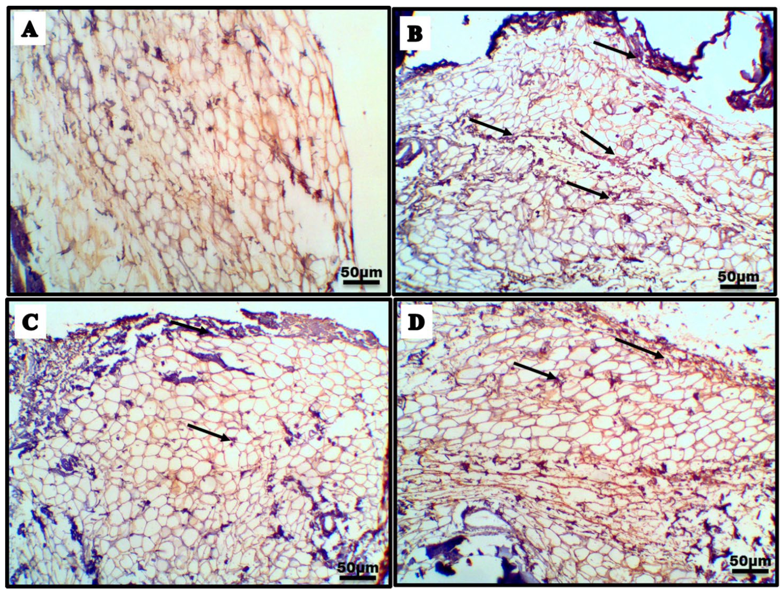
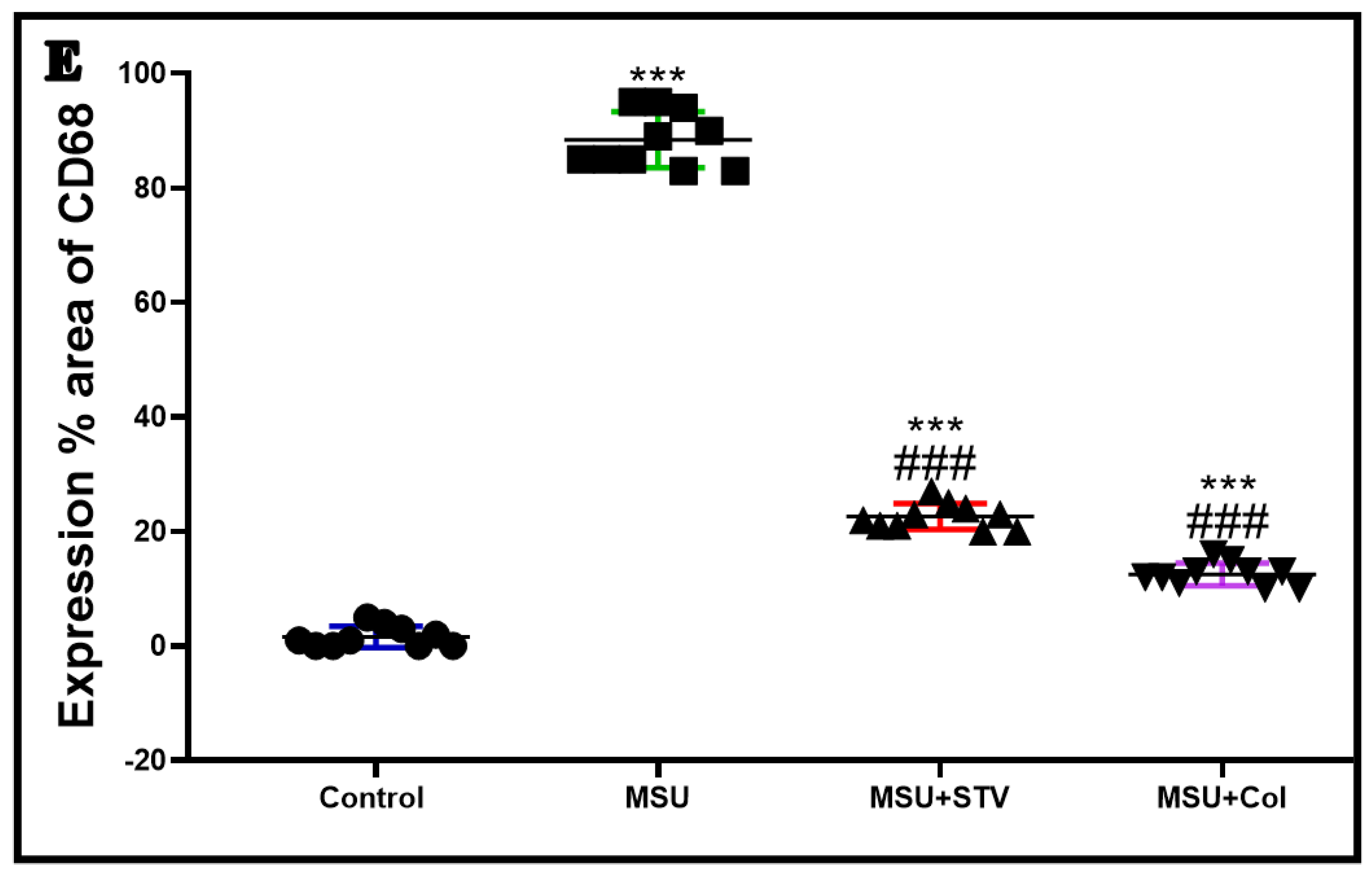
2.10. Immunohistochemical Assay of Nuclear Factor Kappa-B (NFκB), Nuclear Factor Erythroid 2-Related Factor 2 (Nrf2), Caspase 3, CD68, and Caspase 1

An amount of 4 μm of paraffin sections were deparaffinized by xylene. After that, they were rehydrated by ascending concentrations of ethanol. To block the endogenous peroxidase, we used 3% hydrogen peroxide for 30 min. Then, the sections were boiled in 0.01 M citrate buffer (pH6) and incubated in PBS, to expose the antigenic sites. Thereafter, the sections were incubated with primary antibodies of NF-𝜅B p65 (Rabbit polyclonal antibody, 1:100, ab86299, Abcam, Waltham, MA, USA), Caspase 3 (Rabbit monoclonal antibody, 1:100, ab32150, Abcam, USA), Caspase 1 (Rabbit polyclonal antibody, 1:100, ab194986, Abcam, USA) and Nrf2 (Rabbit monoclonal antibody, 1:100, ab32150, Abcam, USA) at 4 °C overnight, and rinsed 3 times with PBS. Then, biotin-labeled anti-rabbit IgG was added, and they were incubated with DAB for brown color detection. Lastly, the slides were washed with PBS and counterstained with hematoxylin for examination with a light microscope [24]. The density of immunostaining was quantified as the percent of synovial tissue occupied by positive staining (calculated by averaging the values from ten fields at 10× magnification) for each synovial tissue by using image J software.

2.11. Tissue Homogenate Measurement of TNFα, IL-1β, IL6, IL18, Cyclooxygenase-2 (Cox-2), Cytochrome c, Bcl-2 Associated X Protein (Bax), B-Cell Lymphoma 2 (Bcl-2), Heme Oxygenase-1 (HO-1) and NAD (P)H Quinone Dehydrogenase (NQO1) in Synovial Tissue

The protein levels of synovial tissue were examined by commercial ELISA kits for the pro-inflammatory markers; TNFα, IL-1β, IL6, and IL18 (catalog numbers; CSB-E11987r, E-EL-R0012, SEA079Ra, and SEA064Ra respectively), cytochrome C (catalog numbers: ab210575) Bax, Bcl-2 (E4513, CSB-E08854r respectively), HO-1 and NQO1 (catalog number E4525-100 and abx155911 respectively) following the instructions of the manufacturer.

2.12. RT-PCR Assessment

Extraction of the total RNA from right paw tissue of all the groups of the experiment was accomplished by TRIzols Reagent (15596026, Life Technologies, Carlsbad, CA, USA), the RNA was then washed by ethanol 75%. An amount of 1 μg of total RNA was reverse-transcribed into single-stranded complementary DNA by using QuantiTects Reverse Transcription Kit (Qiagen, Germantown, MD, USA) using a random primer hexamer in a two-step RT-PCR reaction in which any genomic DNA (gDNA) contamination was eliminated using gDNA Wipeout buffer. Total cDNA (30 ng), used as a template for amplification with the sequence of the specific primers for the NLRP3 gene, was; forward 5′-AGAAGAGACCACGGCAGAAG-3′, and reverse 5′-CCTTGGACCAGGTTCAGTGT-3′, (gene bank accession number is NM_031789.2). Peroxisome proliferator-activated receptor gamma PPARγ gene was; forward 5′-CTATCATTTGCTGTGGAGATCG-3′, and reverse 5′-CTGGTATGGGCCCCACTGGC-3′, (gene bank accession number is NM_012580.2). GADPH gene was; forward 5′-TGGGTGTGAACCATGAGAAG-3′, and reverse 5′-GCTAAGCAGTTGGTGGTGC-3′, (gene bank accession number is NM_199267.2) used at a 300 nM final concentration. Each sample was subjected to real-time PCR in duplicate and the mean values of the duplicates were used for subsequent analysis. Ubiquitin 5 (UBQ5) was used as a reference gene. Rotor-Gene Q collected data automatically and analyzed the value of threshold Cycle (Ct) which normalized to an average Ct value of the house-keeping genes (∆Ct) and the relative expression of each representative was calculated as 2–∆Ct.

2.13. Statistical Analysis

The data of our study were analyzed by GraphPad Prism software (version 9.0; GraphPad Software Inc., La Jolla, CA, USA). They were expressed as mean ± SD. The comparison between the different groups was performed using one-way analysis of variance, followed by Tukey–Kramer post-hoc analysis. While the data from ankle joint swelling were analyzed by repeated ANOVA, p < 0.05 was considered statistically significant.

3. Results

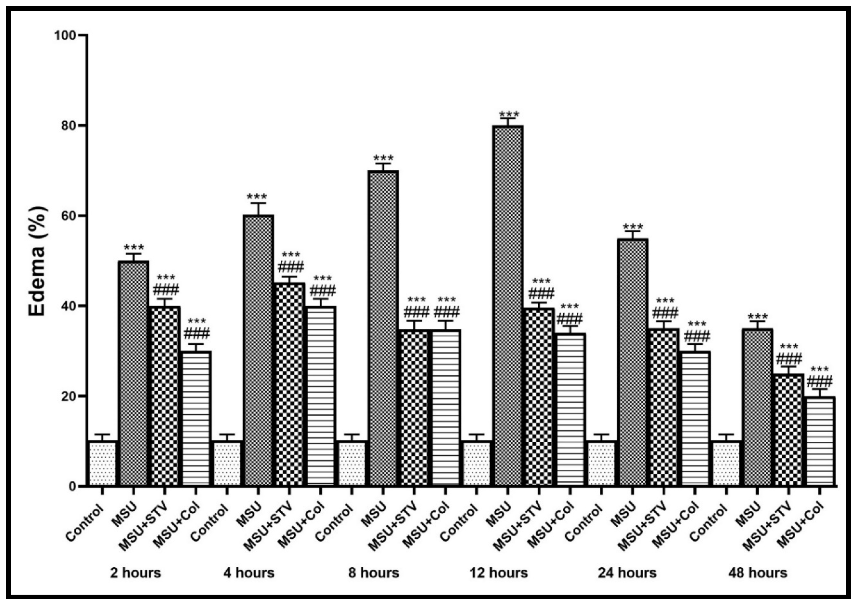
3.1. Effect of Stevia Extracts on the Edema of the Ankle Joint in Monosodium Urates Induced Gouty Arthritis

As shown in (Figure 1) MSU injection in the ankle joint produced knee joint swelling at different times (2, 4, 8, 12, 24, and 48 h) by 45%,60%,75%,80%,55%, and 35% respectively with maximum swelling at 12 h after injection which gradually subsides with time and markedly decreased at 48 h. This swelling was markedly decreased (p < 0.001) by pretreatment with Stevia extract and single Colchicine treatment compared with the MSU-group. This effect was confirmed by X-ray photographing of the ankle joint before scarification as there was mild swelling of soft tissue in the MSU-group, while Stevia and Colchicine groups showed near-normal soft tissue as presented in Figure 2.

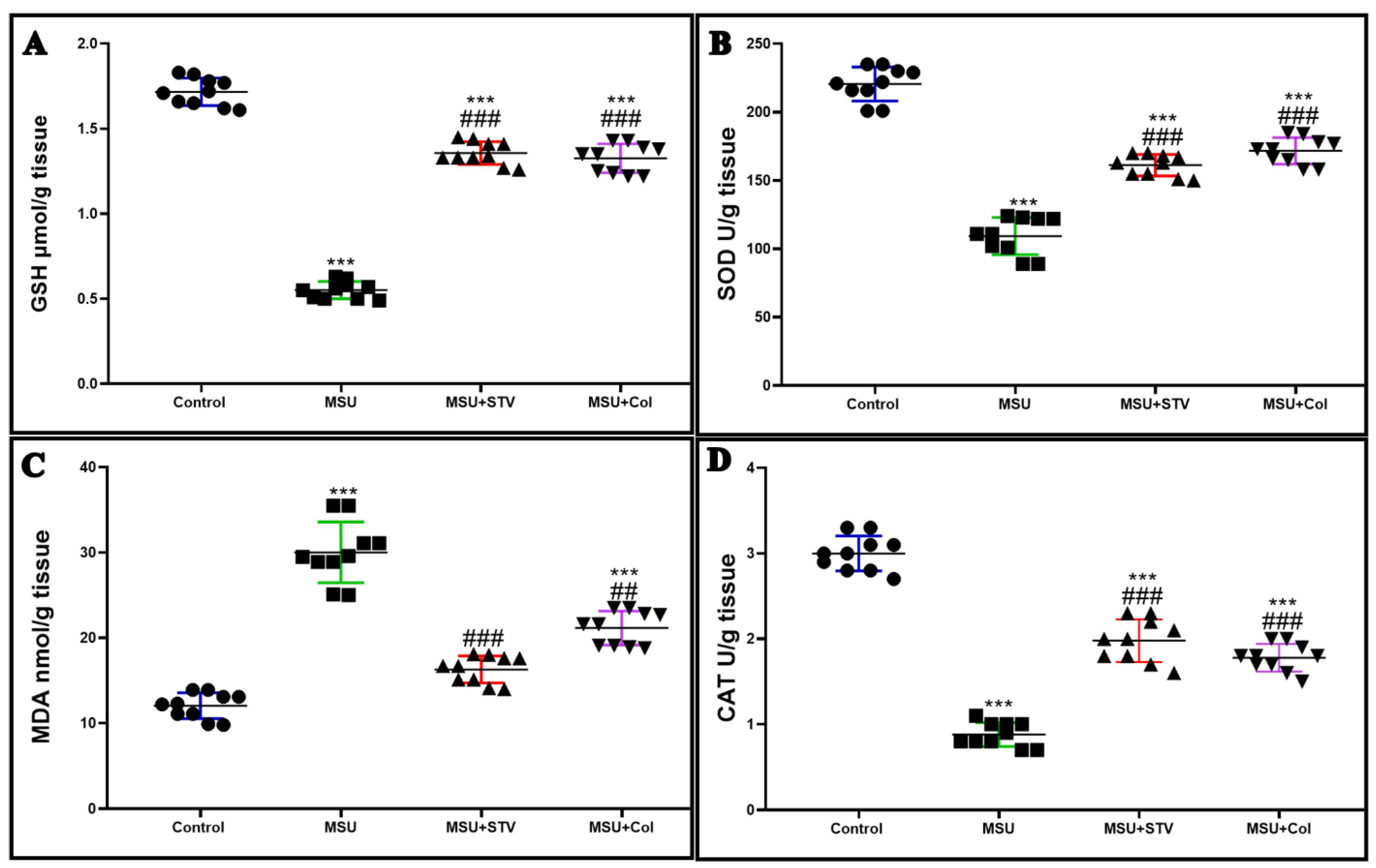
3.2. Ameliorative Effect of Stevia Extract on Oxidative Stress Marker in Synovial Tissues

The level of GSH and the activities of antioxidant enzymes; SOD and CAT in hind paw tissues were significantly decreased (p < 0.001) by 68%, 50%, and 70%, respectively in comparison with control groups. On the other hand, administration of Stevia and colchicine before induction of gouty arthritis significantly (p < 0.001 increased their activities (Table 1, Figure 3A,B,D). In contrast, acute gouty arthritis significantly increased MDA level in hind paw tissues by 141% (p < 0.001) in comparison to control group, while pretreatment with Stevia and colchicine markedly decreased its level by 46.5% (p < 0.001) and 29.7% (p < 0.001), respectively, in comparison to MSU group (Table 1, Figure 3C).

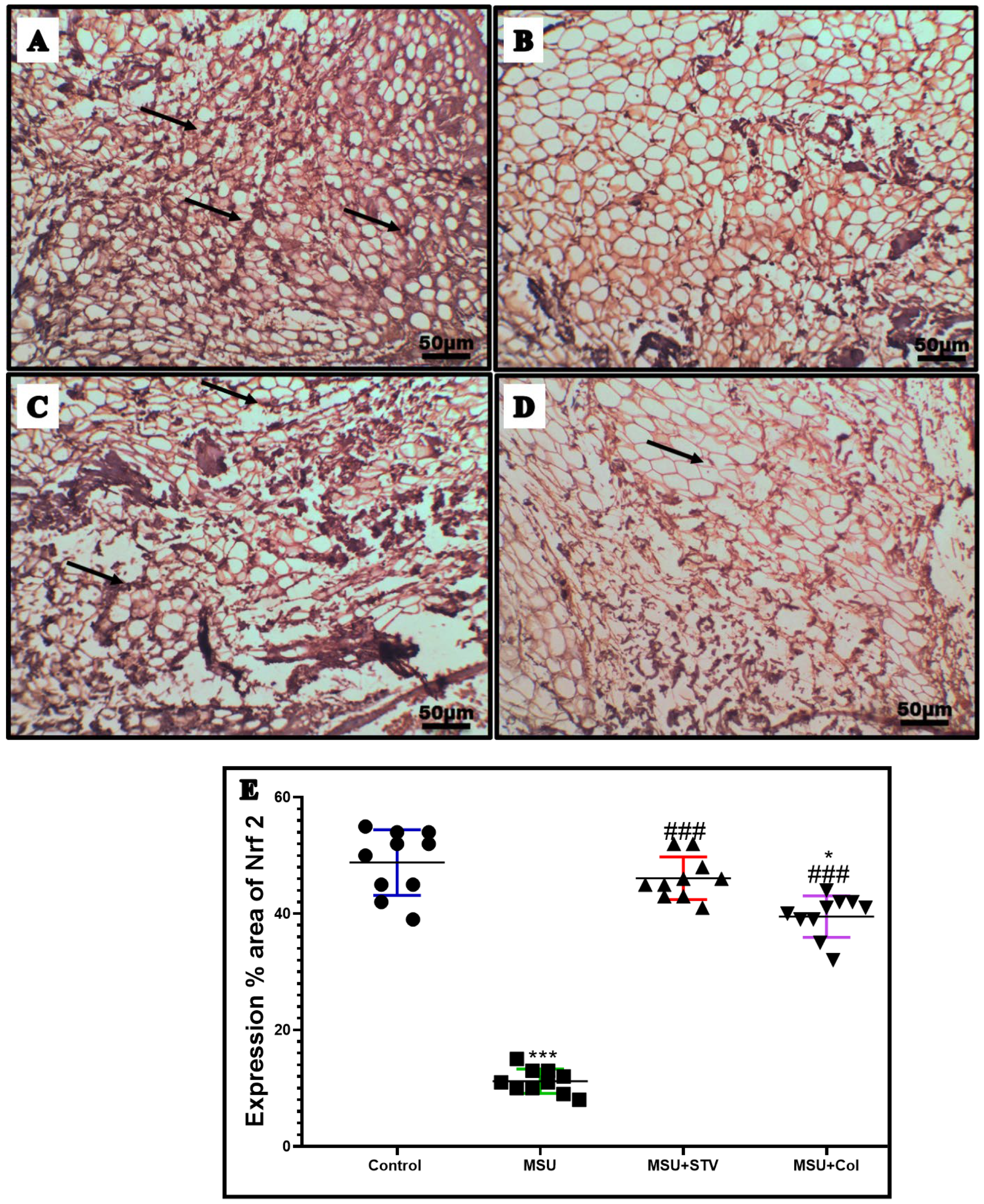
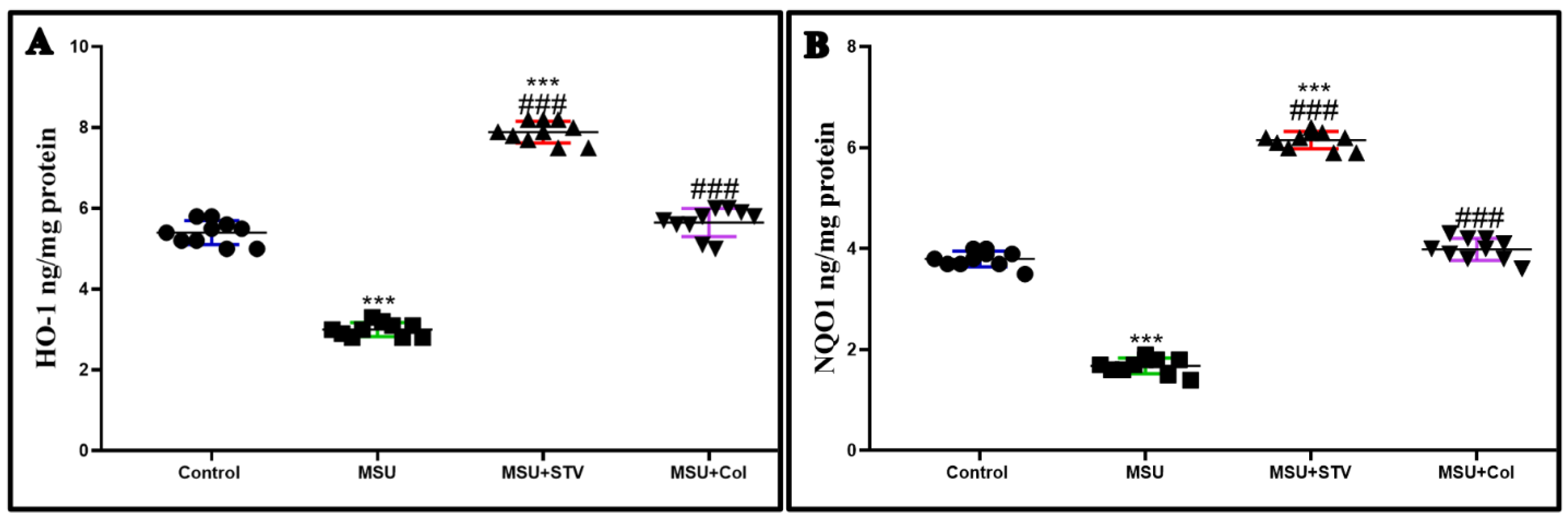
3.3. Impact of Stevia Extract on Nrf2/ARE Pathway in the Ankle Joint Synovial Tissue

MSU intra-articular injection in the ankle joint significantly (p < 0.001) downregulated the immune expression of Nrf2 by 77% as shown in Figure 4A,B, as well as the protein level of its Nrf2 targets HO-1 and NQO1 by 55%, and 57%, respectively, in relation to the control group. Meanwhile, the pretreatment with Stevia and colchicine significantly (p < 0.001) enhanced the immunoexpression of Nrf2 by 309%, and 254%, respectively (Figure 4C,D), and the protein level of HO-1 by 236% and 142%, and NQO1 by 260% and 135%, respectively, in relation to the MSU group as shown in (Figure 5A,B). These results explain the antioxidant effect of Stevia extract by upregulating Nrf2/ARE pathway and its positive impact on the endogenous antioxidant enzymes.

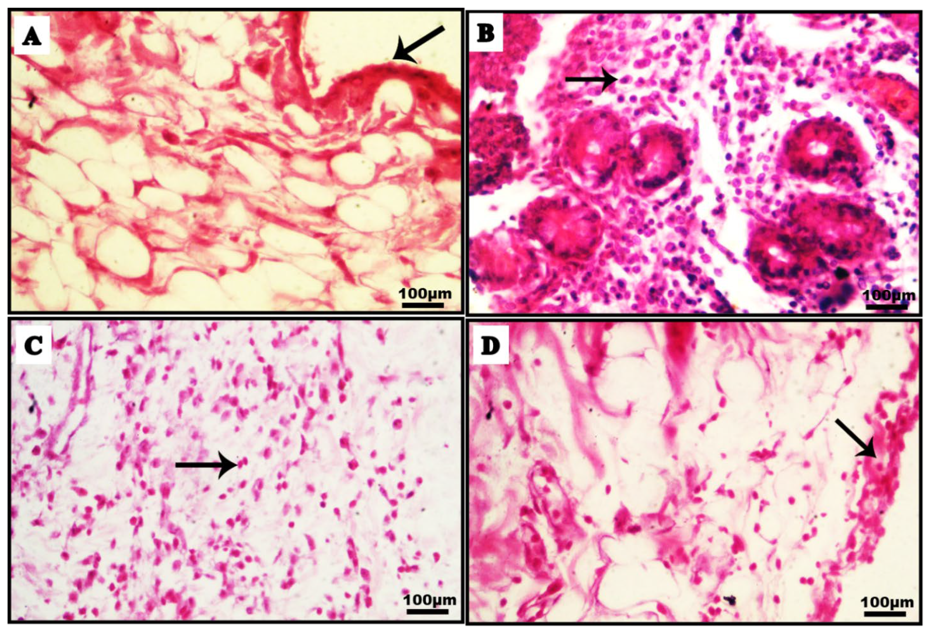
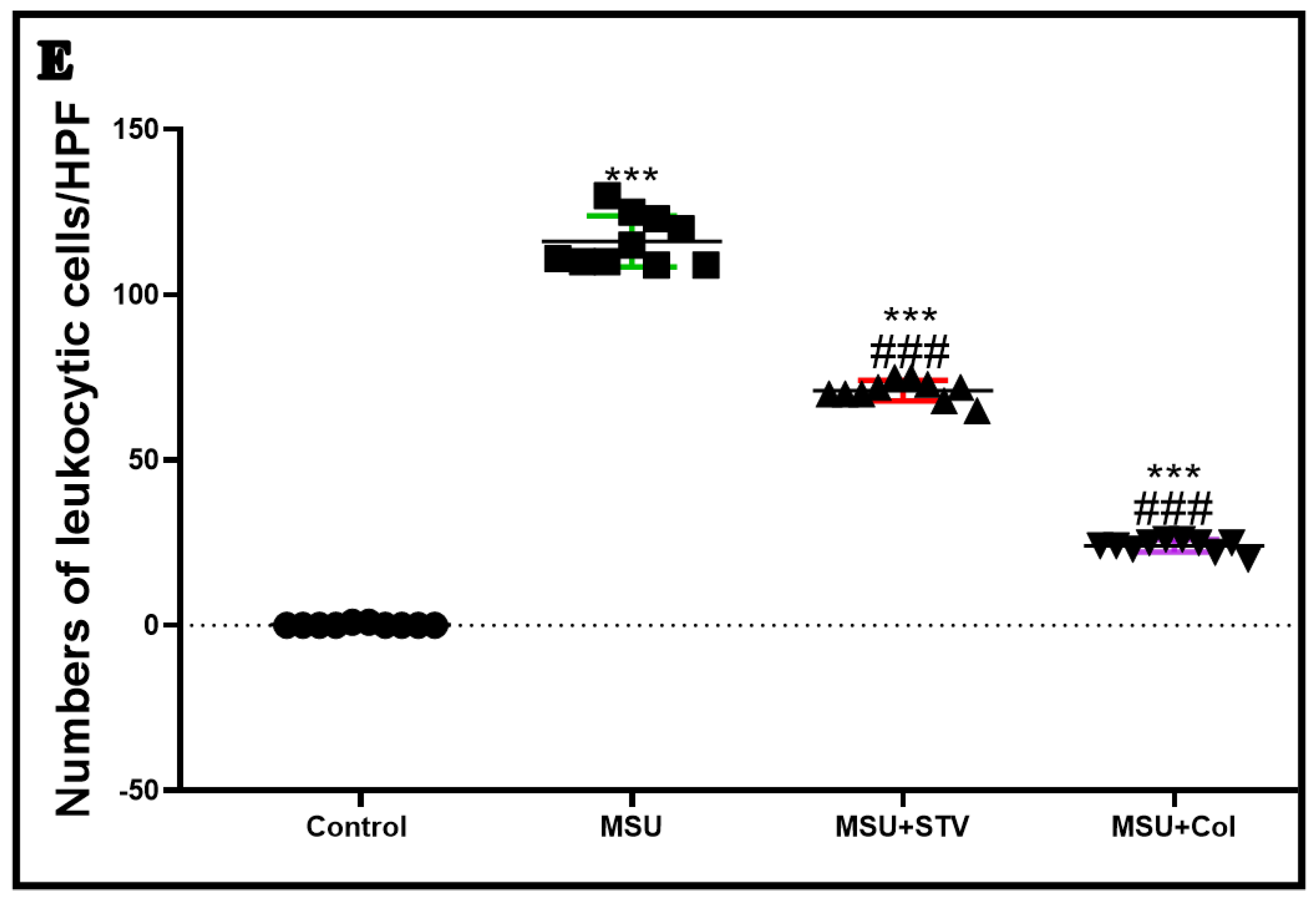
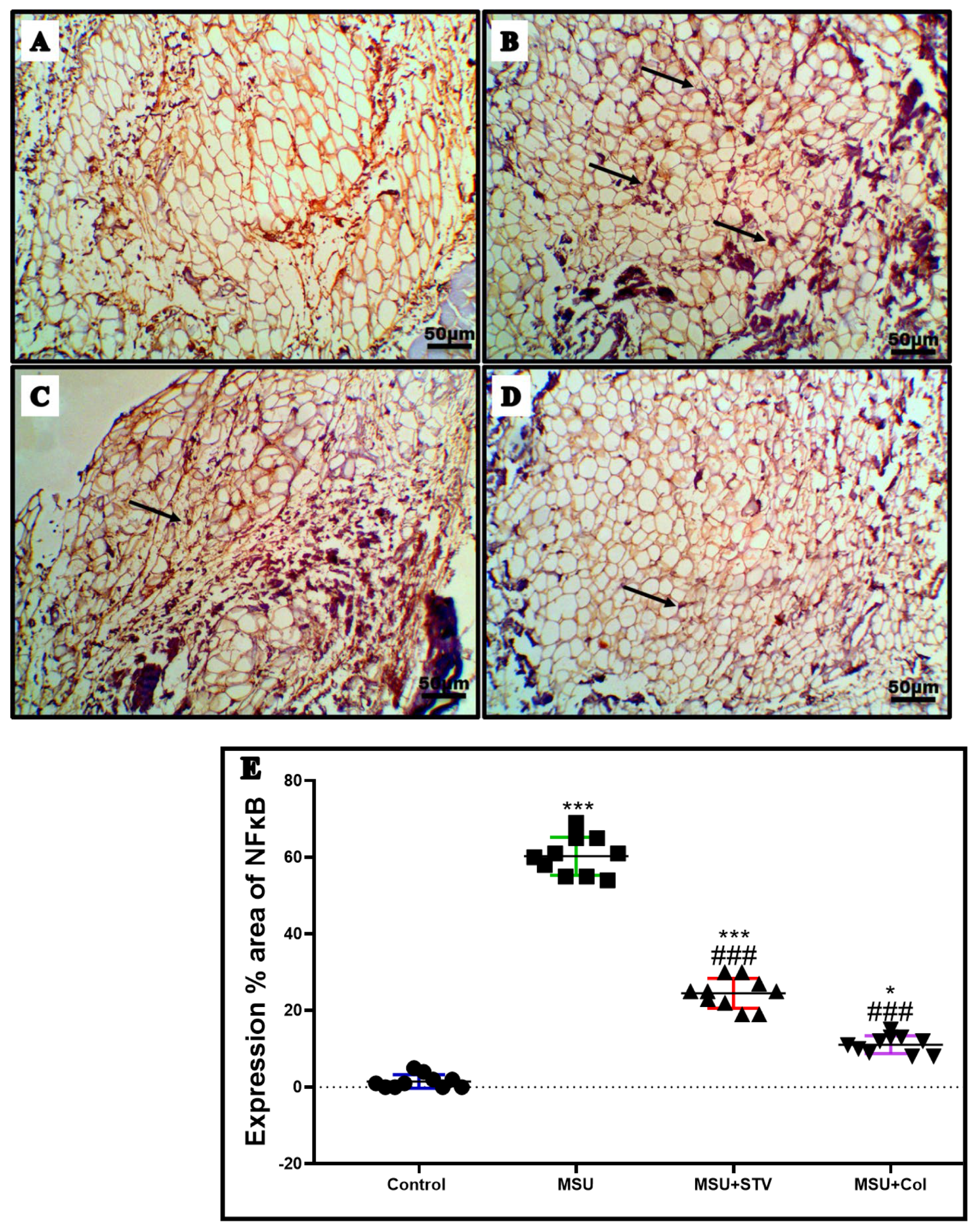
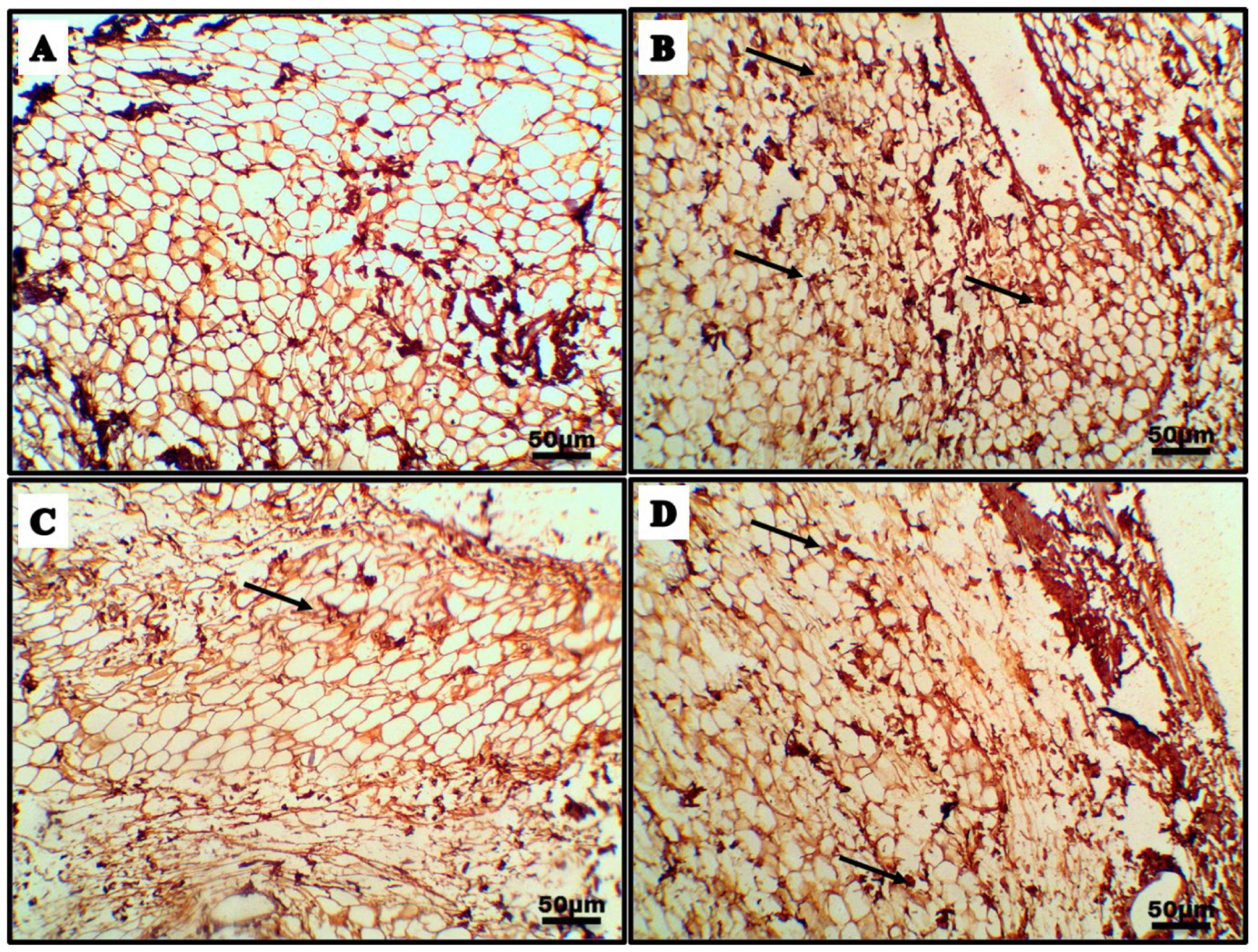
3.4. Ameliorative Effect of Stevia Extract on Synovial Tissue Neutrophil Infiltration of Gouty Arthritis

Examining the ankle joint synovial tissue by H&E staining revealed normal synovial tissue in control rats with severe neutrophil infiltration in the acute gouty arthritis group. On the contrary, pretreatment with Stevia and colchicine significantly (p < 0.001) decreased neutrophil infiltration by 38%, and 80% in comparison to the MSU group (Figure 6). Furthermore, treatment with Stevia and colchicine significantly (p < 0.001) decreased the number of positive CD68 cells, a marker of macrophage infiltration, by 75% and 86%, respectively, in comparison to the MSU group (Figure 7). From these findings we can see that, similar to colchicine, Stevia halts the leucocytic cell infiltration induced by MSU intra-articular injection.

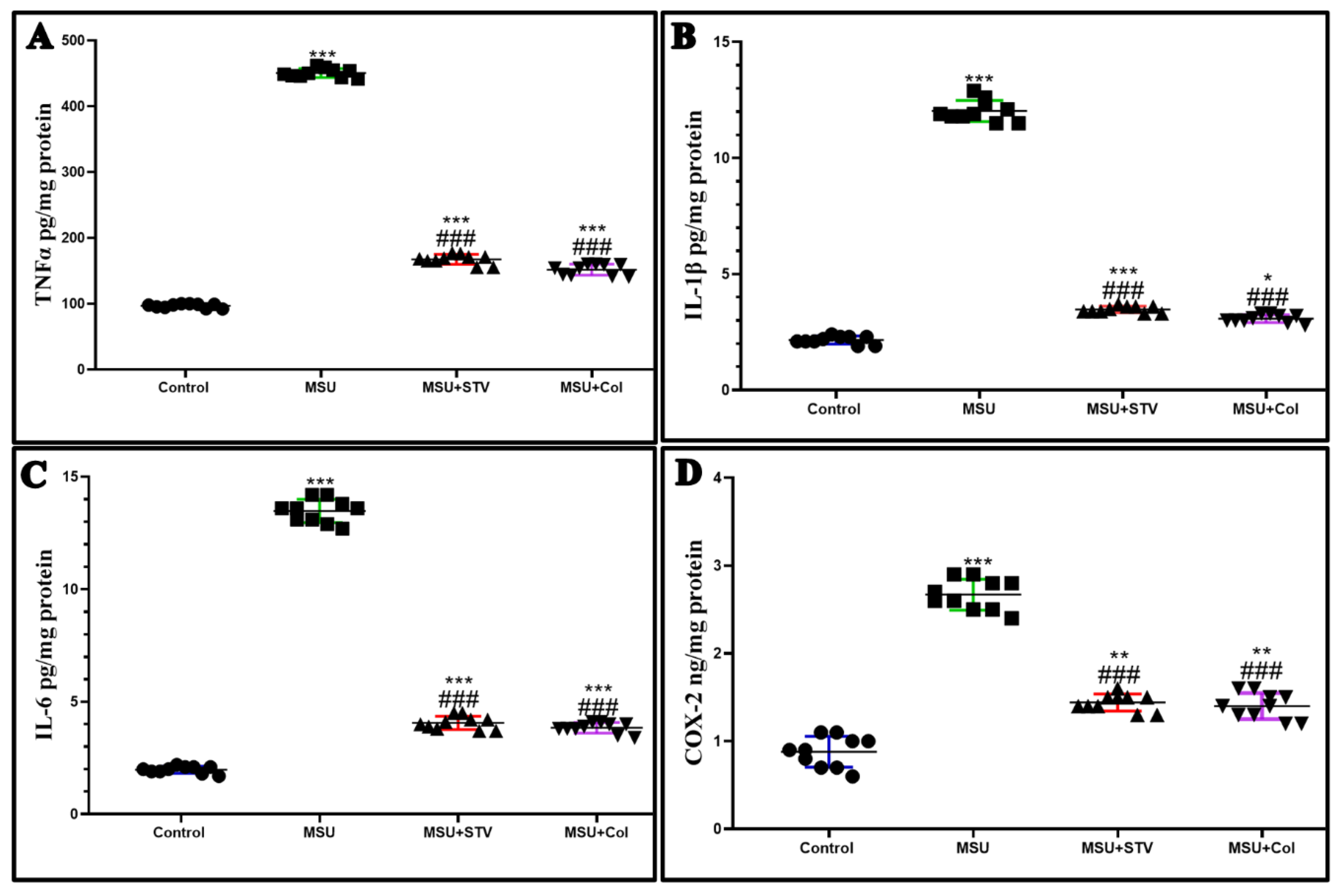
3.5. Ameliorative Effect of Stevia Extract on Synovial Tissue Inflammatory Markers

Injection of the ankle joint with 0.1 mL of MSU significantly increased (p < 0.001) the immune expression of the transcriptional factor NFκB signaling pathway by 97%, which subsequently enhanced the protein level of the proinflammatory markers; TNFα, IL-1β, IL6, and COX-2 by ELIZA by 364%, 454%, 576%, and 200%, respectively, in comparison to the control rats, indicating that the inflammation plays a pivotal role in the pathology of the acute gouty arthritis. On the other hand, pretreatment with Stevia extracts mitigated the inflammatory processes by significant (p < 0.001) downregulation of NFκB immunoexpression by 59% (Figure 8), and proinflammatory markers TNFα, IL-1β, IL6, and COX-2 protein level by 62%, 71%, 69%, and 45%, respectively, (Figure 9A–D) in comparison to MSU group, confirming the powerful anti-inflammatory effect of Stevia alongside its powerful antioxidant effect, similar to the anti-inflammatory effect of the colchicine.

3.6. Impact of Stevia Extract on PPARγ/NLRP3 Pathway in the Ankle Joint Synovial Tissue

MSU significantly decreased (p < 0.001) the gene expression of PPARγ of synovial tissue cells (Figure 10A) by 58%, which independently mediates the significant upregulation (p < 0.001) of gene expression of NLRP3 (Figure 10B) by 144% with a subsequent upregulation of immunoexpression of activated caspase-1 (Figure 11) by 92%, and further secretion of the proinflammatory markers IL-1B and IL18 (Figure 10C,D) by 454%, and 297%, respectively, in comparison to control group. Meanwhile, pretreatment with Stevia methanolic extract significantly (p < 0.001) enhanced the gene expression of PPARγ by 47%, with independent significant downregulation (p < 0.001) in the gene expression of NLRP3, and the immunoexpression of activated caspase 1 by 93% and proinflammatory markers IL-1β, and IL18 secretion by 24%, 71%, 56%, respectively, in relation to MSU group. This indicates that PPARγ/NLRP3 pathway-induced pyroptosis is involved in the pathology of acute gouty arthritis, and that pretreatment with Stevia extracts and colchicine modulates it.

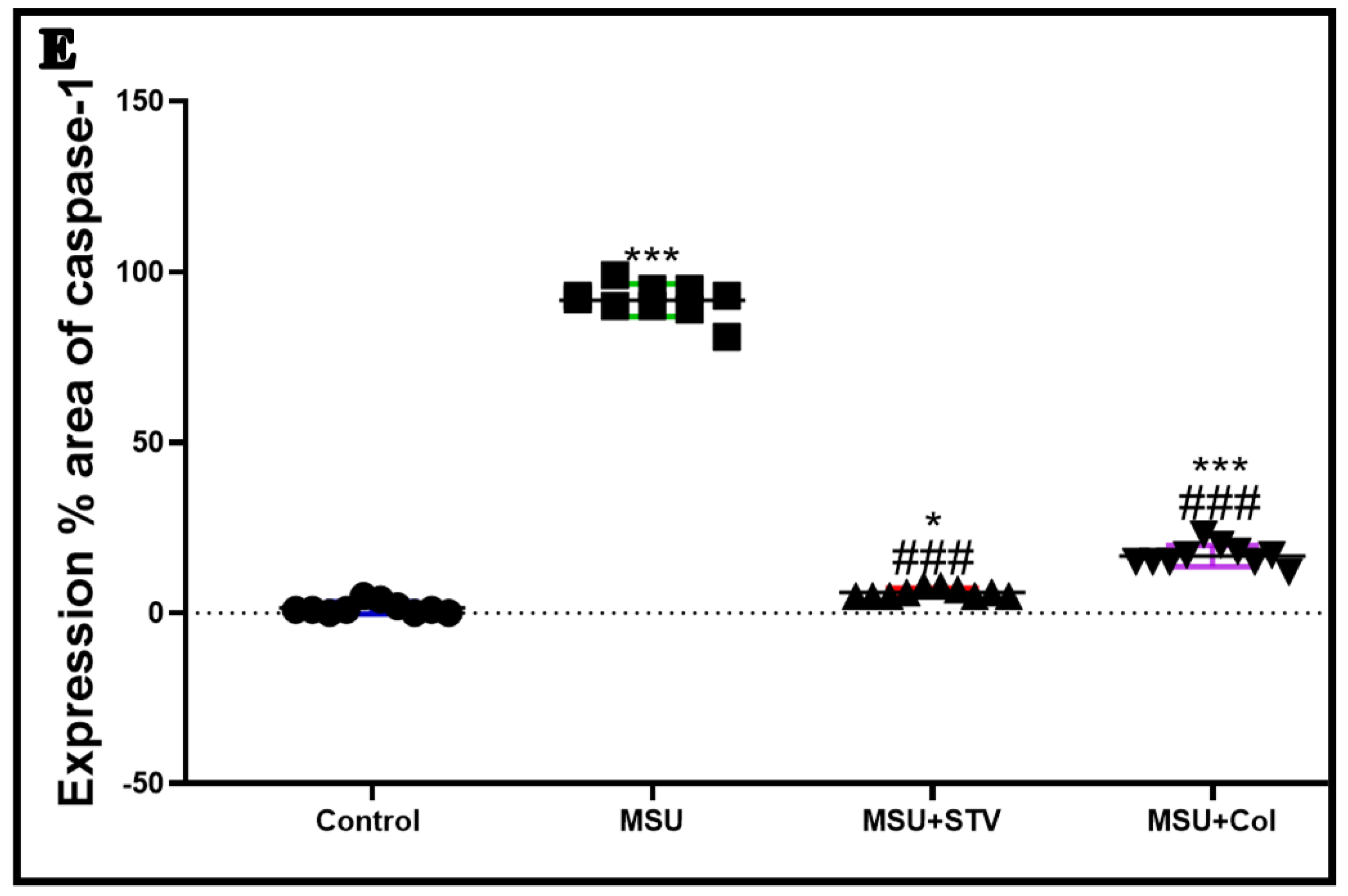
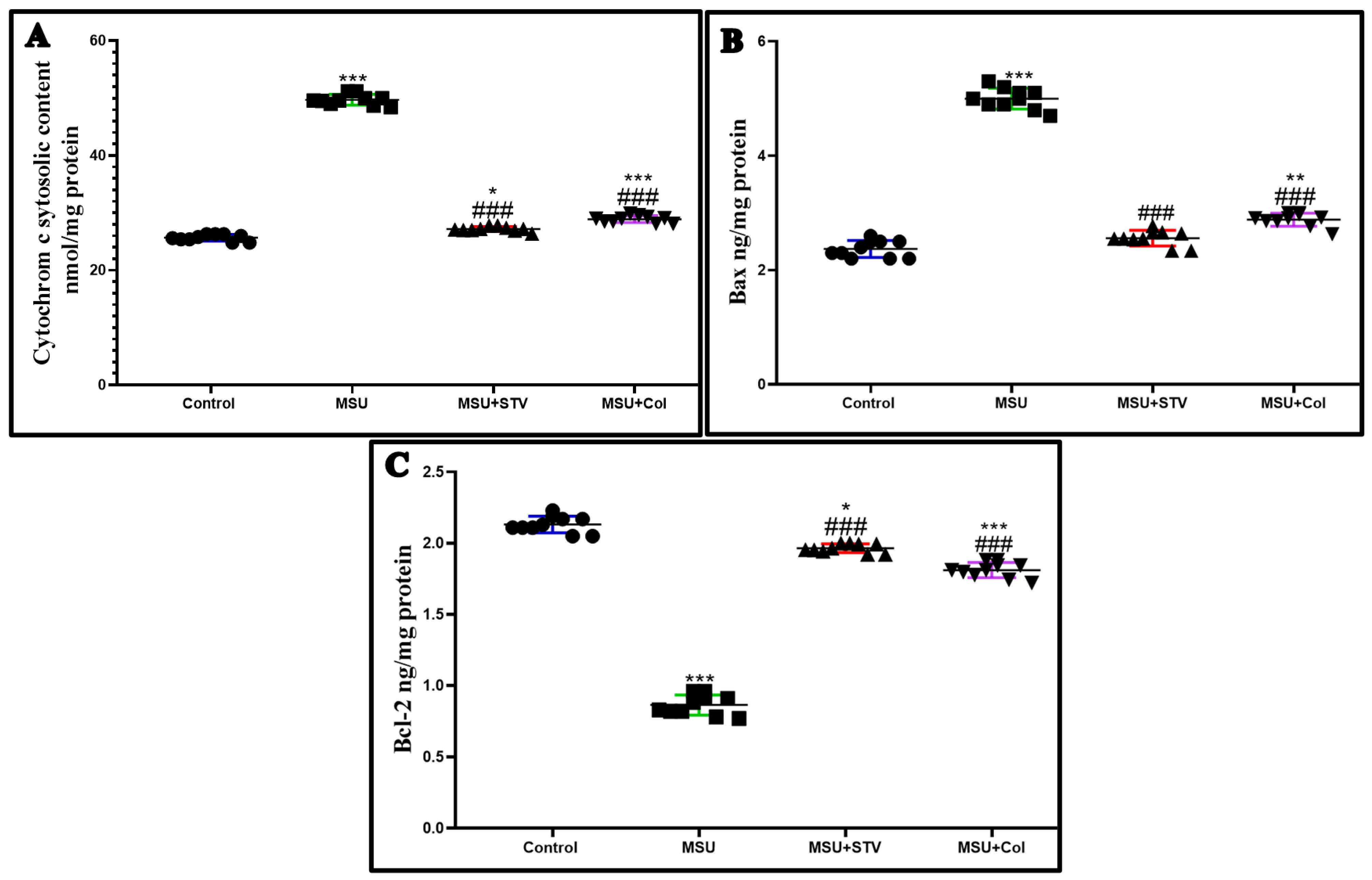
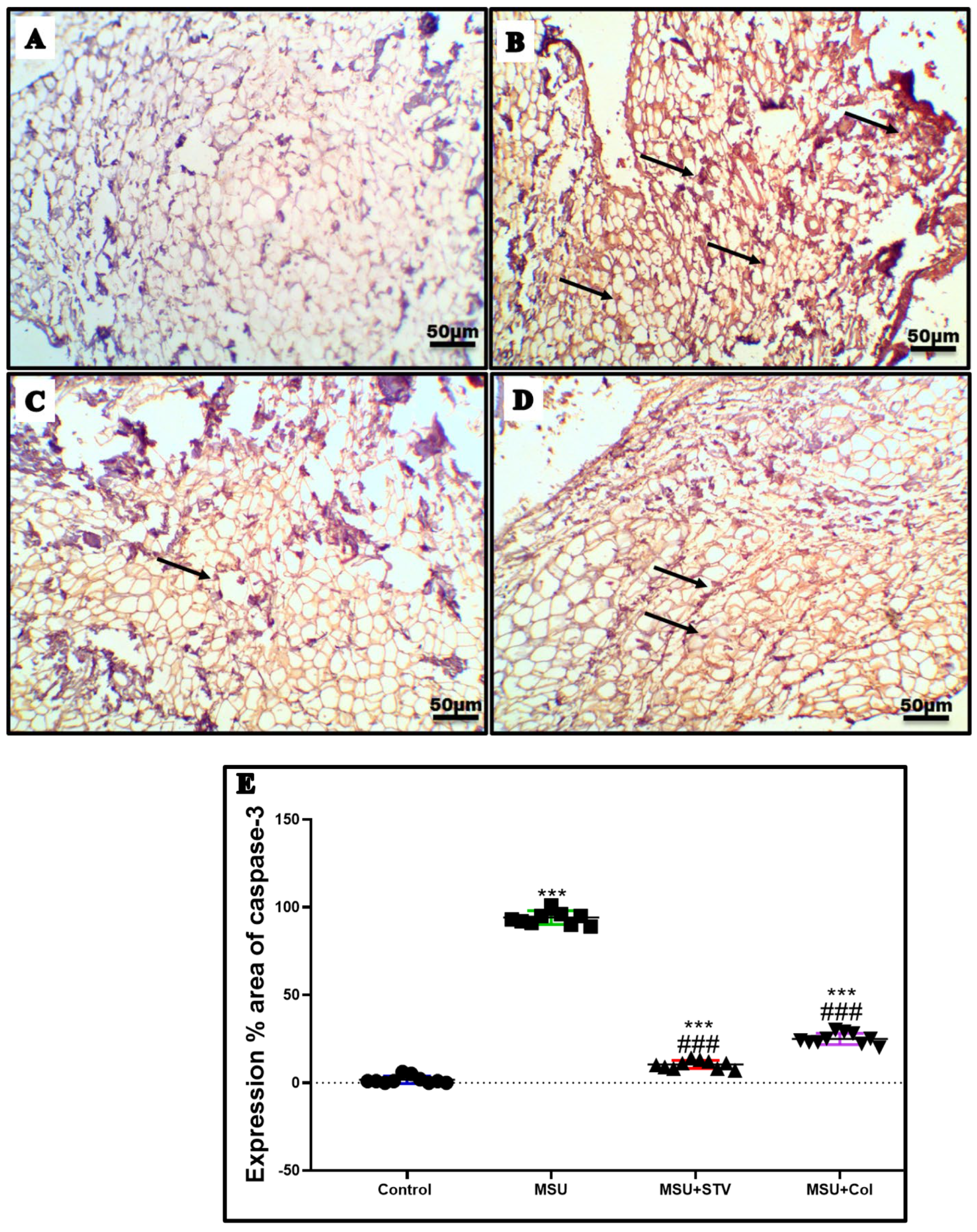
3.7. Stevia Extract Mitigates Synovial Cells’ Proapoptotic Markers Cytochrome C, BAX, BCL2, and Apoptotic Caspase 3 Cells Markers
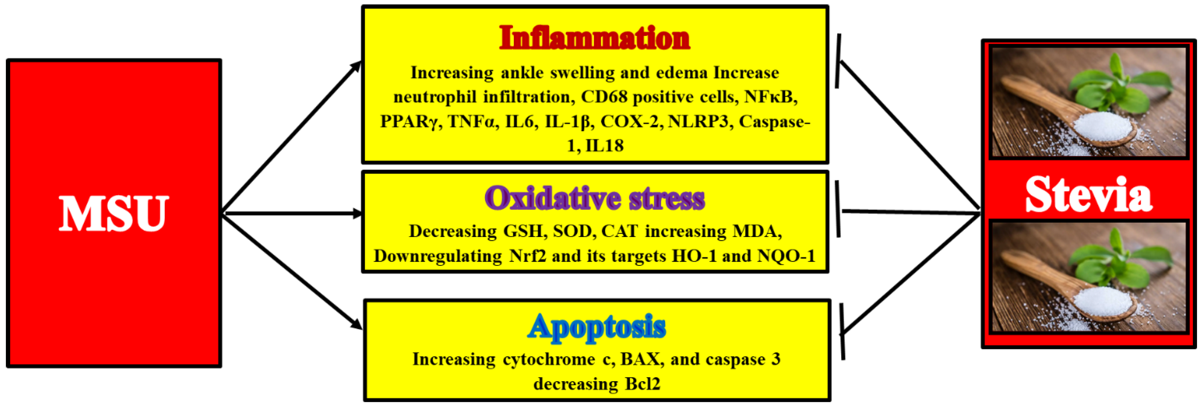
The findings revealed that MSU injection significantly p upregulated (p < 0.001) the protein level of proapoptotic cytosolic cytochrome c, and BAX by 94% and 108%, respectively (Figure 12A,B), and the immunoexpression of caspase-3 apoptotic cells by 98% (Figure 13), with downregulation of the beneficial proapoptotic BCL2 by 59%, in relation to control rats (Figure 12C). Interestingly, pretreatment with Stevia extract showed prominent anti-apoptotic activity by significant downregulation (p < 0.001) of the proapoptotic cytosolic cytochrome c and Bax by 45%, and 48%, respectively, and the immunoexpression of caspase-3 by 89%, with upregulation of the immunoexpression of BCL-2 by 126%, in comparison to MSU group.

4. Discussion

The results of our work demonstrate that treatment of MSU-induced acute gouty arthritis with stevia extract implies an anti-inflammatory effect by a decreasing of ankle joint edema, a histological amendment and decrease in the number of positive CD68 macrophages, a significant depression of NFκB signaling pathway and its TNFα, IL-1β, IL6, and COX-2 inflammatory cytokines, and upregulation of mRNA PPARγ gene expression with subsequent downregulation of NLRP3 inflammasomes, caspase-1, and IL-1β, IL18 cytokines. Secondly, the demonstrate that Stevia extract treatment shows an antioxidant effect by halting gouty arthritis-induced oxidative stress decreasing MAD and increasing GSHX, SOD, and CAT levels with upregulation of Nrf2 and its antioxidant response elements (ARE) HO-1 and NQO1. Lastly, they show that Stevia restrains the apoptosis of synovial cells by significantly downregulating cytochrome c, Bax, and caspase-3 and upregulating of BCL-2. This result confirms the antioxidant, anti-inflammatory, and antiapoptotic effects of Stevia on acute gouty arthritis. In our study, we have two targets: first, to examine the ameliorative effect of stevia extract on acute gouty arthritis; and second, to discuss the possible protective mechanisms.
Hyperuricemia is considered the primary cause of the gouty disease, characterized by the precipitation of MSU crystals inside the joint, causing severe ankle joint swelling [25], which is considered the main indicator of the inflammatory response. This is explained by the infiltration of neutrophils inside the synovial membrane and fluid; these inflammatory cells engulf MSU crystals disrupting its membrane with subsequent releases of proinflammatory cytokines [26]. In our study, MSU crystals stimulate the infiltration of the synovial membrane with inflammatory (Figure 6) cells with enhanced immunoexpression of CD68 positive macrophages (Figure 7), these findings conform to [21]. Alternatively, pretreatment with Stevia extract improves the ankle joint swelling, offers a significant histological decrease in the inflammatory cell’s infiltration, and a marked decrease in the CD68 positive macrophages.
NFκB signaling pathway is involved in many inflammatory diseases, one of which is acute gouty arthritis [27]. NFκB nuclear translocation stimulates proinflammatory cytokines TNFα, IL-1β, and IL6 target genes [28,29], upregulating COX-2 protein level with subsequent overproduction of prostaglandin E2 (PGE2) which induces severe inflammation and activity of the osteoblastic cells [30]. Therefore, NFκB transcription factor activation is considered one of the key regulators for the inflammatory response to gouty arthritis [31]. In our work, ankle joint injection with MSU increases the immunoexpression of NFκB (Figure 8) with elevated protein levels by ELIZA for TNFα, IL-1β, IL6, and COX-2 (Figure 9), similar to the treatment with colchicine, and consistent with Kou et al. [32] who demonstrated that injection of the knee joint with MSU crystals elevates synovial fluid and tissue protein level of NF-𝜅B p65, TNF-𝛼, IL-1𝛽 and IL-8. On the other hand, Stevia extract downregulates the NFκB and its proinflammatory cytokines expressions, and these results conform to the work done by Potočnjak et al. [33] who documented the anti-inflammatory effect of Stevia against cisplatin-induced nephrotoxicity by downregulating NFκB transcription factor with its proinflammatory cytokine TNFα.
At the same time, engulfment of the macrophage to MSU crystals activates the NLRP3 inflammasome [34], which regulates the activation of caspase 1-induced pyroptosis and maturation of pro-inflammatory cytokines IL-1β and IL18 (Figure 10 and Figure 11), these cytokines play a pivotal role in the mobilization of the inflammatory cells, with further release of inflammatory cytokines magnifying the inflammatory insult resulting from MSU crystals injection [35]. It was found that activation of the PPARγ, an endogenous modulator to cellular inflammation, downregulates NLRP3 inflammasome activation in the macrophages [36]. The downregulating effect of the PPARγ on the NLRP3 inflammasome [Figure 10] may be explained by its downregulation to NFκB signaling pathway [37]. In our study, MSU-induced gouty arthritis enhances the gene expression of NLR3 inflammasomes, with caspase-1 activation and cytokines IL-1β, IL18 cytokines secretion, in line with [38], and also downregulated PPARγ gene expression in line with [39]. On the contrary, pretreatment with Stevia extract upregulates the PPARγ gene expression in line with Mostafa et al. [40] who reported that ulcerative colitis treatment with Stevia extract upregulates the PPARγ mRNA expression. Furthermore, Stevia extract pretreatment downregulates the gene expression of NLRP3 inflammasomes. We believe we are the first to shed light on the negative impact of Stevia on NLRP3 inflammasome activation. From the aforementioned, the anti-inflammatory effect of Stevia extract is clear against MSU-induced gouty arthritis, decreasing the ankle joint edema, which is the first hallmark of the inflammatory process, and, in addition, improving the histological structure of the synovial tissue by modulating inflammatory signaling pathways, similar to colchicine treatment.
It was found that ROS overproduction plays a complementary role in the inflammatory response [41]. Infiltration of the synovial fluids with neutrophils after MSU injection induces oxidative insult which potentiates the inflammatory process with vascular permeability and ankle joint edema [42,43]. Therefore, targeting the oxidative stress by Stevia extract shows a beneficial role in combating MSU-induced inflammation. In our study, MSU injection significantly downregulated antioxidant enzymes, Nrf2 and its targets HO-1 and NQo1 (Figure 4 and Figure 5) while increasing the MDA level. This indicates lipid peroxidation, so that these findings conform to the study done Jhang et al. [44] who reported that MSU precipitation in the synovial tissue disrupts the balance between the prooxidant and oxidant, promoting NLR3 inflammasomes-activation as well as downregulating the Nrf2/HO-1 pathway. Fortunately, Stevia extract treatment increases the level of antioxidant enzymes GSHX, SOD, and CAT (Table 1, Figure 3), transcription factor Nrf2 and its targets HO-1 and NQo1, while decreasing the level of MDA lipid peroxidation marker, so that these results are consistent with Zhao et al. [45] who demonstrated the antioxidant effect of Stevia extract on D-galactose-induced aging in mice by scavenging ROS and enhancing the Nrf2/ARE pathway. The antioxidant power of Stevia extract, alongside its anti-inflammatory effect, improves ankle joint edema by decreasing inflammatory cells staffing and NALP3 inflammasomes activation-induced pyroptosis.
There is a very limited amount of research discussing the relationship between gouty arthritis and apoptosis, or on MSU-induced synovial cell apoptosis by the production of an excessive amount of ROS with mitochondrial function impairment [46]. In our study, MSU crystal injection increased cytochrome c, Bax protein levels by ELIZA and caspase-3 immunoexpression with a decrease of BCL-2 protein levels (Figure 12 and Figure 13). However, on the contrary, Stevia extract treatment exhibits an anti-apoptotic effect by downregulating cytochrome c, Bax and caspase-3 as well as upregulating BCL-2, in line with Potočnjak et al. [34] who demonstrated the protective effect of Stevia against nephrotoxicity induced by cisplatin injection by mitigating renal cellular apoptosis, downregulating the expression of Bax, P53 and caspase-9, and upregulating Bcl-2. This work is also in line with El Nashar et al. [47] who have stated that the treatment of phenylbutazone-induced epilepsy with Stevia extracts restrains the apoptosis of hippocampal cells by downregulating the immunoexpression of P53 and caspase-3. Therefore, treating acute gouty arthritis with Stevia attenuates the synovial and vascular endothelial cells’ apoptosis, decreasing the vascular permeability with further joint edema. The antiapoptotic property of the Stevia is explained by its scavenging of ROS production.

5. Conclusions

Stevia extract exhibits anti-inflammatory, antioxidant, and antiapoptotic effects on MSU-induced gouty arthritis. These effects were observed as a downregulating effect on MSU-induced inflammation (downregulating NFκB, TNFα, IL-1β, IL6, NLRP3, caspase-1, and IL18, as well as upregulating PPARγ), oxidative stress (enhancing SOD, GSHX, CAT, Nrf2, HO-1, and NQO1 as well as decreasing MDA) and apoptosis (downregulating cytochrome c, Bax, caspase-3 and upregulating Bcl-2). From all of this, we can see that Stevia, as a safe natural treatment, could be a great protective treatment for acute gouty arthritis. A graphical scheme for the findings of the study is presented in (Figure 14).

Author Contributions

A.M.B.: Data curation, methodology, supervision, visualization, writing & original draft. M.T.: conceptualization, methodology, supervision, visualization, writing & original draft. S.T.E.: methodology, writing—review & editing. I.E.-S.: methodology. B.A.A.: writing—review & editing. H.M.: formal analysis. A.G.A.-K.: supervision, visualization. K.F.: investigation. T.A.S.B.: conceptualization. M.M.I.: methodology, writing—review & editing. All authors have read and agreed to the published version of the manuscript.

Funding

This research did not receive any specific grant from funding agencies in the public, commercial, or not-for-profit sectors.

Institutional Review Board Statement

All animal protocols were approved by the Research Ethics Committee, Faculty of Veterinary Medicine, Mansoura University, with a code number (Code No R/135).

Informed Consent Statement

Not applicable.

Data Availability Statement

Not applicable.

Acknowledgments

The authors would like to thank the Deanship of Scientific Research at Umm Al-Qura University for supporting this work by Grant Code: (22UQU4410180DSR01).

Conflicts of Interest

The authors declare no conflict of interest.

References

  1. Harre, U.; Derer, A.; Schorn, C.; Schett, G.; Herrmann, M. T cells as key players for bone destruction in gouty arthritis? Arthritis Res. Ther. 2011, 13, 135. [Google Scholar] [CrossRef] [PubMed]
  2. Molloy, E.S.; McCarthy, G.M. How crystals damage tissue. Curr. Rheumatol. Rep. 2004, 6, 228–234. [Google Scholar] [CrossRef] [PubMed]
  3. Liu-Bryan, R. Intracellular innate immunity in gouty arthritis: Role of NALP3 inflammasome. Immunol. Cell Biol. 2010, 88, 20–23. [Google Scholar] [CrossRef] [PubMed]
  4. Wei, H.; Hu, C.; Xie, J.; Yang, C.; Zhao, Y.; Guo, Y.; Mei, Z.; Chen, L.; Lan, Z. Doliroside A attenuates monosodium urate crystals-induced inflammation by targeting NLRP3 inflammasome. Eur. J. Pharmacol. 2014, 740, 321–328. [Google Scholar] [CrossRef]
  5. Ponce, L.; Arjona, M.; Blanco, G.; Alvarez, S.; Arcila, E.; Ortega, A.; Nuñez, D.; Verzura, J.; Tovar, R.; Bethencourt, S.; et al. The effect of montelukast in a model of gouty arthritis induced by sodium monourate crystals. Investig. Clin. 2011, 52, 15–22. [Google Scholar]
  6. Desai, J.; Steiger, S.; Anders, H.J. Molecular pathophysiology of gout. Trends Mol. Med. 2017, 23, 756–768. [Google Scholar] [CrossRef]
  7. Kim, Y.; Oh, H.-C.; Park, J.W.; Kim, I.-S.; Kim, J.-Y.; Kim, K.-C.; Chae, D.-S.; Joo-Hyoun, S.; Song, J.-H. Diagnosis and Treatment of Inflammatory Joint Disease. Hip Pelvis 2017, 29, 211–222. [Google Scholar] [CrossRef]
  8. Kim, S.-K.; Choe, J.-Y.; Park, K.-Y. Rebamipide Suppresses Monosodium Urate Crystal-Induced Interleukin-1β Production Through Regulation of Oxidative Stress and Caspase-1 in THP-1 Cells. Inflammation 2016, 39, 473–482. [Google Scholar] [CrossRef]
  9. Pouliot, M.; James, M.J.; McColl, S.R.; Naccache, P.H.; Cleland, L.G. Monosodium urate microcrystals induce cyclooxygenase-2 in human monocytes. Blood 2012, 91, 1769–1776. [Google Scholar] [CrossRef]
  10. Schlesinger, N.; Moore, D.F.; Sun, J.D.; Schumacher, H.R. A survey of current evaluation and treatment of gout. J. Rheumatol. 2006, 33, 2050–2052. [Google Scholar]
  11. Keith, M.P.; Gilliland, W.R. Updates in the Management of Gout. Am. J. Med. 2007, 120, 221–224. [Google Scholar] [CrossRef] [PubMed]
  12. Eleftheriou, G.; Bacis, G.; Fiocchi, R.; Sebastiano, R. Colchicine-induced toxicity in a heart transplant patient with chronic renal failure. Clin. Toxicol. 2008, 46, 827–830. [Google Scholar] [CrossRef] [PubMed]
  13. Jayaprakash, V.; Ansell, G.; Galler, D. Colchicine overdose: The devil is in the detail. N. Z. Med. J. 2007, 120, 1248. [Google Scholar]
  14. Lemus-Mondaca, R.; Vega-Gálvez, A.; Zura-Bravo, L.; Ah-Hen, K. Stevia rebaudiana Bertoni, source of a high-potency natural sweetener: A comprehensive review on the biochemical, nutritional and functional aspects. Food Chem. 2012, 132, 1121–1132. [Google Scholar] [CrossRef]
  15. Chen, T.H.; Chen, S.C.; Chan, P.; Chu, Y.L.; Yang, H.Y.; Cheng, J.T. Mechanism of the hypoglycemic effect of stevioside, a glycoside of Stevia rebaudiana. Planta Med. 2005, 71, 108–113. [Google Scholar] [CrossRef]
  16. Cekic, V.; Vasovic, V.; Jakovljevic, V.; Mikov, M.; Sabo, A. Hypoglycemic action of stevioside and a barley and brewer’s yeast-based preparation in the experimental model on mice. Bosn. J. Basic Med. Sci. 2011, 11, 11–16. [Google Scholar] [CrossRef]
  17. Momtazi, A.A.; Esmaeili, S.-A.; Abdollahi, E.; Sahebkar, A. A Review on the Pharmacology and Toxicology of Steviol Glycosides Extracted from Stevia rebaudiana. Curr. Pharm. Des. 2017, 23, 1616–1622. [Google Scholar] [CrossRef]
  18. Ghaheri, M.; Miraghaee, S.; Babaei, A.; Mohammadi, B.; Kahrizi, D.; Haghighi, Z.M.S.; Bahrami, G. Effect of Stevia rebaudiana Bertoni extract on sexual dysfunction in Streptozotocin-induced diabetic male rats. Cell. Mol. Biol. 2018, 64, 6–10. [Google Scholar] [CrossRef]
  19. Kahrizi, D.; Ghari, S.; Ghaheri, M.; Fallah, F.; Ghorbani, T.; Beheshti, A.A.A.; Kazemi, E.; Ansarypour, Z. Effect of KH2PO4 on gene expression, morphological and biochemical characteristics of Stevia rebaudiana Bertoni under in vitro conditions. Cell. Mol. Biol. 2017, 63, 107–111. [Google Scholar] [CrossRef]
  20. Huang, J.; Zhu, M.; Tao, Y.; Wang, S.; Chen, J.; Sun, W.; Li, S. Therapeutic properties of quercetin on monosodium urate crystal-induced inflammation in rat. J. Pharm. Pharmacol. 2012, 64, 1119–1127. [Google Scholar] [CrossRef]
  21. Han, J.; Xie, Y.; Sui, F.; Liu, C.; Du, X.; Liu, C.; Feng, X.; Jiang, D. Zisheng Shenqi decoction ameliorates monosodium urate crystal-induced gouty arthritis in rats through anti-inflammatory and anti-oxidative effects. Mol. Med. Rep. 2016, 14, 2589–2597. [Google Scholar] [CrossRef]
  22. Alavala, S.; Nalban, N.; Sangaraju, R.; Kuncha, M.; Jerald, M.K.; Kilari, E.K.; Sistla, R. Anti-inflammatory effect of stevioside abates Freund’s complete adjuvant (FCA)-induced adjuvant arthritis in rats. Inflammopharmacology 2020, 28, 1579–1597. [Google Scholar] [CrossRef]
  23. El-Mousalamy, A.M.D.; Hussein, S.A.M.; Hussein, A.M.; Mahmoud, S.A.; El Azab, K.M. Reno Protective Effect of Methanolic Stevia rebaudiana Bertoni Leaves Extract and Its Phenolic Compounds in Type-1-Diabetes. Egypt. J. Chem. 2018, 61, 609–615. [Google Scholar]
  24. Ramos-Vara, J.A. Technical Aspects of Immunohistochemistry. Veter. Pathol. 2005, 42, 405–426. [Google Scholar] [CrossRef]
  25. Martinon, F. Mechanisms of uric acid crystal-mediated autoinflammation. Immunol. Rev. 2010, 233, 218–232. [Google Scholar] [CrossRef]
  26. Sabina, E.P.; Nagar, S.; Rasool, M. A Role of Piperine on Monosodium Urate Crystal-Induced Inflammation—An Experimental Model of Gouty Arthritis. Inflammation 2011, 34, 184–192. [Google Scholar] [CrossRef]
  27. Ghosh, S.; Karin, M. Missing pieces in the NF-κB puzzle. Cell 2002, 109, S81–S96. [Google Scholar] [CrossRef]
  28. Park, M.H.; Hong, J.T. Roles of NF-κB in Cancer and Inflammatory Diseases and Their Therapeutic Approaches. Cells 2016, 5, 15. [Google Scholar] [CrossRef]
  29. Hayden, M.S.; Ghosh, S. Signaling to NF-B. Genes Dev. 2004, 18, 2195–2224. [Google Scholar] [CrossRef]
  30. Laavola, M.; Nieminen, R.; Yam, M.F.; Sadikun, A.; Asmawi, M.Z.; Basir, R.; Welling, J.; Vapaatalo, H.; Korhonen, R.; Moilanen, E. Flavonoids Eupatorin and Sinensetin Present in Orthosiphon stamineus Leaves Inhibit Inflammatory Gene Expression and STAT1 Activation. Planta Med. 2012, 78, 779–786. [Google Scholar] [CrossRef]
  31. Lee, J.; Scagel, C.F. Chicoric acid: Chemistry, distribution, and production. Front. Chem. 2013, 1, 40. [Google Scholar] [CrossRef]
  32. Kou, Y.-Y.; Li, Y.-F.; Xu, M.; Li, W.-Y.; Yang, M.; Li, R.-L. Effects of RuPeng15 Powder (RPP15) on Monosodium Urate Crystal-Induced Gouty Arthritis in Rats. Evid. Based Complement. Altern. Med. 2015, 2015, 527019. [Google Scholar] [CrossRef]
  33. Potočnjak, I.; Broznić, D.; Kindl, M.; Kropek, M.; Vladimir-Knežević, S.; Domitrović, R. Stevia and stevioside protect against cisplatin nephrotoxicity through inhibition of ERK1/2, STAT3, and NF-κB activation. Food Chem. Toxicol. 2017, 107, 215–225. [Google Scholar] [CrossRef]
  34. Schumacher, H.R., Jr. Febuxostat: A non-purine, selective inhibitor of xanthine oxidase for the management of hyperuricemia in patients with gout. Expert Opin. Investig. Drugs 2005, 14, 893–903. [Google Scholar] [CrossRef]
  35. Dalbeth, N.; Haskard, D.O. Mechanisms of inflammation in gout. Rheumatology 2005, 44, 1090–1096. [Google Scholar] [CrossRef]
  36. Yang, C.-C.; Wu, C.-H.; Lin, T.-C.; Cheng, Y.-N.; Chang, C.-S.; Lee, K.-T.; Tsai, P.-J.; Tsai, Y.-S. Inhibitory effect of PPARγ on NLRP3 inflammasome activation. Theranostics 2021, 11, 2424–2441. [Google Scholar] [CrossRef]
  37. Jiang, C.; Ting, A.T.; Seed, B. PPAR-gamma agonists inhibit production of monocyte inflammatory cytokines. Nature 1998, 391, 82–86. [Google Scholar] [CrossRef]
  38. Wang, Q.; Lin, B.; Li, Z.; Su, J.; Feng, Y. Cichoric Acid Ameliorates Monosodium Urate-Induced Inflammatory Response by Reducing NLRP3 Inflammasome Activation via Inhibition of NF-kB Signaling Pathway. Evid. Based Complement. Altern. Med. 2021, 2021, 8868527. [Google Scholar] [CrossRef] [PubMed]
  39. Wang, J.; Chen, G.; Lu, L.; Zou, H. Sirt1 inhibits gouty arthritis via activating PPARγ. Clin. Rheumatol. 2019, 38, 3235–3242. [Google Scholar] [CrossRef] [PubMed]
  40. Mostafa, A.F.; Elalfy, M.M.; Shata, A.; Elhadidy, M.G. Prophylactic effect of aquatic extract of stevia on acetic acid induced-ulcerative colitis in male rats: A possible role of Nrf2 and PPARγ. J. Basic Clin. Physiol. Pharmacol. 2020, 32, 1093–1104. [Google Scholar] [CrossRef]
  41. Rasool, M.; Varalakshmi, P. Suppressive effect of Withania somnifera root powder on experimental gouty arthritis: An in vivo and in vitro study. Chem. Biol. Interact. 2006, 164, 174–180. [Google Scholar] [CrossRef] [PubMed]
  42. Martin, W.J.; Herst, P.M.; Chia, E.W.; Harper, J.L. Sesquiterpene dialdehydes inhibit MSU crystal-induced superoxide production by infiltrating neutrophils in an in vivo model of gouty inflammation. Free Radic. Biol. Med. 2009, 47, 616–621. [Google Scholar] [CrossRef] [PubMed]
  43. Afonso, V.; Champy, R.; Mitrovic, D.; Collin, P.; Lomri, A. Reactive oxygen species and superoxide dismutases: Role in joint diseases. Jt. Bone Spine 2007, 74, 324–329. [Google Scholar] [CrossRef]
  44. Jhang, J.-J.; Cheng, Y.-T.; Ho, C.-Y.; Yen, G.-C. Monosodium urate crystals trigger Nrf2- and heme oxygenase-1-dependent inflammation in THP-1 cells. Cell. Mol. Immunol. 2015, 12, 424–434. [Google Scholar] [CrossRef] [Green Version]
  45. Zhao, L.; Yang, H.; Xu, M.; Wang, X.; Wang, C.; Lian, Y.; Mehmood, A.; Dai, H. Stevia residue extract ameliorates oxidative stress in D-galactose-induced aging mice via Akt/Nrf2/HO-1 pathway. J. Funct. Foods 2019, 52, 587–595. [Google Scholar] [CrossRef]
  46. Zamudio-Cuevas, Y.; Martínez-Flores, K.; Fernández-Torres, J.; Loissell-Baltazar, Y.A.; Medina-Luna, D.; López-Macay, A.; Camacho-Galindo, J.; Hernández-Díaz, C.; Santamaría-Olmedo, M.G.; Villegas, E.O.L.; et al. Monosodium urate crystals induce oxidative stress in human synoviocytes. Arthritis Res. Ther. 2016, 18, 117. [Google Scholar] [CrossRef]
  47. El Nashar, E.M.; Obydah, W.; Alghamdi, M.A.; Saad, S.; Yehia, A.; Maryoud, A.; Kiwan, N.A.; Alasmari, W.A.; Hussein, A.M. Effects of Stevia rebaudiana Bertoni extracts in the rat model of epilepsy induced by pentylenetetrazol: Sirt-1, at the crossroads between inflammation and apoptosis. J. Integr. Neurosci. 2022, 21, 21. [Google Scholar] [CrossRef]
Figure 1. Effects of Stevia extracts on knee swelling in MSU-induced gouty arthritis rats. Rats were pretreated with either vehicle, Stevia extracts, or standard drug colchicine. Knee joint swellings were evaluated at (2, 4, 8, 12, 24, 48 h). All the above table data are expressed as mean ± SD. Repeated one-way ANOVA with Tukey’s post hoc analysis. *** p < 0.001, significant vs. control group ### p < 0.001, significant vs. MSU group.
Processes 10 01751 g001
Figure 2. Therapeutic effects of Stevia on swollen paw rats with monosodium urate-induced gouty arthritis. (A) Representative X-ray picture of rat hind paws before scarification shows normal soft tissue in control rats. (B) Mild paw swelling was seen in monosodium urate-induced gouty arthritis. (C,D) Near-normal soft tissue was observed in rats treated with Stevia 250 mg/kg and colchicine groups respectively.
Processes 10 01751 g002
Figure 3. Antioxidant levels in all animal groups (n = 10) (A) GSH, (B) SOD, (C) MDA and (D) Catalase levels in paw tissues. All data are expressed as mean ± SD. One-way ANOVA test followed by Tukey’s post Hoc test, *** p < 0.001, vs. control group, ### p < 0.001, ## p < 0.01 vs. MSU group.
Processes 10 01751 g003
Figure 4. Microscopic pictures of immunostained sections of ankle joint against Nrf2 showing prominent positive brown staining (n = 10) in the control group (A), marked decrease in positive brown staining (black arrows) in urate group (B), much more increased positive brown staining (black arrows) in Stevia group (C), increased positive brown staining (black arrow) in Col group (D). IHC counterstained with Mayer’s hematoxylin. magnification X: 400 bar 50 (E). The graph represents a statistical analysis of Nrf2 expression % in joint tissues showing significantly decreased percentages in the urate group that significantly increased in treated groups. All data are expressed as mean ± SD. One-way ANOVA test followed by Tukey’s post hoc test, *** p < 0.001, * p < 0.05 vs. control group, ### p < 0.001, vs. MSU group.
Processes 10 01751 g004
Figure 5. Effect of Stevia extract treatment on (A) HO-1, (B) NQO1 protein level by ELISA in synovial tissue of the different groups (n = 10). All data are expressed as mean ± SD. One-way ANOVA test followed by Tukey’s post hoc test, *** p < 0.001, vs. control group, ### p < 0.001, vs. MSU group.
Processes 10 01751 g005
Figure 6. Microscopic pictures of H&E stained sections of ankle joint showing absent inflammation in (A) the control group, (B) massive leukocytic cells infiltration (black arrow) in the urate group, (C) fewer leukocytic cells infiltration (black arrow) in the Stevia group, (D) much fewer leukocytic cells in the colchicine group. X: 100 bar 100. (E) The graph represents a statistical analysis of leukocytic cell numbers in joint tissues showing significantly higher numbers in the urate group that significantly decreased in treated groups (n = 10). All data are expressed as mean ± SD. One-way ANOVA test followed by Tukey’s post hoc test, *** p < 0.001, vs. control group, ### p < 0.001, vs. MSU group.
Processes 10 01751 g006aProcesses 10 01751 g006b
Figure 7. Microscopic pictures of immunostained sections of the ankle joint against CD68 showing negative staining in the control group (A), many CD68-labelled cells (black arrows) in urate group (B), fewer CD68-labelled cells (black arrow) in Stevia group (C), much fewer CD68-labelled cells (black arrows) in col group (D). IHC counterstained with Mayer’s hematoxylin. magnification X: 400 bar 50 (E). The graph represents a statistical analysis of CD68 expression % in joint tissues showing significantly higher percentages in the urate group that significantly decreased in treated groups (n = 10). All data are expressed as mean ± SD. One-way ANOVA test followed by Tukey’s post hoc test, *** p < 0.001, vs. control group, ### p < 0.001, vs. MSU group.
Processes 10 01751 g007aProcesses 10 01751 g007b
Figure 8. Microscopic pictures of immunostained sections of the ankle joint against NF-kb showing negative staining in the control group (A), prominent positive brown staining (black arrows) in urate group (B), moderate positive brown staining (black arrow) in Stevia group (C), mild positive brown staining (black arrow) in col group (D). IHC counterstained with Mayer’s hematoxylin. X: 400 bar 50. (E) The graph represents a statistical analysis of NF expression % in joint tissues showing significantly higher percentages in the urate group that significantly decreased in treated groups (n = 10). All data are expressed as mean ± SD. One-way ANOVA test followed by Tukey’s post hoc test, *** p < 0.001, * p < 0.05 vs. control group, ### p < 0.001, vs. MSU group.
Processes 10 01751 g008
Figure 9. Effect of Stevia extract treatment on (A) TNFα, (B) IL-1β, (C) IL6, and (D) COX-2 protein level by ELISA in synovial tissue of the different groups (n = 10). All data are expressed as mean ± SD. One-way ANOVA test followed by Tukey’s post hoc test, *** p < 0.001, ** p < 0.01 and * p < 0.05 vs. control group, ### p < 0.001, vs. MSU group.
Processes 10 01751 g009
Figure 10. Effect of Stevia extract treatment on gene expression of (A) PPARγ and (B) NLRP3 and protein level by ELISA for (C) IL-1β (D) IL18 in synovial tissue of the different groups (n = 10). All data are expressed as mean ± SD. One-way ANOVA test followed by Tukey’s post hoc test, *** p < 0.001, and * p < 0.05 vs. control group, ### p < 0.001, vs. MSU group.
Processes 10 01751 g010
Figure 11. Microscopic pictures of immunostained sections of the ankle joint against caspase 1 showing negative staining in the control group (A), prominent positive brown staining (black arrows) in urate group (B), mild positive brown staining (black arrow) in Stevia group (C), moderate positive brown staining (black arrows) in Col group (D). IHC counterstained with Mayer’s hematoxylin. magnification X: 400 bar 50. (E) The graph represents a statistical analysis of caspase 1 expression % in joint tissues showing significantly higher percentages in the urate group that significantly decreased in treated groups n = 10). All data are expressed as mean ± SD. One-way ANOVA test followed by Tukey’s post hoc test, *** p < 0.001, * p < 0.05 vs. control group, ### p < 0.001, vs. MSU group.
Processes 10 01751 g011aProcesses 10 01751 g011b
Figure 12. Effect of Stevia extract treatment on protein level of (A) cytochrome c, (B) BAX and (C) BCL-2 by ELISA in synovial tissue of the different groups (n = 10). All data are expressed as mean ± SD. One-way ANOVA test followed by Tukey’s post hoc test, *** p < 0.001, ** p < 0.01 and * p < 0.05 vs. control group, ### p < 0.001, vs. MSU group.
Processes 10 01751 g012
Figure 13. Microscopic pictures of immunostained sections of the ankle joint against caspase 3 showing negative staining in the control group (A), prominent positive brown staining (black arrows) in urate group (B), moderate positive brown staining (black arrow) in Stevia group (C), mild positive brown staining (black arrows) in col group (D). IHC counterstained with Mayer’s hematoxylin. magnification X: 400 bar 50. (E) The graph represents a statistical analysis of caspase 3 expression % in joint tissues showing significantly higher percentages in the urate group that significantly decreased in treated groups (n = 10). All data are expressed as mean ± SD. One-way ANOVA test followed by Tukey’s post hoc test, *** p < 0.001, vs. control group, ### p < 0.001, vs. MSU group.
Processes 10 01751 g013
Figure 14. A graphical scheme for the findings of the study.
Processes 10 01751 g014
Table 1. Effect of Stevia on oxidative stress markers (SOD, GSH, MDA, and CAT) in paw tissues.
Control GroupMSU GroupMSU+STV GroupMSU+Col Group
GSH (μmol/g tissue)1.72. ± 0.0850.55 ± 0.053 ***1.36 ± 0.07 *** ###1.32 ± 0.090 *** ###
SOD (U/g tissue)220 ± 13.2109 ± 14.5 ***161 ± 8.2*** ###172 ± 10.4 *** ###
MDA (nmol/g tissue)12.6 ± 1.5930.47 ± 3.76 ***16.3 ± 1.68 ###21.4 ± 2.1 *** ##
CAT (U/g tissue)3.02 ± 0.190.9 ± 0.15 ***2 ± 0.25 *** ###1.8 ± 0.14 *** ###
All the above table data were expressed as mean ± SD. One-way ANOVA with Tukey’s post hoc analysis. *** p < 0.001, significant vs. control group ### p < 0.001, ## p < 0.01 significant vs. MSU group.
Publisher’s Note: MDPI stays neutral with regard to jurisdictional claims in published maps and institutional affiliations.

