Article

Zinc Oxide Nanoparticles (ZnO NPs), Biosynthesis, Characterization and Evaluation of Their Impact to Improve Shoot Growth and to Reduce Salt Toxicity on Salvia officinalis In Vitro Cultivated

Department of Botany and Microbiology, College of Science, King Saud University, Riyadh 11451, Saudi Arabia
*
Authors to whom correspondence should be addressed.
Processes 2022, 10(7), 1273; https://doi.org/10.3390/pr10071273
Submission received: 2 May 2022 / Revised: 23 June 2022 / Accepted: 24 June 2022 / Published: 28 June 2022
(This article belongs to the Special Issue Green Synthesis of Metallic Nanomaterials and Their Applications)

Abstract

:
Green synthesis of zinc oxide nanoparticles (ZnO NPs) using plant extracts have recently attracted considerable attention due to their environmental protection benefits and their easy and low cost of fabrication. In the current study, ZnO NPS were synthesized using the aqueous extract of Ochradenus arabicus as a capping and reducing agent. The obtained ZnO NPs were firstly characterized using ultraviolet visible (UV-Vis) spectroscopy, Fourier transform infrared (FTIR), transmission electron microscope (TEM), X-ray diffraction (XRD), energy dispersive X-ray absorption (EDX), zeta potential, and zeta size. All these techniques confirmed the characteristic features of the biogenic synthesized ZnO NPs. Then, ZnO NPs were evaluated for their effects on morphological, biochemical, and physiological parameters of Salvia officinalis cultured in Murashige and Skoog medium containing 0, 75, 100, and 150 mM of NaCl. The results showed that ZnO NPs at a dose of 10 mg/L significantly increased the shoot number, shoot fresh weight, and shoot dry weight of Salvia officinalis subjected or not to the salt stress. For the shoot length, a slight increase of 4.3% was recorded in the plant treated by 150 mM NaCl+10 mg/L ZnO NPs compared to the plant treated only with 150 mM of NaCl. On the other hand, without NaCl, the application of both concentrations 10 mg/L and 30 mg/L of ZnO NPs significantly improved the total chlorophyll content by 30.3% and 21.8%, respectively. Under 150 mM of NaCl, the addition of 10 mg/L of ZnO NPs enhanced the total chlorophyll by 1.5 times, whilst a slight decrease of total chlorophyll was recorded in the plants treated by 150 mM NaCl + 30 mg/L ZnO NPs. Additionally, ZnO NPs significantly enhance the proline accumulation and the antioxidative enzyme activities of catalase (CAT), superoxide dismutase (SOD), and glutathione reductase (GR) in plants under salinity. Our findings revealed that green synthesized ZnO NPs, especially at a dose of 10 mg/L, play a crucial role in growth enhancement and salt stress mitigation. Hence, this biosynthesized ZnO NPs at a concentration of 10 mg/L can be considered as effective nanofertilizers for the plants grown in salty areas.

1. Introduction

Salt is the most important devastating stressor that leads to an imbalance in edaphoclimatic conditions [1]. Due to high soil salinity, approximately 6% of fertile land around the world has become uncultivable [2]. Globally, it was noted that about 800 million hectares of arable land are affected by salt. Moreover, freshwater supplies are being strained by the world’s rapidly rising population. As a result, saline water is increasingly being used to irrigate crops [3]. Thus, plant growth can be severely hampered when soil is subjected to salt stress [4]. The vegetative growth parameters including number of branches, plant height, fresh weight, and dry weight of S. offcinalis under saline soil were reduced [5]. Additionally, salinity imposes harmful effects on physiological processes involving oxidative damages in the plant cell [1]. The serious impairment in cell biochemistry and physiological development because of salt stress can disrupt ionic homeostasis in plants. This generates an ion toxicity and osmotic pressure, which leads to oxidative stress at the subcellular level [6]. More absorption and translocation of chlorine (Cl) and sodium (Na+) ions in plant tissue can lead to serious biochemical changes and physiological development [7]. In S. offcinalis, it was noted that salinity tolerance was increased by spraying different concentrations of salicylic acid (SA). On the other hand, for okra plants it was observed that Cochliobolus sp. was used as an efficient biochemical modulator to alleviate salinity stress. Thus, salinity tolerance can be increased through the biological tools using the beneficial microorganisms as well as by chemical compounds applications [8,9,10]. Naturally, plants differ from one to another, with some being able to withstand higher levels of salt stress than others. For instance, the Halophyte plants are better equipped to withstand high salt levels [11,12].
Recently, nanoparticles (NPs) biogenic synthesis has attracted a great deal attention from many researchers since these processes are feasible, less toxic, and ecofriendly. The biogenic synthesis of the NPs is performed through the use of the plant extracts or other biological systems such as fungi, yeast, and bacteria [13]. The biosynthesized nanoparticles are widely used to protect plant growth against abiotic stresses such as salinity drought, heavy metals, flooding, extreme temperatures, etc. [14]. In this regard, it was observed that zinc oxide nanoparticles play an important role in the plant growth and provide protection against salt stress in different plant species like, Abelmoschus esculentus L. [15], soybean [16], Spinacia oleracea L. [17], and Eustoma grandiflorum [18]. Nanoparticles are particles with a diameter of fewer than 100 nanometers in at least one dimension. This distinguishes them from macro-sized particles in terms of physical and chemical properties, as well as a high surface area to volume ratio that facilitates interaction with plant cells [19,20,21]. As nanotechnology advances, more and more designed nanoparticles are released into the environment, which require more credible and precise techniques for their characterization. Many suitable techniques including UV-Vis spectroscopy (UV-Vis), Fourier transformed infrared spectroscopy (FTIR), Transmission Electron Microscopy (TEM), X-ray diffraction (XRD), energy dispersive X-ray absorption (EDX), zeta potential, and zeta size were successfully used to analyze the physicochemical properties of NPs such as the size, the shape, and the crustal structure [22,23,24]. Since nanoparticles (NPs) have distinct physicochemical properties, such as high surface area, high reactivity, tunable pore size, and particle morphology, advancements in the field of nanotechnology open up a variety of possibilities for novel applications in the fields of biotechnology and agricultural industries. Nanoparticles can operate as “magic bullets,” delivering herbicides, nanopesticide, and nanofertilizers. Despite the abundance of data on nanoparticle toxicity in plant systems, few investigations on the processes by which nanoparticles affect plant growth and development have been done [25].
Salvia officinalis L. is a vivacious shrub belonging to the Lamiaceae family. The origin of this plant is the Middle East and the Mediterranean areas, and from there it has been naturalized in different places throughout the world. Due to its flavoring properties, Salvia officinalis can be use in the food preparation. Moreover, in Asian and Latin American folk medicine, S. officinalis L. has been widely used to treat different kinds of diseases [26]. As a medicinal herb to cure a variety of ailments, previous research has revealed that Salvia officinalis may be effective in the treatment of cancer [27]. S. officinalis has a large number of useful secondary metabolites that belong to various chemical classes, such as essential oils, terpenoid compounds, and phenolic derivatives [28]. Additionally, the analysis of S. officinalis extract showed several biological activities such as anti-inflammatory, anti-microbial, hypoglycemic, anti-diabetes, antioxidant potential, and ability to prevent neurovegetative illnesses [26,29,30,31,32].
The plant propagation and production of some important bioactive compounds from the genus Salvia was earlier facilitated by using the biotechnological tools [33]. Plant tissue culture is a biotechnological technique that was widely used for the plant development processes such as functional gene studying, plant micropropagation, transgenic plants regeneration, plant breeding and crops improvement, virus elimination, and healthy plant material preservation [34]. The culture medium generally contains macro and micronutrients, vitamins, carbon sources, and plant growth regulators. The mineral elements optimization in this medium generally enhances the growth and morphogenesis of explants [35]. In the last few years, it was mentioned that the application of nanoparticles (NPs) to the culture medium has successfully eliminated the microbial contaminants from explants. Moreover, NPs showed a positive role in callus induction, organogenesis, somatic embryogenesis, somaclonal variation, genetic transformation, and the production of secondary metabolites [36]. In this context, it was showed that the addition of ZnO nanoparticles to the Murashige and Skoog medium increases the shoot proliferation of the Phoenix dactylifera L. [37]. Furthermore, zinc oxide nanoparticles were also used to reduce abiotic stress especially salt stress [38]. However, ameliorative effect of biogenic zinc oxide nanoparticles has not been tested on in vitro raised shoots of S. offcinalis until now. Therefore, in the present study, we synthesized ZnO NPs and investigated their impacts on morphological, biochemical, and physiological parameters in an in vitro raised shoot of Salvia officinalis under control as well as under salt stress.

2. Materials and Methods

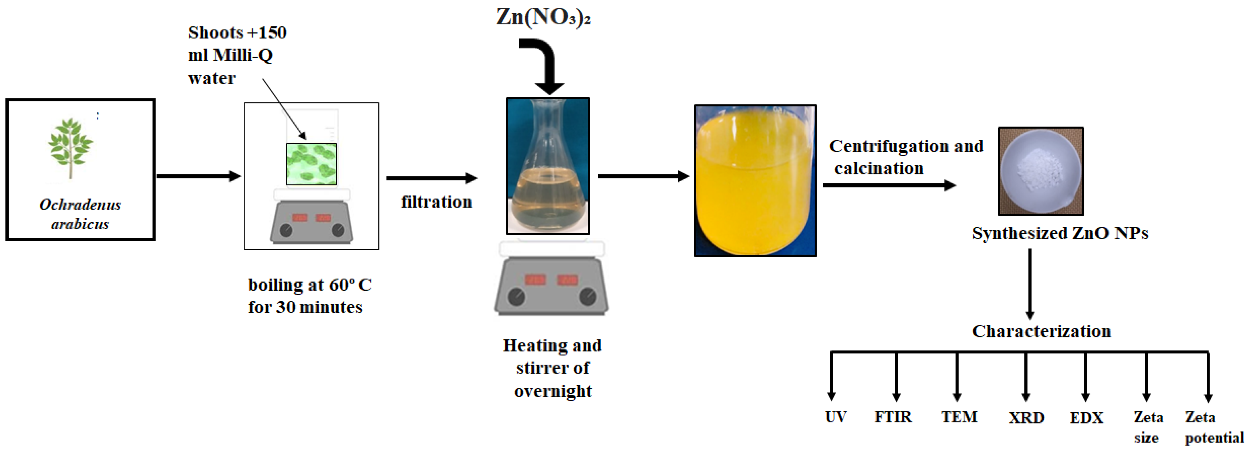
2.1. Plant Extract Preparation and Biosynthesis of Zinc Nanoparticles

Salvia officinalis and Ochradenus arabicus were provided from the plant tissue culture laboratory, botany and microbiology department, college of science King Saud University. The green synthesis of zinc oxide nanoparticles was performed as described [39]. In brief, 5 g of the tissue culture regenerated shoots of Ochradenus arabicus were cut into small pieces and added to 150 mL deionized Milli-Q water in 250 mL conical flask. After boiling at 60 °C for 30 min, the mixture was allowed to cool for 10 min at room temperature. Then after, the plant extract was filtered using Whatman filter paper No. 1 and stored at 4 °C for further use.
In total, 10 mL of 0.1 M of zinc nitrate (Zn(NO3)2) was added to 100 mL of fresh O. arabicus shoot extract, and the mixture was kept in a heating stirrer at 60 °C (Janke & Kunkel, Staufen, Germany) overnight. The synthesis of ZnO NPs was indicated by the appearance of light yellow color. For pellet separation, a centrifugation was carried out at 8000 rpm for 10 min at 4 °C. The obtained pellet was washed three times by distilled water and then dried at 50 °C in a hot air oven (Vaciotem-T oven, JP Selecta SA, Barcelona, Spain). Finally, the calcination was performed at 500 °C for 3 h in a furnace (DKN 602, Yamato Scientific Co., Ltd., Tokyo, Japan), and then the powder was collected and properly stored for further uses.

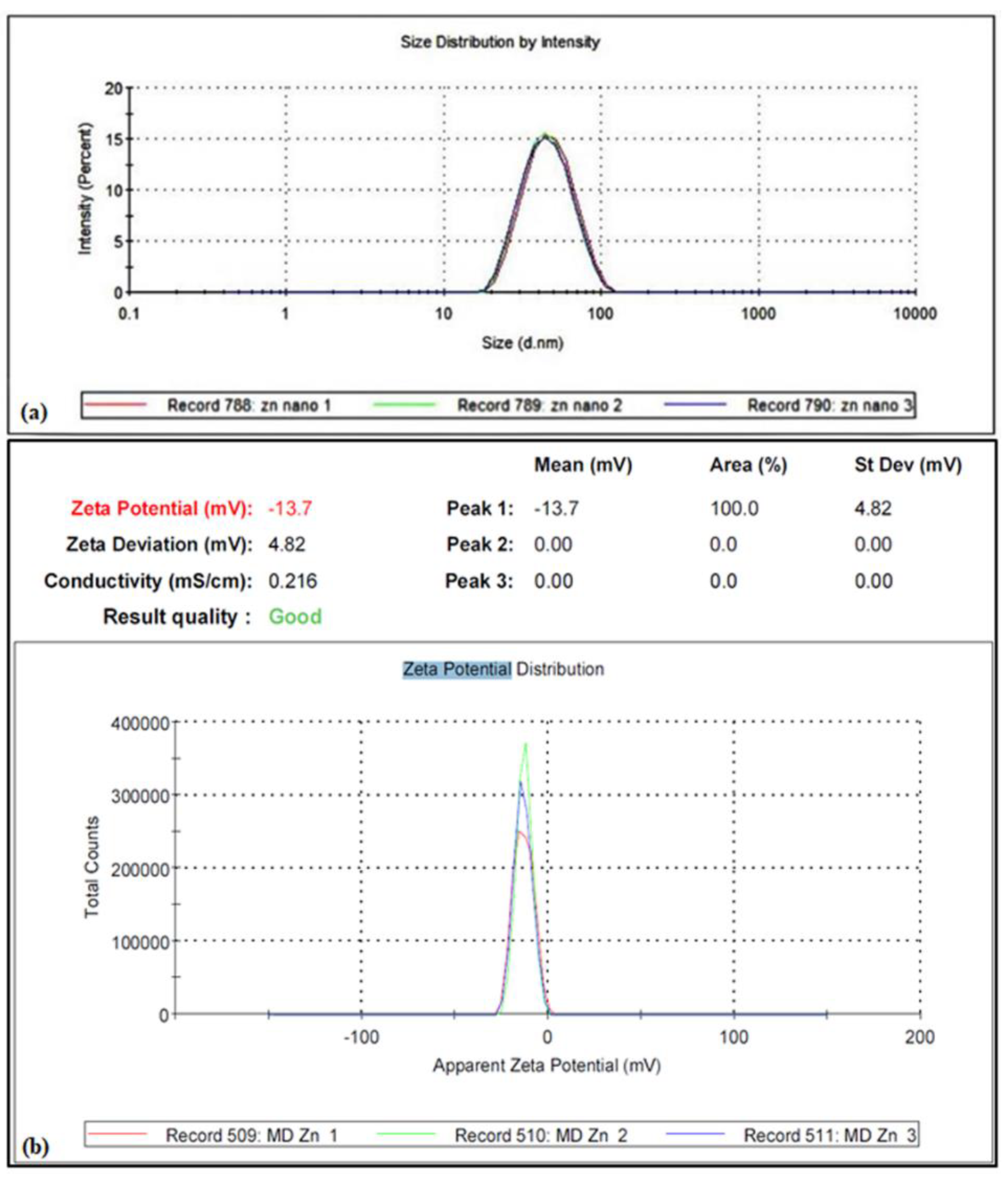
2.2. Biogenic ZnO NPs Characterization

The obtained ZnO NPs were characterized by the use of different techniques. The optical absorption spectrum was firstly analyzed by UV–Vis spectrophotometer (UV-1800, SHIMADZU, Kyoto, Japan) in the range of 200–700 nm. The functional groups in the synthesis ZnO NPs were recorded by Fourier transform infrared spectrometer (SHIMADZU). The transmission electron microscope (TEM) (JEM-1400) was used to characterize the shape and the size of the nanoparticles. The Bruker D8 Discover XRD system was used to analyze the X-ray diffraction of the ZnO NPs powder at a range of 0–80 °C (2θ). The values of XRD were subjected to OriginPro software 2021 and compared with JCPDS Card No. 36-1451. The composition of the bio-produced ZnO NPs was performed using JSM-7500F Field Emission Scanning Electron Microscope (JEOL, Tokyo, Japan) attached with Energy Dispersive X-ray analyzer (EDX). For the particle size distribution and the zeta potential of ZnO NPs, the samples were sonicated for 5 min and then measured by Malvern Zetasizer (Malvern Instruments Ltd., Malvern, UK).

2.3. Shoot Regeneration of Salvia Officinalis

In order to regenerate shoots from S. officinalis, nodal sections (two nodes per explant) were used. Firstly, the explants were surface sterilized for 10 min with sodium hypochlorite 10%, then washed thoroughly 5 times with sterile distilled water. The disinfected explants were then transferred to Magenta™ vessel containing Murashige and Skoog (MS) medium [40] supplemented with 30 g/L sucrose and 0.5, 1.0, 2.0, 3.0, 4.0, and 5.0 µM of benzylaminopurine (BA) and α-naphthalene acetic acid (NAA). The cultures were maintained for 8 weeks in growth chambers at 25 ± 2 °C with a 16-h light/8-h dark photoperiod and a light intensity of 40 μmoles m−2 s−1. Best combination (0.5 µM NAA + 2µM BA) were then selected to assess the effect of two concentrations of zinc oxide nanoparticles ZnO NPs (10 and 30 mg/L) on shoot regeneration with or without 75, 100, and 150 mM of NaCl. The cultures were maintained in the growth chamber as described above. After 6 weeks, samples were harvested and the following parameters were recorded: shoot length, number of shoots, shoot fresh weight, shoot fresh and dry weight, total chlorophyll, proline, catalase, superoxide dismutase, and glutathione reductase.

2.3.1. Estimation of Total Chlorophyll

The estimation of total chlorophyll was carried out according to [41] technique. A total of 0.1 g of leaves were used to extract total chlorophyll in 80% of acetone. The absorbance was measured at 663 nm and 645 nm, and the total chlorophyll content was expressed as mg/g FW.

2.3.2. Proline Estimation

The proline was estimated following the method described by [42]. A total of 500 mg of the fresh samples were extracted in 10 mL of 3% aqueous sulfosalicylic acid. This mixture was filtrated, and 2 mL of filtrate was added to 2 mL of ninhydrin plus 2 mL of glacial acetic acid and heated for 60 min at 100 °C. The reaction was stopped in an ice bath for 5 min, and then 4 mL toluene was added and mixed vigorously. Finally, the absorbance of the upper phase was read at 520 nm using toluene as a blank, and the proline was calculated as µg/g sample fresh weight.

2.3.3. Determination of Antioxidant Enzymes Activities

Enzymes were extracted following the method described by [43]. Briefly, 100 mg of fresh samples were firstly ground in liquid nitrogen and then dissolved in 0.1 M of sodium phosphate buffer (pH 7.4), which contains 100 mM EDTA, 1% (w/v) PVP, and 0.5% (v/v) Triton-X 100. The mixture was centrifuged for 20 min at 4 °C at 14,000 rpm, and then the collected supernatant was analyzed for the activities of the following antioxidant enzymes.

Catalase (EC 1.11.1.6)

The CAT activity was determined using the method reported by [44]. For that 60 mM of H2O2, 1.9 mL distilled water and 100 μL of enzyme extract were mixed properly by pipetting. The enzyme activity was then measured using a UV-vis spectrophotometer (Shimudzu) at 240 nm wavelength, and the enzyme unit was expressed as (U/mg protein).

Superoxide Dismutase (EC 1.15.1.1)

The activity of superoxide dismutase (SOD) was measured using the [45] technique. The reaction mixture consisted of 50 mM of phosphate buffer (pH 7.4), 10 mM methionine, 74 µM Nitrotetrazolium Blue (NBT), 2 µM riboflavin, 0.1 µM EDTA, and 100 μL of enzyme extract in a total volume of 3 mL. The enzyme activity was recorded by measuring the color reduction at 560 nm. One enzyme unit (EU) correspond to 50% inhibition of the reaction.

Glutathione Reductase (EC 1.6.4.2)

The glutathione reductase (GR) activity was determined by measuring the rate of NADPH oxidation at 340 nm as described [46]. A total of 100 µL of the extract was reacted with 2 mL of an assay mixture consisted of 0.1 M tris buffer, 2 mM EDTA, 50 µM NADPH, and 0.5 Mm GSSG. The GR activity was calculated using the molar extinction coefficient 6.2 mM−1 cm−1 of the NADPH and expressed as U mg−1 protein.

2.4. Statistical Analysis

The recorded parameters were statistically analyzed using IBM SPSS statistics 26 software. The data of three replicates were subjected to one-way ANOVA and analyzed with Duncan’s test. The significant difference at p ≤ 0.05 was indicated by different letters.

3. Results

3.1. Characterization of ZnO NPs

In the current study, zinc oxide nanoparticles were synthesized biologically using aqueous leaves extract of Ochradenus arabicus as a reducing and capping agent and zinc nitrate solution as substrate (Figure 1). Different characterization techniques such as UV-Vis spectrophotometer, Fourier transform infrared spectroscopy (FTIR), transmission electron microscope (TEM), X-ray diffraction (XRD), energy dispersive X-ray absorption (EDX), zeta potential, and zeta size were used to ascertain the formation of ZnO NPs. For the UV estimation, zinc oxide nanostructure was dissolved in deionized water, and the optical absorption spectrum was recorded in the range of 200–700 nm. As shown in Figure 2a, the absorption peak ZnO nanoparticle suspension was illustrated at 352 nm. Moreover, the shape and the size of the biosynthesized ZnO nanoparticles were studied and investigated by using transmission electron microscope (TEM). Figure 2b shows that the synthesized ZnO NPs have a spherical morphology, and their sizes range from 16 to 32 nm with an average particle size of 23 nm. Fourier transform infrared analysis was also performed to identify the chemical groups presented in the ZnO nanostructure (Figure 2c). The FTIR profile exhibited a broad absorption band at 439.5 cm−1, which attributes to Zn-O stretching vibration. Meanwhile, the other three functional groups were recorded at 1118, 1629, and 3433 cm−1. The X-ray diffraction pattern of the ZnO NPs powder showed various diffractions peaks at 2 theta degree 31.24°, 34.11°, 35.71°, 47.77°, 56,22°, 62,72°, 66.34°, 67.78°, 72.84°, and 76.79°. As matched with the crystallographic database JCPDS 36-1451, these peaks were assigned to (100), (002), (101), (102), (110), (103), (200), (112), (004), and (202) planes of zinc, respectively (Figure 2d). The elemental composition of the biosynthesized ZnO NPs was tested by the use of The Energy Dispersive X-ray (EDX). The EDX spectrum showed clear signals related to zinc and oxygen, which confirms that the synthesized nanoparticles are principally made of zinc and oxygen (Figure 2e). Finally, the average size and the stability of the nanoparticles were confirmed by the zeta size (Figure 3a) and the zeta potential (Figure 3b) analysis. In these results, we noticed that the mean diameter of ZnO NPs was about 40 nm and the zeta potential was −13.7 mv.

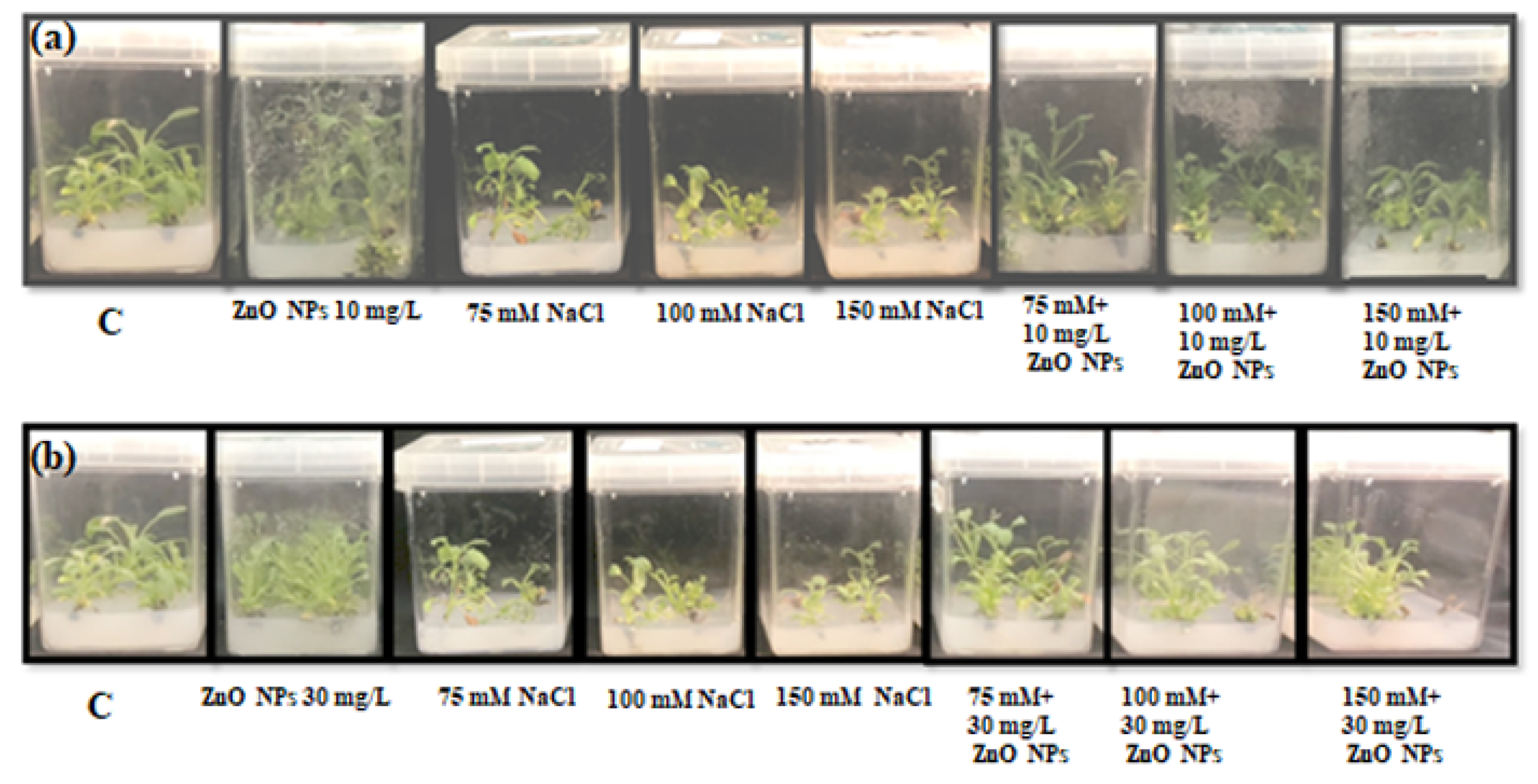
3.2. Effects of ZnO NPS on Salvia officinalis In Vitro Cultivated

3.2.1. Morphological Traits and Biomass Determination

The different concentrations (0.5, 1.0, 2.0, 3.0, 4.0, and 5.0 µM) of α-naphthalene acetic acid (NAA) and benzylaminopurine (BA) were used with shoots on MS media to see the optimum growth (data not shown). After screening, the MS media containing the hormones (0.5µM NAA + 2µM BA) was selected and the ameliorative effect of synthesized ZnO NPs (10 and 30 mg/L) was studied under 0, 75, 100, and 150 mM of NaCl (Figure 4). After six weeks of culture, plants cultivated in the media containing ZnO NPs (10 and 30 mg/L) exhibited higher shoot length, shoot number, and shoot fresh and dry weights compared to the plant cultivated in the media ZnO NPs free. On the other hand, the comparison between the two ZnO NPs treatments showed that the highest values of shoot length (3.6 cm), shoot fresh weight (6.27 cm), and shoot dry weight (0.5 g) were recorded in plants exposed to 10 mg/L of ZnO NPs. However, the three treatments of NaCl (75, 100, and 150 mM) dramatically reduced the mentioned parameters. More reductions of 35%, 29%, 50%, and 23.7%, respectively for the shoot length, shoot number, shoot fresh weight, and shoot dry weight, were recorded in plants under 150 mM of NaCl compared to control plants. We noticed also that the addition of the ZnO NPs (10 mg/L and 30 mg/L) counteracted differently the toxic effect of NaCl stress, and more mitigation was observed in the plants treated with 10 mg/L of ZnO NPs (Figure 5).

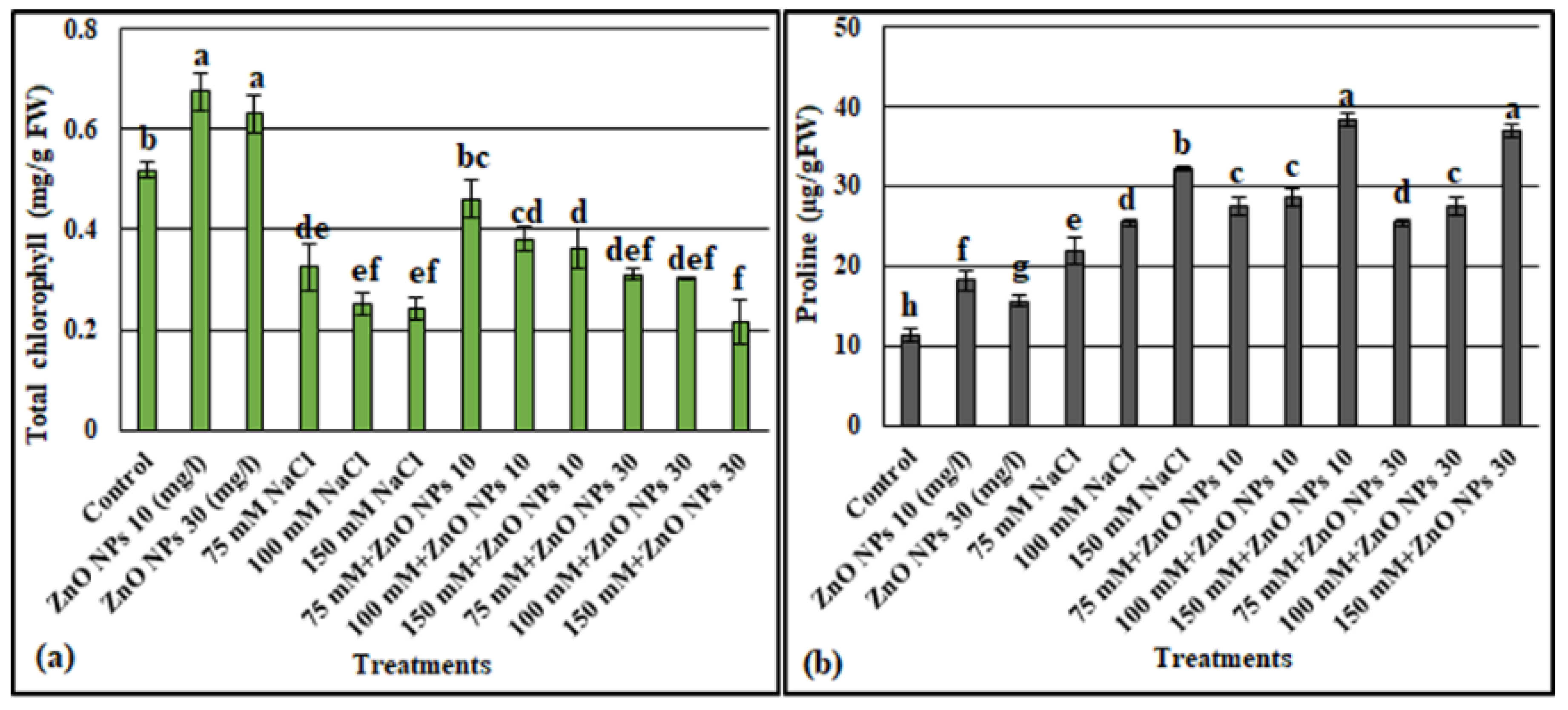
3.2.2. Total Chlorophyll Content

The application of both concentrations 10 mg/L and 30 mg/L of ZnO NPs significantly improved the total chlorophyll content by 30.3% and 21.8%, respectively, compared to the control plants. Whereas the addition of 75, 100, or 150 mM NaCl, decreased the total chlorophyll, and as the concentration of salt increases in MS media, the chlorophyll content decreased accordingly. However, the application of 10 mg/L and 30 mg/L of Zn ONPs recovered the decrease of chlorophyll in the plants subjected to 75 and 100 mM of salt, and the recovery was the highest when the ZnO NPs were applied at 10 mg/L in the media contained 75 mM of NaCl. Under 150 mM of NaCl, the addition of 10 mg/L of ZnO NPs enhanced the total chlorophyll by 1.5 times, whilst a slight decrease of total chlorophyll was recorded in the plants treated by 150 mM NaCl+30 mg/L ZnO NPs (Figure 6a).

3.2.3. Proline Content

The Figure 6b shows a significant increase of the proline amount in response to ZnO NPs and salt treatments compared to non-treated plants. Moreover, the NaCl stressed plants (75, 100, and 150 mM) and treated with ZnO NPs significantly differed from those without ZnO NPs. Higher proline content was noted in 150 mM NaCl stressed plants supplemented with 10 mg/L or 30 mg/L of ZnO NPs.

3.2.4. Antioxidant Enzymes Activities

The catalase (CAT), superoxide dismutase (SOD), and glutathione reductase (GR) were significantly affected in response to ZnO NPs treatments with or without NaCl. The results indicated that stressed plants and applied with 10 mg/L and 30 mg/L of ZnO NPs showed higher enzymes activities compared to those without ZnO NPs treatments. However, the higher improvement of CAT (4.7 times), SOD (5.5 times), and GR (6.5 times) activities were recorded in plants subjected to 150 mM of NaCl and amended with 10 mg/L of ZnO NPs (Table 1).

4. Discussion

In recent years, nanotechnology became the new area of interest for researchers. Nanotechnology is a fabrication, characterization, and utilization of nanomaterials. In different fields such as industry, medicine, energy, and agriculture, the use of the nanotechnology is in augmentation [47]. In addition, the use of nanoparticles (NPs) offers an opportunity for sustainability because of their special physiochemical characteristics in the biosystem [48]. As an eco-friendly approach, plant extracts provide a source for the biological synthesis of several metallic nanoparticles, which are synthesized with well-defined size and shape [49]. Among the all known NPs, zinc oxide nanoparticles (ZnO NPs) have various applications since their green synthesis is simple, inexpensive, viable, and uses less toxic chemicals [50]. In this study, ZnO NPs were green synthesized and assessed for their impact in the improvement of salinity stress of Salvia officinalis in in vitro cultivation. ZnO nanoparticles were prepared using O. Arabicus shoot extract and were characterized by UV-vis absorption, FTIR, TEM, XRD, EDX, zeta potential, and zeta size for confirming the nanostructures for the prepared ZnO nanoparticles. Adding zinc nitrate to the extract of O. Arubicus leads to the color change within few minutes of stirring at 60 °C (Figure 1). This is consistent with the findings of [51], who observed a change of color in the reaction mixture of plant extract and zinc acetate as an indicator to the ZnO NPs formation. When examined by UV spectrophotometry, the biosynthesized ZnO NPs showed a peak at 352 nm (Figure 2a) confirming the presence of ZnO NPs in the reaction mixture [52]. The UV-visible absorption spectrum was largely used to examine the optical properties of nanosized particles [53,54]. The shape and size of the obtained ZnO NPs were characterized using the TEM technique. The result of the Figure 2b showed spherical nanoparticles with sizes ranging from 16 to 32 nm. The size of NPs can be affected by the plant species or the source form which the extracts are used as reducing and capping agents. For instance, the sizes of the ZnO nanoparticles produced from Olea europea and Aloe barbadensis extracts ranged from 18 to 30 nm and 25–40 nm, respectively [55,56]. Using Fourier transform infrared spectroscopy, our sample showed four functional groups detected at 3433.55 cm−1, 1629.98 cm−1, 1118.43 cm−1, and 439.52 cm−1 (Figure 2c). The absorption peak at 439.52 cm−1 mainly represented the ZnO stretching vibrations, while the peak at 1118.43 cm−1 is attributed to primary, secondary alcohol bend or vibration. The absorption peak at 1629.90 cm−1 showed the stretching in proteins. Whereas the fourth peak at 3433.55 cm−1 is attributed to the OH stretching vibration [15,57,58]. The XRD is a rapid test providing information on crystal size and structure [22]. In our findings (Figure 2d), the XRD analysis confirmed that the diffraction peaks planes of the fabricated ZnO NPS were matched well with the quartzite ZnO reported in JCPDS data [59,60]. For the elemental analysis (Figure 2e), the EDX technique revealed 76.53% zinc and 23.47% oxygen, respectively, for the weight%. A similar type of elemental analysis was performed earlier, where a high yield of zinc and oxygen in the fabricated zinc oxide nanoparticles was observed [54,61]. Moreover, the stability of ZnO suspensions was confirmed by measuring the particle size and zeta potential (Figure 3). This technique showed that the average size of the zinc nanoparticles was higher than that measured by TEM. This could be explained by the heterogeneity distribution of ZnO nanoparticles in the colloidal solution, which can increase size of the particles in the zetasizer analysis [62].
Salinity is a critical problem for the productivity of the crops as it can negatively affect the plant growth. The high uptake of Na+ and Cl ions result in an osmotic stress causing a toxicity for the plant. Moreover, salinity induces the generation of reactive oxygen species (ROS) resulting oxidative stress [63]. Nanoparticle application in the agriculture sector is one of the promising techniques that enhance crop productivity under normal as well as under harsh environmental conditions, including salt stress [64]. Among nanoparticles, the application of ZnO NPs is one of the most effective options for improving agricultural yield globally under stressful conditions [14]. While the salt stress at the level of 75, 100, and 150 mM significantly decreased shoot length, shoot number, shoot fresh weight, and shoot dry weight of the in vitro of Salvia officinalis, the application of ZnO NPs significantly boosted these parameters (Figure 5). In this regard, it was demonstrated that treatment with zinc nanoparticles improved plant morphological parameters under normal and salinity stress conditions in various plant species such as potato, cowpea and okra [65,66]. Within plants, it was demonstrated that the Zn positively affected the capacity for water uptake and transport, and it also reduced the harmful effects of abiotic stressors such as salt and heat [67]. At the cell level, zinc is an important micronutrient involved in several physiological processes. Zn is needed for the synthesis of tryptophan, a precursor of the growth hormone IAA. Further, it was noticed that zinc increased the biosynthesis of gibberellin-like substances, and it is the only metal represented in the six enzymes groups. Thus, these crucial roles played by Zn could explain their positive effects to promote the vegetative growth in plants under normal as well as under saline conditions [68,69,70]. The comparison between the two ZnO NPs (10 mg/L and 30 mg/L) showed that higher growth parameters were achieved by the application of 10 mg/L in the culture media. Singh et al. 2021 [71], reported that the uptake and transport of NPs are size dependent. Further, the cell wall poses a great challenge and constitutes a barrier that interferes with the entry of NPs into cells. Thus, efficient methods are needed for the fabrication of beneficial NPs in order to make them available to plants.
Zinc is a necessary micronutrient playing a vital role in the biomass production via its effects on several physiological and metabolic processes such as chlorophyll biosynthesis [68]. In our findings, a decrease of total chlorophyll of Salvia officinalis shoot was observed as the concentration of salt increased in MS culture media. However, ZnO NPs treatments, especially at a dose of 10 mg/L, significantly recover this reduction (Figure 6a). Salt stress inhibited the chlorophyll a, b, and carotenoids, which are the main components of the photosynthetic system [72]. Meanwhile, enhanced levels of photosynthetic pigments were recorded in salt-stressed Lupin ustermis L. and amended with 20, 40, and 60 mg/L of zinc oxide nanoparticles [73]. Al-Zahrani et al. [74] noticed that zinc increased the chlorophyll content via regulating the magnesium transport, which is an important component in the structure of the chlorophyll.
NaCl stress leads to ROS accumulation in the plant cell, thus an increase in the antioxidant enzyme activities and antioxidant metabolism is one of the crucial ways for improving salinity tolerance [75]. In the current study, we observed that the addition of ZnO NPs (10 or 30 mg/L) increased the performance of Salvia officinalis to accumulate more proline (Figure 6b) and to increase the antioxidative enzyme activities of CAT, SOD, and GR in the presence and absence of NaCl (Table 1). In accordance to this, Faizan et al. [14] noted that, in the presence of NaCl, the antioxidative enzyme activities have increased significantly, however the foliar treatment of tomato plants with ZnO NPs further increased the performance of antioxidative enzymes in the plants with or without Nacl. Likewise, the addition of ZnO NPs to salt-stressed Brassica napus mitigated the salt-induced harmful effects via upregulating the antioxidant system, osmolyte biosynthesis, and ionic regulation [76]. Meanwhile, for okra (Abelmoschus esculentus L.), Zn ONPs application increased photosynthetic pigments and CAT and SOD activities, but reduced proline and total soluble sugar contents [47]. Zinc acts in the membrane stabilization against oxidative damages by stabilizing its integrity and permeability. In addition, it is a co-factor for the important enzymes having a pivotal role in the antioxidant defense system [74,77]. Kareem et al. [78] noted that for the plants under stress, the nanoparticles improve the potential of an antioxidant defense system for scavenging the ROS by regulating the microRNA expression.
This work provided an effective method for biogenic of ZnO NPs, which showed better growth amelioration and salt stress alleviation in Salvia officinalis. Thus, further molecular study is required to better understand the mechanisms behind the positive role of ZnO NPs on in vitro cultured plants.

5. Conclusions

It can be concluded that ZnO NPs were well synthesized biologically using shoot extract of Ochradenus arabicus. The efficient fabrication of ZnO NPs was confirmed by UV-Vis, FTIR, TEM, XRD, EDX, zeta potential, and zeta size analysis. Moreover, the application of these ZnO NPs, especially at a dose of 10 mg/L, to the in vitro culture media significantly ameliorated the harmful effect of NaCl in Salvia officinalis. Indeed, this dose improved the plant biomass, total chlorophyll, proline accumulation, and antioxidant enzyme activities in the plants under 0, 75, 100, and 150 mM of NaCl, which indicated that the role of the ZnO NPs in salt stress amelioration can be achieved by the activation of the antioxidant enzymes or by the modulations of the biochemical parameters. Thus, the addition of ZnO NPs at a dose of 10 mg/L can be used as an eco-friendly approach to improve the growth under salt as well as other abiotic stress. Future study at the molecular level is required to better understand the mechanisms behind the roles of ZnO NPs.

Author Contributions

Conceptualization, F.A.-Q. and M.N.; methodology, N.A.A., M.N., M.T. and S.K.; validation, M.T., S.K. and M.N.; formal analysis, M.T. and N.A.A.; investigation, A.M.S., H.O.S. and A.-R.Z.G.; resources, M.N., S.A., N.S.A. and A.A.-H. writing—original draft preparation, M.T., S.K., N.A.A., A.M.S. and M.N.; writing—review and editing, M.T.; visualization, M.T and M.N.; supervision, F.A.-Q. All authors have read and agreed to the published version of the manuscript.

Funding

This research was funded by Deanship of Scientific Research at King Saud University through Project no. RGP-014.

Institutional Review Board Statement

Not applicable.

Informed Consent Statement

Not applicable.

Data Availability Statement

Data are contained within the article.

Acknowledgments

The authors extend their appreciation to the Deanship of Scientific Research at King Saud University for funding this work through the research group Project no. RGP-014.

Conflicts of Interest

The authors declare that they have no conflict of interest.

References

  1. Abid, M.; Zhang, Y.J.; Li, Z.; Bai, D.F.; Zhong, Y.P.; Fang, J.B. Effect of salt stress on growth, physiological and biochemical characters of four kiwifruit genotypes. Sci. Hortic. 2020, 271, 109473. [Google Scholar] [CrossRef]
  2. Acosta-Motos, J.R.; Penella, C.; Hernández, J.A.; Díaz-Vivancos, P.; Sánchez-Blanco, M.J.; Navarro, J.M.; Gómez-Bellot, M.J.; Barba-Espín, G. Towards a sustainable agriculture: Strategies involving phytoprotectants against salt stress. Agronomy 2020, 10, 194. [Google Scholar] [CrossRef] [Green Version]
  3. Tang, X.; Mu, X.; Shao, H.; Wang, H.; Brestic, M. Global plant-responding mechanisms to salt stress: Physiological and molecular levels and implications in biotechnology. Crit. Rev. Biotechnol. 2015, 35, 425–437. [Google Scholar] [CrossRef]
  4. Paul, D. Osmotic stress adaptations in rhizobacteria. J. Basic Microbiol. 2013, 53, 101–110. [Google Scholar] [CrossRef] [PubMed]
  5. Hendawy, S.; Khalid, K.A. Response of sage (Salvia officinalis L.) plants to zinc application under different salinity levels. J. Appl. Sci. Res. 2005, 1, 147–155. [Google Scholar]
  6. Shahbaz, M.; Ashraf, M. Improving salinity tolerance in cereals. Crit. Rev. Plant Sci. 2013, 32, 237–249. [Google Scholar] [CrossRef]
  7. Shelke, D.B.; Nikalje, G.C.; Chambhare, M.R.; Zaware, B.N.; Penna, S.; Nikam, T.D. Na+ and Cl induce differential physiological, biochemical responses and metabolite modulations in vitro in contrasting salt-tolerant soybean genotypes. 3 Biotech 2019, 9, 91. [Google Scholar] [CrossRef]
  8. Bibi, N.; Jan, G.; Jan, F.G.; Hamayun, M.; Iqbal, A.; Hussain, A.; Rehman, H.; Tawab, A.; Khushdil, F. Cochliobolus sp. acts as a biochemical modulator to alleviate salinity stress in okra plants. Plant Physiol. Biochem. 2019, 139, 459–469. [Google Scholar] [CrossRef]
  9. Es-sbihi, F.Z.; Hazzoumi, Z.; Aasfar, A.; Amrani Joutei, K. Improving salinity tolerance in Salvia officinalis L. by foliar application of salicylic acid. Chem. Biol. Technol. Agric. 2021, 8, 25. [Google Scholar] [CrossRef]
  10. Hanin, M.; Ebel, C.; Ngom, M.; Laplaze, L.; Masmoudi, K. New insights on plant salt tolerance mechanisms and their potential use for breeding. Front. Plant Sci. 2016, 7, 1787. [Google Scholar] [CrossRef] [Green Version]
  11. Flowers, T.J.; Colmer, T.D. Plant salt tolerance: Adaptations in halophytes. Ann. Bot. 2015, 115, 327–331. [Google Scholar] [CrossRef] [PubMed] [Green Version]
  12. Hasanuzzaman, M.; Oku, H.; Nahar, K.; Bhuyan, M.; Mahmud, J.A.; Baluska, F.; Fujita, M. Nitric oxide-induced salt stress tolerance in plants: ROS metabolism, signaling, and molecular interactions. Plant Biotechnol. Rep. 2018, 12, 77–92. [Google Scholar] [CrossRef]
  13. Khan, I.; Saeed, K.; Khan, I. Nanoparticles: Properties, applications and toxicities. Arab. J. Chem. 2019, 12, 908–931. [Google Scholar] [CrossRef]
  14. Faizan, M.; Bhat, J.A.; Chen, C.; Alyemeni, M.N.; Wijaya, L.; Ahmad, P.; Yu, F. Zinc oxide nanoparticles (ZnO-NPs) induce salt tolerance by improving the antioxidant system and photosynthetic machinery in tomato. Plant Physiol. Biochem. 2021, 161, 122–130. [Google Scholar] [CrossRef] [PubMed]
  15. Zafar, S.; Hasnain, Z.; Aslam, N.; Mumtaz, S.; Jaafar, H.Z.; Wahab, P.E.M.; Qayum, M.; Ormenisan, A.N. Impact of Zn nanoparticles synthesized via green and chemical approach on Okra (Abelmoschus esculentus L.) growth under salt stress. Sustainability 2021, 13, 3694. [Google Scholar] [CrossRef]
  16. Gaafar, R.; Diab, R.; Halawa, M.; Elshanshory, A.; El-Shaer, A.; Hamouda, M. Role of zinc oxide nanoparticles in ameliorating salt tolerance in soybean. Egypt. J. Bot. 2020, 60, 733–747. [Google Scholar] [CrossRef]
  17. Zafar, S.; Perveen, S.; Kamran Khan, M.; Shaheen, M.R.; Hussain, R.; Sarwar, N.; Rashid, S.; Nafees, M.; Farid, G.; Alamri, S. Effect of zinc nanoparticles seed priming and foliar application on the growth and physio-biochemical indices of spinach (Spinacia oleracea L.) under salt stress. PLoS ONE 2022, 17, e0263194. [Google Scholar] [CrossRef] [PubMed]
  18. Youssef, N.M.; Ghareeb, Z.F.; Ali, A.I.; Taha, L.S. Mitigation of salinity stress on in vitro growth of Eustoma grandiflorum using zinc nanoparticles. Plant Arch. 2020, 2, 4547–4554. [Google Scholar]
  19. Rani, P.U.; Yasur, J.; Loke, K.S.; Dutta, D. Effect of synthetic and biosynthesized silver nanoparticles on growth, physiology and oxidative stress of water hyacinth: Eichhornia crassipes (Mart) Solms. Acta Physiol. Plant. 2016, 38, 58. [Google Scholar] [CrossRef]
  20. Fayez, K.; El-Deeb, B.; Mostafa, N. Toxicity of biosynthetic silver nanoparticles on the growth, cell ultrastructure and physiological activities of barley plant. Acta Physiol. Plant. 2017, 39, 155. [Google Scholar] [CrossRef]
  21. Cvjetko, P.; Zovko, M.; Štefanić, P.P.; Biba, R.; Tkalec, M.; Domijan, A.-M.; Vrček, I.V.; Letofsky-Papst, I.; Šikić, S.; Balen, B. Phytotoxic effects of silver nanoparticles in tobacco plants. Environ. Sci. Pollut. Res. 2018, 25, 5590–5602. [Google Scholar] [CrossRef] [PubMed]
  22. Modena, M.M.; Rühle, B.; Burg, T.P.; Wuttke, S. Nanoparticle characterization: What to measure? Adv. Mater. 2019, 31, 1901556. [Google Scholar] [CrossRef] [PubMed]
  23. Mourdikoudis, S.; Pallares, R.M.; Thanh, N.T. Characterization techniques for nanoparticles: Comparison and complementarity upon studying nanoparticle properties. Nanoscale 2018, 10, 12871–12934. [Google Scholar] [CrossRef] [PubMed] [Green Version]
  24. Rossi, L.; Fedenia, L.N.; Sharifan, H.; Ma, X.; Lombardini, L. Effects of foliar application of zinc sulfate and zinc nanoparticles in coffee (Coffea arabica L.) plants. Plant Physiol. Biochem. 2019, 135, 160–166. [Google Scholar] [CrossRef]
  25. Siddiqui, M.H.; Al-Whaibi, M.H.; Firoz, M.; Al-Khaishany, M.Y. Role of nanoparticles in plants. Nanotechnol. Plant Sci. 2015, 19–35. [Google Scholar] [CrossRef]
  26. Ghorbani, A.; Esmaeilizadeh, M. Pharmacological properties of Salvia officinalis and its components. J. Tradit. Complementary Med. 2017, 7, 433–440. [Google Scholar] [CrossRef]
  27. Valiyari, S.; Baradaran, B.; Abdolalizadeh, J.; Bandehagh, A.; Azadmehr, A.; Hajiaghaee, R. Inhibitory and cytotoxic activities of Salvia officinalis L. extract on human lymphoma and leukemia cells by induction of apoptosis. Adv. Pharm. Bull. 2013, 3, 51. [Google Scholar]
  28. Arikat, N.A.; Jawad, F.M.; Karam, N.S.; Shibli, R.A. Micropropagation and accumulation of essential oils in wild sage (Salvia fruticosa Mill.). Sci. Hortic. 2004, 100, 193–202. [Google Scholar] [CrossRef]
  29. Abd-Elazem, I.S.; Chen, H.S.; Bates, R.B.; Huang, R.C.C. Isolation of two highly potent and non-toxic inhibitors of human immunodeficiency virus type 1 (HIV-1) integrase from Salvia miltiorrhiza. Antivir. Res. 2002, 55, 91–106. [Google Scholar] [CrossRef]
  30. Jung, M.; Lee, H.C.; Ahn, C.W.; Park, W.; Choi, S.; Kim, H.; Cho, D.; Lee, G.T.; Li, H.-R. Effective isolation of magnesium lithospermate B and its inhibition of aldose reductase and fibronectin on mesangial cell line. Chem. Pharm. Bull. 2002, 50, 1135–1136. [Google Scholar] [CrossRef] [Green Version]
  31. Akhondzadeh, S.; Noroozian, M.; Mohammadi, M.; Ohadinia, S.; Jamshidi, A.; Khani, M. Salvia officinalis extract in the treatment of patients with mild to moderate Alzheimer’s disease: A double blind, randomized and placebo-controlled trial. J. Clin. Pharm. Ther. 2003, 28, 53–59. [Google Scholar] [CrossRef] [PubMed]
  32. Garcia, C.S.; Menti, C.; Lambert, A.P.F.; Barcellos, T.; Moura, S.; Calloni, C.; Branco, C.S.; Salvador, M.; Roesch-Ely, M.; Henriques, J.A. Pharmacological perspectives from Brazilian Salvia officinalis (Lamiaceae): Antioxidant, and antitumor in mammalian cells. An. Acad. Bras. Ciências 2016, 88, 281–292. [Google Scholar] [CrossRef] [PubMed] [Green Version]
  33. Kintzios, S.; Nikolaou, A.; Skoula, M. Somatic embryogenesis and in vitro rosmarinic acid accumulation in Salvia officinalis and S. fruticosa leaf callus cultures. Plant Cell Rep. 1999, 18, 462–466. [Google Scholar] [CrossRef]
  34. Loyola-Vargas, V.M.; Ochoa-Alejo, N. An introduction to plant tissue culture: Advances and perspectives. Plant Cell Cult. Protoc. 2018, 1815, 3–13. [Google Scholar] [CrossRef]
  35. Hameg, R.; Arteta, T.A.; Landin, M.; Gallego, P.P.; Barreal, M.E. Modeling and optimizing culture medium mineral composition for in vitro propagation of Actinidia arguta. Front. Plant Sci. 2020, 11, 2088. [Google Scholar] [CrossRef]
  36. Kim, D.H.; Gopal, J.; Sivanesan, I. Nanomaterials in plant tissue culture: The disclosed and undisclosed. RSC Adv. 2017, 7, 36492–36505. [Google Scholar] [CrossRef] [Green Version]
  37. Awad, K.M.; Al-Mayahi, A.M.; Mahdi, M.A.; Al-Asadi, A.; Abass, M.H. In vitro assessment of ZnO nanoparticles on Phoenix dactylifera L. micropropagation. Sci. J. King Faisal Univ. 2020, 21. [Google Scholar] [CrossRef]
  38. Alharby, H.F.; Metwali, E.M.; Fuller, M.P.; Aldhebiani, A.Y. Impact of application of zinc oxide nanoparticles on callus induction, plant regeneration, element content and antioxidant enzyme activity in tomato (Solanum lycopersicum Mill) under salt stress. Arch. Biol. Sci. 2016, 68, 723–735. [Google Scholar] [CrossRef] [Green Version]
  39. Rupa, E.J.; Kaliraj, L.; Abid, S.; Yang, D.-C.; Jung, S.-K. Synthesis of a zinc oxide nanoflower photocatalyst from sea buckthorn fruit for degradation of industrial dyes in wastewater treatment. Nanomaterials 2019, 9, 1692. [Google Scholar] [CrossRef] [Green Version]
  40. Murashige, T.; Skoog, F. A revised medium for rapid growth and bio assays with tobacco tissue cultures. Physiol. Plant. 1962, 15, 473–497. [Google Scholar] [CrossRef]
  41. Arnon, D.I. Copper enzymes in isolated chloroplasts. Polyphenoloxidase in Beta vulgaris. Plant Physiol. 1949, 24, 1. [Google Scholar] [CrossRef] [Green Version]
  42. Bates, L.S.; Waldren, R.P.; Teare, I. Rapid determination of free proline for water-stress studies. Plant Soil 1973, 39, 205–207. [Google Scholar] [CrossRef]
  43. Jogeswar, G.; Pallela, R.; Jakka, N.; Reddy, P.; Venkateswara Rao, J.; Sreenivasulu, N.; Kavi Kishor, P. Antioxidative response in different sorghum species under short-term salinity stress. Acta Physiol. Plant. 2006, 28, 465–475. [Google Scholar] [CrossRef]
  44. Aebi, H. Catalase in vitro. In Methods in Enzymology; Elsevier: Amsterdam, The Netherlands, 1984; Volume 105, pp. 121–126. [Google Scholar]
  45. Dhindsa, R.S.; Plumb-Dhindsa, P.; Thorpe, T.A. Leaf senescence: Correlated with increased levels of membrane permeability and lipid peroxidation, and decreased levels of superoxide dismutase and catalase. J. Exp. Bot. 1981, 32, 93–101. [Google Scholar] [CrossRef]
  46. Rao, M. Cellular detoxifying mechanisms determine the age dependent injury in tropical trees exposed to SO2. J. Plant Physiol. 1992, 140, 733–740. [Google Scholar] [CrossRef]
  47. Alabdallah, N.M.; Alzahrani, H.S. The potential mitigation effect of ZnO nanoparticles on [Abelmoschus esculentus L. Moench] metabolism under salt stress conditions. Saudi J. Biol. Sci. 2020, 27, 3132–3137. [Google Scholar] [CrossRef]
  48. El-Badri, A.M.; Batool, M.; Wang, C.; Hashem, A.M.; Tabl, K.M.; Nishawy, E.; Kuai, J.; Zhou, G.; Wang, B. Selenium and zinc oxide nanoparticles modulate the molecular and morpho-physiological processes during seed germination of Brassica napus under salt stress. Ecotoxicol. Environ. Saf. 2021, 225, 112695. [Google Scholar] [CrossRef]
  49. Vanathi, P.; Rajiv, P.; Narendhran, S.; Rajeshwari, S.; Rahman, P.K.; Venckatesh, R. Biosynthesis and characterization of phyto mediated zinc oxide nanoparticles: A green chemistry approach. Mater. Lett. 2014, 134, 13–15. [Google Scholar] [CrossRef]
  50. Soltanian, S.; Sheikhbahaei, M.; Mohamadi, N.; Pabarja, A.; Abadi, M.F.S.; Tahroudi, M.H.M. Biosynthesis of Zinc Oxide Nanoparticles Using Hertia intermedia and Evaluation of its Cytotoxic and Antimicrobial Activities. BioNanoScience 2021, 11, 245–255. [Google Scholar] [CrossRef]
  51. Shaikhaldein, H.O.; Al-Qurainy, F.; Khan, S.; Nadeem, M.; Tarroum, M.; Salih, A.M.; Gaafar, A.-R.Z.; Alshameri, A.; Alansi, S.; Alenezi, N.A. Biosynthesis and Characterization of ZnO Nanoparticles Using Ochradenus arabicus and Their Effect on Growth and Antioxidant Systems of Maerua oblongifolia. Plants 2021, 10, 1808. [Google Scholar] [CrossRef]
  52. Shamhari, N.M.; Wee, B.S.; Chin, S.F.; Kok, K.Y. Synthesis and characterization of zinc oxide nanoparticles with small particle size distribution. Acta Chim. Slov. 2018, 65, 578–585. [Google Scholar] [CrossRef] [PubMed]
  53. Naseer, M.; Aslam, U.; Khalid, B.; Chen, B. Green route to synthesize Zinc Oxide Nanoparticles using leaf extracts of Cassia fistula and Melia azadarach and their antibacterial potential. Sci. Rep. 2020, 10, 9055. [Google Scholar] [CrossRef] [PubMed]
  54. Rasha, E.; Monerah, A.; Manal, A.; Rehab, A.; Mohammed, D.; Doaa, E. Biosynthesis of Zinc Oxide Nanoparticles from Acacia nilotica (L.) Extract to Overcome Carbapenem-Resistant Klebsiella Pneumoniae. Molecules 2021, 26, 1919. [Google Scholar] [CrossRef] [PubMed]
  55. Awwad, A.; Albiss, B.; Ahmad, A.L. Green synthesis, characterization and optical properties of zinc oxide nanosheets using Olea europea leaf extract. Adv. Mater. Lett. 2014, 5, 520–524. [Google Scholar] [CrossRef]
  56. Gunalan, S.; Sivaraj, R.; Rajendran, V. Green synthesized ZnO nanoparticles against bacterial and fungal pathogens. Prog. Nat. Sci. Mater. Int. 2012, 22, 693–700. [Google Scholar] [CrossRef] [Green Version]
  57. Jayarambabu, N.; Kumari, B.S.; Rao, K.V.; Prabhu, Y. Germination and growth characteristics of mungbean seeds (Vigna radiata L.) affected by synthesized zinc oxide nanoparticles. Int. J. Curr. Eng. Technol. 2014, 4, 2347–5161. [Google Scholar]
  58. Devaraj, P.; Kumari, P.; Aarti, C.; Renganathan, A. Synthesis and characterization of silver nanoparticles using cannonball leaves and their cytotoxic activity against MCF-7 cell line. J. Nanotechnol. 2013, 2013, 598328. [Google Scholar] [CrossRef] [Green Version]
  59. Kalpana, V.; Kataru, B.A.S.; Sravani, N.; Vigneshwari, T.; Panneerselvam, A.; Rajeswari, V.D. Biosynthesis of zinc oxide nanoparticles using culture filtrates of Aspergillus niger: Antimicrobial textiles and dye degradation studies. OpenNano 2018, 3, 48–55. [Google Scholar] [CrossRef]
  60. Salih, A.M.; Al-Qurainy, F.; Khan, S.; Tarroum, M.; Nadeem, M.; Shaikhaldein, H.O.; Gaafar, A.-R.Z.; Alfarraj, N.S. Biosynthesis of zinc oxide nanoparticles using Phoenix dactylifera and their effect on biomass and phytochemical compounds in Juniperus procera. Sci. Rep. 2021, 11, 19136. [Google Scholar] [CrossRef]
  61. Raj, L.; Jayalakshmy, E. Biosynthesis and characterization of zinc oxide nanoparticles using root extract of Zingiber officinale. Orient. J. Chem. 2015, 31, 51–56. [Google Scholar] [CrossRef]
  62. Abdelmigid, H.M.; Hussien, N.A.; Alyamani, A.A.; Morsi, M.M.; AlSufyani, N.M.; Kadi, H.A. Green Synthesis of Zinc Oxide Nanoparticles Using Pomegranate Fruit Peel and Solid Coffee Grounds vs. Chemical Method of Synthesis, with Their Biocompatibility and Antibacterial Properties Investigation. Molecules 2022, 27, 1236. [Google Scholar] [CrossRef] [PubMed]
  63. Spanò, C.; Bottega, S.; Bellani, L.; Muccifora, S.; Sorce, C.; Ruffini Castiglione, M. Effect of zinc priming on salt response of wheat seedlings: Relieving or worsening? Plants 2020, 9, 1514. [Google Scholar] [CrossRef] [PubMed]
  64. Zulfiqar, F.; Ashraf, M. Nanoparticles potentially mediate salt stress tolerance in plants. Plant Physiol. Biochem. 2021, 160, 257–268. [Google Scholar] [CrossRef]
  65. Mahmoud, A.W.M.; Abdeldaym, E.A.; Abdelaziz, S.M.; El-Sawy, M.B.; Mottaleb, S.A. Synergetic effects of zinc, boron, silicon, and zeolite nanoparticles on confer tolerance in potato plants subjected to salinity. Agronomy 2020, 10, 19. [Google Scholar] [CrossRef] [Green Version]
  66. Alabdallah, N.M.; Alzahrani, H.S. Impact of ZnO Nanoparticles on Growth of Cowpea and Okra Plants under Salt Stress Conditions. Biosci. Biotechnol. Res. Asia 2020, 17, 329–340. [Google Scholar] [CrossRef]
  67. Tsonev, T.; Cebola Lidon, F.J. Zinc in plants-an overview. Emir. J. Food Agric. 2012, 24, 322–333. [Google Scholar]
  68. Tondey, M.; Kalia, A.; Singh, A.; Dheri, G.S.; Taggar, M.S.; Nepovimova, E.; Krejcar, O.; Kuca, K. Seed priming and coating by nano-scale zinc oxide particles improved vegetative growth, yield and quality of fodder maize (Zea mays). Agronomy 2021, 11, 729. [Google Scholar] [CrossRef]
  69. Mašev, N.; Kutáček, M. The effect of zinc on the biosynthesis of tryptophan, andol auxins and gibberellins in barley. Biol. Plant. 1966, 8, 142–151. [Google Scholar] [CrossRef]
  70. Estrada-Urbina, J.; Cruz-Alonso, A.; Santander-González, M.; Méndez-Albores, A.; Vázquez-Durán, A. Nanoscale zinc oxide particles for improving the physiological and sanitary quality of a Mexican landrace of red maize. Nanomaterials 2018, 8, 247. [Google Scholar] [CrossRef] [Green Version]
  71. Singh, P.; Arif, Y.; Siddiqui, H.; Sami, F.; Zaidi, R.; Azam, A.; Alam, P.; Hayat, S. Nanoparticles enhances the salinity toxicity tolerance in Linum usitatissimum L. by modulating the antioxidative enzymes, photosynthetic efficiency, redox status and cellular damage. Ecotoxicol. Environ. Saf. 2021, 213, 112020. [Google Scholar] [CrossRef]
  72. El-Tayeb, M. Response of barley grains to the interactive effect of salinity and salicylic acid. Plant Growth Regul. 2005, 45, 215–224. [Google Scholar] [CrossRef]
  73. Abdel Latef, A.A.H.; Abu Alhmad, M.F.; Abdelfattah, K.E. The possible roles of priming with ZnO nanoparticles in mitigation of salinity stress in lupine (Lupinus termis) plants. J. Plant Growth Regul. 2017, 36, 60–70. [Google Scholar] [CrossRef]
  74. Al-Zahrani, H.S.; Alharby, H.F.; Hakeem, K.R.; Rehman, R.U. Exogenous Application of Zinc to Mitigate the Salt Stress in Vigna radiata (L.) Wilczek—Evaluation of Physiological and Biochemical Processes. Plants 2021, 10, 1005. [Google Scholar] [CrossRef] [PubMed]
  75. Guilian, M.; Xing, X.; Zhaozhen, X. Advances in physiological and biochemical research of salt tolerance in plant. Chin. J. Eco-Agric. 2004, 12, 43–46. [Google Scholar]
  76. Farouk, S.; Al-Amri, S.M. Exogenous zinc forms counteract NaCl-induced damage by regulating the antioxidant system, osmotic adjustment substances, and ions in canola (Brassica napus L. cv. Pactol) plants. J. Soil Sci. Plant Nutr. 2019, 19, 887–899. [Google Scholar] [CrossRef]
  77. Marreiro, D.D.N.; Cruz, K.J.C.; Morais, J.B.S.; Beserra, J.B.; Severo, J.S.; De Oliveira, A.R.S. Zinc and oxidative stress: Current mechanisms. Antioxidants 2017, 6, 24. [Google Scholar] [CrossRef]
  78. Kareem, H.A.; Saleem, M.F.; Saleem, S.; Rather, S.A.; Wani, S.H.; Siddiqui, M.H.; Alamri, S.; Kumar, R.; Gaikwad, N.B.; Guo, Z. Zinc Oxide Nanoparticles Interplay with Physiological and Biochemical Attributes in Terminal Heat Stress Alleviation in Mungbean (Vigna radiata L.). Front. Plant Sci. 2022, 13, 842349. [Google Scholar] [CrossRef]
Figure 1. Steps of biogenic synthesis of ZnO NPs from the Ochradenus arabicus extract.
Processes 10 01273 g001
Figure 2. UV−Vis absorption spectra of synthesized ZnO NPs (a), ZnO NPs image under transmission electron microscope (TEM) (b), FTIR spectrum (c), X-ray diffraction (XRD) pattern (d), and EDX spectrum (e) of the synthesized ZnO NPs.
Processes 10 01273 g002
Figure 3. Characterization of zinc oxide nanoparticles by zeta size (a) and zeta potential (b) analysis.
Processes 10 01273 g003
Figure 4. Effect of 10 mg/L (a) and 30 mg/L (b) of zinc oxide nanoparticles on in vitro regeneration of Salvia officinalis subjected to 0, 75, 100, and 150 mM NaCl.
Processes 10 01273 g004
Figure 5. ZnO NPs (10 mg/L and 30 mg/L) effects on shoot length (a), shoot number (b), shoot fresh weight (c), and shoot dry weight (d) of Salvia officinalis subjected to 0, 75, 100, and 150 mM NaCl. Each value is the mean of three replicates ± SD. The significant differences between the treatments are indicated by different letters on bars. The Duncan’s test at p < 0.05 was used for this analysis.
Processes 10 01273 g005
Figure 6. Effect ZnO NPs (10 mg/L and 30 mg/L) on total total chlorophyll content (a) and proline accumulation (b) in the shoot of Salvia officinalis cultivated 0, 75, 100, and 150 mM of NaCl. Each value is the mean of three replicates ± SD. The significant differences between the treatments are indicated by different letters on bars. The Duncan’s test analysis at p < 0.05 was used.
Processes 10 01273 g006
Table 1. Effect ZnO NPs (10 mg/L and 30 mg/L) on catalase (CAT), Superoxide dismutase (SOD), and Glutathione reductase (GR) antioxidant activities (U/mg protein) in the shoot of Salvia officinalis cultivated 0, 75, 100, and 150 mM of NaCl.
Enzymes (U/mg Protein)ControlZnO NPs
10 mg/L
ZnO NPs
30 mg/L
75 mM NaCl100 mM NaCl150 mM NaCl75 Mm + ZnO NPs 10 mg/L100 mM + ZnO NPs 10 mg/L150 mM + ZnO NPs 10 mg/L75 mM + ZnO NPs 30 mg/L100 mM + ZnO NPs 30 mg/L150 mM + ZnO NPs 30 mg/L
CAT3.52 ± 0.43 i5.62 ± 0.41 g4.46 ± 0.031 h5.42 ± 0.3 g7.48 ± 0.26 e8.6 ± 0.38 d 7.57 ± 0.075 e 15.5 ± 0.33 b16.56 ± 0.302 a6.59 ± 0.28 f11.51 ± 0.45 c11.76 ± 0.2 c
SOD1.3 ± 0.25 d1.55 ± 0.1 d1.35 ± 0.18 d1.57 ± 0.4 d 4.5 ± 0.22 c 5.59 ± 0.33 b4.57 ± 0.28 c5.44 ± 0.25 b7.24 ± 0.31 a1.5 ± 0.32 d4.65 ± 0.38 c5.64 ± 0.16 b
GR0.0146 ±
0.00352 h
0.0272 ±
0.00118 g
0.026 ±
0.00321 g
0.0365 ±
0.00125 f
0.047 ±
0.0015 e
0.0574 ±
0.00115 d
0.0844 ±
0.00224 b
0.0857 ±
0.00295 b
0.0951 ±
0.00251 a
0.0672 ±
0.00265 c
0.0672 ±
0.001 c
0.0856 ± 0.00195 b
Values are the means of three replicates ± SD. The significant differences between the treatments are indicated by different letters according to Duncan’s test (p < 0.05).
Publisher’s Note: MDPI stays neutral with regard to jurisdictional claims in published maps and institutional affiliations.

