

Synthesis, Characterization and Antibacterial Efficacy of Catharanthus roseus and Ocimum tenuiflorum-Mediated Silver Nanoparticles: Phytonanotechnology in Disease Management
 ,
, 
Abstract
:1. Introduction
2. Materials and Methods
2.1. Materials
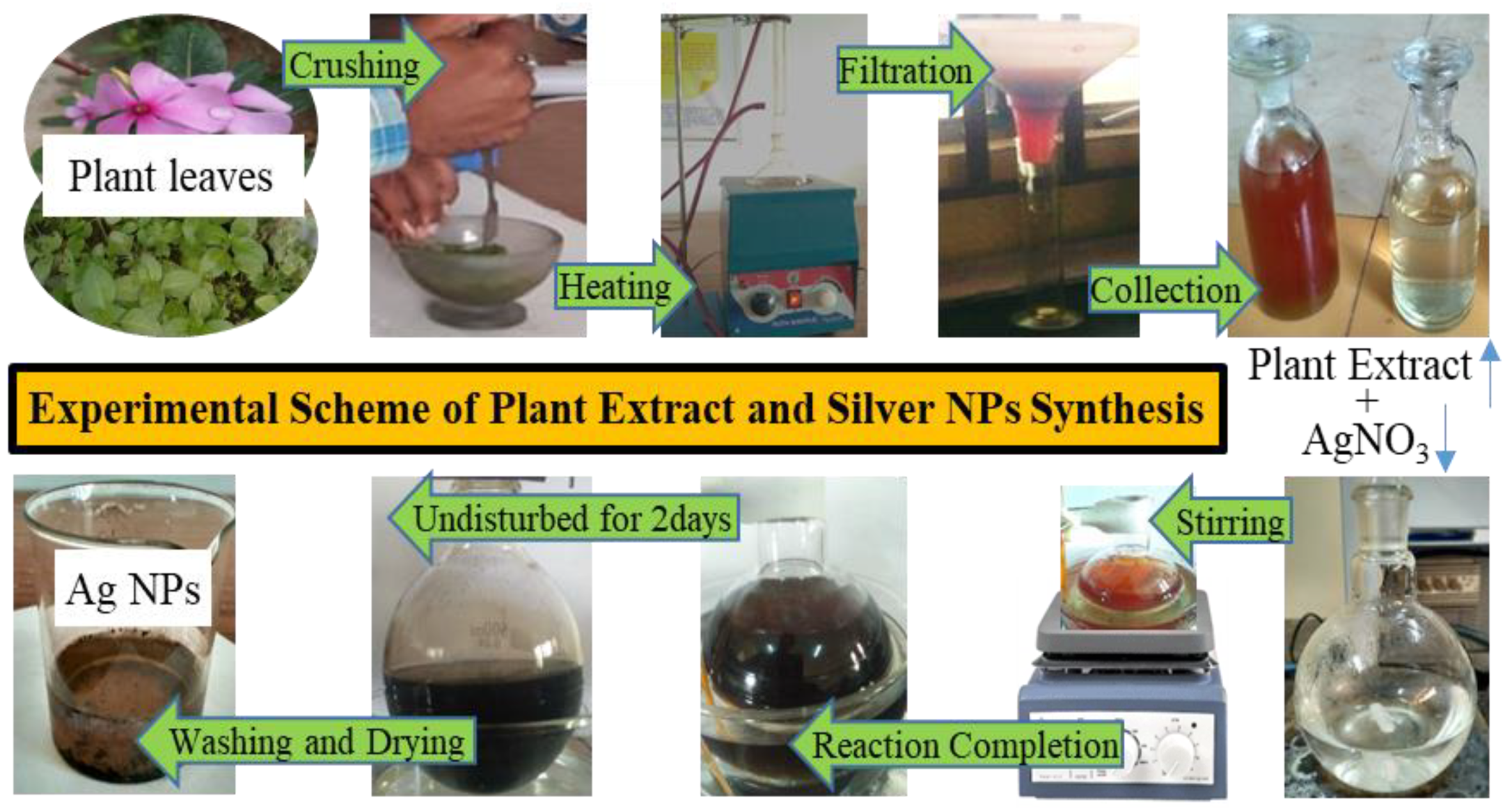
2.2. Preparation of Plant Extracts
2.3. Synthesis of Silver NPs
2.4. Characterization of the NPs
2.5. Nanoparticles Preparation for Antibacterial Study
2.6. Antibacterial Activity
2.6.1. Well Diffusion Assay
2.6.2. Minimum Inhibitory Concentration and Minimum Bactericidal Concentration of Ag NPs
2.7. Statistical Analysis
3. Results and Discussion
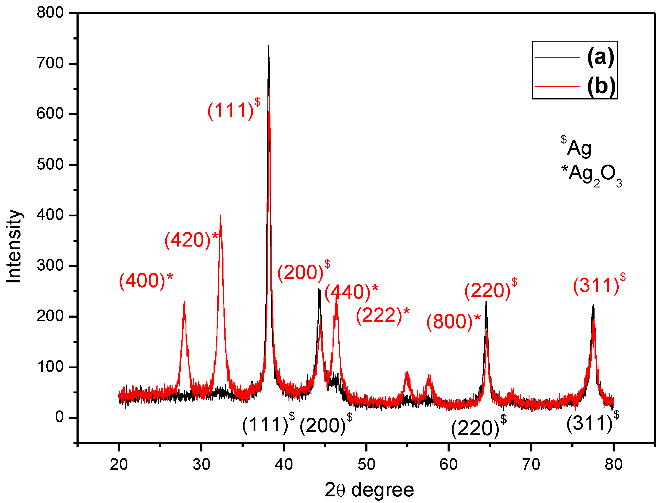
3.1. XRD Analysis
- D = Average crystalline size.
- β = Line broadening in radians.
- θ = Bragg’s angle.
- λ = X-ray wavelength (λ = 1.5418 Ǻ).

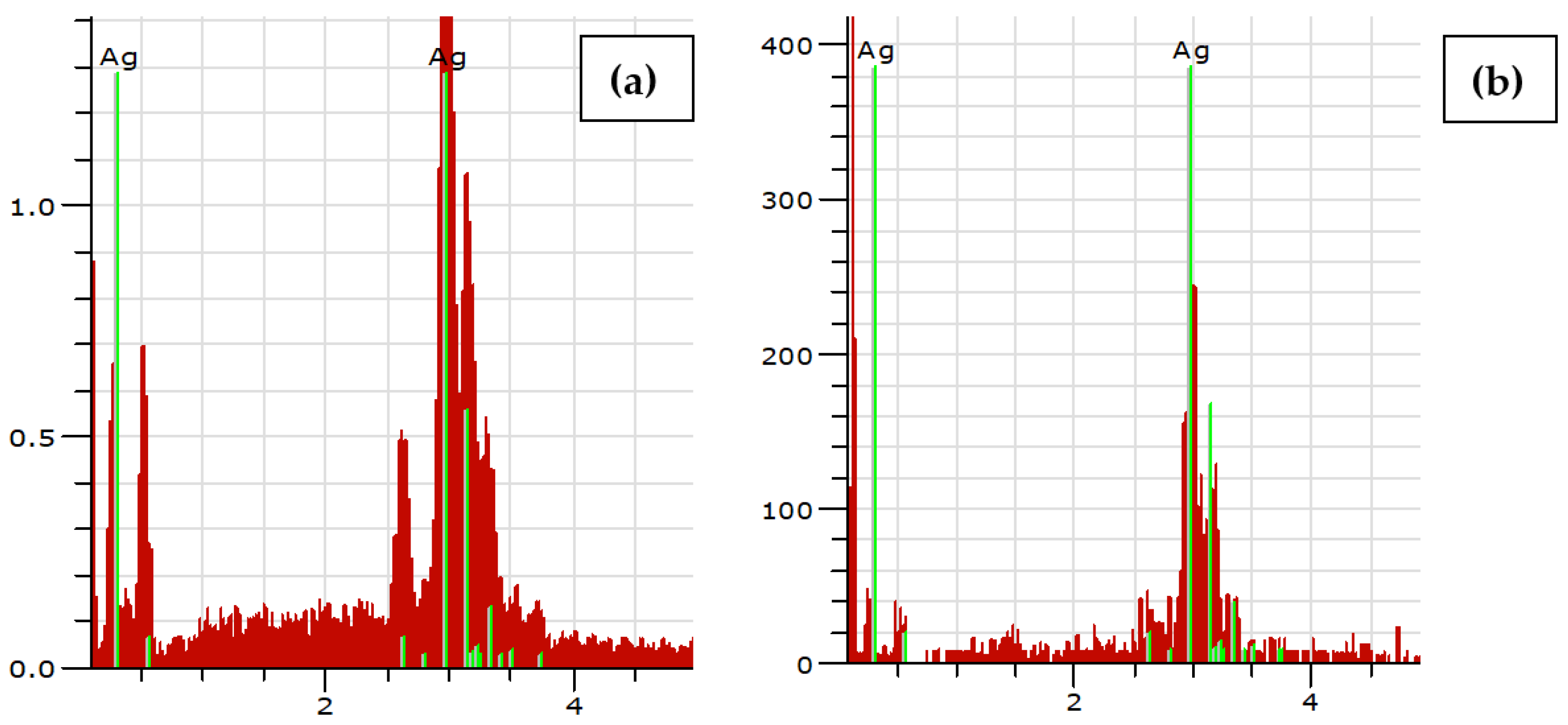
3.2. EDX Analysis
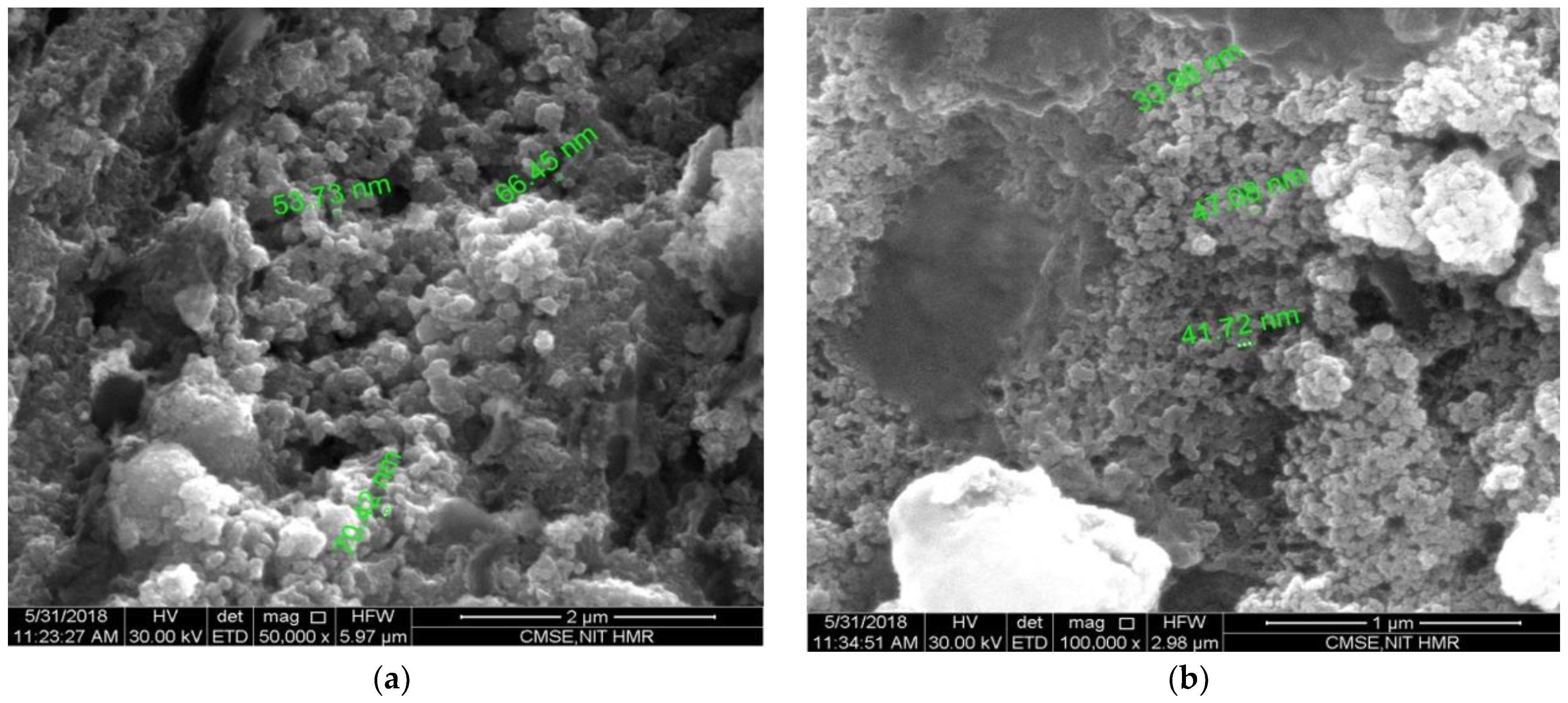
3.3. SEM Analysis
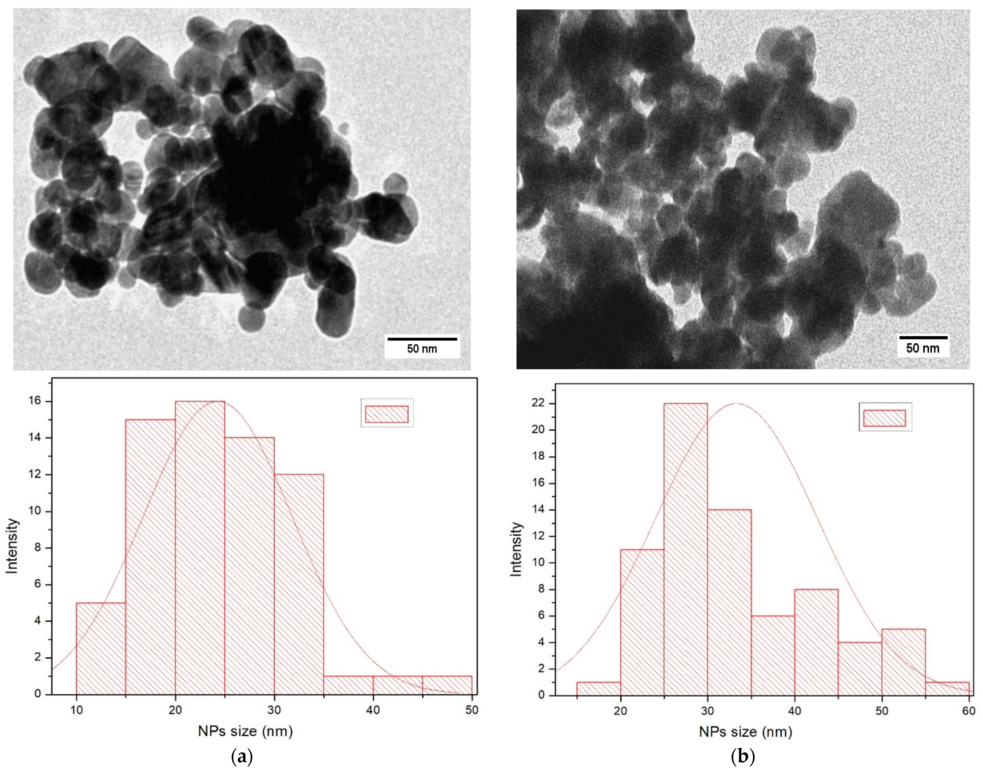
3.4. TEM Analysis
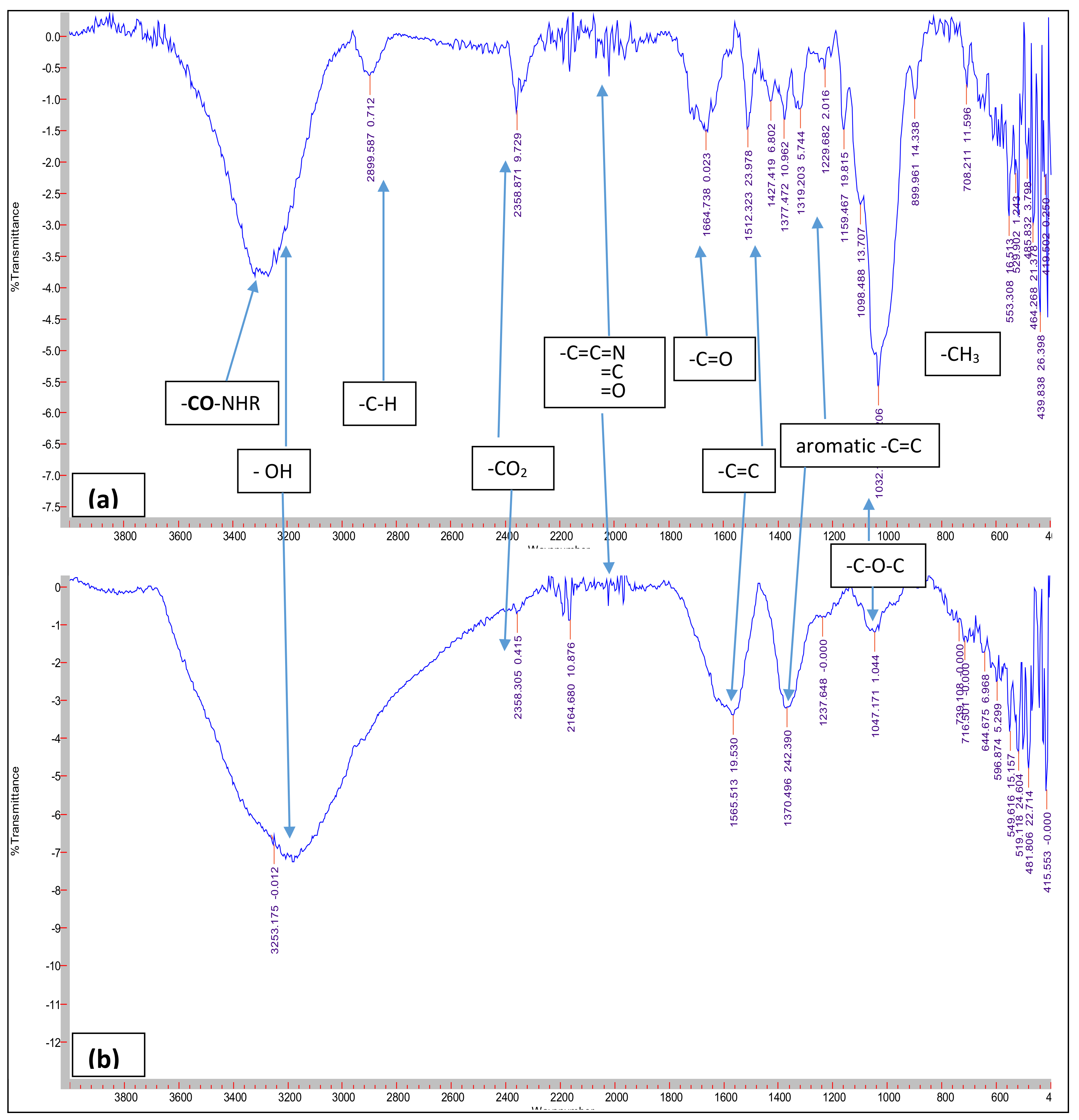
3.5. FTIR Analysis
3.6. Antibacterial Activity
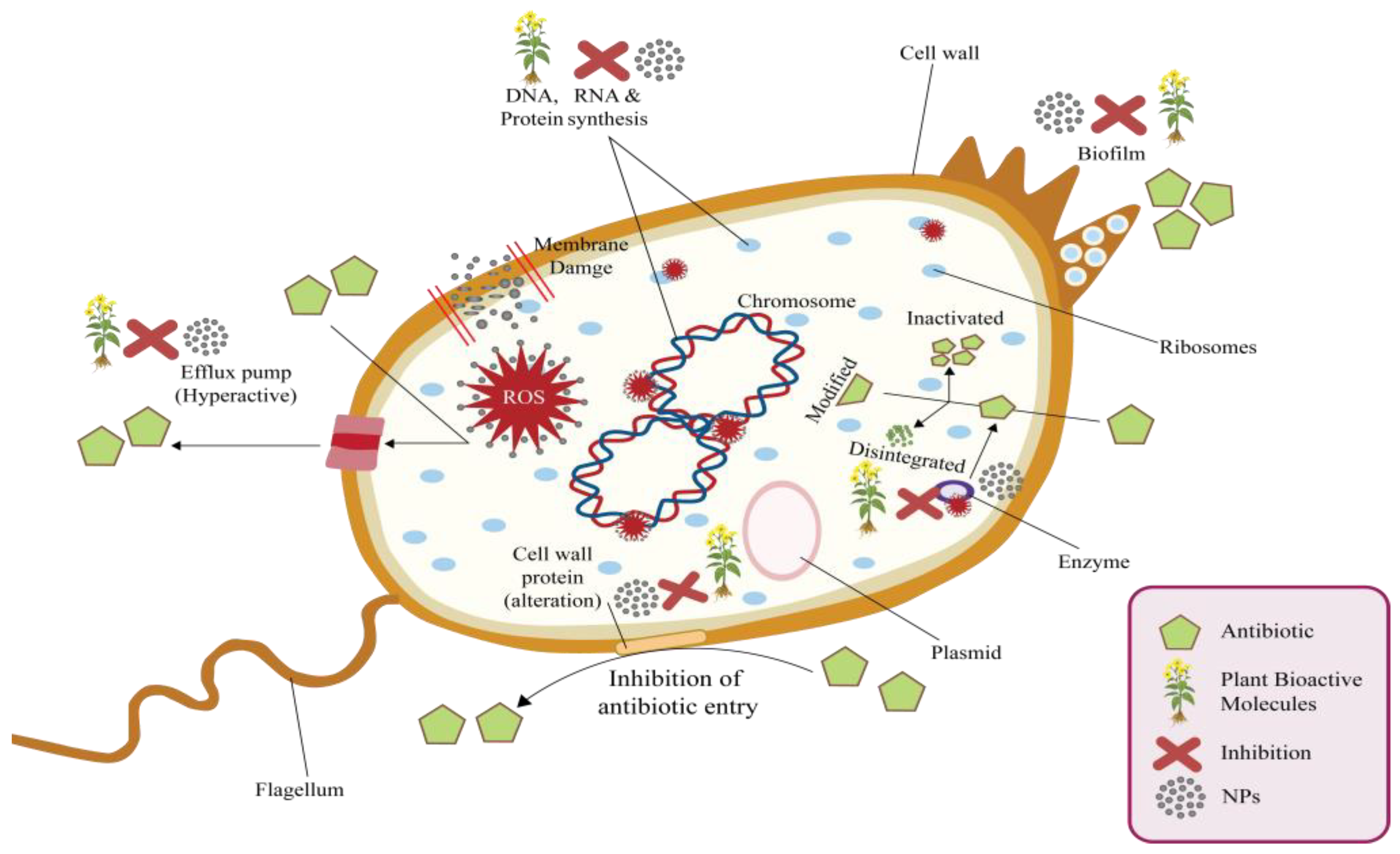
4. Mechanism of Antibacterial Activity: From Plants to NPs
5. Conclusions and Future Perspectives
Supplementary Materials
Author Contributions
Funding
Data Availability Statement
Acknowledgments
Conflicts of Interest
References
- Rajoriya, P.; Misra, P.; Singh, V.K.; Shukla, P.K.; Ramteke, P.W. Green synthesis of silver nanoparticles. Int. J. Biol. Sci. 2017, 7, 7–20. [Google Scholar] [CrossRef]
- Kumar, A.; Chandel, T.; Diwaker; Thakur, N. Predicting the magnetism, structural, thermodynamic and electronic properties of new co-based Heuslers: First principle perspective. Philos. Mag. 2020, 100, 2721–2734. [Google Scholar] [CrossRef]
- Parashar, U.K.; Saxena, P.S.; Srivastava, A. Bioinspired synthesis of silver nanoparticles. Dig. J. Nanomater. Biostruct. 2009, 4, 159–166. [Google Scholar]
- Roy, A. Synthesis of silver nanoparticles from medicinal plants and its biological application: A review. Res. Rev. BioSci. 2017, 12, 138. [Google Scholar]
- Vanitha, G.; Rajavel, K.; Boopathy, G.; Veeravazhuthi, V.; Neelamegam, P. Physiochemical charge stabilization of silver nanoparticles and its antibacterial applications. Chem. Phys. Lett. 2017, 669, 71–79. [Google Scholar] [CrossRef]
- Al-Zahrani, S.S.; Al-Garni, S.M. Biosynthesis of Silver Nanoparticles from Allium ampeloprasum Leaves Extract and Its Antifungal Activity. J. Biomater. Nanobiotechnol. 2019, 10, 11–25. [Google Scholar] [CrossRef] [Green Version]
- Kavitha, K.; Baker, S.; Rakshith, D.; Kavitha, H.U.; Yashwantha Rao, H.C.; Harini, B.P.; Satish, S. Plants as green source towards synthesis of nanoparticles. J. Biol. Sci. 2013, 2, 66–76. [Google Scholar]
- Zahir, A.A.; Chauhan, I.S.; Bagavan, A.; Kamaraj, C.; Elango, G.; Shankar, J.; Arjaria, N.; Roopan, S.M.; Rahuman, A.A.; Singh, N. Synthesis of nanoparticles using Euphorbia prostrata extract reveals a shift from apoptosis to G0/G1 arrest in Leishmania donovani. Nanomed. Nanotechnol. 2014, 5, 4782–4799. [Google Scholar]
- Li, X.; Xu, H.; Chen, Z.S.; Chen, G. Biosynthesis of nanoparticles by microorganisms and their applications. J. Nanomater. 2011, 2011, 270974. [Google Scholar] [CrossRef] [Green Version]
- El-Sherbiny, I.M.; Salih, E. Green Synthesis of Metallic Nanoparticles Using Biopolymers and Plant Extracts. In Green Metal Nanoparticles: Synthesis, Characterization and Their Applications; Suvardhan, K., Shakeel, A., Eds.; Wiley: Hoboken, NJ, USA, 2018; pp. 293–319. [Google Scholar]
- Gokila, V.; Perarasu, V.T.; Rufina, R. Qualitative comparison of chemical and green synthesized Fe3O4 nanoparticles. Adv. Nano Res. 2021, 10, 71–76. [Google Scholar]
- Gour, A.; Jain, N.K. Advances in green synthesis of nanoparticles. Artif. Cells Nanomed. Biotechnol. 2019, 47, 844–851. [Google Scholar] [CrossRef] [Green Version]
- Salem, S.S.; Fouda, A. Green synthesis of metallic nanoparticles and their prospective biotechnological applications: An overview. Biol. Trace Elem. Res. 2021, 199, 344–370. [Google Scholar] [CrossRef]
- Gericke, M.; Pinches, A. Biological synthesis of metal nanoparticles. Hydrometallurgy 2006, 83, 132–140. [Google Scholar] [CrossRef]
- Narayanan, K.B.; Sakthivel, N. Biological synthesis of metal nanoparticles by microbes. Adv. Colloid Interface Sci. 2010, 156, 1–13. [Google Scholar] [CrossRef]
- Singh, P.; Kim, Y.J.; Zhang, D.; Yang, D.C. Biological synthesis of nanoparticles from plants and microorganisms. Trends Biotechnol. 2016, 34, 588–599. [Google Scholar] [CrossRef]
- Husseiny, M.I.; Abd El-Aziz, M.; Badr, Y.; Mahmoud, M.A. Biosynthesis of gold nanoparticles using Pseudomonas aeruginosa. Spectrochim. Acta A Mol. Biomol. Spectrosc. 2007, 67, 1003–1006. [Google Scholar] [CrossRef]
- Yadav, A.; Kon, K.; Kratosova, G.; Duran, N.; Ingle, A.P.; Rai, M. Fungi as an efficient mycosystem for the synthesis of metal nanoparticles: Progress and key aspects of research. Biotechnol. Lett. 2015, 37, 2099–2120. [Google Scholar] [CrossRef]
- Siddiqi, K.S.; Husen, A. Fabrication of metal nanoparticles from fungi and metal salts: Scope and application. Nanoscale Res. Lett. 2016, 11, 98. [Google Scholar] [CrossRef] [Green Version]
- Mishra, A.; Tripathy, S.K.; Yun, S.I. Bio-synthesis of gold and silver nanoparticles from Candida guilliermondii and their antimicrobial effect against pathogenic bacteria. J. Nanosci. Nanotechnol. 2011, 11, 243–248. [Google Scholar] [CrossRef]
- Narayanan, K.B.; Sakthivel, N. Green synthesis of biogenic metal nanoparticles by terrestrial and aquatic phototrophic and heterotrophic eukaryotes and biocompatible agents. Adv. Colloid. Interface. Sci. 2011, 169, 59–79. [Google Scholar] [CrossRef]
- Balkrishna, A.; Kumar, A.; Arya, V.; Rohela, A.; Verma, R.; Nepovimova, E.; Krejcar, O.; Kumar, D.; Thakur, N.; Kuca, K. Phytoantioxidant functionalized nanoparticles: A green approach to combat nanoparticle-induced oxidative stress. Oxid. Med. Cell. Longev. 2021, 2021, 3155962. [Google Scholar] [CrossRef]
- Balkrishna, A.; Arya, V.; Rohela, A.; Kumar, A.; Verma, R.; Kumar, D.; Nepovimova, E.; Kuca, K.; Thakur, N.; Thakur, N.; et al. Nanotechnology interventions in the management of COVID-19: Prevention, Ddiagnosis and virus-like particle vaccines. Vaccines 2021, 9, 1129. [Google Scholar] [CrossRef]
- Silva, L.P.; Reis, I.G.; Bonatto, C.C. Green Synthesis of Metal Nanoparticles by Plants: Current Trends and Challenges. In Green Processes for Nanotechnology; Basiuk, V., Basiuk, E., Eds.; Springer: Cham, Switzerland, 2015; pp. 259–275. [Google Scholar]
- Bhembe, Y.A.; Lukhele, L.P.; Hlekelele, L.; Ray, S.S.; Sharma, A.; Vo, D.V.N.; Dlamini, L.N. Photocatalytic degradation of nevirapine with a heterostructure of few-layer black phosphorus coupled with niobium (V) oxide nanoflowers (FL-BP@Nb2O5). Chemosphere 2020, 261, 128159. [Google Scholar] [CrossRef]
- Solgi, M.; Taghizadeh, M. Biogenic Synthesis of Metal Nanoparticles by Plants. In Biogenic Nano-Particles and Their Use in Agro-Ecosystems; Ghorbanpour, M., Bhargava, P., Varma, A., Choudhary, D., Eds.; Springer: Singapore, 2020; pp. 593–606. [Google Scholar]
- Paul, B.; Bhuyan, B.; Purkayastha, D.D.; Dey, M.; Dhar, S.S. Green synthesis of gold nanoparticles using Pogestemon benghalensis (B) O. Ktz. leaf extract and studies of their photocatalytic activity in degradation of methylene blue. Mater. Lett. 2015, 148, 37–40. [Google Scholar] [CrossRef]
- Kopaczyk, J.M.; Warguła, J.; Jelonek, T. The variability of terpenes in conifers under developmental and environmental stimuli. Environ. Exp. Bot. 2020, 180, 104197. [Google Scholar] [CrossRef]
- Ndaba, B.; Roopnarain, A.; Haripriya, R.A.; Maaza, M. Biosynthesized metallic nanoparticles as fertilizers: An emerging precision agriculture strategy. J. Integr. Agric. 2022, 21, 1225–1242. [Google Scholar] [CrossRef]
- Firdhouse, M.J.; Lalitha, P.; Shubashini, S.K. Novel synthesis of silver nanoparticles using leaf ethanol extract of Pisonia grandis (R. Br). Der Pharma Chem. 2012, 4, 2320–2326. [Google Scholar]
- Thakur, B.K.; Kumar, A.; Kumar, D. Green synthesis of titanium dioxide nanoparticles using Azadirachta indica leaf extract and evaluation of their antibacterial activity. S. Afr. J. Bot. 2019, 124, 223–227. [Google Scholar] [CrossRef]
- Khatana, C.; Kumar, A.; Alruways, M.W.; Khan, N.; Thakur, N.; Kumar, D.; Kumari, A. Antibacterial potential of zinc oxide nanoparticles synthesized using Aloe vera (L.) Burm. f.: A green approach to combat drug resistance. J. Appl. Microbiol. 2021, 15, 1907–1914. [Google Scholar]
- Thakur, N.; Kumar, K.; Kumar, A. Effect of (Ag, Zn) co-doping on structural, optical and bactericidal properties of CuO nanoparticles synthesized by a microwave-assisted method. Dalton Trans. 2021, 50, 6188–6203. [Google Scholar] [CrossRef]
- Anand, K.; Kaur, J.; Singh, R.C.; Thangaraj, R. Preparation and characterization of Ag-doped In2O3 nanoparticles gas sensor. Chem. Phys. Lett. 2017, 682, 140–146. [Google Scholar] [CrossRef]
- Ip, M.; Lui, S.L.; Poon, V.K.; Lung, I.; Burd, A. Antimicrobial activities of silver dressings: An in vitro comparison. J. Med. Microbiol. 2006, 55, 59–63. [Google Scholar] [CrossRef] [Green Version]
- Thakur, N.; Thakur, N.; Chauhan, P.; Sharma, D.P.; Kumar, A.; Jeet, K. Futuristic role of nanoparticles for treatment of COVID-19. Biomat. Polymers Horizon. 2022, 1, 1–22. [Google Scholar] [CrossRef]
- Kumar, M.; Upadhyay, L.S.; Kerketta, A.; Vasanth, D. Extracellular Synthesis of Silver Nanoparticles Using a Novel Bacterial Strain Kocuria rhizophila BR-1: Process Optimization and Evaluation of Antibacterial Activity. Bionanoscience 2022, 12, 423–438. [Google Scholar] [CrossRef]
- Begum, I.; Ameen, F.; Soomro, Z.; Shamim, S.; AlNadhari, S.; Almansob, A.; Al-Sabri, A.; Arif, A. Facile fabrication of malonic acid capped silver nanoparticles and their antibacterial activity. J. King Saud Univ. Sci. 2021, 33, 101231. [Google Scholar] [CrossRef]
- Mani, M.; Okla, M.K.; Selvaraj, S.; Kumar, A.R.; Kumaresan, S.; Muthukumaran, A.; Kaviyarasu, K.; El-Tayeb, M.A.; Elbadawi, Y.B.; Almaary, K.S.; et al. A novel biogenic Allium cepa leaf mediated silver nanoparticles for antimicrobial, antioxidant, and anticancer effects on MCF-7 cell line. Environ. Res. 2021, 198, 111199. [Google Scholar] [CrossRef]
- Prabhu, S.; Poulose, E.K. Silver nanoparticles: Mechanism of antimicrobial action, synthesis, medical applications, and toxicity effects. Int. Nano Lett. 2012, 2, 32. [Google Scholar] [CrossRef] [Green Version]
- POWO. KewScience-Plants of the World Online Home Page. Available online: https://powo.science.kew.org (accessed on 7 April 2023).
- Kumar, S.; Singh, B.; Singh, R. Catharanthus roseus (L.) G. Don: A review of its ethnobotany, phytochemistry, ethnopharmacology and toxicities. J. Ethnopharmacol. 2022, 284, 114647. [Google Scholar]
- Lahare, R.P.; Yadav, H.S.; Bisen, Y.K.; Dashahre, A.K. Estimation of total phenol, flavonoid, tannin and alkaloid content in different extracts of Catharanthus roseus from Durg district, Chhattisgarh, India. Sch. Bull. 2021, 7, 1–6. [Google Scholar] [CrossRef]
- Mandal, A.K.; Poudel, M.; Neupane, N.P.; Verma, A. Phytochemistry, pharmacology, and applications of Ocimum sanctum (Tulsi). In Edible Plants in Health and Diseases: Volume II: Phytochemical and Pharmacological Properties; Masoodi, M.H., Rehman, M.U., Eds.; Springer Nature: Singapore, 2022; pp. 135–174. [Google Scholar]
- Chaudhary, A.; Sharma, S.; Mittal, A.; Gupta, S.; Dua, A. Phytochemical and antioxidant profiling of Ocimum sanctum. J. Food Sci. Technol. 2020, 57, 3852–3863. [Google Scholar] [CrossRef]
- Kuppurangan, G.; Karuppasamy, B.; Nagarajan, K.; Krishnasamy, S.R.; Viswaprakash, N.; Ramasamy, T. Biogenic synthesis and spectroscopic characterization of silver nanoparticles using leaf extract of Indoneesiella echioides: In Vitro assessment on antioxidant, antimicrobial and cytotoxicity potential. Appl. Nanosci. 2016, 6, 973–982. [Google Scholar]
- Mourdikoudis, S.; Pallares, R.M.; Thanh, N.T. Characterization techniques for nanoparticles: Comparison and complementarity upon studying nanoparticle properties. Nanoscale. 2018, 10, 12871–12934. [Google Scholar] [CrossRef] [Green Version]
- M2-A5; Performance Standard for Antimicrobial Disc Susceptibility Test. Approved Standard. National Committee for Clinical Laboratory (NCCL) Standards Publication: Villanova, PA, USA, 1993.
- CLSI Document M7-A7; Methods for Dilution Antimicrobial Susceptibility Tests for Bacteria that Grow Aerobically. Approved Standard—Seventh Addition. Clinical and Laboratory Standards Institute (CLSI): Wayne, PA, USA,, 2006; ISBN 1-56238-587-9.
- Logaranjan, K.; Raiza, A.J.; Gopinath, S.C.; Chen, Y.; Pandian, K. Shape-and size-controlled synthesis of silver nanoparticles using Aloe vera plant extract and their antimicrobial activity. Nanoscale Res. Lett. 2016, 11, 520. [Google Scholar] [CrossRef] [Green Version]
- Janardhanan, R.; Karuppaiah, M.; Hebalkar, N.; Rao, T.N. Synthesis and surface chemistry of nano silver particles. Polyhedron 2009, 28, 2522–2530. [Google Scholar] [CrossRef]
- Femi-Adepoju, A.G.; Dada, A.O.; Otun, K.O.; Adepoju, A.O.; Fatoba, O.P. Green synthesis of silver nanoparticles using terrestrial fern (Gleichenia pectinata (Willd.) C. Presl.): Characterization and antimicrobial studies. Heliyon 2019, 5, e01543. [Google Scholar] [CrossRef] [Green Version]
- Verma, S.K.; Jha, E.; Sahoo, B.; Panda, P.K.; Thirumurugan, A.; Parashare, S.K.S.; Suar, M. Mechanistic insight into the rapid one-step facile biofabrication of antibacterial silver nanoparticles from bacterial release and their biogenicity and concentration-dependent in vitro cytotoxicity to colon cells. RSC Adv. 2017, 7, 40034–40045. [Google Scholar] [CrossRef] [Green Version]
- Banerjee, P.; Satapathy, M.; Mukhopahayay, A.; Das, P. Leaf extract mediated green synthesis of silver nanoparticles from widely available Indian plants: Synthesis, characterization, antimicrobial property and toxicity analysis. Bioresour. Bioprocess. 2014, 1, 3. [Google Scholar] [CrossRef] [Green Version]
- Ankamwar, B.; Damle, C.; Ahmad, A.; Sastry, M. Biosynthesis of gold and silver nanoparticles using Emblica officinalis fruit extract, their phase transfer and transmetallation in an organic solution. J. Nanosci. Nanotechnol. 2005, 5, 1665–1671. [Google Scholar] [CrossRef]
- Kumar, S.; Singh, M.; Halder, D.; Mitra, A. Lippia javanica: A cheap natural source for the synthesis of antibacterial silver nanocolloid. Appl. Nanosci. 2016, 6, 1001–1007. [Google Scholar] [CrossRef] [Green Version]
- Ahmed, S.; Ikram, S. Silver nanoparticles: One pot green synthesis using Terminalia arjuna extract for biological application. J. Nanomed. Nanotechnol. 2015, 6, 4. [Google Scholar]
- Kumar, V.; Yadav, S.C.; Yadav, S.K. Syzygium cumini leaf and seed extract mediated biosynthesis of silver nanoparticles and their characterization. J. Chem. Technol. Biotechnol. 2010, 85, 1301–1309. [Google Scholar] [CrossRef]
- Saxena, A.; Tripathi, R.M.; Zafar, F.; Singh, P. Green synthesis of silver nanoparticles using aqueous solution of Ficus benghalensis leaf extract and characterization of their antibacterial activity. Mater. Lett. 2012, 67, 91–94. [Google Scholar] [CrossRef]
- Jamil, K.; Khattak, S.H.; Farrukh, A.; Begum, S.; Riaz, M.N.; Muhammad, A.; Kamal, T.; Taj, T.; Khan, I.; Riaz, S.; et al. Biogenic synthesis of silver nanoparticles using Catharanthus roseus and its cytotoxicity effect on vero cell lines. Molecules 2022, 27, 6191. [Google Scholar] [CrossRef]
- Ramteke, C.; Chakrabarti, T.; Sarangi, B.K.; Pandey, R.A. Synthesis of silver nanoparticles from the aqueous extract of leaves of Ocimum sanctum for enhanced antibacterial activity. J. Chem. 2013, 2013, 278925. [Google Scholar] [CrossRef] [Green Version]
- Palaniappan, N.; Cole, I.; Caballero-Briones, F.; Manickam, S.; Thomas, K.J.; Santos, D. Experimental and DFT studies on the ultrasonic energy-assisted extraction of the phytochemicals of Catharanthus roseus as green corrosion inhibitors for mild steel in NaCl medium. RSC Adv. 2020, 10, 5399–5411. [Google Scholar] [CrossRef] [Green Version]
- Mandal, S.; Bhattacharya, S.R. UV-VIS and FTIR spectroscopic analysis of phytochemicals and functional group in Ocimum sanctum and a few medicinal plants. Rom. J. Biophys. 2015, 25, 247–257. [Google Scholar]
- Pham, H.N.T.; Vuong, Q.V.; Bowyer, M.C.; Scarlett, C.J. Phytochemicals derived from Catharanthus roseus and their health benefits. Technologies 2020, 8, 80. [Google Scholar] [CrossRef]
- Velmurugan, P.; Iydroose, M.; Lee, S.M.; Cho, M.; Park, J.H.; Balachandar, V.; Oh, B.T. Synthesis of silver and gold nanoparticles using cashew nut shell liquid and its antibacterial activity against fish pathogens. Indian J. Microbiol. 2014, 54, 196–202. [Google Scholar] [CrossRef] [Green Version]
- Rautela, A.; Rani, J. Green synthesis of silver nanoparticles from Tectona grandis seeds extract: Characterization and mechanism of antimicrobial action on different microorganisms. J. Anal. Sci. Technol. 2019, 10, 5. [Google Scholar] [CrossRef] [Green Version]
- Sarwar, A.; Katas, H.; Samsudin, S.N.; Zin, N.M. Regioselective sequential modification of chitosan via azide-alkyne click reaction: Synthesis, characterization, and antimicrobial activity of chitosan derivatives and nanoparticles. PLoS ONE 2015, 10, e0123084. [Google Scholar] [CrossRef] [Green Version]
- Wang, L.; Hu, C.; Shao, L. The antimicrobial activity of nanoparticles: Present situation and prospects for the future. Int. J. Nanomed. 2017, 12, 1227. [Google Scholar] [CrossRef] [Green Version]
- Manikandan, V.; Velmurugan, P.; Park, J.H.; Chang, W.S.; Park, Y.J.; Jayanthi, P.; Oh, B.T. Green synthesis of silver oxide nanoparticles and its antibacterial activity against dental pathogens. 3 Biotech 2017, 7, 72. [Google Scholar] [CrossRef] [Green Version]
- Levy, S.B. Active efflux mechanisms for antimicrobial resistance. Antimicrob. Agents Chemother. 1992, 36, 695–703. [Google Scholar] [CrossRef] [Green Version]
- Savjani, J.K.; Gajjar, A.K.; Savjani, K.T. Mechanisms of resistance: Useful tool to design antibacterial agents for drug-resistant bacteria. Mini. Rev. Med. Chem. 2009, 9, 194–205. [Google Scholar] [CrossRef]
- Osei Sekyere, J.; Govinden, U.; Bester, L.A.; Essack, S.Y. Colistin and tigecycline resistance in carbapenemase-producing Gram-negative bacteria: Emerging resistance mechanisms and detection methods. J. Appl. Microbiol. 2016, 121, 601–617. [Google Scholar] [CrossRef]
- Sekyere, J.O.; Govinden, U.; Essack, S. The molecular epidemiology and genetic environment of carbapenemases detected in Africa. Microb. Drug Resist. 2016, 22, 59–68. [Google Scholar] [CrossRef]
- Balkrishna, A.; Rohela, A.; Kumar, A.; Kumar, A.; Arya, V.; Thakur, P.; Oleksak, P.; Krejcar, O.; Verma, R.; Kumar, D.; et al. Mechanistic insight into antimicrobial and antioxidant potential of Jasminum species: A herbal approach for disease management. Plants. 2021, 10, 1089. [Google Scholar] [CrossRef]
- Dakal, T.C.; Kumar, A.; Majumdar, R.S.; Yadav, V. Mechanistic basis of antimicrobial actions of silver nanoparticles. Front. Microbiol. 2016, 7, 1831. [Google Scholar] [CrossRef] [Green Version]
- Jamdagni, P.; Sidhu, P.K.; Khatri, P.; Nehra, K.; Rana, J.S. Metallic Nanoparticles: Potential Aantimicrobial and Therapeutic Agents. In Advances in Animal Biotechnology and Its Applications; Gahlawat, S.K., Duhan, J.S., Salar, R.K., Siwach, P., Kumar, S., Kaur, P., Eds.; Springer Nature: Singapore, 2018; pp. 143–160. [Google Scholar]
- Bhardwaj, K.; Dhanjal, D.S.; Sharma, A.; Nepovimova, E.; Kalia, A.; Thakur, S.; Bhardwaj, S.; Chopra, C.; Singh, R.; Verma, R.; et al. Conifer-derived metallic nanoparticles: Green synthesis and biological applications. Int. J. Mol. Sci. 2020, 21, 9028. [Google Scholar] [CrossRef]
- Sharmin, S.; Rahaman, M.M.; Sarkar, C.; Atolani, O.; Islam, M.T.; Adeyemi, O.S. Nanoparticles as antimicrobial and antiviral agents: A literature-based perspective study. Heliyon 2021, 7, e06456. [Google Scholar] [CrossRef]
- Durán, N.; Durán, M.; De Jesus, M.B.; Seabra, A.B.; Fávaro, W.J.; Nakazato, G. Silver nanoparticles: A new view on mechanistic aspects on antimicrobial activity. Nanomedicine 2016, 12, 789–799. [Google Scholar] [CrossRef]
- Slavin, Y.N.; Asnis, J.; Hńfeli, U.O.; Bach, H. Metal nanoparticles: Understanding the mechanisms behind antibacterial activity. J. Nanobiotechnol. 2017, 15, 65. [Google Scholar] [CrossRef]
- Yin, I.X.; Zhang, J.; Zhao, I.S.; Mei, M.L.; Li, Q.; Chu, C.H. The antibacterial mechanism of silver nanoparticles and its application in dentistry. Int. J. Nanomed. 2020, 15, 2555–2562. [Google Scholar] [CrossRef] [Green Version]
- Das Mahapatra, A.; Patra, C.; Mondal, J.; Sinha, C.; Chandra Sadhukhan, P.; Chattopadhyay, D. Silver nanoparticles derived from Albizia lebbeck bark extract demonstrate killing of multidrug-resistant bacteria by damaging cellular architecture with antioxidant activity. Chemistryselect 2020, 5, 4770–4777. [Google Scholar] [CrossRef]


| NPs Types | Conc. (µg/mL) | Bacterial Strains | |||||
|---|---|---|---|---|---|---|---|
| Escherichia coli | Bacillus subtilis | ||||||
| IZD (mm) ± SD | MIC (µg/mL) ± SD | MBC (µg/mL) ± SD | IZD (mm) ± SD | MIC (µg/mL) ± SD | MBC (µg/mL) ± SD | ||
| OSAg NPs | 250 | 15.6 ± 1.15 | 3.90 ± 0 | 125 ± 0 | 11.3 ± 0.58 | 6.51 ± 2.26 | 125 ± 0 |
| 500 | 16 ± 0 | 12 ± 0 | |||||
| 1000 | 17 ± 1 | 13.6 ± 1.15 | |||||
| CRAg NPs | 250 | 14.3 ± 0.58 | 7.81 ± 0 | 250 ± 0 | 10 ± 0 | 15.6 ± 0 | 250 ± 0 |
| 500 | 14.6 ± 0.58 | 10.3 ± 0.58 | |||||
| 1000 | 16.6 ± 0.58 | 11 ± 0 | |||||
Disclaimer/Publisher’s Note: The statements, opinions and data contained in all publications are solely those of the individual author(s) and contributor(s) and not of MDPI and/or the editor(s). MDPI and/or the editor(s) disclaim responsibility for any injury to people or property resulting from any ideas, methods, instructions or products referred to in the content. |
© 2023 by the authors. Licensee MDPI, Basel, Switzerland. This article is an open access article distributed under the terms and conditions of the Creative Commons Attribution (CC BY) license (https://creativecommons.org/licenses/by/4.0/).
Share and Cite
Balkrishna, A.; Thakur, N.; Patial, B.; Sharma, S.; Kumar, A.; Arya, V.; Amarowicz, R. Synthesis, Characterization and Antibacterial Efficacy of Catharanthus roseus and Ocimum tenuiflorum-Mediated Silver Nanoparticles: Phytonanotechnology in Disease Management. Processes 2023, 11, 1479. https://doi.org/10.3390/pr11051479
Balkrishna A, Thakur N, Patial B, Sharma S, Kumar A, Arya V, Amarowicz R. Synthesis, Characterization and Antibacterial Efficacy of Catharanthus roseus and Ocimum tenuiflorum-Mediated Silver Nanoparticles: Phytonanotechnology in Disease Management. Processes. 2023; 11(5):1479. https://doi.org/10.3390/pr11051479
Chicago/Turabian StyleBalkrishna, Acharya, Naveen Thakur, Bhavana Patial, Saurabh Sharma, Ashwani Kumar, Vedpriya Arya, and Ryszard Amarowicz. 2023. "Synthesis, Characterization and Antibacterial Efficacy of Catharanthus roseus and Ocimum tenuiflorum-Mediated Silver Nanoparticles: Phytonanotechnology in Disease Management" Processes 11, no. 5: 1479. https://doi.org/10.3390/pr11051479