Article

Immobilization of an Industrial β-Glucosidase from Aspergillus fumigatus and Its Use for Cellobiose Hydrolysis

1
Department of Biochemistry and Molecular Biology, Faculty of Biology, Complutense University Madrid (Spain), 28040 Madrid, Spain
2
Chemical Engineering and Materials Department, Chemical Sciences School, Complutense University Madrid (Spain), 28040 Madrid, Spain
*
Author to whom correspondence should be addressed.
Processes 2022, 10(6), 1225; https://doi.org/10.3390/pr10061225
Submission received: 11 May 2022 / Revised: 10 June 2022 / Accepted: 17 June 2022 / Published: 20 June 2022
(This article belongs to the Special Issue Biochemical Processes for Sustainability)

Abstract

:
In this study, several covalent methods of immobilization based on acrylic supports, Schiff bases and epoxides have been applied to a commercial cocktail with a high β-glucosidase activity secreted by Aspergillus fumigatus. This cocktail was preliminary compared to a commercial secretome of Aspergillus niger, which was also subjected to the aforementioned immobilization methods. Due to its higher activity, the cocktail from A. fumigatus immobilized on ReliZyme™ HA403 activated with glutaraldehyde was employed for pNPG and cellobiose hydrolysis in diverse operational conditions and at diverse enzyme loadings, showing a very high activity at high enzyme load. A kinetic model based on the Michaelis–Menten hypothesis, in which double inhibition occurs due to glucose, has been selected upon fitting it to all experimentally retrieved data with the lowest-activity immobilized enzyme. This model was compared to the one previously established for the free form of the enzyme, observing that cellobiose acompetitive inhibition does not exist with the immobilized enzyme acting as the biocatalyst. In addition, stability studies indicated that the immobilized enzyme intrinsically behaves as the free enzyme, as expected for a one-bond low-interaction protein-support immobilization.

1. Introduction

Biomass can be defined as the matter that has its origin in carbon compounds generated by photosynthesis and the matter derived from it [1]. Its use as a renewable energy source has been widely developed with the aim of substituting, or at least reducing, the use of other energy sources such as oil or coal, which are not renewable and, therefore, their resources are limited. In fact, biomass is envisaged as a sustainable source of energy, materials, chemicals, food and feed in the framework of the integrated biorefinery.
Due to the increasing need for energy and material resources, biomass has positioned itself as an alternative source to fossil fuels and derived chemicals and materials. It can be considered that solar energy is transformed in the biosphere with an efficiency of 0.1%, so it is estimated that 3.1021 J/year [2] is the energy that can be used as an energy source. Thus, annual biomass production is only an order of magnitude smaller than known fossil fuel reserves, which implies that the energy provided by biomass could completely supply human global needs. However, it must also be considered that not all the biomass generated corresponds to terrestrial ecosystems, as algae also participate in its generation. However, most of the available biomass is lignocellulosic biomass, mainly consisting of cellulose (up to 60%), diverse types of hemicelluloses (up to 40%) and lignin (accounting for 7–25%) [3].
The recalcitrance of lignocellulosic biomass (LCB) involves the application of several physical (such as grinding), chemical (acid or basic hydrolysis) and physicochemical (steam explosion, AFEX) pretreatments to render a resource more prone to enzyme action as a result of its higher reactivity and more open structure, adequate for the internal diffusion of reagents, catalysts and biocatalysts in the complex polymeric matrix [4]. Further modification of LCB usually means diverse enzymes that act on all polymers in a synergistic manner; most of these enzymes are secreted by fungi (complex systems) and bacteria or form the cellulosome of some bacteria [5]. Enzymes acting on cellulose are a group of endoglucanases (EC 3.2.1.4), cellobiohydrolases (EC 3.2.1.74) and β-glucosidases (EC 3.2.1.21). The glucanases are depolymerizing enzymes acting in inner sites of the cellulose chain and from both ends, liberating cello-oligosaccharides (COS) and cellobiose. β-glucosidases hydrolyze low molecular weight COS and cellobiose, reducing the competitive inhibition suffered by glucanases due to these compounds [5].
β-glucosidases or β-D-glucoside glycoside hydrolases (EC 3.2.1.21) are a group of enzymes that are capable of breaking bonds of the O-β-glycosidic type, such as those linking glucose molecules in cellobiose. For this reason, they are widely used in the production of ethanol from lignocellulosic residues and in the flavor industry since they release aromatic compounds by hydrolysis during fermentation [6]. These enzymes, under the right conditions, are also capable of performing the reverse reaction, i.e., they can participate in oligosaccharide synthesis reactions, although the products of transglycosylation are composed of several isomers and ideally the catalysis of the reaction should be regioselective [7]. The main difference between hydrolysis and transglycosylation is that while the former reaction is under thermodynamic control, the latter is controlled by kinetics.
Due to its industrial potential, there are many recent reports on fungal β-glucosidase immobilization on different supports by several immobilization methods such as the following: β-glucosidase of Aspergillus versicolor immobilized on magnetic MnO2 nanomaterials [8], β-glucosidase from Aspergillus niger immobilized on diverse amino agarose beads and cross-linked with glutaraldehyde [9], β-glucosidase from Aspergillus awamori using commercial gelatin as support and glutaraldehyde as crosslinker [10], β-glucosidase of Aspergillus niger on amino-based silica via biotin-streptavidin affinity [11], β-glucosidase of Aspergillus niger immobilized on Eupergit® C (an epoxy-activated support) [12], β-glucosidase of Aspergillus japonicus immobilized on anionic exchanger supports (MANAE-agarose and DEAE-cellulose) [13], and β-glucosidase of Aspergillus fumigatus immobilized on cross-linked agarose beads and polymethacrylate Lifetech™ECR8209F particles (Purolite) activated with amino groups (monoaminoethyl-N-aminoethyl: MANAE), polyethylenemine (PEI), or glyoxyl groups (Gly) [14], to name a few.
Among all types of immobilization methods, covalent immobilization is the most used in industry, and different activated supports for that purpose have been developed, based on Eupergit®, Sepabeads™ and ReliZyme™. On one hand, Eupergit® is a methacrylamide polymeric carrier that consists of macroporous beads [15]. It has been identified as one of the most useful carriers for covalent immobilization of enzymes for industrial application due to its ability to stabilize protein conformation by multi-point attachment [16]. On the other hand, Sepabeads™ and ReliZyme™ are highly porous methacrylic polymer matrices. They have low swelling tendency and are available with a wide variety of functional groups for immobilization [17]. Furthermore, when working at high pH values (>10), immobilization also takes place via multipoint covalent attachment through the activated ε-lysine moieties, which is favored by the high superficial density of active groups [18,19,20]. However, when using neutral pH values, most likely the active amino group in the enzyme is the N-terminus amino group of the polypeptide chain [21].
The main objectives of the present work have been (i) a preliminary characterization of the enzymatic extracts supplied by the companies Sigma (St. Louis, MO, USA) (Novozym 188) and ASA Spezialenzyme GmbH (Wolfenbüttel, Germany) (ASA-1000); (ii) the optimization of the immobilization conditions during the covalent binding of the enzyme β-glucosidase to different activated supports; (iii) the study of the kinetic behavior of the most interesting immobilized biocatalyst employing cellobiose as substrate, (iv) the selection of a simple kinetic model that defines the temporal evolution of the chemical composition of the reacting liquid when the best immobilized enzyme, at low loading, is employed in cellobiose hydrolysis, and (v) the analysis of the inherent stability of the best immobilized enzyme.

2. Materials and Methods

2.1. Materials

The enzyme cocktails were a kind gift from Novozymes A/S (Novozym 188) and ASA Spezialenzyme GmbH (β-glukosidase 1000). Novozym 188 is a crude secretome from Aspergillus niger while β-glukosidase 1000 is a crude secretome from Aspergillus fumigatus.
Tribasic sodium citrate buffer was supplied by Panreac (San Fernando de Henares, Madrid, Spain). The β-glucosidase activity assay reagents, such as pNPG and pNP, were supplied by Sigma (St. Louis, MO, USA). Coomassie PlusTM solution was supplied by Thermo Scientific (Waltham, Massachusetts, USA) and BSA (fraction V) was supplied by SERVA Electrophoresis GmbH (Heidelberg, Germany). The dialysis membrane with a pore diameter of 6–8000 Da was from SpectrumLabs (Pireas, Greece). The products used for protein electrophoresis, such as acrylamide, bis-acrylamide, 2-propanol and Coomassie brilliant blue G250 were purchased from Fluka (St. Louis, MO, USA); bromophenol blue, tetramethylethylenediamine (TEMED), sodium dodecyl sulfate (SDS), 2-mercaptoethanol from Sigma-Aldrich (St. Louis, MO, USA); known molecular mass standards from BioRad (Hercules, CA, USA).
The company Resindion S.r.l. (Milan, Italy) provided the supports ReliZyme™ EP403 (batch E104P034C2), ReliZyme™ HA403 (batch E003A057P1) and Sepabeads™ EC-EP303 (batch 501P31/408); and the company Röhm GmbH (Darmstadt, Germany) the support Eupergit® C (batch 1291009609). All supports were a kind gift of both companies.
Glucose and potassium phosphate (mono- and dibasic) were supplied by Sigma-Aldrich. NaCl salt was supplied by Santa Cruz Biotechnology (Dallas, TX, USA). Cellobiose was purchased from Fluka (USA) and hydrochloric acid and NaOH are from Thermo Fisher Scientific (Waltham, MA, USA). Glutaraldehyde was supplied by Sigma (St. Louis, MO, USA).

2.2. Enzyme Activity Assays

The enzymatic activity of β-glucosidase was assessed using p-nitrophenyl-β-D-glucoside (pNPG) as substrate. The hydrolysis of this substrate catalyzed by the enzyme leads to glucose and p-nitrophenol (pNP) as reaction products. The standard pNPG hydrolysis assay was performed at 40 °C in 100 mM citrate buffer pH 5.0. The substrate concentration used in the assay was determined from the Km value of β-glucosidase from Aspergillus niger estimated to be 1.11 mM according to the literature [6,22]. Since the use of a saturating concentration of substrate in the assay is convenient, a concentration of pNPG equivalent to approximately five times this Km value was used, so that the final substrate concentration was 5 mM in a final assay volume of 300 µL. This volume consisted of 100 µL of enzyme solution (the original enzyme was diluted in 100 mM citrate buffer pH 5.0 1000 times by serial dilution) and 200 µL of a solution of 7.5 mM pNPG in the same buffer. The assay was performed at 40 °C for a reaction time of 10 min in a thermostated water bath with back-and-forth stirring. The reaction was stopped with 100 μL of 0.5 M NaOH, and the total volume obtained (400 μL) was transferred to a 96-well plate (Thermo Scientific), and finally the absorbance was measured at 405 nm in a Rayto plate reader model RT-6100. A standard line was constructed from different solutions prepared at increasing concentrations of pNP in 100 mM citrate buffer pH 5.0 in a volume of 300 μL to which 100 μL of 0.5 M NaOH was added. The use of this standard line (Abs405 = 0.05296 nmoles pNP) allowed us to estimate the concentration of the product formed during the reaction and thus calculate the enzyme activity. Under these conditions, an international unit (I.U.) was defined as the amount of enzymes producing one μmol of pNP per minute.

2.3. Protein Concentration Tests

The protein concentration of the enzyme extracts supplied by Novozymes A/S and ASA Spezialenzymes GmbH was tested by the Bradford colorimetric method [22] using Coomassie PlusTM commercial solution (Thermo Scientific). The assays were performed by mixing 200 µL of each sample dilution with 200 µL of the reagent in a plastic vial. The mixture obtained was shaken vigorously and incubated at room temperature for at least 5 min before transferring the contents of the vial to a 96-well plate (Thermo Scientific) and finally measuring the absorbance at 595 nm in a Rayto plate reader model RT-6100. The concentrations were interpolated on a standard line constructed with dilutions of known concentration of bovine serum albumin (BSA) used as standard protein (Abs595 = 0.0245 μg/mL protein).

2.4. Protein Sample Preparation and SDS-PAGE

The two commercial enzyme extracts (Novozym 188 and Glukosidase-1000) were subjected to dialysis against 10 mM citrate buffer pH 5.0. For this purpose, 1 mL of each extract was placed in a dialysis bag prepared with Spectra/Por® Dialysis membrane (Spectrum Labs), whose pore size was 6–8000 Da. Three changes were performed during dialysis using 1 L of 10 mM citrate buffer pH 5.0 in each change, maintaining dialysis for 1 h in the first two changes, 1 h and 20 h in the last change.
PAGE-SDS electrophoresis was performed according to the method of Laemmli [23] in order to identify the main proteins, present in the two commercial enzyme extracts. The Mini-protean Tetra Cell system (Bio-Rad) was used to perform the electrophoretic separation. Ten-well 12.5% polyacrylamide gels of 1 mm thickness and 10 wells were employed. The separation was carried out at room temperature with a constant current intensity of 25 mA per gel. Samples were prepared in sample buffer containing 60 mM Tris-HCl pH 6.8, 10% (v/v) glycerol, 2% (w/v) SDS, 0.01% (w/v) bromophenol blue and 5% (v/v) 2-mercaptoethanol. Once in buffer, samples were heated at 96 °C for 5 min and centrifuged for 10 s at 13,000 rpm in a Labnet Spectrafuge 24D microfuge before being run on polyacrylamide gel. The gels were stained with a solution containing 25% (w/v). Coomassie brilliant blue G-250 in 50% methanol and 10% (v/v) acetic acid and incubated for 15 min with shaking. After staining, the excess dye was removed with deionized water and the gels were subsequently de-stained in a solution of 5% methanol and 10% acetic acid with agitation until the protein bands were clearly distinguished. The SDS-PAGE Molecular Weight Standards, Broad Range from BioRad were selected as molecular mass markers.

2.5. Support Description and Activation for Protein Immobilization

The immobilization of the β-glucosidases present in the enzyme extracts was carried out by covalent attachment to different commercial supports: Eupergit® C (Rhöm, Darmstadt, Germany), Sepabeads™ EC-EP, and ReliZyme™ (Resindion S.r.l., Milan, Italy).
Sepabeads® EC-EP303 presents a particle size of 150 μm, with an average pore size of 10 nm, and an epoxide group as the functional group. Meanwhile, Eupergit® C presents a particle size of 7–225 μm, with an average pore size of 130 nm, and an epoxide group as the functional group. ReliZyme™ EP403 and HA403 supports are S-grade, which refers to a particle size of 100–300 μm. Both are polymethacrylate matrices and have an average pore size of 40–60 nm, but they differ in the functional group, which in the case of EP403 is an epoxide group, while in HA403 it is a hexamethylenediamine group.
ReliZyme™ HA403 support was activated with 0.125% glutaraldehyde for 2.5 h at 175 rpm and 25 °C (as depicted in Figure 1a). After activation it was washed extensively with 100 mM phosphate buffer pH 7.0, and finally stored in refrigerator at 4 °C until use.

2.6. Enzyme Immobilization Procedures

For the preparation of the immobilized biocatalysts, 100 mg of each support were added to 2 mL of an enzyme solution prepared in 1 M potassium phosphate buffer (pH 7.0), which contained 10% (v/v) glucose and the units of β-glucosidase activity (I.U.) required in the immobilization process. Being a competitive reversible inhibitor, apart from the most relevant product of cellobiose processing, glucose was used to protect the active center of β-glucosidase during the immobilization process. The immobilization process was carried out in 12-well plates (Thermo Scientific), setting an orbital shaking at 400 rpm at 25 °C in an ELMI SkyLine thermostatized plate shaker. At the end of the incubation time of the enzyme with the support (24–72 h), the necessary amount of sodium chloride was added in the same well to reach a concentration of 0.5 M (58.4 mg NaCl in 2 mL volume), and the mixture was kept in orbital shaking at 400 rpm at 25 °C for 15 min. This increase in ionic strength allowed us to remove the non-specifically bound enzyme (enzyme not bound by covalent interactions). After this desorption process, the supernatant was recovered and the immobilized derivative was washed three times with 2 mL of 5 mM citrate buffer pH 5.0, maintaining the contact of the biocatalyst with the washing buffer for 2 min under agitation at 400 rpm. and 25 °C and keeping the supernatant of each wash. Until the enzymatic activity was determined, the initial supernatant, the three supernatants obtained in the washes, and the immobilized biocatalyst were kept at 4 °C. Immobilization schemes are provided in Figure 1b,c.

2.7. Preliminary Characterization of Immobilized Enzymes

The amount of enzyme bound to the support (and thus the % immobilization) was estimated by calculating the difference between the enzyme units offered to the support at the beginning of the immobilization process, and the units present in the four supernatants (the initial supernatant and the three supernatants obtained in the washes) collected at the end of the immobilization process.
The activity assay of the immobilized biocatalysts was performed under orbital shaking at 400 rpm and 40 °C, using an ELMI SkyLine thermostatized plate shaker and 12-well plates (Thermo Scientific). In each assay, 100 mg of immobilized biocatalyst and 2 mL of 5 mM pNPG dissolved in 100 mM citrate buffer pH 5.0 were used. In parallel, a control assay was performed with 100 mg of support without enzyme (previously wetted with 2 mL of 5 mM citrate buffer pH 5.0 for 15 min at 25 °C and 400 rpm) to evaluate the contribution of spontaneous pNPG hydrolysis under test conditions. At different reaction times, a 50 μL aliquot of the reaction medium was taken and stored on ice. After completion of the assay, 250 µL of 100 mM citrate buffer pH 5.0 and 100 μL of 0.5 M NaOH were added, and finally the absorbance at 405 nm was measured in the plate reader as described in Section 2.2. The activity was expressed in I.U. (international units), defined as the activity of enzyme that produces 1 μmol of p-nitrophenol per min in the aforementioned conditions.
In some cases, it was necessary to perform dilutions of the 50 μL aliquot of the reaction medium, so that the absorbance obtained could be interpolated on the standard line and thus be able to correctly quantify the PNP formed during the reaction. The retained activity (in %) was calculated as the relative activity of the immobilized enzyme with respect to that which would present the same amount of enzyme in the soluble state [24].

2.8. Enzyme Activity on Cellobiose: Activity Tests

The activity assays were performed also using cellobiose as substrate and were carried out with the two enzyme extracts in soluble form and with the two immobilized biocatalysts prepared from these extracts.
For this purpose, the reactions were carried out with 100 mg immobilized enzyme in a 2 mL reaction volume containing 20 g/L (58.5 mM) cellobiose dissolved in 100 mM citrate buffer pH 5.0 under orbital shaking at 400 rpm and 50 °C. At different reaction times, a 50 μL aliquot of the reaction medium was taken, to which 50 µL of 0.6 M HCl was added. Subsequently, this mixture was diluted after the addition of 400 µL of Milli-Q water and finally the formation of the reaction product was analyzed by HPLC chromatography. In this case, I.U. is defined as the enzyme activity that produces 1 μmol of glucose in 1 min in these conditions.
The equipment used was a JASCO HPLC (LC-2000 series) equipped with a Jasco MD 2010 plus diode-array detector and a RI 2031 detector. A 5 mM H2SO4 solution at a flow rate of 0.5 mL/min was used as the mobile phase, while a Phenomenex Rezex™ ROA-Organic Acid H+ 7 × 300 was the selected column. Compounds eluted in the following order: 1. salts and other impurities in the sample; retention time = 8.1 min. 2. transglycosylation products produced in the active center of the enzyme due to its ping-pong mechanism of action; retention time = 8.9 min. 3. cellobiose; retention time = 9.8 min. 4. citric acid (buffer in which the cellobiose solution was prepared); retention time = 10.5 min. 5. glucose; retention time = 12 min.

2.9. Enzyme Studies on Cellobiose: Kinetic Analysis

As a preliminary study with the best immobilized enzyme, reactions were carried out at different stirring speeds (200, 600 and 800 rpm.) with the immobilized derivative that was prepared from the enzyme extract ASA-1000. The procedure is the same as described before using 100 mg of immobilized derivative and 2 mL of 20 g/L cellobiose in 100 mM citrate buffer pH 5.0 for each of the reactions.
To determine the kinetic model for the hydrolysis of cellobiose with the aforementioned immobilized enzyme, similar runs were carried out at different temperatures (40 °C, 45 °C, 50 °C and 55 °C) and concentrations of cellobiose relevant for the concentration in cellulose saccharification (10 g/L, 20 g/L and 30 g/L).
The results obtained were analyzed and represented using various programs—Aspen Custom Modeler v11, Microsoft Excel 2016 and OriginPro 2019—in order to determine the hydrolysis kinetics of cellobiose catalyzed by the immobilized derivative. A total competitive inhibition kinetic model based on Michaelis and Menten assumptions was fitted to all retrieved data, considering our previous papers [25,26]. Physical and statistical criteria were considered, including standard errors for the kinetic constants and goodness-of-fit statistical parameters. These were the sum of quadratic residues (SQR) (which should be near or equal to zero), Root-Mean-Square-Error (RMSE), the square root (SQR) divided by the difference between data number and number of kinetic parameters in the model (N−K) (same trend or value as SSR), and Fisher’s F (whose value should be maximal, as SQR is in the denominator). They were estimated with these equations:
S Q R = i = 1 N y i , exp y i , calc 2
R M S E = S Q R N K
F = i = 1 N y i , calc 2 / K i = 1 N S Q R / N K

2.10. Enzyme Studies on Cellobiose: Operational Stability

Once the activity on cellobiose of the selected immobilized enzyme was assessed, several consecutive hydrolysis runs at 50 °C using cellobiose as substrate (180 min each, to reach a high conversion) were performed (in triplicate) with the same solid, which was recovered after filtering and thorough washing. By dividing the activity of the solid tested in each cycle by that of the same solid when used for the first time (fresh biocatalyst), the residual or remaining activity was calculated.

3. Results and Discussion

3.1. Preliminary Characterisation of the Enzymatic Extracts

Novozym 188 is a dark-brown solution containing the β-glucosidase from Aspergillus niger according to product specifications. The enzyme activity of Novozym 188 is 96.1 I.U./mL using the standard assay with pNPG as substrate. The titration of the protein concentration by the Bradford colorimetric method allowed us to estimate that Novozym 188 contains 18.7 mg/mL of protein. With these data, it was possible to calculate the specific activity of this product, which was 5.1 I.U./mg.
The product Glukosidase-1000 is also a dark-brown solution from Aspergillus fumigatus [24]. The enzyme activity of this extract is 1234.3 I.U./mL using the standard assay with pNPG as substrate. The evaluation of the protein concentration by the Bradford colorimetric method allowed us to estimate that ASA-1000 contains a concentration of 61.2 mg/mL of protein. Thus, the specific activity of Glukosidase-1000 is 20.2 I.U./mg, approximately four times more active than Novozym 188. Similar values were observed in a previous paper [25].
In the electrophoresis gel obtained after SDS-PAGE for Novozym 188 (Figure 2a), three majority bands corresponding to proteins of different molecular size can be seen. The band that migrates the least would correspond to a protein possessing a molecular mass of 115 KDa, a value that has been described for β-glucosidase from Aspergillus niger according to data consulted in the literature and is close to the molecular mass estimated for almost all β-glucosidases from Aspergillus niger purified so far, which are located in the range of 68 to 220 kDa [24,26]. Therefore, the Novozym 188 product does not exclusively contain the β-glucosidase but is a solution with at least two other proteins whose molecular masses are 86.4 and 62.3 kDa. The electrophoretic profile of glukosidase-1000 is very different: in Figure 2b, at least eight bands corresponding to proteins of different molecular masses between 23 and 117 kDa can be seen. As published previously, this extract is produced by Aspergillus fumigatus, as determined by MS and MS-MS analysis (protein fingerprinting) [25].

3.2. Enzyme Immobilization

A key aspect in the economic profitability of an enzymatic process at the industrial level is the obtaining of immobilized biocatalysts that are active and stable, and that can be reused for several cycles while maintaining their activity [27]. To this end, the covalent immobilization of β-glucosidase (present in Novozym 188 and glukosidase-100 extracts) on different supports activated with epoxide groups (ReliZyme™ EP403, Sepabeads™ EC-EP303 and Eupergit® C) as well as a support activated with aldehyde groups (ReliZyme™ HA403 activated with glutaraldehyde) was addressed. The excellent mechanical properties of these synthetic supports, their low compressibility and high resistance to bacterial biodegradation make them particularly useful for the covalent immobilization of enzymes for subsequent use in industrial bioreactors [17,18,19].
In all cases, the enzyme is covalently bound to the support, since the N-terminus amino groups present on the protein surface (one per subunit in the case of multimeric proteins) can react with the epoxide or aldehyde groups (Figure 1). Some interaction can be due to the ε-amino groups of lysine but, at neutral pH, they are scarcely reactive (due to its high pKa value close to 10). However, the immobilization was performed in a buffer adjusted to pH 7.0 since aldehyde groups are very unstable at pH values higher than 8.5 [28]. Although the use of a buffer at pH 10 and a high ionic strength (1 M) is recommended with supports activated with epoxide groups, a neutral pH was maintained during the immobilization process, as it was found that these conditions were successfully used in the immobilization of β-glucosidase from Novozym 188 on Eupergit® C [28]. Also, a possible denaturation of the enzyme at basic pH values could be avoided. Moreover, glutaraldehyde was used as the activating agent in conditions of high ionic strength, thus ensuring a relatively slow covalent immobilization while avoiding the ionic interactions of the enzyme with free amino groups on the support surface. The low concentration of fresh glutaraldehyde combined with a relatively low-contact-time enzyme support suggest that most bonds involve only one glutaraldehyde molecule (monomeric situation), instead of a dimeric or oligomeric one, which need high glutaraldehyde concentrations and higher contact times [29]. Once the immobilization time was over, NaCl was added in order to determine exclusively the amount of enzyme covalently bound to the support, and to eliminate the possibility that the biocatalysts had absorbed enzyme.
Taking into account the complex mixture of proteins in each enzyme cocktail and the variety of supports and surface functional groups considered, both the contact time of the enzyme preparation with the support and the amount of enzyme added (in I.U.) for the same amount of support (100 mg) were varied, with the final objective of obtaining a biocatalyst with the highest possible specific activity (in I.U./g of immobilized derivative) using Novozym 188 in the first instance. The results of this study are reflected in Table 1. In this table, the activity per mass activated support is aL and it is expressed as U.I. per 100 mg of support, while as is the activity of the preparations immobilized on pNPG expressed as U.I. per gram support. The yields YL and YS can be calculated as:
Y L = A c t i v i t y   o f   e n z y m e   l o s t   i n   l i q u i d   p h a s e I n i t i a l   a c t i v i t y   i n   l i q u i d   p h a s e = a 0 a t a 0 · 100
Y S = A c t i v i t y   o f   e n z y m e   p e r   g r a m   s o l i d F i n a l   a c t i v i t y   l o s t   i n   l i q u i d   p h a s e = a s a 0 a t · 100
where at is the activity remaining at time t in the liquid phase and a0 is the activity present in such phase per gram support just when the immobilization starts.
The results compiled in this table indicate that the ReliZyme™ HA4013 support activated with glutaraldehyde (ReliZyme™ HA403-GA) allows a higher percentage of β-glucosidase bound to the support compared to the other supports used after 24 h of immobilization process. In this sense, it was possible not only to increase the percentage of immobilization by decreasing the amount of enzyme in contact with the support, but also to increase the percentage of retained activity.
As a result of this search for the best possible immobilization method, we prepared an immobilized biocatalyst from Novozym 188 and the ReliZyme™ HA403-GA support (derivative RN4, Table 1) that exhibited a specific activity of 3.30 I.U./g, where 35.7% immobilization and 54.8% retained activity had been achieved. The binding percentage could be improved (71%) by adding fewer total enzyme units in the immobilization process (RN7 derivative, Table 1), but at the expense of a decrease in the specific activity of the biocatalyst (1.5 I.U./g). Taking into account these optimal immobilization conditions, a single immobilized biocatalyst was prepared from Glukosidase-1000 and the ReliZyme HA403-GA support (derivative RN8, Table 1). This biocatalyst presented a specific activity of 3.60 I.U./g, achieving 100% immobilization and 42.3% retained activity. This specific activity is higher than that described for immobilized biocatalysts of β-glucosidase from A. niger (Novozym 188) in Eupergit® C [12], which showed as best result an activity of 2.20 I.U./g in citrate buffer pH 4.8 at 50 °C with the same concentration of pNPG (5 mM).
For the calculation of the retained activity (%) and specific activity of all immobilized biocatalysts, the corresponding kinetic progress curves up to 120 min of the pNPG substrate hydrolysis reaction were performed under the same experimental conditions as the soluble enzyme. The loss of β-glucosidase activity observed in the best biocatalysts (54.8% retained activity in RN4, and 73.8% retained activity in RN8) could be a result of multipoint binding of the enzyme to the support. In this sense, the ReliZyme™ HA403 support contains at least 500 µmoles of amino groups per g of support (which are subsequently transformed into aldehyde groups upon activation with glutaraldehyde), and this high degree of activation would result in a high number of binding points of the enzyme to the support and, as a result, a possible distortion of the enzyme structure and its active center. Another reason that could explain this loss of activity would be the rate of substrate transfer to the enzyme immobilized on the support. The hydrolysis reaction of pNPG occurs at a high rate, so that the total rate of the process would be determined by the arrival of more substrate to the active center of the enzyme, which in this case would be the slow and, therefore, limiting stage of catalysis. It should be noted that the ReliZyme™ HA403 support has been used in the immobilization of other enzymes of biotechnological interest, appearing in several literature citations to date [15,30,31,32,33,34].
Table 2 shows the results of several immobilizations carried out with increasing enzyme activities in the liquid medium, as measured by the parameter aL (I.U. of enzyme offered per 100 mg of support). The objective of these experiments was to determine the maximum capacity of the selected support (ReliZyme™ HA403). Subsequently, the activity of the preparations immobilized on pNPG and on cellobiose, the natural substrate (as and as cel), was analyzed, determining the activity that, with respect to the equivalent free enzyme, the immobilized preparations showed (Ys and Ys cel). It can be seen that the β-glucosidase of A. fumigatus, when immobilized, loses more activity against pNPG than against cellobiose. This is a curious fact, very convenient for an enzymatic saccharification process in biorefinery, which indicates that a hydrophilic substrate, such as cellobiose, is not repelled by the surface of the support. However, pNPG has a more hydrophobic character and seems to be repelled at high enzyme loadings on the support. As the support surface is being covered by proteins, this repulsion could be due to them, not to the support surface itself.
If the actual activity of β-glucosidase immobilized on cellobiose at different enzyme loads is taken into account, it can be seen that, even at the maximum load, 68.3% of the enzyme activity offered in the liquid phase is recovered. However, the time evolution of the enzyme activity in the liquid phase (data not shown) indicates that this activity is the maximum that the chosen support and immobilization system would accept. Being the maximum, it is certainly very high: 324.8 I.U./g support, which is a promising result for the application of these immobilized products in combination with glucanases in the hydrolysis process of cellulose contained in lignocellulosic substrates. In any case, for further studies in the present work, we continued with the lower enzyme activity to understand what its innate properties are, both related to activity and stability.

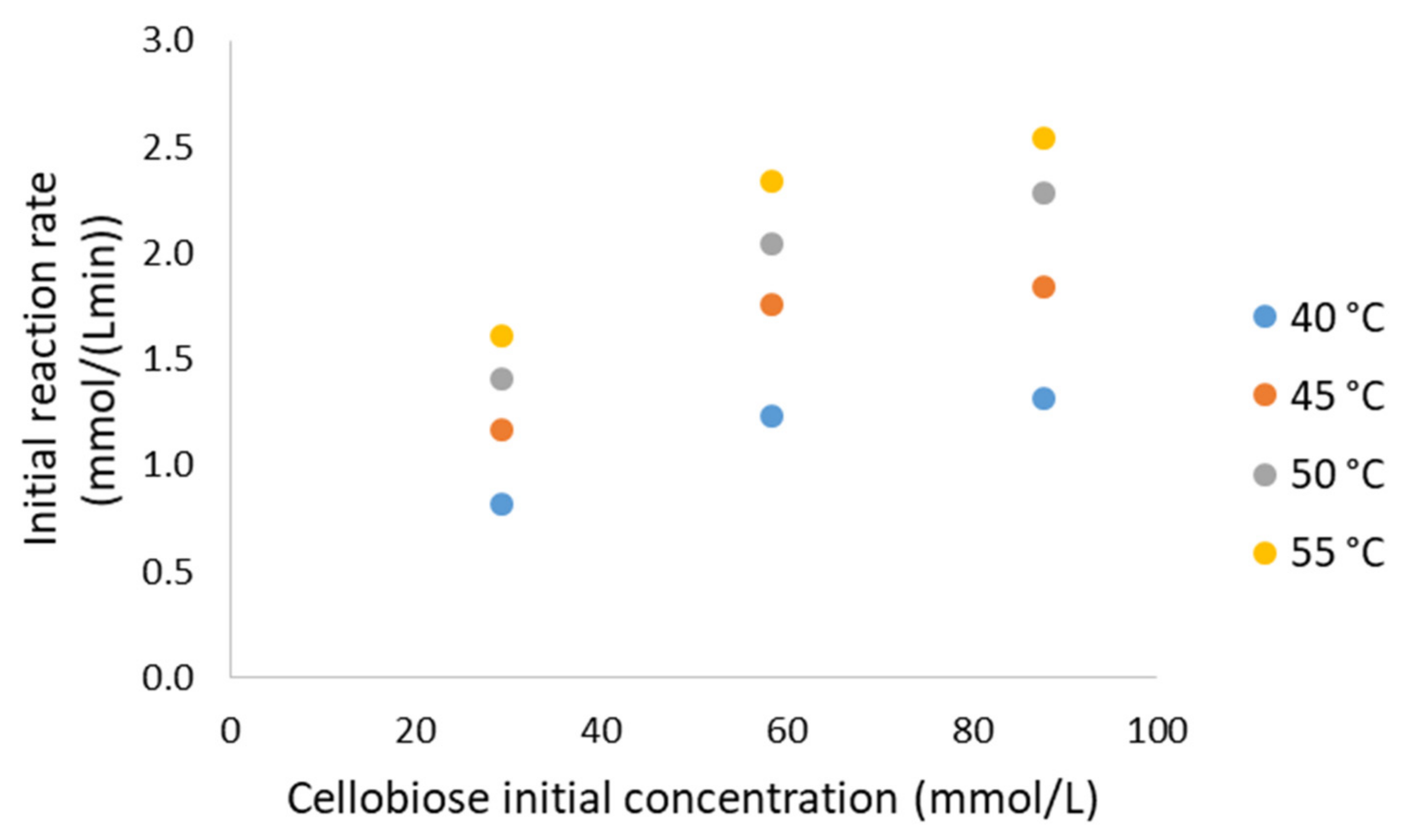
3.3. Cellobiose Hydrolysis: Preliminary Analysis

To test the potential use of the biocatalysts obtained in the treatment of food waste, the supports with the best characteristics for each enzyme (RN4 and RN8 for Novozym 188 and Glukosidase-1000, respectively) were used in a hydrolysis reaction of the natural substrate cellobiose, a disaccharide consisting of two glucose units linked by β(1,4) glycosidic linkage, as discussed above. Its action was compared with that of the free enzyme by reacting the same units of enzymatic activity of immobilized and free enzyme. Considering the initial reaction rate, immobilization activated the β-glucosidases when cellobiose is used as a substrate: Novozym 188 activity increased 177% when immobilized, while the activity of the enzyme from Aspergillus fumigatus increased 55% after immobilization.
It is usual to observe mass transfer limitations when using enzymes that are supported on porous solids. On the first side, external mass transfer limitations are due to the reduction of kinetic energy inside the Prandtl layer as the fluid phase impacts the solid. Thus, the rate at which the molecules move is low near the surface of the particles and this phenomenon can restrict the contact between the immobilized enzyme on the particle surface and the substrate moving from the liquid phase. We should take into account that the slowest dynamic phenomenon, either mass transfer or chemical reaction, is the one that controls the observed global rate and, thus, the system productivity. In order to optimize the process conditions, the stirring rate was varied to determine whether the hydrolysis process was controlled by the external diffusion rate or by the rate at which the enzyme carried out the chemical reaction.
In Figure 3a, the conversion of cellobiose versus reaction time at several stirring speeds is observed, being the conversion the ratio of the concentration of substrate converted divided by the concentration of initial substrate. In the right side of Figure 3, we can observe the initial reaction rate (activity of the enzyme) versus the stirring speed. Taking into account the experimental error carried out in the determination of the reaction rate for each of the stirring speeds (Figure 3), for 400, 600 and 800 rpm, the reaction rate is practically the same, while the rate corresponding to 200 rpm deviates notably; it is much lower. This figure shows that there is a zone where the external matter transfer controls the process rate (for stirring speeds below 400 rpm) and another zone (stirring speeds below 400 rpm) where the catalytic process determines the global rate. Thus, since the same result was obtained in the cellobiose hydrolysis reaction at a stirring speed of 400 rpm, this was chosen as the optimum speed to determine the kinetic model of the reaction.

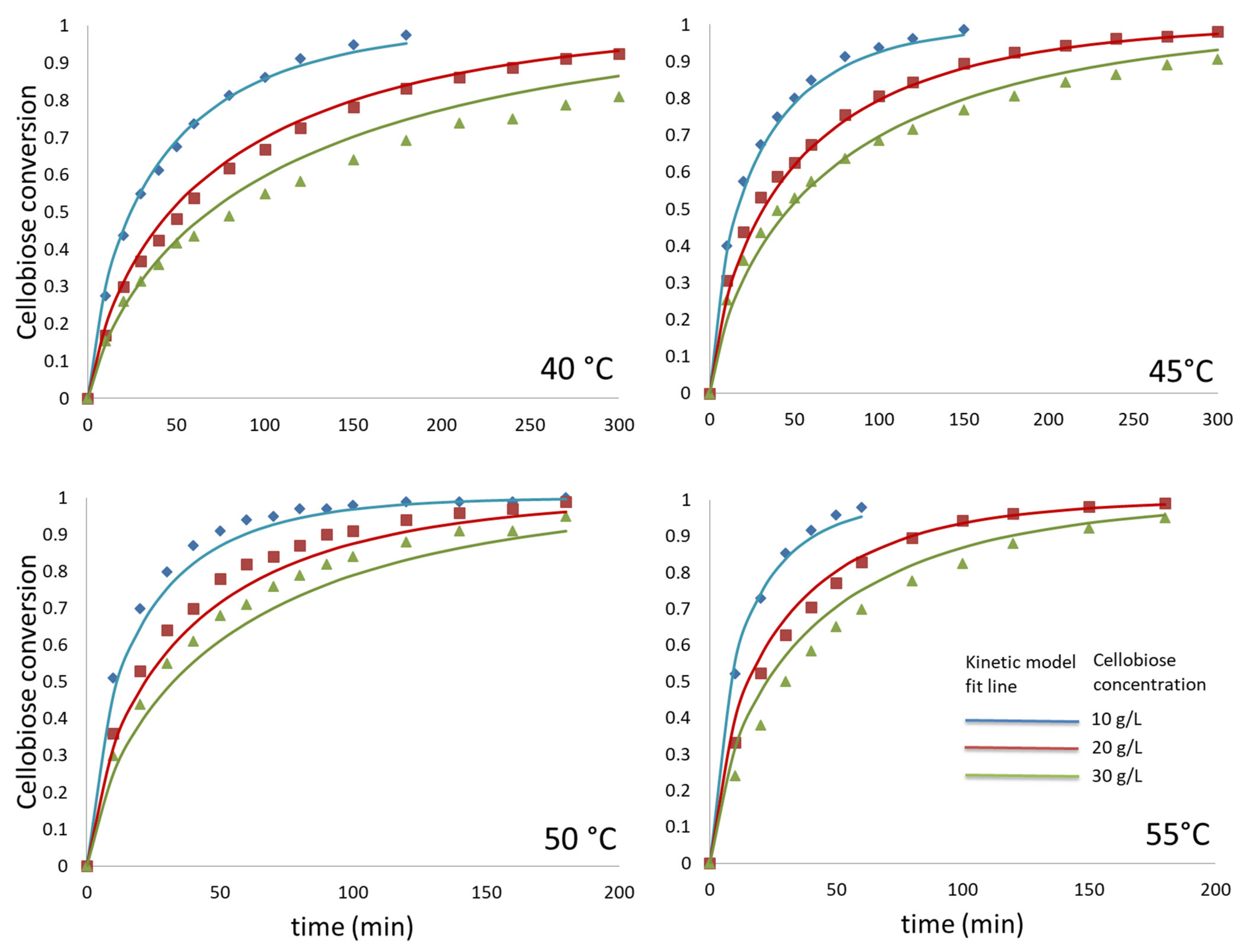
3.4. Cellobiose Hydrolysis: Kinetic Modelling

To describe the behavior of the enzyme under different reaction conditions and thus to determine the kinetic model governing the reaction catalyzed by the enzyme, several reactions were carried out at different temperatures (40, 45, 50 and 55 °C) and concentrations of cellobiose substrate (10, 20 and 30 g/L—29.3, 58.5, and 87.7 mmol/L). As expected, as the temperature rises, total conversion of substrate is reached before, while at higher substrate initial concentration a longer time for total conversion is needed.
As a first approach, initial reaction rates (a magnitude proportional to enzyme activity on cellobiose) were estimated for all runs and represented versus initial cellobiose concentration and displayed in Figure 4. It is evident that the reaction rate trends towards a maximum value (Vmax) at high values of the concentration of substrate, a hyperbolic behavior very common to enzymes showing a Michaelis–Menten behavior. When comparing with results obtained with the free enzyme [25], where a maximum value of reaction rate is obtained at relatively low concentration values of substrate, with a fast reduction at higher values, one gets the impression that no inhibition due to substrate is present in the process catalyzed by the immobilized enzyme, in clear contrast with what happens with the free form of the enzyme, notably inhibited by cellobiose. Thus, immobilization reduces, if not eliminates, this inhibition. This inhibitory effect, however, was also observed in a recent paper by our group when working with cross-linked agarose beads activated with polyethylenemine (PEI), or glyoxyl groups (Gly) [14], suggesting that support and immobilization chemistry play a role in the inhibitory role of cellobiose.
The kinetic models proposed, and fitted to all data, are Michaelis–Menten models with competitive inhibition by glucose, a type of inhibition very common to hydrolases acting on poly-, oligo- and disaccharides. The notable size of the active center of this β-glucosidase allows for the inhibition due to more than one molecule of glucose [25].
r = k 1 · a E · c e l o b i o s e 0 · 1 X K M · 1 + 2 · c e l o b i o s e 0   · X / K I n + c e l o b i o s e 0 · 1 X
k 1 = e x p ln k 1   0 E a R · 1 T
where k1 is the kinetic constant of product yield from the enzyme-substrate complex; aE is the enzyme activity; X is the conversion of cellobiose; KM corresponds to the Michaelis–Menten constant for cellobiose; KI is the competitive inhibition constant due to the product glucose; and n is the number of glucose molecules acting as competitive inhibitors. The kinetic constant k1 was expressed as an exponential function of temperature through a modified version of the well-known Arrhenius equation, a form most convenient when fitting a kinetic model to data retrieved at several operational temperatures. Here, k1 0 is the pre-exponential term of k1, while Ea is the activation energy and R is the constant for ideal gases.
All kinetic models tested fit closely to all data (N = 174) at the same time, as clearly shown in Figure 5 and in Table 3, where the values of the goodness-of-fit parameters defined in Equations (1)–(3) are collected. Very high F values and low values for SQR and RMSE indicate a good fit of the model. However, wider intervals for the kinetic constants are observed if we consider that only one glucose molecule acts as the competitive inhibitor per active center at a given time. When n = 2, that is, two glucose molecules are present in the active center simultaneously, F value is higher, SQR and RMSE are lower and very narrow error intervals for all kinetic constants are observed. If n > 2, goodness-of-fit parameter values worsen, indicating that, from a statistical perspective, the presence of more than two glucose molecules in the active center acting as inhibitors is less likely than the presence of only two of these molecules. This is in agreement with what was obtained and published previously for the free form of the enzyme [25].
When comparing the kinetic constant values for both forms of the enzyme, the higher value of activation energy is evident for the immobilized enzyme, indicating a higher rigidity of the solid-enzyme structure in comparison to the free enzyme. Immobilization also affects KM and KI: the Michaelis–Menten constant for the free enzyme is 2.8 mM, while its value is 20 mM for the immobilized enzyme, showing that immobilization has reduced the capacity of the enzyme to link a substrate in the active center and/or partition effects near the surface reduce the substrate concentration surrounding the enzyme. The effect on the inhibition constant is affected in a similar way: 8 mM for the free enzyme versus 42 mM for the immobilized enzyme; thus, either the ligand-active center is compromised or partition effects reduce glucose concentration near the enzyme, though it is more likely the first consideration as glucose is produced in the active center (it is not a compound being transferred and bound into the enzyme, as with cellobiose). Das et al. [35] found a similar behavior of the β-glucosidase from Aspergillus fumigatus ABK9 when immobilizing it on alginate beads: Dixon plots indicate that KI values for the free and immobilized forms of the enzyme were 310 and 430 mM, clearly showing a low inhibition by glucose further reduced by immobilization.
Finally, a curious, but important, effect when comparing initial values for the cellobiose hydrolysis per U.I. activity for both forms of the enzyme is that, at 40 °C and for the lowest value of cellobiose initial concentration, the immobilized enzyme is much more active (2.44 versus 0.39 mmol/(U.I. min), so a notable surface activation happens after immobilization.

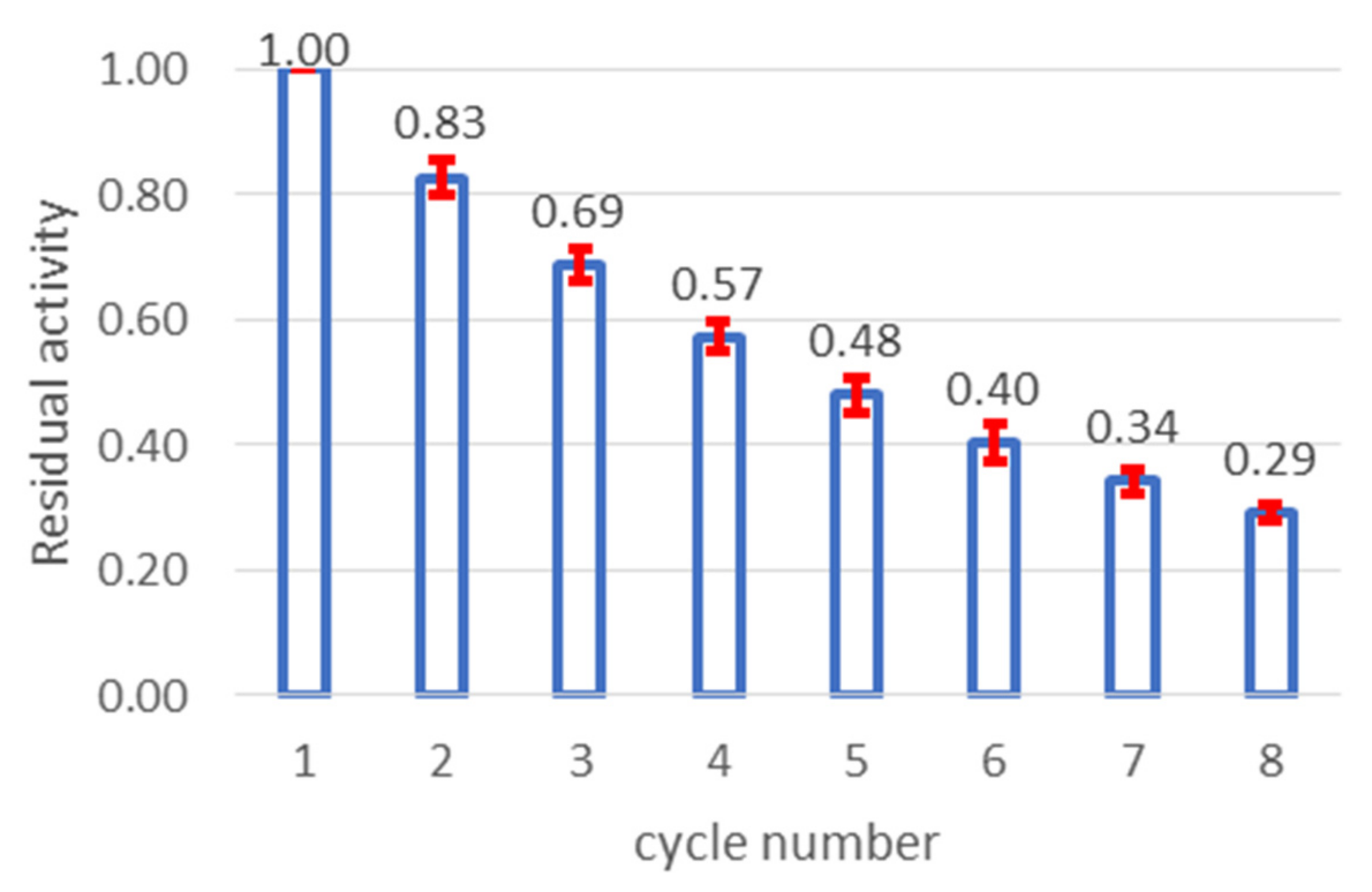
3.5. Immobilized Enzyme: Operational Stability

In Figure 6 we have collected the results of the evolution of remaining or residual activity in the immobilized enzyme with the cycle number. It can be observed that there is a notable deactivation of the enzyme during recycling, as expected when there is only a slight interaction between the support and the enzyme (only one covalent bond via the terminal amino group of the polypeptide chain). In fact, this stability is similar to that of the free enzyme [36]. In contrast, when multiple interaction happens, even if no covalent bonds are present, enzyme stabilization can be attained [37]. The low-interaction enzyme support found here is ensured by the high ionic strength during immobilization, as Betancor et al. observed for diverse supports activated with glutaraldehyde when immobilizing β-galactosidase and D-amino acid oxidase [29].

4. Conclusions

This research compared Glukosidase-1000 from ASA-spezialenzymes with Novozym 188, both commercial preparations from Aspergillus fumigatus and Aspergillus niger, respectively. It has been proved that the first one shows higher specific enzymatic activity in the hydrolysis of pNPG.
Using different acrylate supports, the best support for this activity immobilization was ReliZyme™ HA403 activated with glutaraldehyde in terms of total immobilized activity from the liquid and immobilized enzyme activity towards pNPG. It has also been proved that the best performance in cellobiose hydrolysis was displayed by this immobilized β-glucosidase. A first kinetic analysis under reaction-controlled conditions led to the conclusion that no apparent acompetitive inhibition due to cellobiose is present when using the immobilized version of the enzyme, in contrast to what was published for the free extract or even for other immobilized preparations of the same commercial product. It is also observed that the immobilized biocatalyst displays better specific activity than its soluble counterpart. At high enzyme loading, the immobilized β-glucosidase showed a notable activity on cellobiose, while immobilization was affected more negatively due to its activity on pNPG, a more hydrophobic substrate.
Finally, a kinetic model of cellobiose hydrolysis by immobilized β-glucosidase from ASA-spezialenzymes has been established for a low-loading immobilized enzyme. No apparent acompetitive inhibition due to cellobiose is observed in this immobilized version of the enzyme, in contrast to what was published for the free extract. In both cases, a notable double inhibition due to the product (glucose) is present, though much lower than for the free enzyme, while an apparent lower affinity of the immobilized enzyme is perceived when comparing KM values. This can be due to structural changes of the enzyme upon immobilization and/or partition effects affecting glucose and cellobiose near the surface of the support. This low-loading enzymatic preparation was as stable as its free counterpart according to remaining activity temporal evolution in several consecutive hydrolysis cycles, indicating only a slight stabilization when compared to the free enzyme if using high ionic strength during immobilization.

Author Contributions

Conceptualization, M.A. and M.L.; methodology, M.A. and C.Y.; software, M.L.; validation, C.Y., M.A., M.L. and J.E.; formal analysis, M.A. and M.L.; investigation, C.Y., J.E., M.A.; data curation, M.L.; writing—original draft preparation, C.Y. and M.L.; writing—review and editing, C.Y., M.A. and M.L.; supervision, M.A. and M.L.; project administration, M.L.; funding acquisition, M.L. All authors have read and agreed to the published version of the manuscript.

Funding

This research was partially funded by MINECO and AEI under contracts CTQ2017-84963-C2-1-R and PID2020-114365RB-C21, funding that is gratefully acknowledged.

Institutional Review Board Statement

Not applicable.

Informed Consent Statement

Not applicable.

Data Availability Statement

All relevant data are provided within this manuscript.

Acknowledgments

The authors are indebted to Novozymes A/S, to ASA Spezialenzymes and to Resindion srL, for the kind gift of enzymatic cocktails and the Sepabeads and ReliZyme supports.

Conflicts of Interest

The authors declare no conflict of interest. The funders had no role in the design of the study; in the collection, analyses, or interpretation of data; in the writing of the manuscript, or in the decision to publish the results.

References

  1. Demirbaş, A. Biomass resource facilities and biomass conversion processing for fuels and chemicals. Energy Conv. Manag. 2001, 42, 1357–1378. [Google Scholar] [CrossRef]
  2. Bolton, J.R. Conversion and storage of solar energy. Ann. Rev. Energy 1979, 4, 353–401. [Google Scholar] [CrossRef]
  3. Calsavara, L.P.V.; Morales, F.F.; Zanin, G.M. Comparison of catalytic properties of free and immobilized cellobiase Novozym 188. Appl. Biochem. Biotechnol. 2001, 91–93, 615–626. [Google Scholar] [CrossRef]
  4. Agbor, V.B.; Cicek, N.; Sparling, R.; Berlin, A.; Levin, D.B. Biomass pretreatment: Fundamentals toward application. Biotechnol. Adv. 2011, 29, 675–685. [Google Scholar] [CrossRef]
  5. Chandel, A.K.; Chandrasekhar, G.; Silva, M.B.; da Silva, S.S. The realm of cellulases in biorefinery development. Crit. Rev. Biotechnol. 2012, 32, 187–202. [Google Scholar] [CrossRef]
  6. Srivastava, N.; Rathour, R.; Jha, S.; Pandey, K.; Srivastava, M.; Thakur, V.K.; Sengar, R.S.; Gupta, V.K.; Mazumder, P.B.; Khan, A.F.; et al. Microbial beta glucosidase enzymes: Recent advances in biomass conversation for biofuels application. Biomolecules 2019, 9, 220. [Google Scholar] [CrossRef] [Green Version]
  7. Singhania, R.R.; Patel, A.K.; Pandey, A.; Ganansounou, E. Genetic modification: A tool for enhancing beta-glucosidase production for biofuel application. Biores. Technol. 2017, 245, 1352–1361. [Google Scholar] [CrossRef]
  8. Huang, C.; Feng, Y.; Patel, G.; Xu, X.Q.; Qian, J.; Liu, Q.; Kai, G.Y. Production, immobilization and characterization of beta-glucosidase for application in cellulose degradation from a novel Aspergillus versicolor. Int. J. Biol. Macromol. 2021, 177, 437–446. [Google Scholar] [CrossRef]
  9. Vazquez-Ortega, P.G.; Alcaraz-Fructuoso, M.T.; Rojas-Contreras, J.A.; López-Miranda, J.; Fernandez-Lafuente, R. Stabilization of dimeric β-glucosidase from Aspergillus niger via glutaraldehyde immobilization under different conditions. Enz. Microb. Technol. 2018, 110, 38–45. [Google Scholar] [CrossRef]
  10. Nishida, V.S.; de Oliveira, R.F.; Brugnari, T.; Correa, R.C.G.; Peralta, R.A.; Castoldi, R.; de Souza, C.G.; Bracht, A.; Peralta, R.M. Immobilization of Aspergillus awamori β-glucosidase on commercial gelatin: An inexpensive and efficient process. Int. J. Biol. Macromol. 2018, 111, 1206–1213. [Google Scholar] [CrossRef]
  11. Wu, X.; Qu, B.; Liu, Y.; Ren, X.; Wang, S.; Quan, Y. Highly enhanced activity and stability via affinity induced immobilization β-glucosidase from Aspergillus niger onto amino-based silica for the biotransformation of ginsenoside Rb1. J. Chromatog. A 2021, 1653, 462388. [Google Scholar] [CrossRef]
  12. Tu, M.; Zhang, X.; Kurabi, A.; Gilkes, N.; Mabee, W.; Saddler, J. Immobilization of β-glucosidase on Eupergit C for lignocellulose hydrolysis. Biotechnol. Lett. 2006, 28, 151–156. [Google Scholar] [CrossRef]
  13. Silva, T.M.; Pessela, B.C.; Silva, J.C.R.; Lima, M.S.; Jorge, J.A.; Guisan, J.M.; Polizeli, M.L.T.M. Immobilization and high stability of an extracellular β-glucosidase from Aspergillus japonicus by ionic interactions. J. Mol. Catal. B Enzym. 2014, 104, 95–100. [Google Scholar] [CrossRef]
  14. Alvarez-Gonzalez, C.; Santos, V.E.; Ladero, M.; Bolivar, J.M. Immobilization-Stabilization of β-Glucosidase for Implementation of Intensified Hydrolysis of Cellobiose in Continuous Flow Reactors. Catalysts 2022, 12, 80. [Google Scholar] [CrossRef]
  15. Katchalski-Katzir, E.; Kraemer, D.M. Eupergit® C, a carrier for immobilization of enzymes of industrial potential. J. Mol. Catal. B Enzym. 2000, 10, 157–176. [Google Scholar] [CrossRef]
  16. Hilterhaus, L.; Minow, B.; Müller, J.; Berheide, M.; Quitmann, H.; Katzer, M.; Thum, O.; Antranikian, G.; Zeng, A.P.; Liese, A. Practical application of different enzymes immobilized on Sepabeads. Bioproc. Biosys. Eng. 2008, 31, 163–171. [Google Scholar] [CrossRef]
  17. Mateo, C.; Abian, O.; Fernández-Lorente, G.; Pedroche, J.; Fernández-Lafuente, R.; Guisan, J.M.; Tam, A.; Daminiti, M. Epoxy sepabeads: A novel epoxy support for stabilization of industrial enzymes via very intense multipoint covalent attachment. Biotechnol. Progr. 2002, 18, 629–634. [Google Scholar] [CrossRef]
  18. Kahar, U.M.; Sani, M.H.; Chan, K.-G.; Goh, K.M. Immobilization of α-amylase from Anoxybacillus sp. SK3-4 on ReliZyme and immobead supports. Molecules 2016, 21, 1196. [Google Scholar] [CrossRef] [Green Version]
  19. Bu, Y.; Zhang, T.; Jiang, B.; Chen, J. Improved performance of d-psicose 3-epimerase by immobilisation on amino-epoxide support with intense multipoint attachment. Foods 2021, 10, 831. [Google Scholar] [CrossRef]
  20. Rashid, M.H.; Siddiqui, K.S. Carboxy-group modification: High-temperature activation of charge-neutralized and charge-reversed beta-glucosidases from Aspergillus niger. Biotechnol. Appl. Biochem. 1998, 27, 231–237. [Google Scholar]
  21. Bezbradica, D.I.; Mateo, C.; Guisan, J.M. Novel support for enzyme immobilization prepared by chemical activation with cysteine and glutaraldehyde. J. Mol. Catal. B Enzym. 2014, 102, 218–224. [Google Scholar] [CrossRef]
  22. Bradford, M. A rapid and sensitive method for the quantitation of microgram quantities of protein utilizing the principle of protein-dye binding. Anal. Biochem. 1976, 72, 248–254. [Google Scholar] [CrossRef]
  23. Laemmli, U.K. Cleavage of structural proteins during the assembly of the head of bacteriophage T4. Nature 1970, 277, 680–685. [Google Scholar] [CrossRef]
  24. Lima, M.A.; Oliveira-Neto, M.; Kadowaki, M.A.S.; Rosseto, F.R.; Prates, E.T.; Squina, F.M.; Leme, A.F.P.; Skaf, M.S.; Polikarpov, I. Aspergillus niger β-glucosidase has a cellulase-like tadpole molecular shape: Insights into glycoside hydrolase family 3 (GH3) β-glucosidase structure and function. J. Biol. Chem. 2013, 288, 32991–33005. [Google Scholar] [CrossRef] [Green Version]
  25. Wojtusik, M.; Yepes, C.M.; Villar, J.C.; Cordes, A.; Arroyo, M.; Garcia-Ochoa, F.; Ladero, M. Kinetic modeling of cellobiose by a β-glucosidase from Aspergillus fumigatus. Chem. Eng. Res. Design 2018, 136, 502–512. [Google Scholar] [CrossRef]
  26. Wojtusik, M.; Villar, J.C.; Ladero, M.; Garcia-Ochoa, F. Physico-chemical kinetic modelling of hydrolysis of a steam-explosion pre-treated corn stover: A two-step approach. Bioresour. Technol. 2018, 268, 592–598. [Google Scholar] [CrossRef]
  27. Sheldon, R.A. Enzyme immobilization: The quest for optimum performance. Adv. Synth. Catal. 2007, 349, 1289–1307. [Google Scholar] [CrossRef]
  28. López-Gallego, F.; Montes, T.; Fuentes, M.; Alonso, N.; Grazu, V.; Betancor, L.; Guisán, J.M.; Fernández-Lafuente, R. Improved stabilization of chemically aminated enzymes via multipoint covalent attachment on glyoxyl supports. J. Biotechnol. 2005, 116, 1–10. [Google Scholar] [CrossRef] [PubMed]
  29. Betancor, L.; López-Gallego, F.; Hidalgo, A.; Alonso-Morales, N.; Mateo, G.D.O.C.; Fernández-Lafuente, R.; Guisán, J.M. Different mechanisms of protein immobilization on glutaraldehyde activated supports: Effect of support activation and immobilization conditions. Enz. Microbiol. Technol. 2006, 39, 877–882. [Google Scholar] [CrossRef]
  30. Mallin, H.; Wulf, H.; Bornscheuer, U.T. A self-sufficient Baeyer–Villiger biocatalysis system for the synthesis of ɛ-caprolactone from cyclohexanol. Enzyme Microb. Technol. 2013, 53, 283–287. [Google Scholar] [CrossRef] [PubMed]
  31. Le Traon-Masson, M.P.; Pellerin, P. Purification and characterization or two β-D-glucosidases from an Aspergillus niger enzyme preparation: Affinity and specificity toward glucosylated compounds of the processing of fruits. Enzyme Microb. Technol. 1998, 22, 374–382. [Google Scholar] [CrossRef]
  32. Nagy, F.; Gyujto, I.; Tasnádi, G.; Barna, B.; Balogh-Weiser, D.; Faber, K.; Poppe, L.; Hall, M. Design and application of a bi-functional redox biocatalyst through covalent co-immobilization of ene-reductase and glucose dehydrogenase. J. Biotechnol. 2020, 323, 246–253. [Google Scholar] [CrossRef]
  33. Bilgin, R.; Yalcin, M.S.; Yildirim, D. Optimization of covalent immobilization of Trichoderma reesei cellulase onto modified ReliZyme HA403 and Sepabeads EC-EP supports for cellulose hydrolysis, in buffer and ionic liquids/buffer media. Artif. Cells Nanomed. Biotechnol. 2016, 44, 1276–1284. [Google Scholar] [CrossRef]
  34. D’Arrigo, P.; Allegretti, C.; Fiorati, A.; Piubelli, L.; Rosini, E.; Tessaro, D.; Valentino, M.; Pollegioni, L. Immobilization of L-aspartate oxidase from Sulfolobus tokodaii as a biocatalyst for resolution of aspartate solutions. Catal. Sci. Technol. 2015, 5, 1106–1114. [Google Scholar] [CrossRef]
  35. Das, A.; Paul, T.; Ghosh, P.; Halder, S.K.; Das Mohapatra, P.K.; Pati, B.R.; Mondal, K.C. Kinetic study of a glucose tolerant β-glucosidase from Aspergillus fumigatus ABK9 entrapped into alginate beads. Waste Biomass Valor. 2015, 6, 53–61. [Google Scholar] [CrossRef]
  36. Wojtusik, M.; Vergara, P.; Villar, J.C.; Garcia-Ochoa, F.; Ladero, M. Thermal and operational deactivation of Aspergillus fumigatus β-glucosidase in ethanol/water pretreated wheat straw enzymatic hydrolysis. J. Biotechnol. 2019, 292, 32–38. [Google Scholar] [CrossRef]
  37. Fathi, Z.; Doustkhah, E.; Ebrahimipour, G.; Darvishi, F. Noncovalent immobilization of Yarrowia lipolytica lipase on dendritic-like amino acid-functionalized silica nanoparticles. Biomolecules 2019, 9, 502. [Google Scholar] [CrossRef] [Green Version]
Figure 1. (a) Activation of ReliZyme™ HA403 support particle with glutaraldehyde. The support has a hexamethylamine arm that reacts with one of the aldehyde groups of the glutaraldehyde, releasing a water molecule in the reaction and leaving the support particle activated with the glutaraldehyde bound; (b) Immobilization of the enzyme via external amino groups (the N-terminus amino in particular) to epoxide groups on support surface; (c) Immobilization of the enzyme by Schiff-base formation between aldehyde groups on the support (provided by glutaraldehyde) and external amino groups on the enzyme (usually, the N-terminus amino group at neutral pH).
Processes 10 01225 g001
Figure 2. (a) Electrophoretic profile of Novozym 188. Lane 1: 3.74 µg from Novozym 188. Lane 2: standard proteins of known molecular mass: myosin, β-galactosidase, phosphorylase b, BSA, ovalbumin, carbonic anhydrase, soybean trypsin inhibitor, lysozyme. (b) Electrophoretic profile of Glukosidase-1000. Lane 1: 3.02 µg from the commercial enzyme preparation. Lane 2: the mentioned standard proteins of known molecular mass.
Processes 10 01225 g002
Figure 3. Effect of stirring speed on hydrolysis of cellobiose catalyzed by immobilized β-glucosidase (ASA-1000) at various reactor stirring speeds. Reaction conditions: 100 mg of immobilized RN8 derivative; temperature 40 °C; 2 mL sodium citrate buffer 100 mM pH 5.0; 20 g/L cellobiose. (a) Progress curves and (b) Initial observed rate versus agitation speed.
Processes 10 01225 g003
Figure 4. Initial reaction rate versus initial concentration of cellobiose at several temperatures.
Processes 10 01225 g004
Figure 5. Fit of the proposed model (in lines) to data retrieved at all temperatures (points) tested and several cellobiose initial concentrations. Other reaction conditions: 100 mg of immobilized RN8 derivative; 2 mL sodium citrate buffer 100 mM pH 5.0.
Processes 10 01225 g005
Figure 6. Residual activity evolution for several consecutive hydrolysis cycles. Conditions: 20 g/L (58.5 mM) cellobiose in 100 mM citrate buffer pH 5.0 under orbital shaking at 400 rpm and 50 °C.
Processes 10 01225 g006
Table 1. Covalent immobilization of β-glucosidases from the commercial preparations Novozym 188 (Novozymes A/S) and Glukosidase-1000 (ASA-spezialenzymes GmbH).
Immobilized PreparationSelected SupportEnzyme CocktailImmobilization Process TimeaLYLYSaS
RN1ReliZyme™ EP403Novozym 18824 h8.624.7%0.18%0.04
RN2ReliZyme™ EP403Novozym 18872 h2.728.9%11.9%0.9
RN3ReliZyme™ HA403-GANovozym 18824 h2.748.5%5%0.7
RN4ReliZyme™ HA403-GANovozym 18824 h1.435.7%54.8%3.3
RN5Eupergit® CNovozym 18872 h2.414.3%2.8%0.09
RN6Sepabeads™ EC-EP303Novozym 18872 h2.45.7%17.9%0.2
RN7ReliZyme™ HA403-GANovozym 18824 h0.4571%46%1.5
RN8ReliZyme™ HA403-GAASA-100024 h0.8100%73.8%6.2
Note: aL Activity (U.I.) in liquid per 100 mg support; YL Immobilization yield in terms of activity lost from liquid; YS Immobilization final yield in the support; aS Specific enzymatic activity (U.I./g support). The selected method is in bold letters.
Table 2. Covalent immobilization of the β-glucosidase in the commercial preparation Glukosidase-1000 (ASA-Spezialenzymes GmbH) on ReliZyme™ HA403-GA at several enzyme loading at 24 h.
Immobilized PreparationaLYLYSaSYScelaScel
RN80.8100%73.8%6.294.2%3.5
RN91.6100%71.8%11.591.4%6.8
RN103.2100%67.4%21.692.1%13.7
RN116.4100%63.2%40.489.5%26.6
RN1212.8100%58.4%74.890.2%53.6
RN1325.6100%45.1%11687.3%103.8
RN1451.280%37.3%19179.5%189.0
RN15102.445%33.4%34268.3%324.8
Note: aL Activity (U.I.) in liquid per 100 mg support; YL Immobilization yield in terms of activity lost from liquid; YS Immobilization final yield in the support; aS Specific enzymatic activity (U.I./g support); YScel Immobilization final yield in the support in terms of activity towards cellobiose; aScel Specific enzymatic activity (U.I./g support) on cellobiose. The RN8 runs, in bold, is the benchmark immobilization, at low enzyme activity, as selected in Table 1.
Table 3. Kinetic constants (with their standard errors) and goodness-of-fit statistical parameters for the kinetic model fitted to all data from cellobiose hydrolysis runs performed at several temperatures and initial substrate concentrations. The selected model is in bold letters.
Kinetic ModelLn k10Ea/R (k1)KMKISQRRMSEF-Value
M1 n = 116.26 ± 2.457632 ± 5360.0025 ± 0.0080.006 ± 0.0020.1980.039213,879
M2 n = 215.78 ± 0.717516 ± 2210.020 ± 0.0030.042 ± 0.0030.1930.037814,389
M3 n = 315.72 ± 0.727493 ± 2250.025 ± 0.0040.091 ± 0.0050.2020.038413,742
M4 n = 415.69 ± 0.737478 ± 2270.028 ± 0.0040.142 ± 0.0060.2070.039313,320
Note: Ln k10 Neperian of the pre-exponential value of the catalytic constant k1 (mmol cellobiose/(U.I. enzyme x minute)). Ea/R Activation energy of the catalytic constant k1 divided by the Ideal Gas constant R (K). KM Michaelis–Menten constant for cellobiose (mol/L); KI Inhibition constant for glucose (mol/L).
Publisher’s Note: MDPI stays neutral with regard to jurisdictional claims in published maps and institutional affiliations.

