Article

Assessment of a Formulation Containing a Castanea sativa Shells Extract on Skin Face Parameters: In Vivo Evaluation

by
Ana Margarida Silva
1,
Paulo C. Costa
2,3,
Cristina Delerue-Matos
1 and
Francisca Rodrigues
1,*
1
REQUIMTE/LAQV, Polytechnic of Porto—School of Engineering, Rua Dr. António Bernardino de Almeida, 4249-015 Porto, Portugal
2
REQUIMTE/UCIBIO, MedTech-Laboratory of Pharmaceutical Technology, Department of Drug Sciences, Faculty of Pharmacy, University of Porto, Rua de Jorge Viterbo Ferreira, 228, 4050-313 Porto, Portugal
3
Associate Laboratory i4HB—Institute for Health and Bioeconomy, Faculty of Pharmacy, University of Porto, 4050-313 Porto, Portugal
*
Author to whom correspondence should be addressed.
Processes 2022, 10(11), 2230; https://doi.org/10.3390/pr10112230
Submission received: 28 September 2022 / Revised: 24 October 2022 / Accepted: 26 October 2022 / Published: 31 October 2022
(This article belongs to the Special Issue Bioactive Compounds from Food Waste and By-Products)

Abstract

:
In the last years, the demand for new eco-friendly ingredients has increased on the cosmetic market. Consumers are more aware of sustainable principles and, simultaneously, more conscious regarding the skin aging process. Chestnut (Castanea sativa) shells are a food by-product produced in high quantities in Europe. This waste has been described as a rich source of phenolic compounds with skin biological effects, such as antioxidant, antiradical, and anti-inflammatory activities. Despite the huge number of assays reporting the richness of chestnut shell extracts in bioactive compounds as well as the development of cosmetic formulations containing these extracts, no in vivo assays have assessed their clinical efficacy in human volunteers. The aim of this study was to evaluate the effect on skin face parameters of a formulation containing a chestnut shell extract in human volunteers (n = 22) who applied the product twice per day, for 56 days. For that, biophysical techniques, including Corneometer®, Cutometer®, and PrimosPremium, were employed, allowing the quantification of skin hydration and firmness, as well as of wrinkles’ depth and volume and wrinkles’ skin roughness. The results demonstrated that the formulation led to a slight decrease in roughness and wrinkles’ depth, although no significant differences with respect to a placebo were observed. In addition, a clear improvement of skin hydration was achieved (t0 = 54.00 Arbitrary Units (A.U.) and t56 = 58.62 A.U.). In contrast to the placebo, the active formulation increased skin firmness up to 31.76% in 50.00% of the volunteers, but without significant differences with respect to the placebo, probably due to the short period of treatment. A long-term use of the product is recommended to possibly observe significant differences in all parameters.

1. Introduction

The interest in new cosmetic ingredients obtained from agro-food by-products has been rapidly growing in the cosmetic field [1]. Chestnut (Castanea sativa) by-products, particularly shells, are a good example of new cosmetic ingredients that have been intensely explored in the last years [2,3]. Different studies reported the richness of chestnut shells extracts in phenolic compounds, such as pyrogallol, protocatechuic acid, ellagic acid, caffeic acid, gallic acid, epigallocatechin, and catechin [4,5,6,7]. These extracts were obtained by different eco-friendly extraction techniques, such as Subcritical Water Extraction (SWE), Supercritical Fluid Extraction-CO2 (SC-CO2), Microwave-Assisted Extraction (MAE), or Ultrasound-Assisted Extraction (UAE) [4,5,6,7]. Particularly, Lameirão et al. employed UAE to recover phenolic compounds from chestnut shells, aiming to explore new cosmetic applications [7]. The optimal extract reported by the authors achieved a high scavenging efficiency against Reactive Oxygen and Nitrogen Species (ROS and RNS, respectively), namely, nitric oxide (NO) and hypochlorous acid (HOCl) (IC50 = 0.1 µg/mL and 0.7 µg/mL, respectively), species enrolled in the wrinkle formation process [7]. This activity is probably associated with the phenolic compounds present in the extract, particularly procyanidins and flavonols, being ellagic acid (40.4 µg/mg of dry weight (dw), epigallocatechin (15.3 µg/mg dw), and a caffeic acid derivative (15.4 µg/mg dw) the principal ones [7]. Ellagic acid has been described as a potent antioxidant that acts as a melanogenesis suppressor with photoaging prevention ability [8,9]. In vivo studies on the skin of hairless mice demonstrated that the topical application of ellagic acid can decrease skin damage associated with the Ultraviolet (UV)-B radiation, by decreasing matrix metalloproteinases (MMP) production and mitigating collagen fiber degradation [9]. These roles lead to the inhibition of pro-inflammatory cytokines production (interleukin (IL) release, such as IL-1β and IL-6), preventing wrinkles formation [9]. In addition, an ex vivo permeation assay performed in Franz diffusion cells coupled to human skin reported that ellagic acid achieved a permeation of 732.1 µg/g of dry weight (dw) after 8 h of topical application [1]. Epigallocatechin, another phenolic compound present in the extract, has photoprotective properties, reducing skin damage such as roughness, sagginess, and erythema and counteracting the dermal collagen decrease induced by UVB and UVA radiations [10]. An in-vivo Solar Protection Factor (SPF) study demonstrated that the topical application of (-) epigallocatechin-3-gallate can block the UVB-induced infiltration of leukocytes and decrease myeloperoxidase activity, exhibiting preventive effects on diseases such as inflammatory dermatoses, photoaging, and photocarcinogenesis [11]. Caffeic acid, another polyphenol quantified in C. sativa shells extracts obtained by UAE, is well known for its anti-aging activity [7,12]. Yamada et al. reported that the topical application of caffeic acid in hairless mice had a suppressive effect against UVA radiation and ROS generation in the skin [13]. In addition, caffeic acid was able to permeate in all skin sections of the pig ear, achieving a flux value of 0.48 µg/cm2/h [14]. Considering the composition of the chestnut shell extract obtained by UAE and the possible associated skin benefits, Pinto et al. and Oliveira et al. developed a semisolid formulation (O/W cream) [2,3] and an hydrogel [15] containing this active ingredient, reporting good stability properties. However, in vivo information is still needed, particularly clinical studies that confirm the efficacy of the final formulations on volunteers. Therefore, the goal of this study was to evaluate the anti-aging efficacy of a cosmetic formulation containing a chestnut shell extract as the active ingredient through clinical studies, aiming to validate a new cosmetic ingredient obtained from a food by-product on the basis of a circular economy concept.

2. Materials and Methods

2.1. Chemicals

Decyl oleate was purchased from Guinama SL (Valencia, Spain), while white petrolatum, glycerin, and cetearyl alcohol were provided by Acofarma distribucion AS (Barcelona, Spain). Microcare® PHDG was supplied by THOR Personal Care (Barcelona, Spain). All ingredients were of pharmaceutical and cosmetic grade. Water was purified by ion exchange using synthetic resins.

2.2. Samples

The C. sativa shells were kindly provided by Sortegel (Sortes, Bragança, Portugal). The shells were dehydrated (Excalibur Food Dehydrator, Sacramento, CA, USA) at 41 °C for 24 h and subsequently grinded in a miller (Ultra Centrifugal Mill ZM 200, Retsch, Germany) and stored at 20 °C until extraction.

2.3. Preparation of the Extract

The C. sativa shells extract was obtained according to Lameirão et al. using their optimal extract conditions [7]. The UAE technique was performed using an ultrasonic probe processor (Sonic Vibracell, model VCX50, Newtown, CT, USA) associated with a probe tip No. 630-0219 with 13 mm of diameter. Briefly, 5 g of powder sample was mixed with 100 mL of water, and the extraction was performed at 70 °C for 40 min. After preparation, the extract was filtered through Whatman n° 1 paper, centrifuged, and incorporated in the formulation as an aqueous phase.

2.4. Preparation of the Formulation

The formulation was developed and prepared according to Pinto et al. [2]. The excipients used and their respective quantities are summarized in Table 1. A base reference without extract (placebo) was also prepared. Briefly, the water and oil phases were separately heated in a water bath at 60–80 °C. At the same temperature, the water phase was added to the oil phase, while stirring at 700–1000 rpm, using a propeller agitator (Heidolph, RZR 2041, Wehr, Germany). The mixer speed was reduced to complete the homogenization, followed by cooling to room temperature. Subsequently, the formulation was conditioned and stored at room temperature (18–28 °C).

2.5. Clinical Evaluation in Human Volunteers

A double-blinded randomized study was performed to evaluate the effect of the formulations with and without extract on the face skin of volunteers. Twenty-two healthy females, with a mean age of 53 ± 7 years, were randomly enrolled in the study. All volunteers received the study information and signed an informed consent. To be included in the study, the volunteers had to fulfil the following inclusion criteria: (i) present visible wrinkles on the crow’s feet area of both sides of the face; (ii) have a Fitzpatrick skin phototype between II and IV; (iii) not present allergy or hypersensitivity to the components of the cosmetic product; (iv) not present cutaneous alterations on the face; and (v) not have performed anti-aging treatments on the 30 days preceding the study beginning. The study was performed following the Declaration of Helsinki, ICH-GCP, and legal requirements, fulfilling independent ethics committee’s requirements, previously approved by INFARMED, I.P., through the National Registry for Clinical Studies (RNEC), number 102124.
Each subject applied around 0.704 ± 0.263 mg/cm2 of the formulation with the extract (active cream) on cleansed skin of the randomized hemiface and 0.776 ± 0.236 mg/cm2 of the formulation without the extract (placebo) on the other hemiface, twice daily, during 56 consecutive days. The potential reduction in wrinkles and skin roughness by the formulation with the extract was assessed and compared to the baseline values of these parameters and to the effects of the placebo. The measurements were performed before (t0) and after 56 (t56) consecutive days of the products’ application (twice daily), under controlled atmospheric conditions, namely, at room temperature of 23.0 ± 1.0 °C and relative humidity of 50.0 ± 10.0%, after 20 min of acclimatization.

2.5.1. Instrumental Assessments

Wrinkles’ Depth and Volume Evaluation

The mean depth and mean volume of the wrinkles were evaluated on the crow’s feet area of both hemifaces of each volunteer with PrimosPremium (Canfield Scientific, Inc., Parsippany, NJ, USA), in a black room under standardized lighting and position conditions.

Skin Roughness Evaluation

The skin roughness average (Ra) was assessed on the crow’s feet area of both hemifaces of each volunteer with PrimosPremium (Canfield Scientific, Inc., Parsippany, NJ, USA), in a black room under standardized lighting and position conditions.

Skin Firmness Evaluation

The skin firmness and elasticity were evaluated by the suction method with the equipment Cutometer® SEM 575 (Courage + Khazaka electronic GmbH, Köln, Germany) on each hemiface of each volunteer (along the cheekbone).

Skin Hydration Evaluation

The skin capacitance was assessed instrumentally with a Corneometer® CM825 (Courage + Khazaka electronic GmbH, Köln, Germany) by applying a slight pressure on each side of the forehead of each volunteer. Each skin site was measured in triplicate, not exactly on the same spot, but in a neighboring skin area. Repeated measurements on the same skin area led to a moisture increase due to occlusion, as water accumulated under the probe head and could not evaporate.

2.6. Statistical Analysis

All measurements were performed in triplicate, and the results are presented as mean and standard deviation. A descriptive statistical analysis of the wrinkles’ depth and volume, as well as of skin roughness, firmness, and hydration was performed at each timepoint of the evaluation, using a normality test (Shapiro–Wilk test) to assure the normal distribution of the objective data. For non-normal distributions, non-parametric tests (Wilcoxon test or Mann–Whitney) were applied. A value of p < 0.05 was considered significant, through IBM SPSS Statistics software (SPSS Inc., Chicago, IL, USA).

3. Results and Discussion

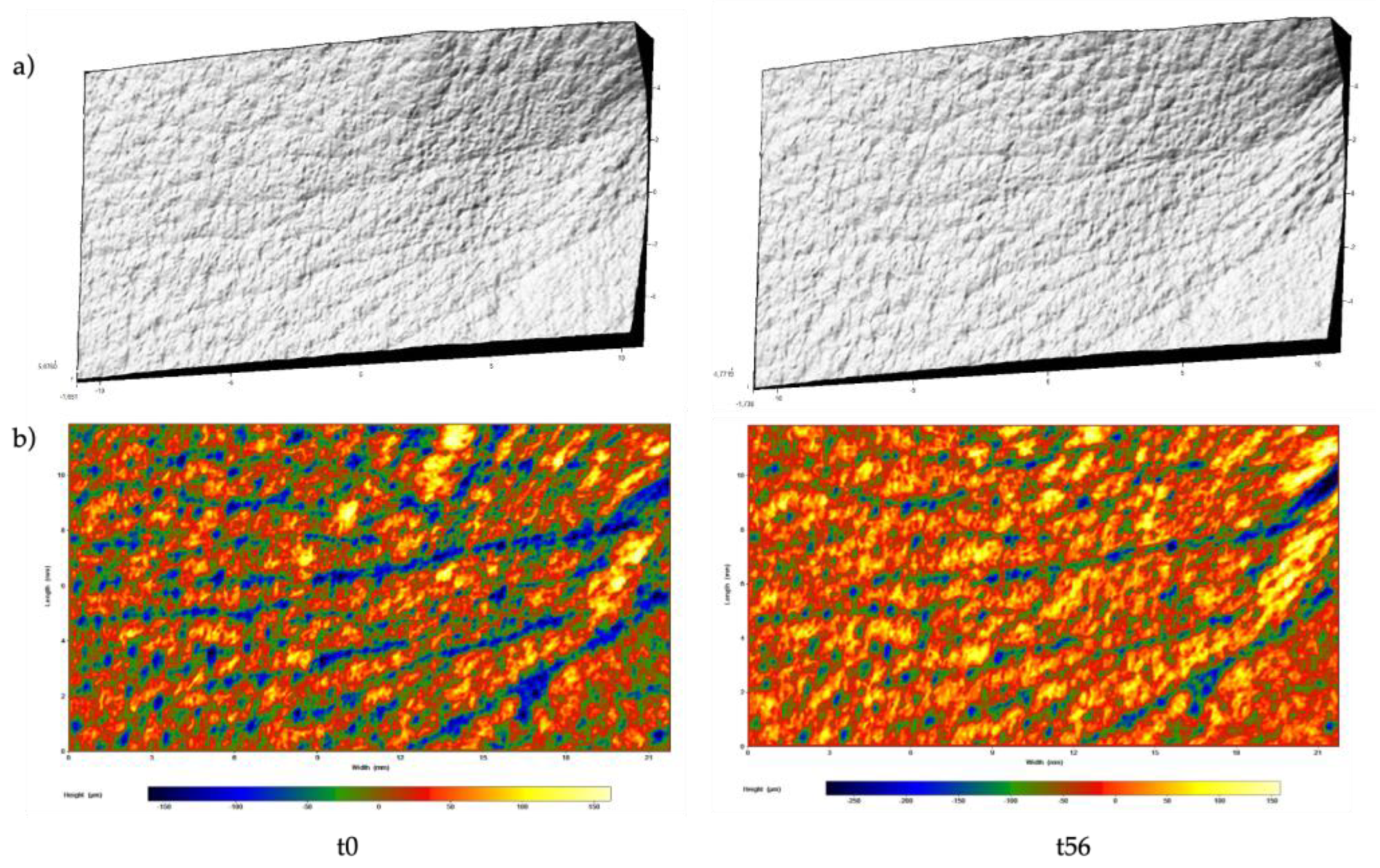
3.1. Anti-Wrinkle Potential Effect

Wrinkles are a consequence of the skin aging process, particularly due to the gradual loss of skin physiological integrity [16]. In the last years, plants extracts have been a target of the cosmetic industry due to their chemical composition that can prevent the skin aging process. In fact, the compounds present in plants extracts may reduce ROS reactivity, absorb UV light, suppress enzymes enrolled in the aging process, and reduce wrinkle formation [17]. Based on that, the chestnut shell extract obtained by UAE was incorporated in a face cream formulation at a concentration of 55% (w/w), aiming to evaluate its efficacy. The PrimosPremium equipment was applied at the corner of the eye to evaluate the wrinkles’ depth, volume, and roughness after 56 days of the products’ application, twice daily. Figure 1 and Figure 2 show images of a detailed skin surface in the crow’s feet area obtained during the analyses, before and after 56 days of study, respectively, for the active and the placebo formulations.
The wrinkles’ depth and volume and the skin roughness in the hemiface areas treated with the active formulation and the placebo were compared before (t0) and after (t56) the application of the products (Table 2).
According to Table 2, the wrinkles’ depth decreased from 78.82 to 75.27 µm after 56 days of twice-daily application of the active formulation. In addition, a mean decrease of −3.55 μm was observed, varying from −24.09% to 54.55% in the volunteers; however, these differences were not significant (p = 0.250). As concerns the placebo, a decrease in the wrinkle depth from 79.14 to 77.73 µm was observed; 54.55% of the volunteers showed a decrease up to −36.59%, and a mean decrease of −1.41 μm was achieved; however, the differences were not significant (p = 0.782). When comparing the active formulation with the placebo, no significant differences (p = 0.581) were observed.
Regarding the wrinkles’ volume, the active formulation led to a decrease from 2.91 to 2.78 mm3 after 56 consecutive days of treatment, while the placebo reduced it from 3.22 to 3.17 mm3 (Table 2). A mean of −0.13 mm3 was observed for the wrinkles’ volume, decreasing up to −28.84% in 59.09% of the volunteers, but the differences were not significant (p = 0.256). In the placebo-treated hemiface, the wrinkles’ volume decreased by −29.74% in 54.55% of the volunteers, though the differences were not significant (p = 0.625). No significant differences (p = 0.431) were observed between the active formulation and the placebo.
The skin roughness was also analyzed by the PrimosPremium equipment. As shown in Table 2, the crow’s feet treated with the active formulation presented a slight decrease in roughness after 56 days (t0 = 13.38 µm and t56 = 12.76 µm), though the differences were not significant (p = 0.069). A decreased up to −23.12% was observed in 61.90% of the volunteers. Similarly, the hemiface area treated with the placebo showed a roughness reduction (from 13.39 to 12.73 µm), achieving a mean decrease of −0.66 μm, though the differences were not significant (p = 0.173).
In the PrimosPremium analyses, it appeared that the active formulation had a good capacity to reduce the wrinkles present on the crow’s feet area, although the differences measured with respect to the placebo were not significant. Figure 1 and Figure 2 show an example of a detailed analysis of the skin surface in the eye contour area, carried out for the active and the placebo formulations, respectively.
Leelapornpisid et al. evaluated the anti-wrinkle effect of a gel containing a nanoemulsion loaded with an ethyl acetate marigold flower extract in 30 healthy human volunteers [18]. The skin wrinkles parameters, namely, surface, volume, and roughness, were evaluated by the Visiometer® equipment in areas treated with the gel extract and a placebo and in an untreated area (intact skin) of the lower forearms, at time 0 and after 8 weeks of treatment [18]. The results demonstrated that the wrinkles in the area treated with the gel extract showed significantly reduced surface (4.44 to 4.21%), volume (43.85 to 41.28%), and roughness (9.71 to 9.25%); however, the best results were achieved in the untreated and placebo areas [18]. In another study, Leevutinun et al. evaluated the beneficial effects of a 5% Momordica cochinchinensis extract incorporated in an O/W emulsion through PrimosPremium analysis, determining the surface roughness in 22 female healthy volunteers with slight wrinkles or fine lines (crow’s feet) [19]. The formulation decreased the skin roughness average, allowing the conclusion that the extract may reduce the wrinkles after 8 weeks of treatment [19]. Rodrigues et al. also evaluated the potentialities of a coffee silverskin extract as a new anti-wrinkle ingredient [20]. The authors developed and compared two formulations, one containing the coffee silverskin extract (2.5%), and the other containing 2.5% of the extract and 1.5% of HyaCare® Filler CL. The wrinkles’ depth, roughness, and volume were assessed in 20 volunteers during 28 days [20]. The authors concluded that after 28 days of treatment, the wrinkles’ depth decreased, but no significant differences were observed between the formulations [20]. A very slight reduction of volume and roughness were reported [20]. These studies are in line with the present one, revealing a potential refilling of the eye’s wrinkles using the chestnut shell extract as a cosmetic ingredient. However, promising results could only be produced if a long-term treatment was performed.

3.2. Firmness Effects

The metabolism of extracellular matrix proteins is strongly affected by the aging process, which causes an increase in protein degradation and the degradation of collagen and elastin functions. This phenomenon results in a reduction of the skin elasticity and firmness and an increase of its laxity [21]. Thus, it is imperative to develop formulations that act on the dermis and on collagen synthesis, improving skin firmness [22]. This viscoelastic skin parameter can be measured by noninvasive devices, such as Cutometer®, which allows a quick, easy, and painlessly evaluation [23]. In the present study, the influence of the chestnut shell extract formulation was assessed with the Cutometer® SEM 575 equipment along the cheekbone of each volunteer’s hemiface and compared to the baseline values and the results after the administration of the placebo. The formulations were applied for 56 days, twice daily, and the analysis focused on the Ur/Uf parameter (that evaluates skin’s biological elasticity). As demonstrated in Table 3, skin firmness improved after 56 days of treatment with the active formulation, increasing up to 31.76% in 50.00% of the volunteers, with a mean increase of 2.34%. However, no significant differences (p = 0.638) were observed between t0 and t56. As concerns the placebo, skin firmness was reduced from 34.84 to 34.02%, after 56 days of application. A mean variation of −0.82% was observed, but the differences were not significant (p = 0.445). Although the skin firmness improved after applying the active formulation, no significant differences (p = 0.455) were observed between the formulations.
In a study conducted by Rodrigues et al., the active formulation as well as a hyaluronic acid formulation increased the skin firmness, leading to a higher Ur/Uf parameter after 28 days of application compared to the baseline value [20]. However, no significant differences were observed between the formulations [20]. Mahmood et al. also evaluated the skin elasticity effects (using a Cutometer®) of a formulation containing a green tea extract (3%) in 10 healthy male volunteers, after 8 weeks of application [24]. According to the authors, the biological elasticity of the skin did not improve with the active formulation [24]. In contrast, the results of the present study demonstrated that the formulation containing 55% of the chestnut shell extract achieved better firmness effects than the placebo. These results support the capacity of the extract to penetrate the skin and act in the collagen support matrix, leading to a more firm, thicker, and elastic skin [25].

3.3. Hydration Effects

Skin hydration improves the natural barrier function of the skin, protecting against external factors and preventing skin aging [26]. The Corneometer® equipment is a standardized tool that can be employed to evaluate skin hydration, by measuring the skin electrical properties, such as conductance and capacitance [26]. In the present study, the Corneometer® CM825 was used at t0 and t56 to measure the hydrating efficacy of the active formulation, compared to the placebo. As shown in Table 3, after 56 days of application of the active formulation, twice daily, the mean skin hydration increased from 54.00 to 58.62 Arbitrary Units (A.U.), and the difference was significant (p = 0.006). A mean increase of 4.62 A.U. was observed for this parameter, rising from 54.91% in 72.73% of the volunteers. Regarding the placebo, the hydration results were higher at t56, reaching a value of 63.37 A.U. (Table 3); an increase up to 87.57% was measured in 61.90% of the volunteers, and the difference was significant (p = 0.013). When comparing the two formulations, no significant differences (p = 0.403) were observed. Considering the results obtained and the previous results reported by our team regarding the extract composition and it good skin compatibility [1,7], the improvement of skin hydration could result from the polyphenols and minerals present in the chestnut shell extract. The presence in the formulation of glycerin and white petrolatum, ingredients associated with a protective effect of the skin’s barrier function [27,28] and with occlusive effects that create a hydrophobic layer on the skin blocking transepidermal water loss [28], may also justify the improvement of skin hydration that we measured.
Other authors evaluated topical formulations containing natural extracts. For example, Rodrigues et al. achieved a better hydration, compared to baseline hydration, after 28 days of use of a formulation containing a coffee silverskin extract, namely, 65.54 A.U., a value in line with the one obtained in the present study [20]. A clinical study performed in 22 healthy women volunteers revealed that the application of a cream containing an M. cochinchinensis extract induced an increase of 9% and 10% in the skin hydration rate after 28 and 56 days of use, respectively, values that are inferior to the one reported in the present study [19]. In addition, Wineman et al. evaluated the hydration effect of two facial formulations on the arm skin of female volunteers (n = 10), testing an extreme day cream and an extreme night treatment, containing a complex of Dead Sea water and three Himalayan extracts (Tibetan goji berries, moss lichen, and Himalayan raspberry root extracts) [29]. Skin hydration varied from 16% to 29% and from 29% to 63%, respectively, for the extreme day cream and the extreme night treatment [29]. Similarly, in the present study, skin hydration increased after the application of the two tested formulations. Nevertheless, most of the studies do not compare the results of active formulations with those of a placebo; therefore, it is difficult to make conclusions on the measured effects in relation to the composition of the extracts, as reported in the present study.
This clinical study has strengths and limitations that should be highlighted. Concerning its strengths, for the first time a formulation containing an active ingredient obtained from a chestnut by-product was evaluated in human volunteers, adding value to an industrial waste generated in huge amounts and, simultaneously, protecting the environment and generating data that may increase the cosmetic industry interest in these new green active ingredients. However, the expected effects in the volunteers were not evident, particularly in regard to wrinkles’ depth, which is a clear limitation of this study. This could be explained by the short time of the clinical study. Therefore, a comparison with a formulation containing not only the extract but also a well-stablished anti-wrinkles active ingredient (such as hyaluronic acid) could explore the potentialities of these ingredients in synergism, an approach widely used by the cosmetic industry when testing natural extracts.

4. Conclusions

This study allowed assessing the clinical effect of a chestnut shell extract incorporated in a skin care formulation in 20 volunteers. The formulation containing the extract led to a reduction in wrinkles’ depth and volume, as well as in skin roughness, after 56 days of treatment, although these results were similar to those obtained with the placebo. The formulation with the chestnut shells extract improved the skin firmness in 50.00% of the volunteers. Nevertheless, a study examining the effects of a long continuous use of the active formulation in volunteers (at least 180 days) will be crucial to make more precise conclusion about the skin effects of this product.

Author Contributions

Conceptualization, F.R.; methodology, A.M.S.; software, A.M.S.; investigation, A.M.S. and F.R.; resources, F.R.; writing—original draft preparation, A.M.S.; writing—review and editing, F.R., P.C.C. and C.D.-M.; supervision, F.R., P.C.C. and C.D.-M.; project administration, F.R.; funding acquisition, F.R. All authors have read and agreed to the published version of the manuscript.

Funding

This research was funded by the project PTDC/ASP-AGR/29277/2017—Castanea sativa shells as a new source of active ingredients for Functional Food and Cosmetic applications: a sustainable approach, supported by national funds by FCT/MCTES and co-supported by the Fundo Europeu de Desenvolvimento Regional (FEDER) throughout COMPETE 2020—Programa Operacional Competitividade e Internacionalização (POCI-01-0145-FEDER-029277). It was also funded by the projects UIDB/50006/2020 and UIDP/50006/2020 through national funds. This work was also financed by national funds from FCT—Fundação para a Ciência e a Tecnologia, I.P., in the scope of the project UIDP/04378/2020 and UIDB/04378/2020 of the Research Unit on Applied Molecular Biosciences—UCIBIO and the project LA/P/0140/2020 of the Associate Laboratory Institute for Health and Bioeconomy—i4HB.

Data Availability Statement

Not applicable.

Acknowledgments

The authors are thankful to Sortegel for the chestnut samples and their availability during this work. The authors are thankful to Evonik for the samples. Ana Margarida Silva is thankful for the Ph.D. grant (SFRH/BD/144994/2019) financed by POPH-QREN and subsidized by the European Science Foundation and Ministério da Ciência, Tecnologia e Ensino Superior. Francisca Rodrigues (CEECIND/01886/2020) is thankful for her contract financed by FCT/MCTES—CEEC Individual Program Contract.

Conflicts of Interest

The authors declare no conflict of interest.

References

  1. Pinto, D.; Cádiz-Gurrea, M.d.l.L.; Garcia, J.; Saavedra, M.J.; Freitas, V.; Costa, P.; Sarmento, B.; Delerue-Matos, C.; Rodrigues, F. From soil to cosmetic industry: Validation of a new cosmetic ingredient extracted from chestnut shells. SM&T 2021, 29, e00309. [Google Scholar]
  2. Pinto, D.; Lameirão, F.; Delerue-Matos, C.; Rodrigues, F.; Costa, P. Characterization and Stability of a Formulation Containing Antioxidants-Enriched Castanea sativa Shells Extract. Cosmetics 2021, 8, 49. [Google Scholar] [CrossRef]
  3. Oliveira, N.; Cádiz-Gurrea, M.d.l.L.; Silva, A.M.; Macedo, C.; Rodrigues, F.; Costa, P. Development and Optimization of a Topical Formulation with Castanea sativa Shells Extract Based on the Concept “Quality by Design”. Sustainability 2022, 14, 129. [Google Scholar] [CrossRef]
  4. Pinto, D.; Vieira, E.F.; Peixoto, A.F.; Freire, C.; Freitas, V.; Costa, P.; Delerue-Matos, C.; Rodrigues, F. Optimizing the extraction of phenolic antioxidants from chestnut shells by subcritical water extraction using response surface methodology. Food Chem. 2021, 334, 127521. [Google Scholar] [CrossRef] [PubMed]
  5. Pinto, D.; Cádiz-Gurrea, M.D.L.L.; Sut, S.; Ferreira, A.S.; Leyva-Jimenez, F.J.; Dall’acqua, S.; Segura-Carretero, A.; Delerue-Matos, C.; Rodrigues, F. Valorisation of underexploited Castanea sativa shells bioactive compounds recovered by supercritical fluid extraction with CO2: A response surface methodology approach. J. CO2 Util. 2020, 40, 101194. [Google Scholar] [CrossRef]
  6. Pinto, D.; Silva, A.M.; Freitas, V.; Vallverdú-Queralt, A.; Delerue-Matos, C.; Rodrigues, F. Microwave-assisted extraction as a green technology approach to recover polyphenols from Castanea sativa shells. Food Sci. Technol. 2021, 1, 229–241. [Google Scholar] [CrossRef]
  7. Lameirão, F.; Pinto, D.; Vieira, E.F.; Peixoto, A.F.; Freire, C.; Sut, S.; Dall’Acqua, S.; Costa, P.; Delerue-Matos, C.; Rodrigues, F. Green-sustainable recovery of phenolic and antioxidant compounds from industrial chestnut shells using ultrasound-assisted extraction: Optimization and evaluation of biological activities in vitro. Antioxidants 2020, 9, 267. [Google Scholar] [CrossRef] [Green Version]
  8. Shimogaki, H.; Tanaka, Y.; Tamai, H.; Masuda, M. In vitro and in vivo evaluation of ellagic acid on melanogenesis inhibition. Int. J. Cosmet. Sci. 2000, 22, 291–303. [Google Scholar] [CrossRef] [PubMed]
  9. Bae, J.Y.; Choi, J.S.; Kang, S.W.; Lee, Y.J.; Park, J.; Kang, Y.H. Dietary compound ellagic acid alleviates skin wrinkle and inflammation induced by UV-B irradiation. Exp. Dermatol. 2010, 19, e182–e190. [Google Scholar] [CrossRef]
  10. Kim, J.; Hwang, J.-S.; Cho, Y.-K.; Han, Y.; Jeon, Y.-J.; Yang, K.-H. Protective effects of (−)-epigallocatechin-3-gallate on UVA-and UVB-induced skin damage. Skin Pharmacol. Physiol. 2001, 14, 11–19. [Google Scholar] [CrossRef]
  11. Katiyar, S.K.; Matsui, M.S.; Elmets, C.A.; Mukhtar, H. Polyphenolic Antioxidant (-)-Epigallocatechin-3-Gallate from Green Tea Reduces UVB-lnduced Inflammatory Responses and Infiltration of Leukocytes in Human Skin. Photochem. Photobiol. 1999, 69, 148–153. [Google Scholar] [CrossRef] [PubMed]
  12. Rodrigues, F.; Alves, A.C.; Nunes, C.; Sarmento, B.; Amaral, M.H.; Reis, S.; Oliveira, M.B.P. Permeation of topically applied caffeine from a food by—product in cosmetic formulations: Is nanoscale in vitro approach an option? Int. J. Pharm. 2016, 513, 496–503. [Google Scholar] [CrossRef] [PubMed]
  13. Yamada, Y.; Yasui, H.; Sakurai, H. Suppressive effect of caffeic acid and its derivatives on the generation of UVA-induced reactive oxygen species in the skin of hairless mice and pharmacokinetic analysis on organ distribution of caffeic acid in ddY mice. Photochem. Photobiol. 2006, 82, 1668–1676. [Google Scholar] [CrossRef] [PubMed]
  14. Marti-Mestres, G.; Mestres, J.; Bres, J.; Martin, S.; Ramos, J.; Vian, L. The in vitro percutaneous penetration of three antioxidant compounds. Int. J. Pharm. 2007, 331, 139–144. [Google Scholar] [CrossRef]
  15. Pinto, D.; Braga, N.; Rodrigues, F.; Oliveira, M.B.P.P. Castanea sativa Bur: An Undervalued By-Product but a Promising Cosmetic Ingredient. Cosmetics 2017, 4, 50. [Google Scholar] [CrossRef] [Green Version]
  16. Rodrigues, F.; Nunes, M.A.d.M.; Oliveira, M.B.P.P. Applications of recovered bioactive compounds in cosmetics and health care products. In Olive Mill Waste: Recent Advances for Sustainable Management; Galanakis, C.M., Ed.; Elsevier Inc.: Amsterdam, The Netherlands, 2017; pp. 255–274. [Google Scholar]
  17. Abdul Karim, A.; Azlan, A.; Ismail, A.; Hashim, P.; Abd Gani, S.S.; Zainudin, B.H.; Abdullah, N.A. Phenolic composition, antioxidant, anti-wrinkles and tyrosinase inhibitory activities of cocoa pod extract. BMC Complement. Altern. Med. 2014, 14, 381. [Google Scholar] [CrossRef] [Green Version]
  18. Leelapornpisid, P.; Kiattisin, K.; Jantrawut, P.; Phrutivorapongkul, A. Nanoemulsion loaded with marigold flower extract (Tagetes erecta linn) in gel preparation as anti-wrinkles cosmeceutical. Int. J. Pharm. Pharm. Sci. 2014, 6, 231–236. [Google Scholar]
  19. Leevutinun, P.; Krisadaphong, P.; Petsom, A. Clinical evaluation of Gac extract (Momordica cochinchinensis) in an antiwrinkle cream formulation. J. Cosmet. Sci 2015, 66, 175–187. [Google Scholar]
  20. Rodrigues, F.; Matias, R.; Ferreira, M.; Amaral, M.H.; Oliveira, M.B.P.P. In vitro and in vivo comparative study of cosmetic ingredients Coffee silverskin and hyaluronic acid. Exp. Dermatol. 2016, 25, 572–574. [Google Scholar] [CrossRef] [Green Version]
  21. Gillbro, J.; Merinville, E.; Cattley, K.; Al-Bader, T.; Hagforsen, E.; Nilsson, M.; Mavon, A. In vivo topical application of acetyl aspartic acid increases fibrillin-1 and collagen IV deposition leading to a significant improvement of skin firmness. Int. J. Cosmet. Sci. 2015, 37, 41–46. [Google Scholar] [CrossRef] [Green Version]
  22. Maia Campos, P.M.; Gonçalves, G.M.; Gaspar, L.R. In vitro antioxidant activity and in vivo efficacy of topical formulations containing vitamin C and its derivatives studied by non-invasive methods. Skin Res. Technol. 2008, 14, 376–380. [Google Scholar] [CrossRef] [PubMed]
  23. Jacobs, S.W.; Culbertson, E.J. Effects of topical mandelic acid treatment on facial skin viscoelasticity. Facial Plast. Surg. 2018, 34, 651–656. [Google Scholar] [CrossRef] [PubMed]
  24. Mahmood, T.; Akhtar, N.; Khan, B.A.; Shoaib Khan, H.M.; Saeed, T. Changes in skin mechanical properties after long-term application of cream containing green tea extract. Aging Clin. Exp. Res. 2011, 23, 333–336. [Google Scholar] [CrossRef] [PubMed]
  25. Husein el Hadmed, H.; Castillo, R.F. Cosmeceuticals: Peptides, proteins, and growth factors. J. Cosmet. Dermatol. 2016, 15, 514–519. [Google Scholar] [CrossRef]
  26. Verdier-Sévrain, S.; Bonté, F. Skin hydration: A review on its molecular mechanisms. J. Cosmet. Dermatol. 2007, 6, 75–82. [Google Scholar] [CrossRef]
  27. Milani, M.; Sparavigna, A. The 24-hour skin hydration and barrier function effects of a hyaluronic 1%, glycerin 5%, and Centella asiatica stem cells extract moisturizing fluid: An intra-subject, randomized, assessor-blinded study. Clin. Cosmet. Investig. Dermatol. 2017, 10, 311. [Google Scholar] [CrossRef] [Green Version]
  28. Kovács, A.; Péter-Héderi, D.; Perei, K.; Budai-Szűcs, M.; Léber, A.; Gácsi, A.; Csányi, E.; Berkó, S. Effects of Formulation Excipients on Skin Barrier Function in Creams Used in Pediatric Care. Pharmaceutics 2020, 12, 729. [Google Scholar] [CrossRef]
  29. Wineman, E.; Portugal-Cohen, M.; Soroka, Y.; Cohen, D.; Schlippe, G.; Voss, W.; Brenner, S.; Milner, Y.; Hai, N.; Ma’or, Z. Photo-damage protective effect of two facial products, containing a unique complex of Dead Sea minerals and Himalayan actives. J. Cosmet. Dermatol. 2012, 11, 183–192. [Google Scholar] [CrossRef]
Figure 1. Shown is a 3D picture of wrinkles in the eye contour, obtained with PRIMOSPremium equipment from one of the volunteers before (t0) and after 56 (t56) consecutive days of application of the active formulation, twice daily. (a) Photography; (b) Representation of the relief in a colored image, where the blue color represents the deepest areas, and depth decreases moving from green to red, orange, and yellow (the highest areas).
Processes 10 02230 g001
Figure 2. Shown is a 3D picture of wrinkles in the eye contour obtained with PrimosPremium equipment from one of the volunteers before (t0) and after 56 (t56) consecutive days of application of the placebo formulation, twice daily. (a) Photography; (b) Representation of the relief in a colored image, where the blue color represents the deepest areas, and depth decreases moving from green to red, orange, and yellow (the highest areas).
Processes 10 02230 g002
Table 1. Percentage of raw materials present in each formulation (active and placebo).
Raw MaterialsPercentage in Each Formulation (w/w)
Active CreamPlacebo
Purified water-54.9
C. sativa shell extract54.9-
Decyl oleate17.017.0
Cetearyl alcohol12.012.0
Glycerin10.510.5
White petrolatum5.05.0
Microcare® PHDG0.60.6
Table 2. Mean values of the wrinkles’ depth and volume and the skin roughness in the crow’s feet area before (t0) and after 56 (t56) consecutive days of application of the active cream and the placebo, twice daily, through PrimosPremium analysis. The results are expressed as mean ± SD (n = 22).
Active Cream Hemiface Area TreatmentPlacebo Hemiface Area
Treatment
t0t56t0t56
Wrinkles’ depth (µm)78.82 ± 27.8375.27 ± 25.0979.14 ± 22.5977.73 ± 29.04
Wrinkles’ volume (mm3)2.91 ± 1.352.78 ± 1.363.22 ± 1.293.17 ± 1.47
Wrinkles’ roughness (µm)13.38 ± 2.4112.76 ± 2.1913.39 ± 1.7512.73 ± 2.25
Table 3. Mean values of face skin firmness obtained through the Cutometer® SEM 575 analysis and mean values of face skin hydration obtained through the Corneometer® CM825 analysis. The measurements performed before (t0) and after 56 (t56) consecutive days of application, twice daily, of the active cream and the placebo. The results are expressed as mean ± SD (n = 22). * in the same row indicates significant differences between timepoints (p < 0.05).
Active Cream Hemiface Area TreatmentPlacebo Hemiface Area
Treatment
t0t56t0t56
Skin firmness (%)33.74 ± 4.4734.23 ± 4.5334.84 ± 3.1934.02 ± 5.12
Skin hydration (A.U.)54.00 ± 9.71 *58.62 ± 9.1653.25 ± 12.41 *63.37 ± 17.96
A.U.—Arbitrary Units.
Publisher’s Note: MDPI stays neutral with regard to jurisdictional claims in published maps and institutional affiliations.

