Article

Anti-Obesity Activity in 3T3-L1 Cells of Cornus officinalis Fruits Harvested at Different Times

1
Department of Herbal Crop Research, National Institute of Horticultural and Herbal Science, Rural Development Administration, Eumseong 27709, Korea
2
Department of Food Science and Biotechnology, Chungbuk National University, Cheongju 28644, Korea
3
Department of Korean Medicine, Graduate School, Kyung Hee University, Seoul 02447, Korea
*
Author to whom correspondence should be addressed.
Processes 2022, 10(10), 2008; https://doi.org/10.3390/pr10102008
Submission received: 31 August 2022 / Revised: 23 September 2022 / Accepted: 29 September 2022 / Published: 5 October 2022
(This article belongs to the Special Issue Plants as Functional Food Ingredients and Food Preservative)

Abstract

:
Obesity, a metabolic disease linked to several other diseases, is emerging as a global problem. We determined the harvest time that maximized the anti-obesity effect by evaluating the inhibition of lipid accumulation in 3T3-L1 cells treated with fruits of Cornus officinalis. FS1 (pericarp + seeds, harvested 15 September) showed excellent anti-obesity activity (Oil Red O; 26.12 ± 1.37% vs. MDI) and inhibited the expression of PPAR-γ (0.05 ± 0.01 ford vs. MDI), C/EBPα (0.03 ± 0.02 ford vs. MDI), and C/EBPβ (0.33 ± 0.07 ford vs. MDI) at 200 μg/mL. The antioxidant activity of FS1 was also the most effective. In addition, FS1 contained higher levels of active ingredients than samples harvested in other periods. Especially, content of loganin, verbenalin, and sweroside was high. In terms of anti-obesity activity and functional components, immature COF (FS1) was the best, and these results indicate that it is necessary to adjust the harvest time, when used as an anti-obesity agent.

1. Introduction

The fruit of Cornus officinalis (COF) is traditionally used for the prevention and treatment of various diseases in East Asian countries, such as Korea, Japan, and China [1]. It is harvested in autumn, and the seeds are removed and dried [2]. Components of COF include malic acid, tartaric acid, gallic acid, ursolic acid [3], and iridoid glycosides, such as morroniside, loganin, and sweroside [4]. COF extract and active components have immune-enhancing, cardioprotective, antibacterial, anti-hyperglycemic, antiaging, antioxidant, and renal- and neuro-protective effects [5].
The World Health Organization (WHO, Geneva, Switzerland) defines obesity as an excessive amount of adipose tissue in the body. Obesity, which occurs when the number of calories consumed is greater than the number of calories used, is caused by decreased physical activity, genetic factors, and changes in diet. Obesity is a metabolic disease that causes hypertension, cardiovascular disease, and diabetes [6]. Due to overeating and lack of exercise, obesity is increasing worldwide, and it is emerging as a risk factor for personal and social health issues [7].
Adipocyte differentiation, i.e., the differentiation of adipocytes to mature adipocytes, is closely related to obesity because it is involved in fat production and accumulation [8]. Adipogenesis is achieved, in part, by the C/EBP (CCAAT/enhancer binding protein) and PPAR-γ (peroximal proliferator-activated receptor-γ) [9,10]. Hormonal stimulation induces the PPAR-γ and C/EBPα expression by increasing the C/EBPβ expression at the early stage of differentiation and decreases the C/EBPβ expression at the late stage. Adipogenesis is regulated by the expression levels of these factors [11,12]. 3T3-L1 cells have been used to investigate the differentiation of preadipocytes into adipocytes and evaluate adipogenesis.
The anti-obesity activities of COF [13] are limited to the pericarp, and studies on fruits (including seeds) and anti-obesity effects, according to harvest time, are scarce. Therefore, we used 3T3-L1 to analyze lipid accumulation and related protein expression level to assess the effects of COF on adipocyte differentiation, lipid production, and identify the harvest time that maximizes the inhibition of adipocyte differentiation.

2. Materials and Methods

2.1. Sample Preparation

COFs were harvested on the 15th of each month, from September to December, 2020, respectively, in Gurye-Gun, Korea (Figure 1). The pericarp (seeds removed from the fruit) and fruits (with seeds) were freeze-dried and crushed into powder. The samples (pericarp and fruits containing seeds, 10 g) were extracted three times with 100 mL of 70% ethanolic solution for 1 h by ultrasonication. After filtration (0.45 µm regenerated cellulose filter, Phenomenex, Torrance, CA, USA), the extract was concentrated under vacuum and freeze-dried (Table 1).

2.2. Cell Culture of 3T3-L1 Pre-Adipocytes

The 3T3-L1 adipocytes were purchased from the ATCC (Manassas, VA, USA) and incubated in Dulbecco’s modified Eagle’s medium (DMEM; Gibco, Grand Island, NY, USA) with 10% bovine serum (Gibco), 100 µg/mL streptomycin (Gibco), and 100 units/mL penicillin at 37 °C in a 5% CO2 incubator.

2.3. Cell Viability of Pre-Adipocytes

Cell viability was measured by methylthiazolyldiphenyl-tetrazolium bromide (MTT) assay. The 3T3-L1 pre-adipocytes were cultured in 48-well plates at 5 × 10⁴ cells/mL for 24 h and treated with COF extracts (50, 100, 200 µg/mL). After 24 h, the media was removed and replaced with 0.5 mg/mL MTT reagent (Sigma-Aldrich, St. Louis, MO, USA) in growth medium, and reacted at 37 °C in a CO2 incubator for 1 h. The media was removed, and 100 µL of dimethyl sulfoxide was added to elute formazan crystal. The absorbance of samples was measured at 550 nm by using a microplate reader (Synergy H1; Biotek, Winooski, VT, USA).

2.4. Differentiation of Pre-Adipocytes

Differentiation was induced over 2 days in post-confluent cells, using DMEM with 10% FBS (fetal bovine serum, Gibco), streptomycin (100 µg/mL), and penicillin (100 units/mL) containing MDI (1 µM dexamethasone, 500 µM isobutylmethylxanthine, and 10 µg/mL insulin from Sigma-Aldrich). The cultures were replenished with DMEM containing 10% FBS, 100 units/mL penicillin, 100 µg/mL streptomycin, COF extract (50, 100, 200 µg/mL), and 10 µg/mL insulin medium every 2 days. The experiments were performed using adipocytes at 8 days from post-differentiation.

2.5. Oil Red O Staining of Adipocytes

After differentiation induction of adipocytes, the media was removed. The cells were washed with PBS (phosphate-buffered saline). The cells were fixed with 3.7% formalin for 15 min. Next, the cells were washed with distilled water and stained with oil red O solution (Sigma-Aldrich, St. Louis, MO, USA) for 15 min at 25 °C. The oil red O solution was then removed. The cells were washed with distilled water before being photomicrographed. To quantify the eluted reagent, isopropanol was added, and the absorbance of the samples was measured at 520 nm using a microplate reader.

2.6. Western Blot Analysis of Adipocytes

Adipocytes were washed with cold PBS. The cells were lysed in RIPA buffer containing protease and phosphatase inhibitor cocktail (GenDEPOT, Katy, TX, USA). After centrifugation at 12,000 rpm, the supernatant was obtained. The protein concentration in the supernatant was quantified by using the bicinchoninic acid protein assay kit (Pierce Biotechnology Inc., Rockford, IL, USA). The protein samples were separated in SDS-polyacrylamide gels and transferred to NC (nitrocellulose) membranes (Bio-Rad, Hercules, CA, USA). The NC membranes were blocked in Tris-buffered saline Tween 20 (TBST, 20 mmol/L Tris-HCl, 150 mmol/L NaCl, 0.1% Tween 20, pH 7.5) containing 2% bovine serum albumin (GenDEPOT, Katy, TX, USA). The primary antibody (Cell Signaling Technology, Beverly, MA) was added for incubation for 12 h at 4 °C, followed by the secondary antibody (Cell Signaling Technology, Danvers, MA, USA) for 30 min. The NC membranes were washed with TBST. The membranes were reacted with enhanced chemiluminescence solution (Bio-Rad, Hercules, CA, USA); then, the visualized data were taken using a ChemiDoc Imaging System (Bio-Rad, Hercules, CA, USA). Quantitative analysis was performed using ImageJ (NIH, Bethesda, MD, USA).

2.7. Antioxidant Activity Assays

2.7.1. DPPH Free Radical Scavenging Assay

A DPPH (2,2-diphenyl-picrylhydrazyl, Sigma-Aldrich) radical scavenging assay of COF was performed using the method of Tepe et al. [14]. Two-hundred microliters of 0.2 mM DPPH solution in ethanol were added to 50 μL of COF extract and reacted in darkness for 30 min; the absorbance of samples at 520 nm was measured using a microplate reader. Trolox standard (Sigma-Aldrich, St. Louis, MO, USA) was used as the positive control.

2.7.2. ABTS Radical Scavenging Assay

An ABTS (2,2′-azino-bis-3-ethylbenzthiazoline-6-sulphonic acid, Sigma-Aldrich) radical scavenging assay of COF was performed by modifying the method of Re et al. [15]. One-hundred-ninety microliters of ABTS solution were added to 10 μL of COF extract and reacted in darkness for 30 min, and the absorbance of samples at 735 nm was measured using a microplate reader. Ascorbic acid standard (Sigma-Aldrich, St. Louis, MO, USA) was used as the positive control.

2.7.3. Determination of TP Content

Total phenolic content was measured by modifying the method of Dewanto et al. [16]. After adding 2% Na2CO3 (Sigma-Aldrich, St. Louis, MO, USA) solution to COF extract, 1N Folin-Ciocalteu reagent (Sigma-Aldrich, St. Louis, MO, USA) was added. After the samples had been allowed to stand for 30 min, the absorbance of samples was measured at 750 nm using a microplate reader. Gallic acid (Sigma-Aldrich, St. Louis, MO, USA) was used as a standard.

2.7.4. FRAP Assay

Ferric reducing antioxidant power (FRAP) was measured by modifying the Mau et al. method [17]. A total of 0.2 M sodium phoshate buffer (pH 6.6) and potassium ferricyanide (Sigma-Aldrich) were mixed and reacted with COF extract, followed by mixing with trichloroacetic acid (Sigma-Aldrich, St. Louis, MO, USA). After centrifuging for 10 min, the absorbance was measured at 700 nm by mixing the supernatant, distilled water, and ferric chloride (Sigma-Aldrich, St. Louis, MO, USA). Trolox (Sigma-Aldrich, St. Louis, MO, USA) was used as a standard.

2.8. HPLC Analysis of Major Active Components

Quantitative analyses were performed on an Agilent 1200 series HPLC (Agilent Technologies Inc., Santa Clara, CA, USA) by modifying the method of Liu et al. [4]. Chromatography was carried out at 35 °C on an C18 column (YMC-Triart; 250 × 4.6 mm, S-5 µm, 12 nm, YMC, Kyoto, Japan). The mobile phase consisted of 0.2% (v/v) acetic acid in water (A) and 0.2% (v/v) acetic acid in acetonitrile (B) with gradient elution: 0–4 min, isocratic 1% B; 4–18 min, 1–12% B; 18–22 min, 12–20% B; 22–35 min, 20–30% B, 35–40 min, and 30–100% B. The gradient elution re-equilibration time was 10 min. The flow rate and UV detector were set at 1.2 mL/min and 254 nm, respectively. The injection volume of samples was 10 µL. The standards of active compounds were dissolved in methanol to 10 mg/mL and diluted. Loganin, morroniside, verbenalin, sweroside, 7-O-methyl morroniside, loganetin, and sarracenin analytical standards were purchased from ChemFaces (Wuhan, China). Gallic acid and cornuside analytical standards were purchased from Sigma-Aldrich (Table 2).

2.9. Statistical Analysis

The experiments were repeated three or four times and expressed as mean and standard deviation values. The significant difference between non-treated (VC, MDI) and treated (samples) cells was determined using the two-sample Student’s t-test. The significant difference among concentrations of same sample in anti-obesity activity and samples in antioxidant properties and active components was determined using the ANOVA by SPSS ver. 22 (SPSS Institute, IL, USA), with a significance level p < 0.05 by DMRT.

3. Results

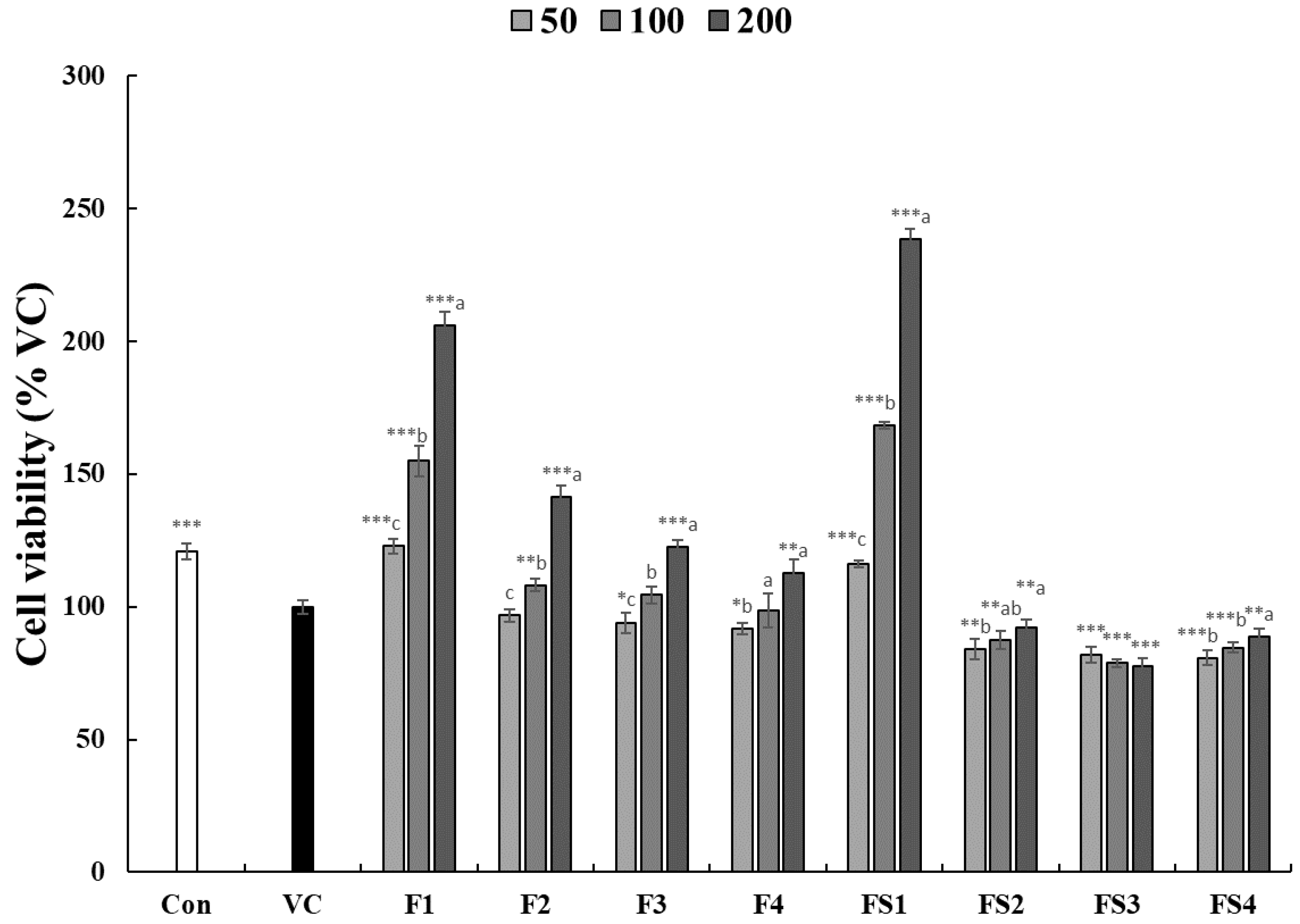
3.1. Effect of COF on Cell Viability

The effect of COF extract on 3T3-L1 preadipocyte viability (50, 100, and 200 μg/mL) was evaluated by MTT assay. The survival rate was >80% (Figure 2); therefore, toxicity was insignificant.

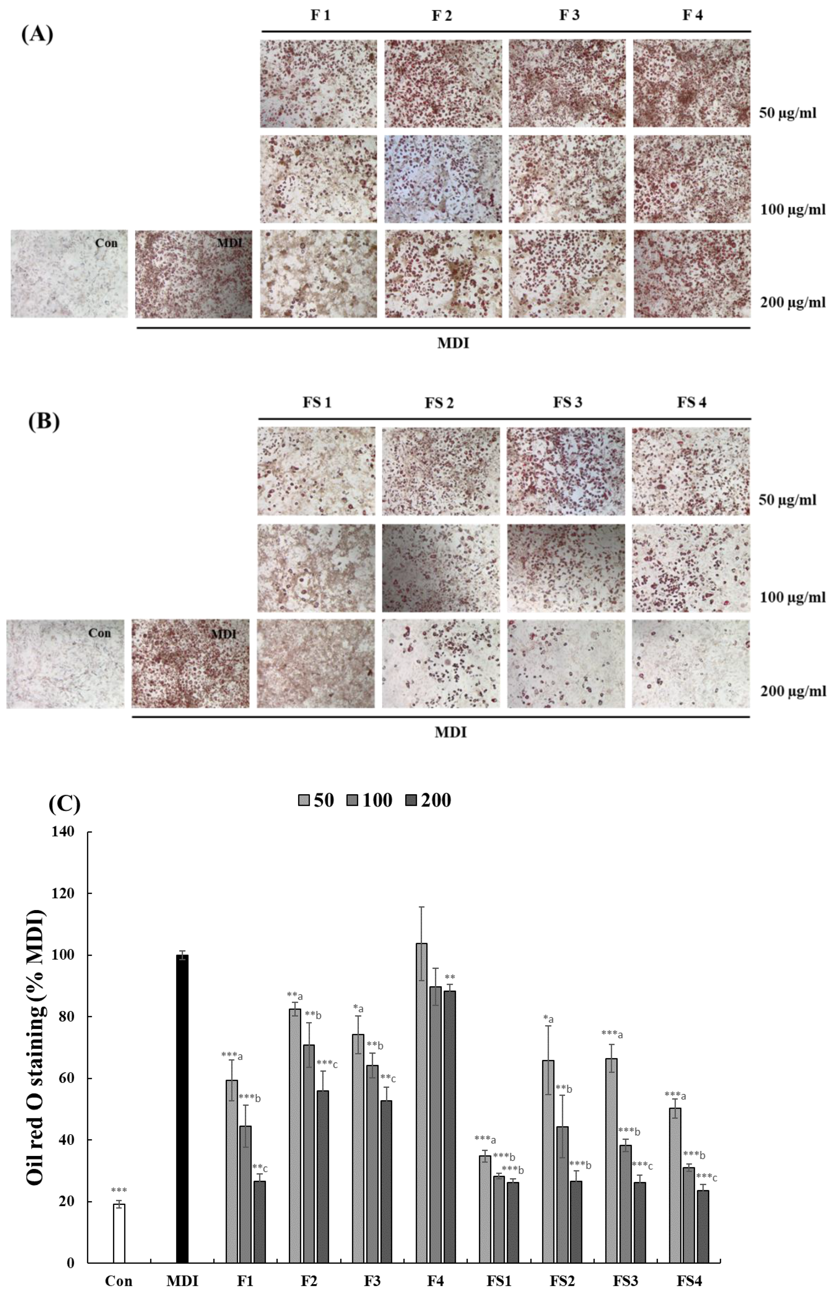
3.2. Effect of COF on 3T3-L1 Cell Differentiation

ORO staining was performed to investigate the effect of COF on the adipocyte differentiation of 3T3-L1 preadipocytes (Figure 3). The pericarp (F) and pericarp + seeds (FS) decreased the differentiation rate of adipocytes in a concentration-dependent manner (MDI (methylisobutylxanthine, dexamethasone, and insulin): 100% vs. F4 50 μg/mL: 103.69%~FS1 200 μg/mL: 26.12%). In terms of parts, the effect was greater in pericarp + seeds (FS) than pericarp (F); in terms of the harvest period, the effect was greater in the first period. All samples, except F4, had a concentration-dependent anti-obesity effect (p < 0.05).

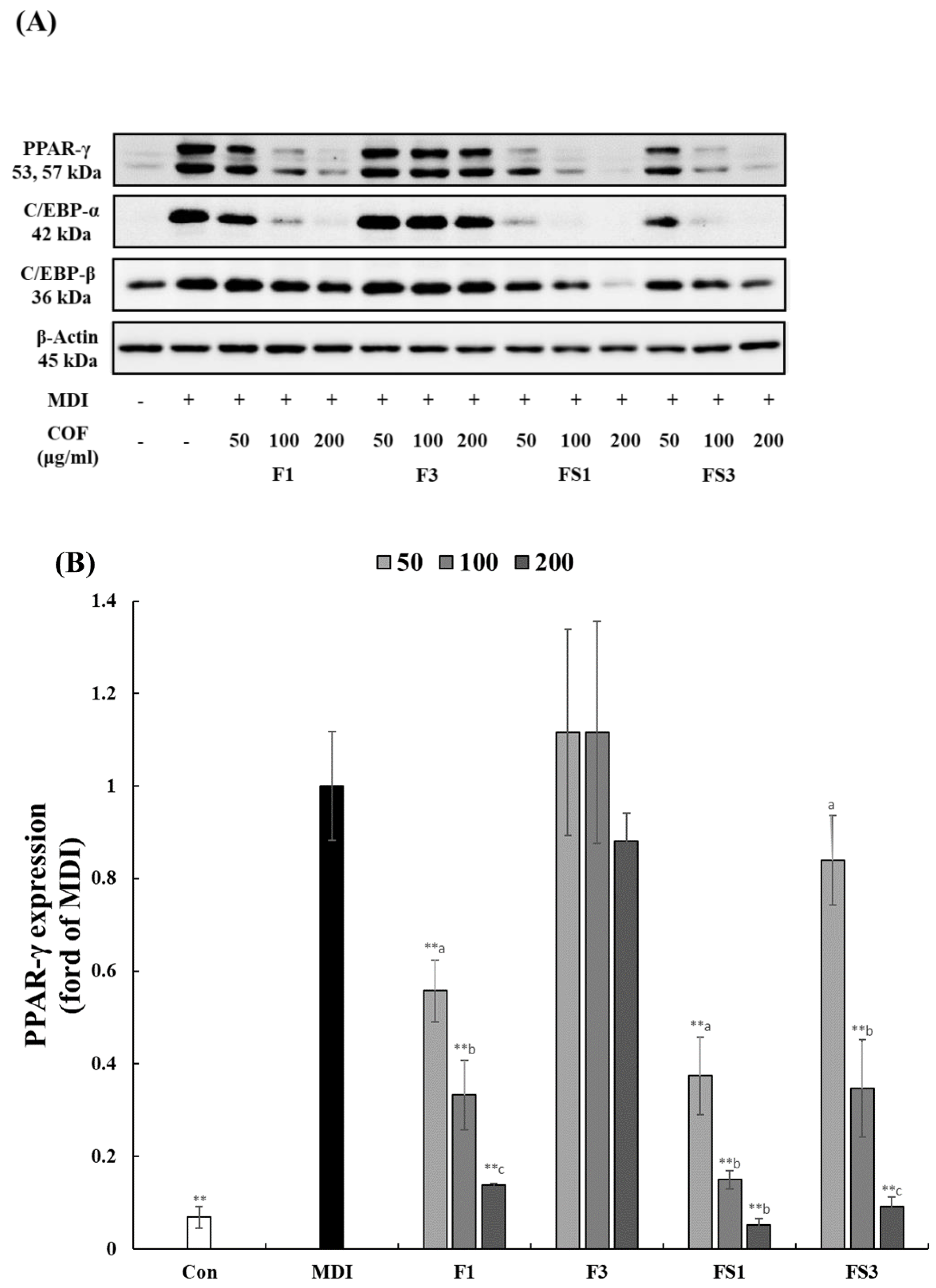
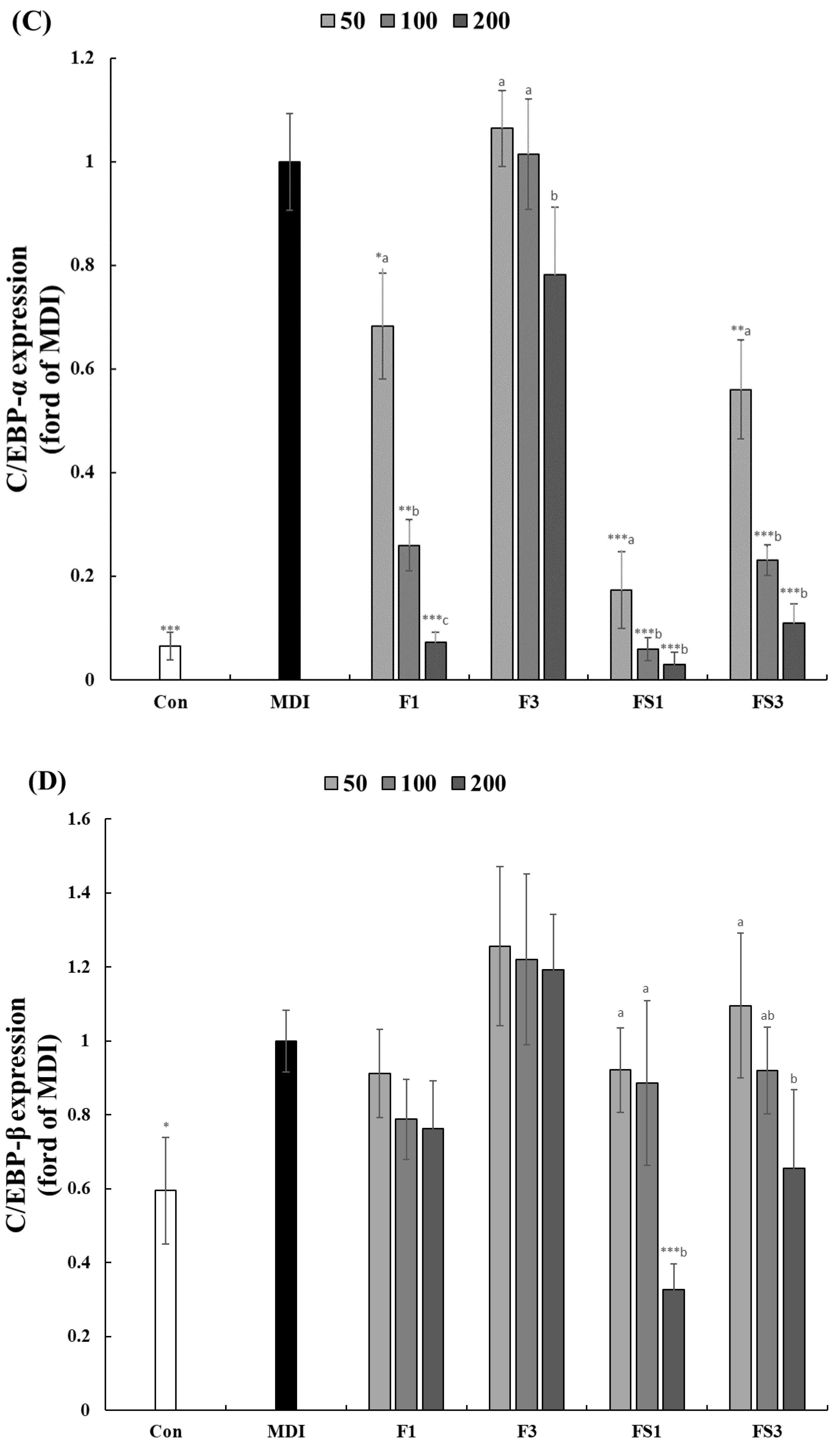
3.3. COF Inhibits the Expression of C/EBPα, β, and PPAR-γ

Western blotting was performed to evaluate the expression of proteins related to adipocyte differentiation and lipid accumulation. Protein expression of C/EBPα, β, and PPAR-γ was compared between the first and third periods using ORO staining (Figure 3). The expression of PPAR-γ (200 μg/mL) during the first and third periods was 0.14- and 0.88-fold higher vs. MDI in Fs and 0.05- and 0.09-fold higher vs. MDI in FSs, respectively. C/EBPα (200 μg/mL) expression was 0.07- and 0.78-fold higher vs. MDI in Fs and 0.03- and 0.11-fold vs. MDI in FSs, respectively. In PPAR-γ and C/EBPα expression levels, F1 and FS1 showed significant differences from MDI at all concentrations (p < 0.05). Finally, C/EBPβ (200 μg/mL) expression was 0.76- and 1.19-fold higher vs. MDI in Fs and 0.33- and 0.66-fold higher vs. MDI in FSs, respectively (Figure 4). Similar to the results of ORO staining, protein expression levels were lower in FSs than Fs, and in the first rather than third period.

3.4. Antioxidant Activity of COF

The DPPH radical scavenging activities (IC50 values) of the extracts were 15.89–219.60 μg/mL, and the ABTS radical scavenging activities were 13.28–151.44 μg/mL. FRAP were 71.67–871.16 mg/g. FS1 and F4 had the highest and lowest activities (p < 0.05), respectively. The TPCs of the extracts were 39.37–288.96 mg/g; the TPCs were highest in FS1 and lowest in F4 (p < 0.05) (Table 3).

3.5. Active Components

The levels of all active components were highest in COF extracts (F1 and FS1) harvested in September, and the levels of all components, except morroniside, were higher in FS1 than F1 (p < 0.05). Morroniside showed higher contents of F1 (63.95 mg/g) and FS1 (48.28 mg/g) than other samples (p < 0.05) (Table 4, Figure S1). The loganin, verbenalin, and sweroside contents differed, according to the inclusion of seeds.

4. Discussion

Preadipocytes differentiate into adipocytes via various hormones and transcription factors and produce and accumulate intracellular lipid [18]. Lipid accumulation, as a marker of adipogenesis, was quantified by ORO staining. The first pericarp and pericarp + seed showed the greatest inhibition of lipid accumulation. COF is an immature green fruit; green fruits, such as green apples, contain high levels of active ingredients, such as polyphenols and iridoids, and they have antioxidant [19] and anti-obesity [20] effects. ROS suppresses the reduction of the energy consumption of adipocytes by inducing the dysfunction of the mitochondria by inhibiting the breathing process [21]. Substances with high antioxidant activity (such as N-acetyl cysteine) ameliorate obesity by inhibiting lipid accumulation [22,23]. Regarding antioxidant activity, the effect of unripe green fruit was superior to that of fruit containing seeds, rather than pericarp, which is consistent with the inhibition of lipid accumulation. Loganin and sweroside, the contents of which were high in FS1, modulate fat accumulation by regulating AMPKα, SIRT1, and PPARα [24,25]. Polyphenols inhibit adipocyte differentiation and lipid accumulation by regulating the expression of the transcription factors linked to adipogenesis [26]. The polyphenol and iridoid components of the COF had anti-obesity and -oxidant effects in this study. The sine this study did not measure the anti-obesity effect under the condition of increasing ROS, and the anti-obesity effect of COF is more likely to be the effect of iridoids, rather than antioxidants.
The adipogenesis of preadipocytes is mediated by transcription factors and hormones, including C/EBPα, C/EBPβ, and PPARγ [27]. Adipogenesis is achieved by the sequential expression of transcription factors, and C/EBPβ and C/EBPδ mediate the transcriptional activation of PPARγ and C/EBPα [28]. C/EBPβ is expressed at an early stage of differentiation, and its expression decreases at later stages. By contrast, PPARγ and C/EBPα expression levels increase as differentiation progresses. Regarding the effect of COF on transcription factors, there was little difference in C/EBPβ expression between the control and MDI groups. There was also little difference in the expression level of COF among the groups. C/EBPβ was expressed in the early stage of differentiation, and its expression decreased at later stages. However, FS1 suppressed the expression of C/EBPβ in a concentration-dependent manner. PPARγ and C/EBPα expression by C/EBPβ showed a significant difference between the control and MDI groups. Therefore, FS1 ameliorates obesity because it inhibits the transcription factors related to fat differentiation.
These results indicate that the main functional components and anti-obesity activities can vary greatly, depending on the harvest time of COF. In Korea, immature COF harvested earlier may be more valuable, in terms of functionality, than fruits at the normal harvest time.

5. Conclusions

Immature COF had anti-obesity and -oxidant effects, which were greater in COF containing seeds. The anti-obesity effect was caused by the suppression of C/EBPα, C/EBPβ, and PPARγ expression by various components of immature fruits. Therefore, immature pericarp + seeds of COF have therapeutic potential for obesity. In Korea, the typical harvest time for COF is November. However, in this study, it was found that COF harvested in September contained excellent activity and a large amount of active ingredients. Therefore, there is a need to advance the harvest time of COF; immature COF could be used as a material for anti-obesity agents. In addition, COF generally uses only the pericarp; it is also necessary to consider the use of seeds.

Supplementary Materials

The following supporting information can be downloaded at: https://www.mdpi.com/article/10.3390/pr10102008/s1, Figure S1: Typical chromatograms of COF with different harvest times and parts.

Author Contributions

Conceptualization, E.S.L. and G.Y.J.; methodology, E.S.L., S.J.C., M.H.K., H.D.K. and Y.-J.J.; data curation, G.Y.J. and G.-S.K.; resources, G.Y.J. and S.J.C.; writing-original draft, E.S.L. and G.Y.J.; writing-review and editing, G.Y.J. All authors have read and agreed to this manuscript.

Funding

This study was supported by the Cooperative Research Program for Agriculture Science and Technology Development (PJ01495201) of the RDA, Korea.

Institutional Review Board Statement

Not applicable.

Informed Consent Statement

Not applicable.

Data Availability Statement

Not applicable.

Conflicts of Interest

The authors declare no conflict of interest.

References

  1. Park, E.K.; Lee, C.G.; Kim, J.; Lim, E.; Hwang, S.; Yun, S.H.; Jeong, S.Y. Anti-osteoporotic effects of the herbal mixture of Cornus officinalis and Achyranthes japonica in vitro and in vivo. Plants 2020, 9, 1114. [Google Scholar] [CrossRef] [PubMed]
  2. Chung, S.R.; Jeune, K.H.; Park, S.Y.; Jang, S.J. Toxicity and lectins constituents from the seed of Cornus officinalis. Kor. J. Pharmacogn. 1993, 24, 177–182. [Google Scholar]
  3. Hwang, K.A.; Hwang, Y.J.; Song, J. Antioxidant activities and oxidative stress inhibitory effects of ethanol extracts from Cornus officinalis on raw 264.7 cells. BMC Complement Altern. Med. 2016, 16, 1–9. [Google Scholar] [CrossRef] [PubMed] [Green Version]
  4. Liu, Z.; Zhu, Z.; Zhang, H.; Tan, G.; Chen, X.; Chai, Y. Qualitative and quantitative analysis of Fructus corni using ultrasound-assisted microwave extraction and high-performance liquid chromatography coupled with diode array UV detection and time-of-flight mass spectrometry. J. Pharm. Biomed. Anal. 2011, 55, 557–562. [Google Scholar] [CrossRef] [PubMed]
  5. Huang, J.; Zhang, Y.; Dong, L.; Gao, Q.; Yin, L.; Quan, H.; Chen, R.; Fu, X.; Lin, D. Ethnopharmacology, phytochemistry, and pharmacology of Cornus officinalis Sieb. et Zucc. J. Ethnopharmacol. 2018, 213, 280–301. [Google Scholar] [CrossRef]
  6. Hwang, J.H.; Lee, S.W.; Han, K.H.; Seo, H.J.; Kim, J.D. Anti-angiogenesis and anti-adipogenesis effects of Anthrisci radix extract. Biotechnol. Bioproc. Eng. 2013, 18, 164–172. [Google Scholar] [CrossRef]
  7. Lee, W.J.; Koh, E.H.; Won, J.C.; Kim, M.S.; Park, J.Y.; Lee, K.U. Obesity: The role of hypothalamic AMP-activated protein kinase in body weight regulation. Int. J. Biochem. Cell Biol. 2005, 37, 2254–2259. [Google Scholar] [CrossRef]
  8. Park, C.H.; Rhyu, D.Y.; Sharma, B.R.; Yokozawa, T. Inhibition of preadipocyte differentiation and lipid accumulation by 7-O-galloyl-d-sedoheptulose treatment in 3T3-L1 adipocytes. Biomed. Prev. Nutr. 2013, 3, 319–324. [Google Scholar] [CrossRef]
  9. Farmer, S.R. Regulation of PPARc activity during adipogenesis. Int. J. Obes. 2005, 29, 13–16. [Google Scholar] [CrossRef] [Green Version]
  10. Guan, H.P.; Ishizuka, T.; Chui, P.C.; Lehrke, M.; Lazar, M.A. Corepressors selectively control the transcriptional activity of PPARc in adipocytes. Genes. Dev. 2005, 19, 453–461. [Google Scholar] [CrossRef] [Green Version]
  11. Christy, R.J.; Kaestner, K.H.; Geiman, D.E.; Lane, M.D. CCAAT/enhancer binding protein gene promoter: Binding of nuclear factors during differentiation of 3T3-L1 preadipocytes. Proc. Natl. Acad. Sci. USA 1991, 88, 2593–2597. [Google Scholar] [CrossRef] [PubMed]
  12. Hamm, J.K.; Park, B.H.; Farmer, S.R. A role for C/EBPβ in regulating peroxisome proliferator-activated receptor γ activity during adipogenesis in 3T3-L1 preadipocytes. J. Biol. Chem. 2001, 276, 18464–18471. [Google Scholar] [CrossRef] [Green Version]
  13. Kang, C.H.; Kwon, Y.J.; So, J.S. Anti-adipogenic effects of Corni fructus in 3T3-L1 preadipocytes. Biotechnol. Bioprocess Eng. 2014, 19, 52–57. [Google Scholar] [CrossRef]
  14. Tepe, B.; Sokmen, M.; Akpulat, H.A.; Sokmen, A. Screening of the antioxidant potentials of six Salvia species from Turkey. Food Chem. 2006, 95, 200–204. [Google Scholar] [CrossRef]
  15. Re, R.; Pellegrini, N.; Proteggente, A.; Pannala, A.; Yang, M.; Rice Evans, C. 1999 Antioxidant activity applying an im-proved ABTS radical cation decolorization assay. Free Radicals Biol. Med. 1999, 26, 1231–1237. [Google Scholar] [CrossRef]
  16. Dewanto, V.; Wu, X.; Adom, K.K.; Liu, R.H. Thermal processing enhances the nutritional value of tomatoes by increasing total antioxidant activity. J. Agric. Food Chem. 2002, 50, 3010–3014. [Google Scholar] [CrossRef]
  17. Mau, J.L.; Lin, H.C.; Song, S.F. Antioxidant properties of several specialty mushrooms. Food Res Int. 2002, 35, 519–526. [Google Scholar] [CrossRef]
  18. Jang, Y.; Kang, H.; Kim, J.; Lee, S.H. Anti-obesity effects of an enzymatic extract of mandarin (Citrus unshiu) peel in 3T3-L1 adipocytes. Korean J. Food Sci. Technol. 2021, 53, 149–153. [Google Scholar]
  19. Park, M.W.; Park, Y.K.; Kim, E.S. Properties of phenolic compounds in unripe apples. J. East Asian Soc. Dietary Life 2004, 14, 343–347. [Google Scholar]
  20. Danielewski, M.; Matuszewska, A.; Szeląg, A.; Sozański, T. The impact of anthocyanins and iridoids on transcription factors crucial for lipid and cholesterol homeostasis. Int. J. Mol. Sci. 2021, 22, 6074. [Google Scholar] [CrossRef]
  21. Boccellino, M.; D’ Angelo, S. Anti-obesity effects of polyphenol intake: Current status and future possibilities. Int. J. Mol. Sci. 2020, 21, 5642. [Google Scholar] [CrossRef] [PubMed]
  22. Pablo, C.; Daiana, S.; Soledad, C.; Virginia, D.; Lelia, D.; Juan, C.C.; Liliana, N.G. N-Acetylcysteine reduces markers of differentiation in 3T3-L1 adipocytes. Int. J. Mol. Sci. 2011, 12, 6936–6951. [Google Scholar]
  23. Lee, Y.J.; Yoon, B.R.; Choi, H.S.; Lee, B.Y.; Lee, O.H. Effect of Sargassum micracanthum extract on lipid accumulation and reactive oxygen species (ROS) production during differentiation of 3T3-L1 preadipocytes. Korean J. Food Preserv. 2012, 19, 455–461. [Google Scholar] [CrossRef] [Green Version]
  24. Xu, B.; Ye, Z.; Tian, T.; Zhu, R.; Liu, C.; Fang, X.; Zhang, D.; Fu, M.; Gao, S.; Zhao, D. Loganin regulates glycolipid metabolism by influencing intestinal microbiota and AMPK signaling in obese mice. J. Tradit. Chin. Med. Sci. 2022, 9, 321–329. [Google Scholar] [CrossRef]
  25. Yang, Q.; Shu, F.; Gong, J.; Ding, P.; Cheng, R.; Li, J.; Tong, R.; Ding, L.; Sun, H.; Huang, W.; et al. Sweroside ameliorates NAFLD in high-fat diet induced obese mice through the regulation of lipid metabolism and inflammatory response. J. Ethnopharmacol. 2020, 255, 112556. [Google Scholar] [CrossRef]
  26. Kim, N.H.; Jegal, J.; Kim, Y.N.; Heo, J.D.; Rho, J.R.; Yang, M.H.; Jeong, E.J. Chokeberry extract and its active polyphenols suppress adipogenesis in 3T3-L1 adipocytes and modulates fat accumulation and insulin resistance in diet-induced obese mice. Nutrients 2018, 10, 1734. [Google Scholar] [CrossRef] [Green Version]
  27. Rosen, E.D.; Sarraf, P.; Troy, A.E.; Bradwin, G.; Moore, K.; Milstone, D.S.; Spiegelman, B.M.; Mortensen, R.M. PPAR is required for differentiation of adipose tissue in vivo and in vitro. Mol. Cell 1999, 4, 611–617. [Google Scholar] [CrossRef]
  28. Gustafson, B.; Smith, U. Cytokines promote Wnt signaling and inflammation and impair the normal differentiation and lipid accumulation in 3T3-L1 preadipocytes. J. Biol. Chem. 2006, 281, 9507–9516. [Google Scholar] [CrossRef]
Figure 1. Harvest times of Cornus officinalis fruits. First, 15 September; second, 15 October; third, 15 November; fourth, 15 December (2020).
Processes 10 02008 g001
Figure 2. Effect of Cornus officinalis fruit extracts (50, 100, and 200 µg/mL) on 3T3-L1 preadipocyte viability, as revealed by MTT assay. The 3T3-L1 cells were incubated for 24 h with C. officinalis fruit extracts. Data are means ± SD. (n = 4). VC, vehicle control (DMSO). * p < 0.05; ** p < 0.01; *** p < 0.001. Significant difference by the two-sample Student’s t-test between non-treated (VC) and treated cells. Different small letters on the bars indicate a significant difference (p < 0.05) among concentrations of the same samples. Samples are listed in Table 1.
Processes 10 02008 g002
Figure 3. Effect of Cornus officinalis fruit extracts (50, 100, and 200 µg/mL) on oil red O staining in 3T3-L1 cells. Effect of Cornus officinalis on fat droplet formation in 3T3-L1 cells. (A) Pericarp, (B) pericarp + seeds, and (C) quantification of oil red O staining. The 3T3-L1 cells were incubated for 8 days with MDI, with or without Cornus officinalis extracts. The cells were stained with oil red O and examined under a light microscope. Relative lipid content was evaluated by counting oil red O-stained cells. Data are means ± SD (n = 3). * p < 0.05; ** p < 0.01; *** p < 0.001. Significant difference by the two-sample Student’s t-test between non-treated (MDI) and treated cells. Different small letters on the bars indicate a significant difference (p < 0.05) among concentrations of the same samples. Samples are listed in Table 1. MDI: methylisobutylxanthine, dexamethasone, and insulin.
Processes 10 02008 g003
Figure 4. Effects of Cornus officinalis fruit extracts on the expression of proteins linked to adipocyte differentiation in 3T3-L1 cells, as revealed by Western blotting. (A) Western blot, (B) PPAR-γ, (C) C/EBP-α, and (D) C/EBP-β. 3T3-L1 preadipocytes were incubated in differentiation medium, with or without CO, from days 0 to 8. Values are means ± SD of triplicate experiments. * p < 0.05; ** p < 0.01; *** p < 0.001. Significant difference by the two-sample Student’s t-test between non-treated (MDI) and treated cells. Different small letters on the bars indicate a significant difference (p < 0.05) among concentrations of the same samples. Samples are listed in Table 1.
Processes 10 02008 g004aProcesses 10 02008 g004b
Table 1. Sample preparation.
SamplesHarvest Time
F1Pericarp15 September
F215 October
F315 November
F415 December
FS1Pericarp + seeds15 September
FS215 October
FS315 November
FS415 December
Table 2. Calibration equations and correlation coefficients.
CompoundLinear Range (μg/mL)Calibration EquationCorrelation Coefficient (r2)
Gallic acid2.5–100y = 10.5884x + 7.5749R2 = 0.9998
Morroniside40–200y = 4.1525x + 11.5427R2 = 0.9998
Loganin2.5–100y = 4.4595x − 0.3537R2 = 1.0000
Verbenalin10–400y = 4.6197x − 18.8144R2 = 0.9996
Sweroside2.5–200y = 7.8192x − 6.9401R2 = 0.9997
Cornuside2.5–100y = 5.7834x − 0.4795R2 = 1.0000
Table 3. Antioxidant activities and total phenolic contents of Cornus officinalis fruit extracts.
DPPH (IC50, μg/mL)ABTS (IC50, μg/mL)FRAP (mg TE/g)TPC (mg GAE/g)
F155.93 ± 0.36 d41.37 ± 0.27 d539.95 ± 1.57 c135.84 ± 3.08 d
F2106.56 ± 0.29 c81.72 ± 0.57 c193.92 ± 1.57 f68.51 ± 1.68 e
F3130.51 ± 0.81 b93.48 ± 0.64 b140.41 ± 0.00 g69.40 ± 1.33 e
F4219.60 ± 1.54 a151.44 ± 2.52 a71.67 ± 0.20 h39.37 ± 1.67 f
FS115.89 ± 0.03 g13.28 ± 0.10 g871.16 ± 2.36 a288.96 ± 4.23 a
FS235.83 ± 0.05 e31.58 ± 0.09 e413.42 ± 0.79 d142.07 ± 3.33 c
FS335.24 ± 0.16 e30.77 ± 0.35 e401.63 ± 1.36 e139.18 ± 3.67 cd
FS425.54 ± 0.08 f21.97 ± 0.16 f549.02 ± 1.57 b187.84 ± 3.42 b
Trolox9.35 ± 0.13
Ascorbic acid 7.89 ± 0.08
Different small letters in the same items indicate a significant difference (p < 0.05) among the samples. Samples are listed in Table 1.
Table 4. Major components of Cornus officinalis fruit extracts.
Gallic Acid (mg/g)Morroniside (mg/g)Loganin (mg/g)Verbenalin (mg/g)Sweroside (mg/g)Cornuside (mg/g)
F11.49 ± 0.08 a63.95 ± 0.49 a25.85 ± 0.14 c3.19 ± 0.51 d2.71 ± 0.14 d4.07 ± 0.07 b
F20.28 ± 0.03 e25.38 ± 0.62 d10.09 ± 0.22 f0.66 ± 0.04 e0.94 ± 0.10 e1.62 ± 0.02 d
F30.14 ± 0.01 f23.56 ± 0.76 e11.79 ± 0.36 f0.75 ± 0.06 e0.98 ± 0.06 e1.61 ± 0.07 d
F41.02 ± 0.01 b31.15 ± 0.95 c15.42 ± 0.38 e0.57 ± 0.06 e1.02 ± 0.10 e1.85 ± 0.03 d
FS11.50 ± 0.07 a48.28 ± 0.32 b38.95 ± 0.39 a37.10 ± 0.80 a12.52 ± 0.47 a5.68 ± 0.10 a
FS20.82 ± 0.11 c20.11 ± 1.50 f20.53 ± 2.53 d17.55 ± 2.77 c6.95 ± 1.05 c2.20 ± 0.28 c
FS30.49 ± 0.11 d20.00 ± 1.51 f20.44 ± 1.25 d16.90 ± 0.46 c6.36 ± 0.38 c1.82 ± 0.22 d
FS41.39 ± 0.06 a26.76 ± 1.38 d28.49 ± 1.08 b27.05 ± 1.16 b8.82 ± 0.53 b2.33 ± 0.10 c
Different small letters in the same items indicate a significant difference (p < 0.05) among the samples. Samples are listed in Table 1.
Publisher’s Note: MDPI stays neutral with regard to jurisdictional claims in published maps and institutional affiliations.

