
Metronidazole-Induced Encephalopathy in a 16-Year-Old Girl with Crohn’s Disease: Case Report and Review of the Pediatric Literature
Abstract
:1. Introduction
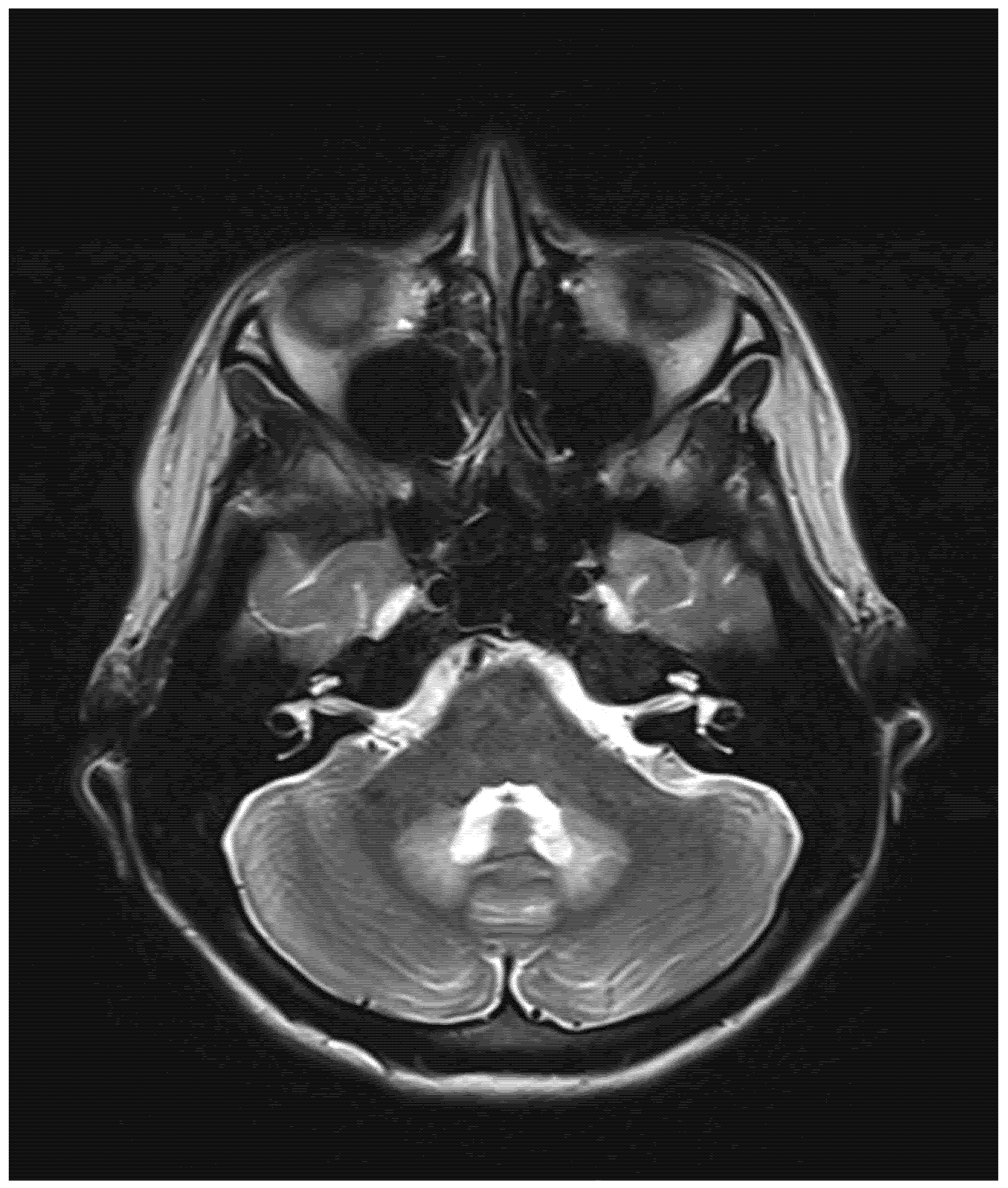
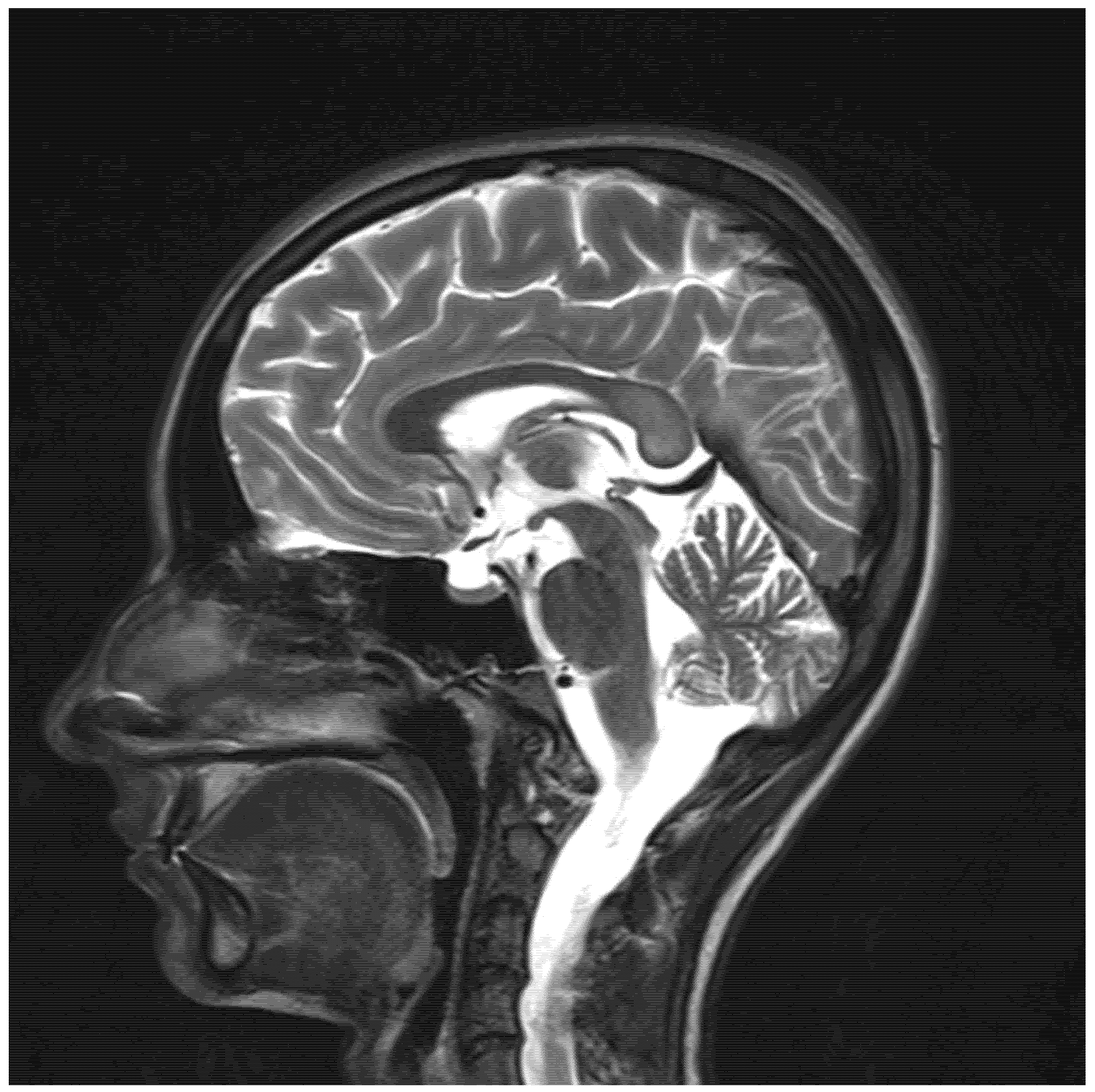
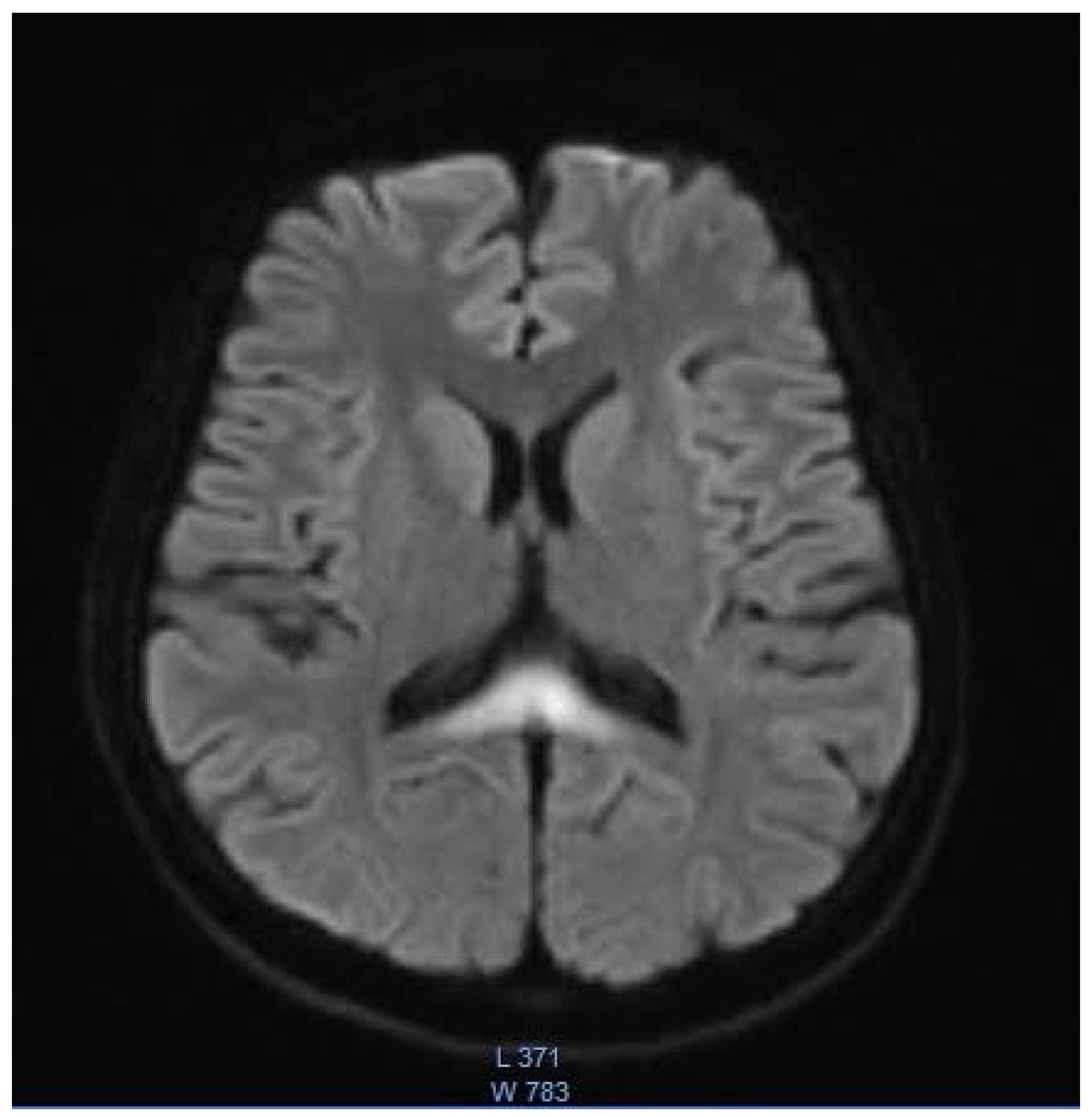
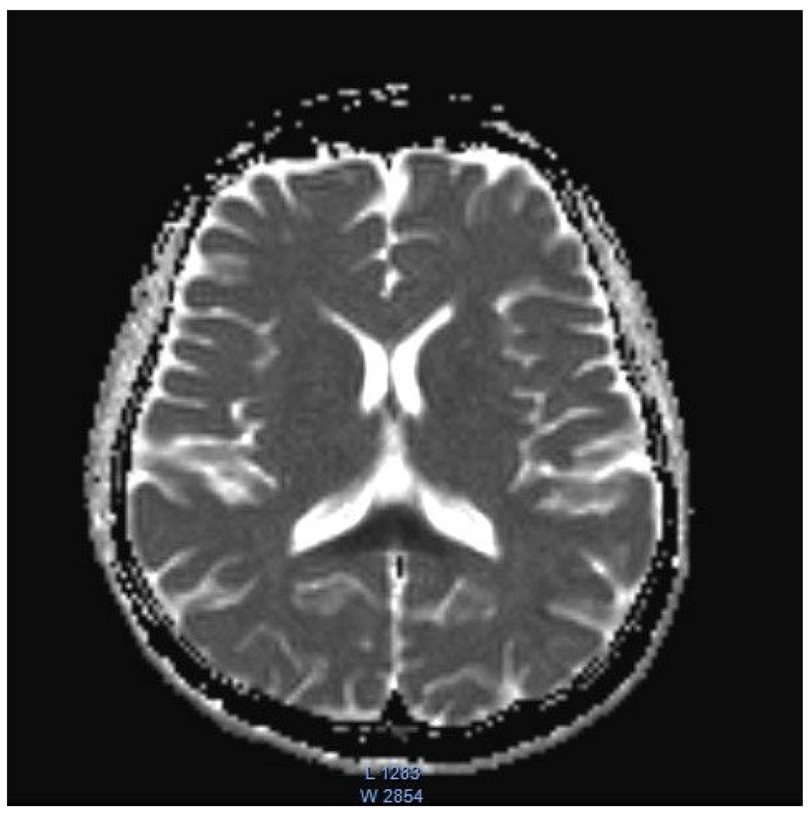
2. Case Presentation
3. Discussion
4. Conclusions
Author Contributions
Funding
Institutional Review Board Statement
Informed Consent Statement
Data Availability Statement
Conflicts of Interest
References
- Bailes, J.; Willis, J.; Priebe, C.; Strub, R. Encephalopathy With Metronidazole in a Child. Arch. Pediatr. Adolesc. Med. 1983, 137, 290–291. [Google Scholar] [CrossRef] [PubMed]
- Bates, J.E.; Almast, J.; Augustine, E.F. Neonatal dentate nucleus T2 hyperintensity after in utero metronidazole exposure. Neurology 2015, 85, 1006. [Google Scholar] [CrossRef] [PubMed]
- Cecil, K.M.; Halsted, M.J.; Schapiro, M.; Dinopoulos, A.; Jones, B.V. Reversible MR Imaging and MR Spectroscopy Abnormalities in Association with Metronidazole Therapy. J. Comput. Assist. Tomogr. 2002, 26, 948–951. [Google Scholar] [CrossRef] [PubMed]
- Chang, M.Y.; Borchert, M.S. Cortical Visual Impairment Treated by Plasmapheresis in a Child With Metronidazole-Induced Encephalopathy. J. Neuro-Ophthalmol. 2019, 41, e66–e68. [Google Scholar] [CrossRef] [PubMed]
- Chatzkel, J.A.; Vossough, A. Metronidazole-induced cerebellar toxicity. Pediatr. Radiol. 2009, 40, 1453. [Google Scholar] [CrossRef]
- Gaye, A.; Sanghavi, A. Metronidazole-Induced Encephalopathy in a Pediatric Patient. J. Allergy Clin. Immunol. 2007, 119, S38. [Google Scholar] [CrossRef]
- Kafadar, I.; Moustafa, F.; Yalçn, K.; Klç, B.A. A Rare Adverse Effect of Metronidazole. Pediatr. Emerg. Care 2013, 29, 751–752. [Google Scholar] [CrossRef]
- Omrani, A.; Rohani, M.; Hosseinpour, S.; Tavasoli, A.R. Persistent dystonia and basal ganglia involvement following metronidazole induced encephalopathy. Neurol. Sci. 2019, 41, 957–959. [Google Scholar] [CrossRef]
- Patel, L.; Batchala, P.; Almardawi, R.; Morales, R.; Raghavan, P. Acute metronidazole-induced neurotoxicity: An update on MRI findings. Clin. Radiol. 2019, 75, 202–208. [Google Scholar] [CrossRef]
- Starrs, M.E.; Yenigun, O.M. Metronidazole, an Uncommon Cause of Dizziness and Ataxia in the Emergency Department: A Case Report. Clin. Pract. Cases Emerg. Med. 2021, 2, 239–241. [Google Scholar] [CrossRef]
- Garg, A.; Sudan, Y.S.; Gupta, R.; Bansal, A.R. Headphone sign: Metronidazole-induced encephalopathy. Neurol. India 2016, 64, 1374–1376. [Google Scholar] [CrossRef] [PubMed]
- Sun, Y.; Overby, P.J.; Mehta, H. Case 271: Metronidazole-induced Encephalopathy. Radiology 2019, 293, 473–479. [Google Scholar] [CrossRef] [PubMed]
- Yazdani, R.M.; Kayfan, S.; Cao, J.; Clarke, R.L.; Pfeifer, C.M. MRI findings of metronidazole neurotoxicity in a pediatric patient with chronic diarrhea. Radiol. Case Rep. 2018, 14, 75–78. [Google Scholar] [CrossRef] [PubMed]
- Van Rheenen, P.F.; Aloi, M.; Assa, A.; Bronsky, J.; Escher, J.C.; Fagerberg, U.L.; Gasparetto, M.; Gerasimidis, K.; Griffiths, A.; Henderson, P.; et al. The Medical Management of Paediatric Crohn’s Disease: An ECCO-ESPGHAN Guideline Update. J. Crohn’s Coliti 2021, 15, 171–194. [Google Scholar] [CrossRef] [PubMed]
- Turner, D.; Ricciuto, A.; Lewis, A.; D’Amico, F.; Dhaliwal, J.; Griffiths, A.M.; Bettenworth, D.; Sandborn, W.J.; Sands, B.E.; Reinisch, W.; et al. STRIDE-II: An Update on the Selecting Therapeutic Targets in Inflammatory Bowel Disease (STRIDE) Initiative of the International Organization for the Study of IBD (IOIBD): Determining Therapeutic Goals for Treat-to-Target strategies in IBD. Gastroenterology 2021, 160, 1570–1583. [Google Scholar] [CrossRef] [PubMed]
- Sørensen, C.G.; Karlsson, W.K.; Amin, F.M.; Lindelof, M. Metronidazole-induced encephalopathy: A systematic review. J. Neurol. 2018, 267, 1–13. [Google Scholar] [CrossRef]
- Kuriyama, A.; Jackson, J.L.; Doi, A.; Kamiya, T. Metronidazole-Induced Central Nervous System Toxicity. Clin. Neuropharmacol. 2011, 34, 241–247. [Google Scholar] [CrossRef]
- Dainer, M.J. Untoward reaction to Flagyl (metronidazole). Am. J. Obstet. Gynecol. 1979, 133, 939–940. [Google Scholar] [CrossRef]
- Roy, U. Clinical and Neuroradiological Spectrum of Metronidazole Induced Encephalopathy: Our Experience and the Review of Literature. J. Clin. Diagn. Res. 2016, 10, OE01. [Google Scholar] [CrossRef]
- Bergogne-Berezin, E.; Bryskier, A. The suppository form of antibiotic administration: Pharmacokinetics and clinical application. J. Antimicrob. Chemother. 1999, 43, 177–185. [Google Scholar] [CrossRef] [Green Version]
- Mathew, R.P.; Kunhimohammed, S.P.; Joseph, M. A Case of Topical Metronidazole–Induced Encephalopathy. JAMA Neurol. 2020, 77, 1318–1319. [Google Scholar] [CrossRef] [PubMed]
- Sechi, G.; Serra, A. Wernicke’s encephalopathy: New clinical settings and recent advances in diagnosis and management. Lancet Neurol. 2007, 6, 442–455. [Google Scholar] [CrossRef]
- Oudman, E.; Wijnia, J.W.; Oey, M.J.; van Dam, M.; Postma, A. Wernicke-Korsakoff syndrome despite no alcohol abuse: A summary of systematic reports. J. Neurol. Sci. 2021, 426, 117482. [Google Scholar] [CrossRef] [PubMed]
- Hahn, J.S.; Berquist, W.; Alcorn, D.M.; Chamberlain, L.; Bass, D. Wernicke Encephalopathy and Beriberi During Total Parenteral Nutrition Attributable to Multivitamin Infusion Shortage. Pediatrics 1998, 101, e10. [Google Scholar] [CrossRef] [PubMed]
- Fernandes, L.; Bezerra, F.R.; Monteiro, M.; Silva, M.L.; De Oliveira, F.R.; Lima, R.R.; Fontes-Júnior, E.A.; Maia, C.S.F. Thiamine deficiency, oxidative metabolic pathways and ethanol-induced neurotoxicity: How poor nutrition contributes to the alcoholic syndrome, as Marchiafava–Bignami disease. Eur. J. Clin. Nutr. 2017, 71, 580–586. [Google Scholar] [CrossRef]
- Bhattacharyya, S.; Darby, R.R.; Raibagkar, P.; Castro, L.N.G.; Berkowitz, A.L. Antibiotic-associated encephalopathy. Neurology 2016, 86, 963–971. [Google Scholar] [CrossRef]
- Ferro, J.M.; Santos, M.O. Neurology of inflammatory bowel disease. J. Neurol. Sci. 2021, 424, 117426. [Google Scholar] [CrossRef]
- Bond, K.; Brinjikji, W.; Eckel, L.; Kallmes, D.; McDonald, R.; Carr, C. Dentate Update: Imaging Features of Entities That Affect the Dentate Nucleus. Am. J. Neuroradiol. 2017, 38, 1467–1474. [Google Scholar] [CrossRef]
- Couce, M.; Ramos, F.; Bueno, M.; Díaz, J.; Meavilla, S.; Bóveda, M.; Fernández-Marmiesse, A.; García-Cazorla, A. Evolution of maple syrup urine disease in patients diagnosed by newborn screening versus late diagnosis. Eur. J. Paediatr. Neurol. 2015, 19, 652–659. [Google Scholar] [CrossRef]
- Kohlschütter, A.; Eichler, F. Childhood leukodystrophies: A clinical perspective. Expert Rev. Neurother. 2011, 11, 1485–1496. [Google Scholar] [CrossRef] [Green Version]
- Wang, Q.; Li, X.; Ding, Y.; Liu, Y.; Song, J.; Yang, Y. Clinical and mutational spectra of 23 Chinese patients with glutaric aciduria type 1. Brain Dev. 2014, 36, 813–822. [Google Scholar] [CrossRef] [PubMed]
- Khadilkar, S.; Jaggi, S.; Patel, B.; Yadav, R.; Hanagandi, P.; Amaral, L.F.D. A practical approach to diseases affecting dentate nuclei. Clin. Radiol. 2015, 71, 107–119. [Google Scholar] [CrossRef] [PubMed]
- Boy, N.; Contributors, A.I.; Mühlhausen, C.; Maier, E.M.; Heringer, J.; Assmann, B.; Burgard, P.; Dixon, M.; Fleissner, S.; Rockman-Greenberg, C.; et al. Proposed recommendations for diagnosing and managing individuals with glutaric aciduria type I: Second revision. J. Inherit. Metab. Dis. 2016, 40, 75–101. [Google Scholar] [CrossRef] [PubMed]
- Baertling, F.; Rodenburg, R.J.; Schaper, J.; Smeitink, J.A.; Koopman, W.J.H.; Mayatepek, E.; Morava, E.; Distelmaier, F. A guide to diagnosis and treatment of Leigh syndrome. J. Neurol. Neurosurg. Psychiatry 2013, 85, 257–265. [Google Scholar] [CrossRef] [PubMed]
- Li, L.; Tang, X.; Li, W.; Liang, S.; Zhu, Q.; Wu, M. A case of methylprednisolone treatment for metronidazole-induced encephalopathy. BMC Neurol. 2019, 19, 49. [Google Scholar] [CrossRef] [PubMed]


| Author, Year, Country | Age (Years), Sex | Indication | Cumulative Dose of Metronidazole | Days to First Symptoms; Total Duration of Treatment | Symptoms and Clinical Findings | MRI Findings | Follow Up MRI | Outcome |
|---|---|---|---|---|---|---|---|---|
| Bailes, 1983, USA [1] | 12, M | Perforated appendicitis | 4 g | 4; 4 | Altered mental status; seizures | N/A | N/A | Resolution |
| Bates, 2015, USA [2] | 36-day-old, M | Mother’s vaginosis (utero exposure) | N/A | N/A; N/A | Hypothermia; bradycardia; failure to thrive; decreased tone and strength | T2: symmetrical hyperintensity in the dentate nuclei | Not done | Resolution |
| Cecil, 2002, USA [3] | 17, M | Crohn’s disease | N/A | N/A; N/A | Gait instability; Polyneuropathy; visual disturbance; tremor | T2: Symmetrical hyperintensity in the substantia nigra, red nucleus, globus pallidus, the putamen, caudate body, caudate heads and medial thalami | Near complete resolution | Near complete resolution |
| Chang, 2021, USA [4] | 14, M | Clostridium difficile enterocolitis | N/A | N/A; N/A | Altered mental status; stiffening of 4 extremities; visual disturbance; slurred speech; gait instability | T2: symmetrical hyperintensity with corresponding diffusion restriction on DWI in posterior frontal, parietal, and occipital periventricular white matter and splenium of the corpus callosum | Near complete resolution | Near complete resolution |
| Chatzkel, 2010, USA [5] | 15, F | Crohn’s disease | N/A | 7; N/A | Ataxia; dysmetria | T2: Symmetrical hyperintensity in the dentate nuclei | Resolution | N/A |
| Gaye, USA, 2007 [6] | Teenager, M | appendectomy | N/A | N/A; N/A | Unresponsiveness; respiratory distress; decerebrate posturing | Left parietal flair signal | N/A | Resolution |
| Kafadar, 2013, Turkey [7] | 3, M | amoebiasis diarrhea | N/A | 14; N/A | Loss of vision; ataxia, dizziness | Normal | N/A | Resolution |
| Omrani, Iran, 2020 [8] | 11, M | Febrile bloody diarrhea | 12 g | N/A; N/A | Tinnitus; hearing loss; aggressive behavior; generalized dystonia; generalized tonic-clonic seizure; decreased level of consciousness. | T2: Symmetrical hyperintensity in dentate nuclei, substantia nigra, globus pallidi, splenium of the corpus callosum, and centrum semiovale | Improvement | Partial improvement |
| Patel, USA, 2020 [9] | 8, M | Prophylaxis after small bowel transplantation | 1378.8 g | Three years; three years | Ataxia | T2: Symmetrical hyperintensity in the dentate nuclei, inferior olivary nuclei, putamen, and corpus callosum | Resolution | Resolution |
| Starrs, 2021, USA [10] | 12, M | Clostridium deficile infection | N/A | 75; 75 | Vertigo; nausea; vomiting; ataxia; gait instability | T2: Symmetrical hyperintensity in the dentate nuclei | N/A | Resolution |
| Sudan, 2016, India [11] | 14, M | Acute abdominal pain | N/A | 3; 5 | Dysarthria; altered mental status; seizures. | T2: symmetrical hyperintensity in the optic tracts, dorsal midbrain, inferior olivary nuclei, peri-aqueductal white matter, superior and inferior colliculi, superior cerebellar peduncle, dentate nuclei, medulla oblongata, and cervical spinal cord segment extending from the cervicomedullary junction to C6-C7 level DWI: restricted diffusion in the splenium of the corpus callosum | N/A | Resolution |
| Sun, 2019, USA [12] | 11, M | Fusobacterium menigitis | N/A | 3 months; N/A | Vomiting; dizziness; vertigo; gait instability; bilateral lower extremity paresthesia | T2: Symmetrical hyperintensity in the dentate nuclei, dorsal pons, and medulla. DWI: no restricted diffusion | Resolution | Resolution |
| Yazdani, 2019, USA [13] | 17, M | Chronic diarrhea | N/A | N/A | Gait instability; abnormal unilateral lean | T2: Symmetrical signal hyperintensity in dorsal pons, dentate nuclei, dorsal medulla | Improvement | Resolution |
Publisher’s Note: MDPI stays neutral with regard to jurisdictional claims in published maps and institutional affiliations. |
© 2022 by the authors. Licensee MDPI, Basel, Switzerland. This article is an open access article distributed under the terms and conditions of the Creative Commons Attribution (CC BY) license (https://creativecommons.org/licenses/by/4.0/).
Share and Cite
Rybak, K.; Warchoł, A.; Drobczyński, Ł.; Banaszkiewicz, A. Metronidazole-Induced Encephalopathy in a 16-Year-Old Girl with Crohn’s Disease: Case Report and Review of the Pediatric Literature. Children 2022, 9, 1408. https://doi.org/10.3390/children9091408
Rybak K, Warchoł A, Drobczyński Ł, Banaszkiewicz A. Metronidazole-Induced Encephalopathy in a 16-Year-Old Girl with Crohn’s Disease: Case Report and Review of the Pediatric Literature. Children. 2022; 9(9):1408. https://doi.org/10.3390/children9091408
Chicago/Turabian StyleRybak, Karolina, Aleksandra Warchoł, Łukasz Drobczyński, and Aleksandra Banaszkiewicz. 2022. "Metronidazole-Induced Encephalopathy in a 16-Year-Old Girl with Crohn’s Disease: Case Report and Review of the Pediatric Literature" Children 9, no. 9: 1408. https://doi.org/10.3390/children9091408