
Should We Be Screening for Ischaemic Heart Disease Earlier in Childhood?

 ,
,
Abstract
:1. Introduction
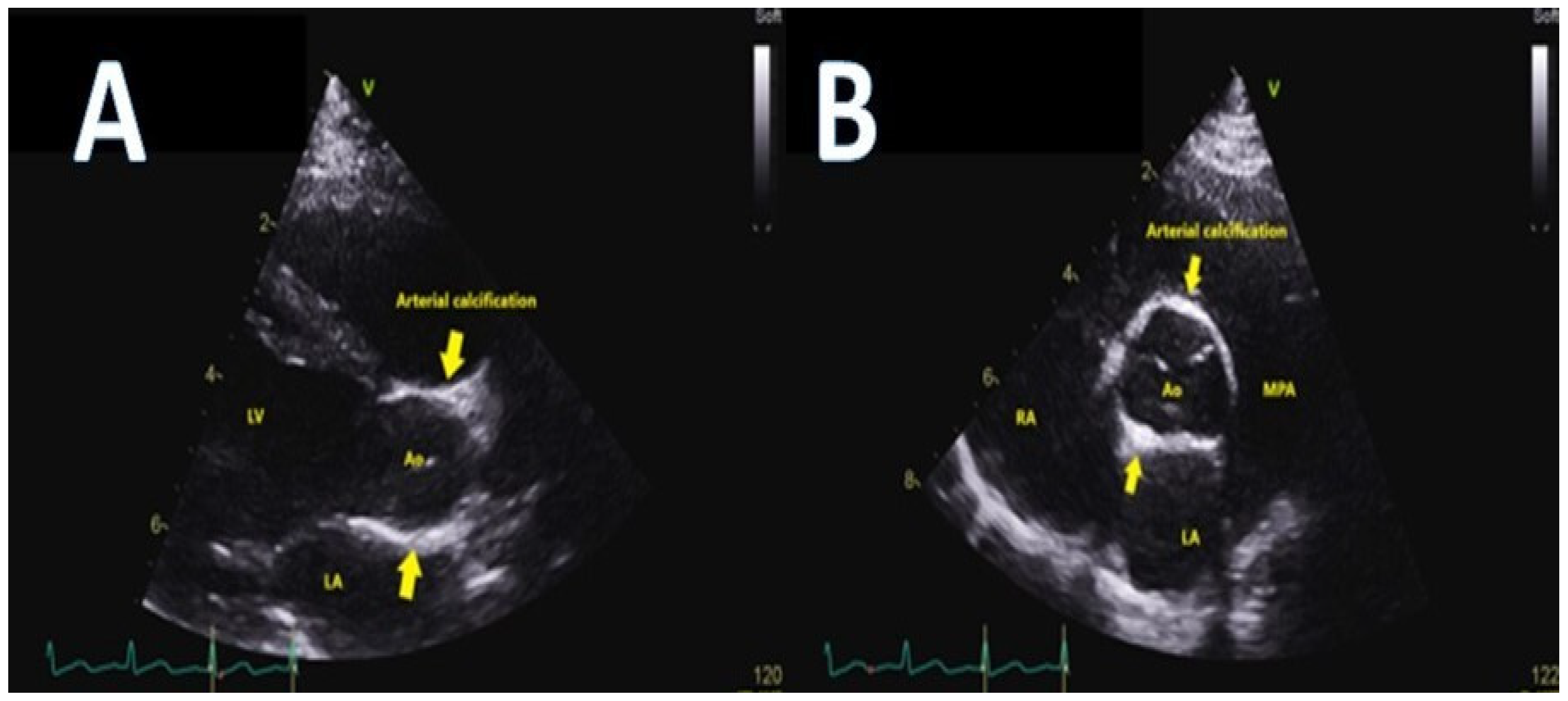
2. What Forms of Coronary Disease Do We Encounter in Childhood?
3. What Predisposing Factors Give Rise to Atherosclerosis and What Strategies in Childhood Could We Employ to Detect and Reduce Atherosclerosis Development in Later Life?
4. Discussion
5. Conclusions
Funding
Institutional Review Board Statement
Informed Consent Statement
Data Availability Statement
Conflicts of Interest
Abbreviations
| ALCAPA | anomalous left coronary artery from the pulmonary artery |
| KD | Kawasaki disease |
| FHC | familial hypercholesterolemia |
| LV | left ventricular |
| ECG | electrocardiogram |
| Echo | echocardiogram |
| CT | computed tomography |
| MI | myocardial infarction |
| LDL | low density lipoprotein |
| HoFHC | homozygous family hypercholesterolemia |
| HeFHC | heterozygous hypercholesterolemia |
| LDL-R | low density lipoprotein receptor |
| PCSK9 | proprotein convertase subtilisin/kexin type 9 |
References
- Khan, M.A.; Hashim, M.J.; Mustafa, H.; Baniyas, M.Y.; Al Suwaidi, S.K.B.M.; AlKatheeri, R.; Alblooshi, F.M.K.; Almatrooshi, M.E.A.H.; Alzaabi, M.E.H.; Al Darmaki, R.S.; et al. Global Epidemiology of Ischemic Heart Disease: Results from the Global Burden of Disease Study. Cureus 2020, 12, e9349. [Google Scholar] [CrossRef]
- Lusis, A.J. Atherosclerosis. Nature 2000, 407, 233–241. [Google Scholar] [CrossRef]
- Strong, J.P. Landmark perspective: Coronary atherosclerosis in soldiers. A clue to the natural history of atherosclerosis in the young. JAMA 1986, 256, 2863–2866. [Google Scholar] [CrossRef]
- Hertiš, P.T.; Petek, T.; Močnik, M.; Marčun Varda, N. Systemic Inflammation, Oxidative Stress and Cardiovascular Health in Children and Adolescents: A Systematic Review. Antioxidants 2022, 11, 894. [Google Scholar] [CrossRef]
- Fazeli Farsani, S.; van der Aa, M.P.; van der Vorst, M.M.J.; Knibbe, C.A.; de Boer, A. Global trends in the incidence and prevalence of type 2 diabetes in children and adolescents: A systematic review and evaluation of methodological approaches. Diabetologia 2013, 56, 1471–1488. [Google Scholar] [CrossRef] [Green Version]
- Nadeau, K.J.; Anderson, B.J.; Berg, E.G.; Chiang, J.L.; Chou, H.; Copeland, K.C.; Hannon, T.S.; Huang, T.T.; Lynch, J.L.; Powell, J.; et al. Youth-Onset Type 2 Diabetes Consensus Report: Current Status, Challenges, and Priorities. Diabetes Care 2016, 39, 1635–1642. [Google Scholar] [CrossRef] [Green Version]
- Copeland, K.C.; Zeitler, P.; Geffner, M.; Guandalini, C.; Higgins, J.; Hirst, K.; Kaufman, F.R.; Linder, B.; Marcovina, S.; McGuigan, P.; et al. Characteristics of adolescents and youth with recent-onset type 2 diabetes: The TODAY cohort at baseline. J. Clin. Endocrinol. Metab. 2011, 96, 159–167. [Google Scholar] [CrossRef]
- Raj, M. Obesity and cardiovascular risk in children and adolescents. Indian J. Endocrinol. Metab. 2012, 16, 13–19. [Google Scholar] [CrossRef]
- Keith, J.D. Anomalous origin of the coronary artery from the pulmonary artery. Br. Heart J. 1959, 21, 149–161. [Google Scholar] [CrossRef] [Green Version]
- Bland, E.F.; White, P.D.; Garland, J. Congenital anomalies of the coronary arteries? Report of an unusual case associated with cardiac hypertrophy. Am. Heart J. 1933, 8, 787–801. [Google Scholar] [CrossRef]
- Fontana, R.S.; Edwards, J.E. Congenital Cardiac Disease: A Review of 357 Cases Studied Pathologically; WB Saunders: Philadelphia, PA, USA, 1962. [Google Scholar]
- Wesselhoeft, H.; Fawcett, J.S.; Johnson, A.L. Anomalous origin of the left coronary artery from the pulmonary trunk. Its clinical spectrum, pathology and pathophysiology, based on a review of 140 cases with seven further cases. Circulation 1968, 38, 403425. [Google Scholar] [CrossRef] [Green Version]
- Frommelt, M.A.; Miller, W.; Williamson, J.; Bergstrom, S. Detection of septal coronary collaterals by color flow Doppler imaging is a marker for anomalous origin of a coronary artery from the pulmonary artery. J. Am. Soc. Echocardiogr. 2002, 15, 259–263. [Google Scholar] [CrossRef]
- Alsara, O.; Kalavakunta, J.K.; Hajjar, V.; Alsarah, A.; Cho, N.; Dhar, G. Surviving sudden cardiac death secondary to anomalous left coronary artery from the pulmonary artery: A case report and literature review. Heart Lung 2014, 43, 476–480. [Google Scholar] [CrossRef]
- Patel, S.G.; Frommelt, M.A.; Frommelt, P.C.; Kutty, S.; Cramer, J.W. Echocardiographic diagnosis, surgical treatment, and outcomes of anomalous left coronary artery from the pulmonary artery. J. Am. Soc. Echocardiogr. 2017, 30, 896–903. [Google Scholar] [CrossRef]
- Ojha, V.; Sh, C.; Vadher, A.; Singh Malhi, A.; Kothari, S.; Jagia, P. Anomalous left coronary artery from right pulmonary artery (ALCARPA) with dual left anterior descending arteries- a hitherto unreported combination of coronary anomalies diagnosed on dual source CT. J. Cardiovasc. Comput. Tomogr. 2020, 14, e69–e70. [Google Scholar] [CrossRef]
- Alexi-Meskishvili, V.; Hetzer, R.; Weng, Y.; Lange, P.E.; Jin, Z.; Berger, F.; Loebe, M. Anomalous origin of the left coronary artery from the pulmonary artery. Early results with direct aortic reimplantation. J. Thorac. Cardiovasc. Surg. 1994, 108, 354–362. [Google Scholar] [CrossRef] [Green Version]
- Fehrenbacher, T.A.; Mitchell, M.E.; Ghanayem, N.S.; Tweddell, J.S. Surgery and critical care for anomalous coronary artery from the pulmonary artery. Cardiol. Young 2010, 20 (Suppl. S3), 35–43. [Google Scholar] [CrossRef]
- Takeuchi, S.; Imamura, H.; Katsumoto, K.; Hayashi, I.; Katohgi, T.; Yozu, R.; Ohkura, M.; Inoue, T. New surgical method for repair of anomalous left coronary artery from pulmonary artery. J. Thorac. Cardiovasc. Surg. 1979, 78, 7–11. [Google Scholar] [CrossRef]
- Chin, A.J.; Larsen, R.L.; Seliem, M.A.; Andrews, B.; Jones, A.; Vetter, J.; Lieb, D. Noninvasive imaging of intraarterial baffles in infants and children. J. Am. Soc. Echocardiogr. 1993, 6, 45–50. [Google Scholar] [CrossRef]
- Naqvi, N.; Babu-Narayan, S.V.; Krupickova, S.; Muthialu, N.; Maiya, S.; Chandershekar, P.; Cheang, M.H.; Kostolny, M.; Tsang, V.; Marek, J. Myocardial Function Following Repair of Anomalous Origin of Left Coronary Artery from the Pulmonary Artery in Children. J. Am. Soc. Echocardiogr. 2020, 33, 622–630. [Google Scholar] [CrossRef]
- Radman, M.; Mastropietro, C.W.; Costello, J.M.; Amula, V.; Flores, S.; Caudill, E.; Karki, K.; Migally, K.; Narasimhulu, S.; Piggott, K.; et al. Collaborative Research from the Pediatric Cardiac Intensive Care Society (CoRe-PCICS) Investigators. Intermediate Outcomes After Repair of Anomalous Left Coronary Artery From the Pulmonary Artery. Ann. Thorac. Surg. 2021, 112, 1307–1315. [Google Scholar] [CrossRef]
- Sasikumar, D.; Dharan, B.S.; Arunakumar, P.; Gopalakrishnan, A.; Sivasankaran, S.; Krishnamoorthy, K.M. The outcome of mitral regurgitation after the repair of anomalous left coronary artery from the pulmonary artery in infants and older children. Interact. Cardiovasc. Thorac. Surg. 2018, 27, 238–242. [Google Scholar] [CrossRef]
- Kececioglu, D.; Deng, M.C.; Schmid, C.; Kehl, H.G.; Baba, H.A.; Yelbuz, M.; Scheld, H.H.; Vogt, J. Anomalous origin of the left coronary artery from the pulmonary artery with large anterior myocardial infarction and ischemia: Successful tunnel repair and concomitant heterotopic heart transplantation as biological bridge to recovery. Transpl. Int. 1997, 10, 161–163. [Google Scholar] [CrossRef]
- Hault, K.; Sebire, N.J.; Ho, S.Y.; Sheppard, M.N. The difficulty in diagnosing idiopathic arterial calcification of infancy, its variation in presentation, and the importance of autopsy. Cardiol. Young 2008, 18, 624–627. [Google Scholar] [CrossRef]
- Sebire, N.J.; Ramsay, A.; Sheppard, M. Idiopathic arterial calcification presenting with cardiac failure and sudden death in an 11-year-old girl. Pediatr. Dev. Pathol 2002, 5, 412–414. [Google Scholar] [CrossRef]
- Guimarães, S.; Lopes, J.M.; Oliveira, J.B.; Santos, A. Idiopathic infantile arterial calcification: A rare cause of sudden unexpected death in childhood. Patholog. Res. Int. 2010, 2010, 185314. [Google Scholar] [CrossRef] [Green Version]
- Rutsch, F.; Ruf, N.; Vaingankar, S.; Toliat, M.R.; Suk, A.; Höhne, W.; Schauer, G.; Lehmann, M.; Roscioli, T.; Schnabel, D.; et al. Mutations in ENPP1 are associated with ‘idiopathic’ infantile arterial calcification. Nat. Genet. 2003, 34, 379–381. [Google Scholar] [CrossRef]
- Chong, C.R.; Hutchins, G.M. Idiopathic infantile arterial calcification: The spectrum of clinical presentations. Pediatr. Dev. Pathol. 2008, 11, 405–415. [Google Scholar] [CrossRef]
- Kutty, S.; Cava, J.R.; Frommelt, M.A. Idiopathic infantile arterial calcification: A case report of prenatal and postnatal echocardiographic diagnosis. Echocardiography 2009, 26, 862–864. [Google Scholar] [CrossRef]
- Van der Sluis, I.M.; Boot, A.M.; Vernooij, M.; Meradji, M.; Kroon, A.A. Idiopathic infantile arterial calcification: Clinical presentation, therapy and long-term follow-up. Eur. J. Pediatr. 2006, 165, 590–593. [Google Scholar] [CrossRef]
- Patel, M.; Andronikou, S.; Solomon, R.; Sinclair, P.; McCulloch, M. Idiopathic arterial calcification in childhood. Pediatr. Radiol. 2004, 34, 652–655. [Google Scholar] [CrossRef]
- Nitschke, Y.; Rutsch, F. Inherited Arterial Calcification Syndromes: Etiologies and Treatment Concepts. Curr. Osteoporos. Rep. 2017, 15, 255–270. [Google Scholar] [CrossRef]
- Inwald, D.P.; Yen Ho, S.; Shepherd, M.N.; Daubeney, P.E. Idiopathic infantile arterial calcification presenting as fatal hypertensive cardiomyopathy. Arch. Dis. Child 2006, 91, 928. [Google Scholar] [CrossRef] [Green Version]
- Glatz, A.C.; Pawel, B.R.; Hsu, D.T.; Weinberg, P.; Chrisant, M.R. Idiopathic infantile arterial calcification: Two case reports, a review of the literature and a role for cardiac transplantation. Pediatr. Transpl. 2006, 10, 225–233. [Google Scholar] [CrossRef]
- Bonhoeffer, P.; Bonnet, D.; Piéchaud, J.F.; Stümper, O.; Aggoun, Y.; Villain, E.; Kachaner, J.; Sidi, D. Coronary artery obstruction after the arterial switch operation for transposition of the great arteries in newborns. J. Am. Coll. Cardiol. 1997, 29, 202–206. [Google Scholar] [CrossRef] [Green Version]
- Ou, P.; Celermajer, D.S.; Marini, D.; Agnoletti, G.; Vouhé, P.; Brunelle, F.; Le Quan Sang, K.H.; Thalabard, J.C.; Sidi, D.; Bonnet, D. Safety and accuracy of 64-slice computed tomography coronary angiography in children after the arterial switch operation for transposition of the great arteries. JACC Cardiovasc. Imaging 2008, 1, 331–339. [Google Scholar] [CrossRef]
- Ou, P.; Khraiche, D.; Celermajer, D.S.; Agnoletti, G.; Le Quan Sang, K.H.; Thalabard, J.C.; Quintin, M.; Raisky, O.; Vouhe, P.; Sidi, D.; et al. Mechanisms of coronary complications after the arterial switch for transposition of the great arteries. J. Thorac. Cardiovasc. Surg. 2013, 145, 1263–1269. [Google Scholar] [CrossRef] [Green Version]
- White, P.H.; Cooley, W.C.; Transitions Clinical Report Authoring Group; American Academy of Pediatrics; American Academy of Family Physicians; American College of Physicians. Supporting the health care transition from adolescence to adulthood in the medical home. Pediatrics 2018, 142, e20182587. [Google Scholar] [CrossRef] [Green Version]
- Cooley, W.C.; Sagerman, P.J.; American Academy of Pediatrics; American Academy of Family Physicians; American College of Physicians; Transitions Clinical Report Authoring Group. Supporting the health care transition from adolescence to adulthood in the medical home. Pediatrics 2011, 128, 182–200. [Google Scholar] [CrossRef] [Green Version]
- Sawicki, G.S.; Lukens-Bull, K.; Yin, X.; Demars, N.; Huang, I.C.; Livingood, W.; Reiss, J.; Wood, D. Measuring the transition readiness of youth with special healthcare needs: Validation of the TRAQ–Transition Readiness Assessment Questionnaire. J. Pediatr. Psychol. 2011, 36, 160–171. [Google Scholar] [CrossRef] [Green Version]
- Kamiyama, H.; Ayusawa, M.; Ogawa, S.; Saji, T.; Hamaoka, K. Health-care transition after Kawasaki disease in patients with coronary artery lesion. Pediatr. Int. 2018, 60, 232–239. [Google Scholar] [CrossRef]
- Dahdah, N.; Kung, S.C.; Friedman, K.G.; Marelli, A.; Gordon, J.B.; Belay, E.D.; Baker, A.L.; Kazi, D.S.; White, P.H.; Tremoulet, A.H.; et al. Falling Through the Cracks: The Current Gap in the Health Care Transition of Patients With Kawasaki Disease: A Scientific Statement From the American Heart Association. J. Am. Heart Assoc. 2021, 10, e023310. [Google Scholar] [CrossRef]
- Tsuda, E.; Tsujii, N.; Hayama, Y. Stenotic lesions and the maximum diameter of coronary artery aneurysms in Kawasaki disease. J. Pediatr 2018, 194, 165–170. [Google Scholar] [CrossRef]
- Kovacs, A.H.; McCrindle, B.W. So hard to say goodbye: Transition from paediatric to adult cardiology care. Nat. Rev. Cardiol. 2014, 11, 51–62. [Google Scholar] [CrossRef]
- Makino, N.; Nakamura, Y.; Yashiro, M.; Kosami, K.; Matsubara, Y.; Ae, R.; Aoyama, Y.; Yanagawa, H. Nationwide epidemiologic survey of Kawasaki disease in Japan, 2015–2016. Pediatr. Int. 2019, 61, 397–403. [Google Scholar] [CrossRef]
- Nakamura, Y.; Yashiro, M.; Yamashita, M.; Aoyama, N.; Otaki, U.; Ozeki, Y.; Sano, T.; Kojo, T.; Ae, R.; Aoyama, Y.; et al. Cumulative incidence of Kawasaki disease in Japan. Pediatr. Int. 2018, 60, 19–22. [Google Scholar] [CrossRef] [Green Version]
- Lin, Y.T.; Manlhiot, C.; Ching, J.C.; Han, R.K.; Nield, L.E.; Dillenburg, R.; Pepelassis, D.; Lai, L.S.; Smythe, J.F.; Chahal, N.; et al. Repeated systematic surveillance of Kawasaki disease in Ontario from 1995 to 2006. Pediatr. Int. 2010, 52, 699–706. [Google Scholar] [CrossRef]
- Daniels, L.B.; Gordon, J.B.; Burns, J.C. Kawasaki disease: Late cardiovascular sequelae. Curr. Opin. Cardiol. 2012, 27, 572–577. [Google Scholar] [CrossRef]
- Dionne, A.; Ibrahim, R.; Gebhard, C.; Benovoy, M.; Leye, M.; Dery, J.; Lapierre, C.; Girard, P.; Fournier, A.; Dahdah, N. Difference between persistent aneurysm, regressed aneurysm, and coronary dilation in Kawasaki disease: An optical coherence tomography study. Can. J. Cardiol. 2018, 34, 1120–1128. [Google Scholar] [CrossRef]
- McCrindle, B.W.; Rowley, A.H.; Newburger, J.W.; Burns, J.C.; Bolger, A.F.; Gewitz, M.; Baker, A.L.; Jackson, M.A.; Takahashi, M.; Shah, P.B.; et al. Diagnosis, treatment, and long-term management of Kawasaki disease: A scientific statement for health professionals from the American Heart Association. Circulation 2017, 135, e927–e999. [Google Scholar] [CrossRef]
- Mackie, A.S.; Fournier, A.; Swan, L.; Marelli, A.J.; Kovacs, A.H. Transition and transfer from pediatric to adult congenital heart disease care in Canada: Call for strategic implementation. Can. J. Cardiol. 2019, 35, 1640–1651. [Google Scholar] [CrossRef]
- Nakra, N.A.; Blumberg, D.A.; Herrera-Guerra, A.; Lakshminrusimha, S. Multi-System Inflammatory Syndrome in Children (MIS-C) Following SARS-CoV-2 Infection: Review of Clinical Presentation, Hypothetical Pathogenesis, and Proposed Management. Children 2020, 7, 69. [Google Scholar] [CrossRef]
- Bassareo, P.P.; Calcaterra, G.; Fanos, V. Coronavirus disease 2019, Kawasaki disease, and multisystem inflammatory syndrome in children. J. Pediatr. 2020, 224, 184. [Google Scholar] [CrossRef]
- Calcaterra, G.; Mehta, J.L.; Fanos, V.; Bassareo, P.P. Insights on Kawasaki disease and multisystem inflammatory syndrome: Relationship with COVID-19 infection. Minerva. Pediatr. 2021, 73, 203–208. [Google Scholar] [CrossRef]
- Cinteză, E.; Voicu, C.; Filip, C.; Ioniță, M.; Popescu, M.; Bălgrădean, M.; Nicolescu, A.; Mahmoud, H. Myocardial Infarction in Children after COVID-19 and Risk Factors for Thrombosis. Diagnostics 2022, 12, 884. [Google Scholar] [CrossRef]
- Expert Panel on Integrated Guidelines for Cardiovascular Health and Risk Reduction in Children and Adolescents; National Heart, Lung, and Blood Institute. Expert panel on integrated guidelines for cardiovascular health and risk reduction in children and adolescents: Summary report. Pediatrics 2011, 128 (Suppl. S5), S213–S256. [Google Scholar] [CrossRef] [Green Version]
- Mercuro, G.; Bassareo, P.P.; Flore, G.; Fanos, V.; Dentamaro, I.; Scicchitano, P.; Laforgia, N.; Ciccone, M.M. Prematurity and low weight at birth as new conditions predisposing to an increased cardiovascular risk. Eur. J. Prev. Cardiol. 2013, 20, 357–367. [Google Scholar] [CrossRef]
- Hadi, H.A.; Carr, C.S.; Al Suwaidi, J. Endothelial dysfunction: Cardiovascular risk factors, therapy, and outcome. Vasc. Health Risk Manag. 2005, 1, 183–198. [Google Scholar]
- Martino, F.; Loffredo, L.; Carnevale, R.; Sanguigni, V.; Martino, E.; Catasca, E.; Zanoni, C.; Pignatelli, P.; Violi, F. Oxidative stress is associated with arterial dysfunction and enhanced intima-media thickness in children with hypercholesterolemia: The potential role of nicotinamide-adenine dinucleotide phosphate oxidase. Pediatrics 2008, 122, e648–e655. [Google Scholar] [CrossRef]
- Loffredo, L.; Martino, F.; Carnevale, R.; Pignatelli, P.; Catasca, E.; Perri, L.; Calabrese, C.M.; Palumbo, M.M.; Baratta, F.; Del Ben, M.; et al. Obesity and hypercholesterolemia are associated with NOX2 generated oxidative stress and arterial dysfunction. J. Pediatr. 2012, 161, 1004–1009. [Google Scholar] [CrossRef]
- Masoura, C.; Pitsavos, C.; Aznaouridis, K.; Skoumas, I.; Vlachopoulos, C.; Stefanadis, C. Arterial endothelial function and wall thickness in familial hypercholesterolemia and familial combined hyperlipidemia and the effect of statins. A systematic review and meta-analysis. Atherosclerosis 2011, 214, 129–138. [Google Scholar] [CrossRef]
- Narverud, I.; Retterstøl, K.; Iversen, P.O.; Halvorsen, B.; Ueland, T.; Ulven, S.M.; Ose, L.; Aukrust, P.; Veierød, M.B.; Holven, K.B. Markers of atherosclerotic development in children with familial hypercholesterolemia: A literature review. Atherosclerosis 2014, 235, 299–309. [Google Scholar] [CrossRef]
- Awan, Z.; Alrasadi, K.; Francis, G.A.; Hegele, R.A.; McPherson, R.; Frohlich, J.; Valenti, D.; de Varennes, B.; Marcil, M.; Gagne, C.; et al. Vascular calcifications in homozygote familial hypercholesterolemia. Arterioscler. Thromb. Vasc. Biol. 2008, 28, 777–785. [Google Scholar] [CrossRef]
- Rader, D.J.; Cohen, J.; Hobbs, H.H. Monogenic hypercholesterolemia: New insights in pathogenesis and treatment. J. Clin. Investig. 2003, 111, 1795–1803. [Google Scholar] [CrossRef]
- Hu, H.; Cheng, J.; Lin, S.; Wang, S.; Chen, X. Calcified Aortic Valve Disease in Patients With Familial Hypercholesterolemia. J. Cardiovasc. Pharmacol. 2020, 76, 506–513. [Google Scholar] [CrossRef]
- Goldstein, J.L.; Brown, M.S. The LDL receptor. Arterioscler. Thromb. Vasc. Biol. 2009, 29, 431–438. [Google Scholar] [CrossRef] [Green Version]
- Defesche, J.C.; Gidding, S.S.; Harada-Shiba, M.; Hegele, R.A.; Santos, R.D.; Wierzbicki, A.S. Familial hypercholesterolaemia. Nat. Rev. Dis. Primers 2017, 3, 17093. [Google Scholar] [CrossRef]
- Borén, J.; Chapman, M.J.; Krauss, R.M.; Packard, C.J.; Bentzon, J.F.; Binder, C.J.; Daemen, M.J.; Demer, L.L.; Hegele, R.A.; Nicholls, S.J.; et al. Low-density lipoproteins cause atherosclerotic cardiovascular disease: Pathophysiological, genetic, and therapeutic insights: A consensus statement from the European Atherosclerosis Society Consensus Panel. Eur. Heart J. 2020, 41, 2313–2330. [Google Scholar] [CrossRef]
- Benn, M.; Watts, G.F.; Tybjærg-Hansen, A.; Nordestgaard, B.G. Mutations causative of familial hypercholesterolaemia: Screening of 98 098 individuals from the Copenhagen General Population Study estimated a prevalence of 1 in 217. Eur. Heart J. 2016, 37, 1384–1394. [Google Scholar] [CrossRef] [Green Version]
- Fellin, R.; Arca, M.; Zuliani, G.; Calandra, S.; Bertolini, S. The history of Autosomal Recessive Hypercholesterolemia (ARH). From clinical observations to gene identification. Gene 2015, 555, 23–32. [Google Scholar] [CrossRef]
- Goldstein, J.L.; Schrott, H.G.; Hazzard, W.R.; Bierman, E.L.; Motulsky, A.G. Hyperlipidemia in coronary heart disease. II. Genetic analysis of lipid levels in 176 families and delineation of a new inherited disorder, combined hyperlipidemia. J. Clin. Investig. 1973, 52, 1544–1568. [Google Scholar] [CrossRef] [Green Version]
- Beheshti, S.O.; Madsen, C.M.; Varbo, A.; Nordestgaard, B.G. Worldwide Prevalence of Familial Hypercholesterolemia: Meta-Analyses of 11 Million Subjects. J. Am. Coll. Cardiol. 2020, 75, 2553–2566. [Google Scholar] [CrossRef]
- EAS Familial Hypercholesterolaemia Studies Collaboration (FHSC). Global perspective of familial hypercholesterolaemia: A cross-sectional study from the EAS Familial Hypercholesterolaemia Studies Collaboration (FHSC). Lancet 2021, 398, 1713–1725. [Google Scholar] [CrossRef]
- Nordestgaard, B.G.; Chapman, M.J.; Humphries, S.E.; Ginsberg, H.N.; Masana, L.; Descamps, O.S.; Wiklund, O.; Hegele, R.A.; Raal, F.J.; Defesche, J.C.; et al. Familial hypercholesterolaemia is underdiagnosed and undertreated in the general population: Guidance for clinicians to prevent coronary heart disease: Consensus statement of the European Atherosclerosis Society. Eur. Heart J. 2013, 34, 3478–3490. [Google Scholar] [CrossRef] [Green Version]
- Hopkins, P.N.; Toth, P.P.; Ballantyne, C.M.; Rader, D.J.; National Lipid Association Expert Panel on Familial Hypercholesterolemia. Familial hypercholesterolemias: Prevalence, genetics, diagnosis and screening recommendations from the National Lipid Association Expert Panel on Familial Hypercholesterolemia. J. Clin. Lipidol. 2011, 5 (Suppl. S3), S9–S17. [Google Scholar] [CrossRef]
- Singh, S.; Bittner, V. Familial hypercholesterolemia--epidemiology, diagnosis, and screening. Curr. Atheroscler. Rep. 2015, 17, 482. [Google Scholar] [CrossRef]
- Singh, A.; Gupta, A.; Collins, B.L.; Qamar, A.; Monda, K.L.; Biery, D.; Lopez, J.A.G.; de Ferranti, S.D.; Plutzky, J.; Cannon, C.P.; et al. Familial Hypercholesterolemia Among Young Adults with Myocardial Infarction. J. Am. Coll. Cardiol. 2019, 73, 2439–2450. [Google Scholar] [CrossRef]
- Pang, J.; Martin, A.C.; Mori, T.A.; Beilin, L.J.; Watts, G.F. Prevalence of Familial Hypercholesterolemia in Adolescents: Potential Value of Universal Screening? J. Pediatr. 2016, 170, 315–316. [Google Scholar] [CrossRef]
- Avis, H.J.; Kusters, D.M.; Vissers, M.N.; Huijgen, R.; Janssen, T.H.; Wiegman, A.; Kindt, I.; Kastelein, J.J.; Wijburg, F.A.; Hutten, B.A. Follow-up of children diagnosed with familial hypercholesterolemia in a national genetic screening program. J. Pediatr. 2012, 161, 99–103. [Google Scholar] [CrossRef]
- O’Brien, E.C.; Roe, M.T.; Fraulo, E.S.; Peterson, E.D.; Ballantyne, C.M.; Genest, J.; Gidding, S.S.; Hammond, E.; Hemphill, L.C.; Hudgins, L.C.; et al. Rationale and design of the familial hypercholesterolemia foundation CAscade SCreening for Awareness and DEtection of Familial Hypercholesterolemia registry. Am. Heart J. 2014, 167, 342–349.e17. [Google Scholar] [CrossRef]
- Marks, D.; Thorogood, M.; Neil, H.A.; Humphries, S.E. A review on the diagnosis, natural history, and treatment of familial hypercholesterolaemia. Atherosclerosis 2003, 168, 1–14. [Google Scholar] [CrossRef]
- Wiegman, A.; Gidding, S.S.; Watts, G.F.; Chapman, M.J.; Ginsberg, H.N.; Cuchel, M.; Ose, L.; Averna, M.; Boileau, C.; Borén, J.; et al. Familial hypercholesterolaemia in children and adolescents: Gaining decades of life by optimizing detection and treatment. Eur. Heart J. 2015, 36, 2425–2437. [Google Scholar] [CrossRef] [Green Version]
- Ramaswami, U.; Futema, M.; Bogsrud, M.P.; Holven, K.B.; Roeters van Lennep, J.; Wiegman, A.; Descamps, O.S.; Vrablik, M.; Freiberger, T.; Dieplinger, H.; et al. Comparison of the characteristics at diagnosis and treatment of children with heterozygous familial hypercholesterolaemia (FH) from eight European countries. Atherosclerosis 2020, 292, 178–187. [Google Scholar] [CrossRef] [Green Version]
- Bertolini, S.; Calandra, S.; Arca, M.; Averna, M.; Catapano, A.L.; Tarugi, P.; Italian Study Group of Homozygous Familial Hypercholesterolemia. Homozygous familial hypercholesterolemia in Italy: Clinical and molecular features. Atherosclerosis 2020, 312, 72–78. [Google Scholar] [CrossRef]
- Sturm, A.C.; Knowles, J.W.; Gidding, S.S.; Ahmad, Z.S.; Ahmed, C.D.; Ballantyne, C.M.; Baum, S.J.; Bourbon, M.; Carrié, A.; Cuchel, M.; et al. Clinical Genetic Testing for Familial Hypercholesterolemia: JACC Scientific Expert Panel. J. Am. Coll Cardiol 2018, 72, 662–680. [Google Scholar] [CrossRef]
- Molven, I.; Retterstøl, K.; Andersen, L.F.; Veierød, M.B.; Narverud, I.; Ose, L.; Svilaas, A.; Wandel, M.; Holven, K.B. Children and young adults with familial hypercholesterolaemia (FH) have healthier food choices particularly with respect to dietary fat sources compared with non-FH children. J. Nutr. Sci. 2013, 2, e32. [Google Scholar] [CrossRef] [Green Version]
- Cicero, A.F.G.; Colletti, A.; Bajraktari, G.; Descamps, O.; Djuric, D.M.; Ezhov, M.; Fras, Z.; Katsiki, N.; Langlois, M.; Latkovskis, G.; et al. Lipid-lowering nutraceuticals in clinical practice: Position paper from an International Lipid Expert Panel. Nutr. Rev. 2017, 75, 731–767. [Google Scholar] [CrossRef]
- Steinberg, D. The rationale for initiating treatment of hypercholesterolemia in young adulthood. Curr. Atheroscler. Rep. 2013, 15, 296. [Google Scholar] [CrossRef]
- Ras, R.T.; Geleijnse, J.M.; Trautwein, E.A. LDL-cholesterol-lowering effect of plant sterols and stanols across different dose ranges: A meta-analysis of randomised controlled studies. Br. J. Nutr. 2014, 112, 214–219. [Google Scholar] [CrossRef] [Green Version]
- Gylling, H.; Plat, J.; Turley, S.; Ginsberg, H.N.; Ellegård, L.; Jessup, W.; Jones, P.J.; Lütjohann, D.; Maerz, W.; Masana, L.; et al. Plant sterols and plant stanols in the management of dyslipidaemia and prevention of cardiovascular disease. Atherosclerosis 2014, 232, 346–360. [Google Scholar] [CrossRef]
- Martino, F.; Martino, E.; Morrone, F.; Carnevali, E.; Forcone, R.; Niglio, T. Effect of dietary supplementation with glucomannan on plasma total cholesterol and low density lipoprotein cholesterol in hypercholesterolemic children. Nutr. Metab. Cardiovasc. Dis. 2005, 15, 174–180. [Google Scholar] [CrossRef]
- Martino, F.; Puddu, P.E.; Pannarale, G.; Colantoni, C.; Martino, E.; Niglio, T.; Zanoni, C.; Barillà, F. Low dose chromium-polynicotinate or policosanol is effective in hypercholesterolemic children only in combination with glucomannan. Atherosclerosis 2013, 228, 198–202. [Google Scholar] [CrossRef]
- Guardamagna, O.; Abello, F.; Baracco, V.; Stasiowska, B.; Martino, F. The treatment of hypercholesterolemic children: Efficacy and safety of a combination of red yeast rice extract and policosanols. Nutr. Metab. Cardiovasc. Dis. 2011, 21, 424–429. [Google Scholar] [CrossRef]
- Manousaki, D.; Barnett, T.A.; Mathieu, M.E.; Maximova, K.; Simoneau, G.; Harnois-Leblanc, S.; Benedetti, A.; McGrath, J.J.; Henderson, M.; QUALITY Cohort Collaborative Group. Tune out and turn in: The influence of television viewing and sleep on lipid profiles in children. Int. J. Obes. 2020, 44, 1173–1184. [Google Scholar] [CrossRef]
- Authors/Task Force Members; ESC Committee for Practice Guidelines (CPG); ESC National Cardiac Societies. 2019 ESC/EAS guidelines for the management of dyslipidaemias: Lipid modification to reduce cardiovascular risk. Atherosclerosis 2019, 290, 140–205. [Google Scholar] [CrossRef] [Green Version]
- Luirink, I.K.; Wiegman, A.; Kusters, D.M.; Hof, M.H.; Groothoff, J.W.; de Groot, E.; Kastelein, J.J.P.; Hutten, B.A. 20-Year Follow-up of Statins in Children with Familial Hypercholesterolemia. N. Engl. J. Med. 2019, 381, 1547–1556. [Google Scholar] [CrossRef]
- Grabarczyk, M.; Pomianowska, K.; Zaręba-Głód, T.; Zachurzok, A.; Małecka-Tendera, E.; Matusik, P. Statin therapy and lipids-lowering supplements-safe and effective treatment of lipids disturbances in children. Pediatr. Endocrinol. Diabetes Metab. 2022, 28, 46998. [Google Scholar] [CrossRef]
- Dombalis, S.; Nash, A. The Effect of Statins in Children and Adolescents with Familial Hypercholesterolemia: A Systematic Review. J. Pediatr. Health Care 2021, 35, 292–303. [Google Scholar] [CrossRef]
- Van der Graaf, A.; Cuffie-Jackson, C.; Vissers, M.N.; Trip, M.D.; Gagné, C.; Shi, G.; Veltri, E.; Avis, H.J.; Kastelein, J.J. Efficacy and safety of coadministration of ezetimibe and simvastatin in adolescents with heterozygous familial hypercholesterolemia. J. Am. Coll. Cardiol. 2008, 52, 1421–1429. [Google Scholar] [CrossRef] [Green Version]
- Catapano, A.L.; Graham, I.; De Backer, G.; Wiklund, O.; Chapman, M.J.; Drexel, H.; Hoes, A.W.; Jennings, C.S.; Landmesser, U.; Pedersen, T.R.; et al. 2016 ESC/EAS Guidelines for the Management of Dyslipidaemias: The Task Force for the Management of Dyslipidaemias of the European Society of Cardiology (ESC) and European Atherosclerosis Society (EAS) Developed with the special contribution of the European Association for Cardiovascular Prevention & Rehabilitation (EACPR). Atherosclerosis 2016, 253, 281–344. [Google Scholar]
- Santos, R.D.; Ruzza, A.; Hovingh, G.K.; Wiegman, A.; Mach, F.; Kurtz, C.E.; Hamer, A.; Bridges, I.; Bartuli, A.; Bergeron, J.; et al. Evolocumab in Pediatric Heterozygous Familial Hypercholesterolemia. N. Engl. J. Med. 2020, 383, 1317–1327. [Google Scholar] [CrossRef]
- Luirink, I.K.; Hutten, B.A.; Greber-Platzer, S.; Kolovou, G.D.; Dann, E.J.; de Ferranti, S.D.; Taylan, C.; Bruckert, E.; Saheb, S.; Oh, J.; et al. Practice of lipoprotein apheresis and short-term efficacy in children with homozygous familial hypercholesterolemia: Data from an international registry. Atherosclerosis 2020, 299, 24–31. [Google Scholar] [CrossRef]
- Bassareo, P.P.; Abella, R.; Fanos, V.; Mercuro, G. Biomarkers of corticosteroid-induced hypertrophic cardiomyopathy in preterm babies. Front. Biosci. 2010, 2, 1460–1471. [Google Scholar] [CrossRef] [Green Version]
- Peña, E.; Nguyen, E.T.; Merchant, N.; Dennie, C. ALCAPA syndrome: Not just a pediatric disease. Radiographics 2009, 29, 553–565. [Google Scholar] [CrossRef]
- Palmas, G.; Tumbarello, R.; Abbruzzese, P.; Fanos, V. Idiopathic infantile arterial calcification: Case report. Minerva. Pediatr. 2008, 60, 457–460. [Google Scholar]
- De Ferranti, S.D.; Rodday, A.M.; Mendelson, M.M.; Wong, J.B.; Leslie, L.K.; Sheldrick, R.C. Prevalence of Familial Hypercholesterolemia in the 1999 to 2012 United States National Health and Nutrition Examination Surveys (NHANES). Circulation 2016, 133, 1067–1072. [Google Scholar] [CrossRef]
- Skinner, A.C.; Perrin, E.M.; Skelton, J.A. Prevalence of obesity and severe obesity in US children, 1999-2014. Obesity 2016, 24, 1116–1123. [Google Scholar] [CrossRef] [Green Version]
- Garrido-Miguel, M.; Cavero-Redondo, I.; Álvarez-Bueno, C.; Rodríguez-Artalejo, F.; Moreno, L.A.; Ruiz, J.R.; Ahrens, W.; Martínez-Vizcaíno, V. Prevalence and Trends of Overweight and Obesity in European Children From 1999 to 2016: A Systematic Review and Meta-analysis. JAMA Pediatr. 2019, 173, e192430. [Google Scholar] [CrossRef]
- Belay, B.; Belamarich, P.F.; Tom-Revzon, C. The use of statins in pediatrics: Knowledge base, limitations, and future directions. Pediatrics 2007, 119, 370–380. [Google Scholar] [CrossRef]
- Newman, W.P., III; Freedman, D.S.; Voors, A.W.; Gard, P.D.; Srinivasan, S.R.; Cresanta, J.L.; Williamson, G.D.; Webber, L.S.; Berenson, G.S. Relation of serum lipoprotein levels and systolic blood pressure to early atherosclerosis. The Bogalusa Heart Study. N. Engl. J. Med. 1986, 314, 138–144. [Google Scholar] [CrossRef]
- NCD Risk Factor Collaboration (NCD-RisC). Worldwide trends in body-mass index, underweight, overweight, and obesity from 1975 to 2016: A pooled analysis of 2416 population-based measurement studies in 128·9 million children, adolescents, and adults. Lancet 2017, 390, 2627–2642. [Google Scholar] [CrossRef] [Green Version]
- Berenson, G.S.; Srinivasan, S.R.; Bao, W.; Newman, W.P., III; Tracy, R.E.; Wattigney, W.A. Association between multiple cardiovascular risk factors and atherosclerosis in children and young adults. The Bogalusa Heart Study. N. Engl. J. Med. 1998, 338, 1650–6165. [Google Scholar] [CrossRef] [PubMed]
- Koskinen, J.; Juonala, M.; Dwyer, T.; Venn, A.; Thomson, R.; Bazzano, L.; Berenson, G.S.; Sabin, M.A.; Burns, T.L.; Viikari, J.S.A.; et al. Impact of Lipid Measurements in Youth in Addition to Conventional Clinic-Based Risk Factors on Predicting Preclinical Atherosclerosis in Adulthood: International Childhood Cardiovascular Cohort Consortium. Circulation 2018, 137, 1246–1255. [Google Scholar] [CrossRef] [PubMed]
- Skilton, M.R.; Celermajer, D.S. Endothelial dysfunction and arterial abnormalities in childhood obesity. Int. J. Obes. 2006, 30, 1041–1049. [Google Scholar] [CrossRef] [Green Version]
- Canas, J.A.; Sweeten, S.; Balagopal, P.B. Biomarkers for cardiovascular risk in children. Curr. Opin. Cardiol. 2013, 28, 103–114. [Google Scholar] [CrossRef]
- Tonstad, S.; Joakimsen, O.; Stensland-Bugge, E.; Leren, T.P.; Ose, L.; Russell, D.; Bønaa, K.H. Risk factors related to carotid intima-media thickness and plaque in children with familial hypercholesterolemia and control subjects. Arterioscler. Thromb. Vasc. Biol. 1996, 16, 984–991. [Google Scholar] [CrossRef]
- Dalla Pozza, R.; Ehringer-Schetitska, D.; Fritsch, P.; Jokinen, E.; Petropoulos, A.; Oberhoffer, R.; Association for European Paediatric Cardiology Working Group Cardiovascular Prevention. Intima media thickness measurement in children: A statement from the Association for European Paediatric Cardiology (AEPC) Working Group on Cardiovascular Prevention endorsed by the Association for European Paediatric Cardiology. Atherosclerosis 2015, 238, 380–387. [Google Scholar] [CrossRef]
- Pintus, R.; Bassareo, P.P.; Dessì, A.; Deidda, M.; Mercuro, G.; Fanos, V. Metabolomics and Cardiology: Toward the Path of Perinatal Programming and Personalized Medicine. Biomed. Res. Int. 2017, 2017, 6970631. [Google Scholar] [CrossRef] [Green Version]


| Criterion | Title 2 |
|---|---|
| 1 | Occasional finding of LDL ≥ 4 mmol/L (160 mg/dL) without any bodily sign of note and with a parent suffering from hypercholesterolemia or ischaemic heart disease (<55 years in males and <60 years in females) |
| 2 | LDL-C ≥ 5.0 mmol/L (190 mg/dL) in two different checks after 3 months of low cholesterol diet |
| 3 | LDL-C ≥ 3.5 mmol/L (130 mg/dL) and one parent with a FHC genetic diagnosis |
| 4 | LDL-C ≥ 13 mmol/L (500 mg/dL) with cutaneous xanthomas (dominant or recessive HoFHC) |
Publisher’s Note: MDPI stays neutral with regard to jurisdictional claims in published maps and institutional affiliations. |
© 2022 by the authors. Licensee MDPI, Basel, Switzerland. This article is an open access article distributed under the terms and conditions of the Creative Commons Attribution (CC BY) license (https://creativecommons.org/licenses/by/4.0/).
Share and Cite
Bassareo, P.P.; O’Brien, S.T.; Dunne, E.; Duignan, S.; Martino, E.; Martino, F.; Mcmahon, C.J. Should We Be Screening for Ischaemic Heart Disease Earlier in Childhood? Children 2022, 9, 982. https://doi.org/10.3390/children9070982
Bassareo PP, O’Brien ST, Dunne E, Duignan S, Martino E, Martino F, Mcmahon CJ. Should We Be Screening for Ischaemic Heart Disease Earlier in Childhood? Children. 2022; 9(7):982. https://doi.org/10.3390/children9070982
Chicago/Turabian StyleBassareo, Pier Paolo, Stephen T. O’Brien, Esme Dunne, Sophie Duignan, Eliana Martino, Francesco Martino, and Colin J. Mcmahon. 2022. "Should We Be Screening for Ischaemic Heart Disease Earlier in Childhood?" Children 9, no. 7: 982. https://doi.org/10.3390/children9070982