Permanent Maxillary and Mandibular Central Incisor Width as Predictor of Permanent Maxillary Canine Width in a Kurdish Population: A Pilot Study


Abstract
:1. Introduction
2. Materials and Methods
2.1. Study Design
2.2. Study Sample
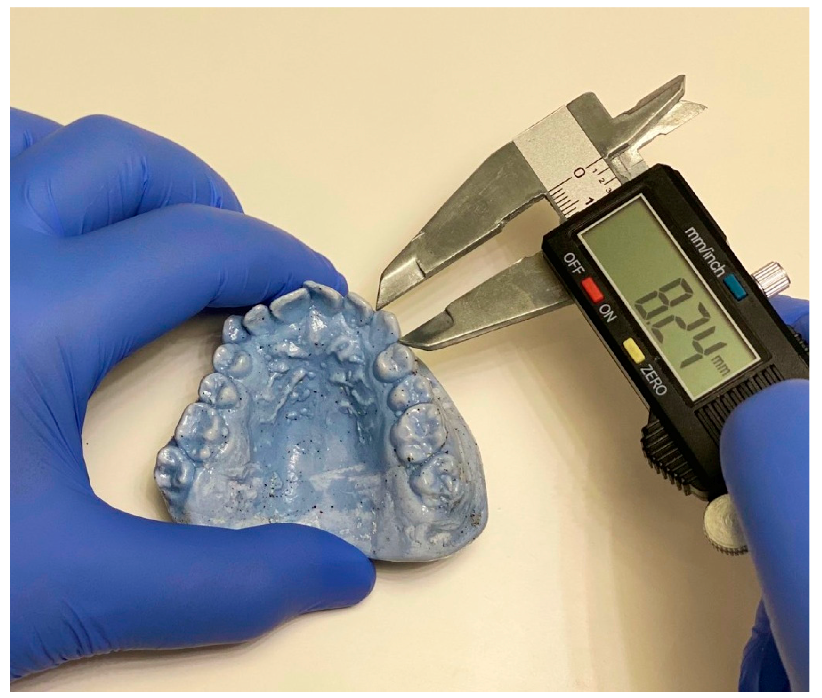
2.3. Measurements and Study Outcomes
2.4. Statistical Analysis
3. Results
4. Discussion
5. Conclusions
Funding
Acknowledgments
Conflicts of Interest
References
- McDonald, R.E.; Avery, D.R.; Dean, J.A. Management of the Developing Occlusion. Dentistry for the Child and the Adolescent, 8th ed.; Mosby: St Louis, MO, USA, 2004; p. 627. [Google Scholar]
- Ling, J.Y.; Wong, R.W. Tanaka-Johnston mixed dentition analysis for southern Chinese in Hong Kong. Angle Orthod. 2006, 76, 632–636. [Google Scholar] [PubMed]
- Diagne, F.; Diop-Ba, K.; Ngom, P.I.; Mbow, K. Mixed dentition analysis in a Senegalese population: Elaboration of prediction tables. Am. J. Orthod. Dentofac. Orthop. 2003, 124, 178–183. [Google Scholar] [CrossRef]
- Philip, N.I.; Prabhakar, M.; Arora, D.; Chopra, S. Applicability of the Moyers mixed dentition probability tables and new prediction aids for a contemporary population in India. Am. J. Orthod. Dentofac. Orthop. 2010, 138, 339–345. [Google Scholar] [CrossRef] [PubMed]
- Abate, A.; Cavagnetto, D.; Fama, A.; Matarese, M.; Bellincioni, F.; Assandri, F. Efficacy of Operculectomy in the Treatment of 145 Cases with Unerupted Second Molars: A Retrospective Case-Control Study. Dent. J. (Basel) 2020, 8, 65. [Google Scholar] [CrossRef]
- Maspero, C.; Abate, A.; Cavagnetto, D.; Fama, A.; Stabilini, A.; Farronato, G.; Farronato, M. Operculectomy and spontaneous eruption of impacted second molars: A retrospective study. J. Biol. Regul. Homeost. Agents 2019, 33, 1909–1912. [Google Scholar]
- Lee-Chan, S.; Jacobson, B.N.; Chwa, K.H.; Jacobson, R.S. Mixed dentition analysis for Asian-Americans. Am. J. Orthod. Dentofac. Orthop. 1998, 113, 293–299. [Google Scholar] [CrossRef]
- Proffit, W.R.; Fields, H.W. Contemporary Orthodontics, 3rd ed.; Elsevier: Missouri, USA, 2000; pp. 165–170. [Google Scholar]
- Moyers, R.E. Handbook of Orthodontics, 4th ed.; Year Book Medical Publishers Inc.: Chicago, IL, USA, 1988; pp. 235–239. [Google Scholar]
- Tanaka, M.M.; Johnston, L.E. The prediction of the size of unerupted canines and premolars in a contemporary orthodontic population. J. Am. Dent. Assoc. 1974, 88, 798–801. [Google Scholar] [CrossRef]
- Brito, F.C.; Nacif, V.C.; Melgaço, C.A. Mandibular permanent first molars and incisors as predictors of mandibular permanent canine and premolar widths: Applicability and consistency of the method. Am. J. Orthod. Dentofac. Orthop. 2014, 145, 393–398. [Google Scholar] [CrossRef]
- Bherwani, A.K.; Fida, M. Development of a prediction equation for the mixed dentition in a Pakistani sample. Am. J. Orthod. Dentofac. Orthop. 2011, 140, 626–632. [Google Scholar] [CrossRef] [Green Version]
- Uysal, T.; Sari, Z. Intermaxillary tooth size discrepancy and mesiodistal crown dimensions for a Turkish population. Am. J. Orthod. Dentofac. Orthop. 2005, 128, 226–230. [Google Scholar] [CrossRef]
- Uysal, T.; Sari, Z.; Basciftci, F.A.; Memili, B. Intermaxillary tooth size discrepancy and malocclusion: Is there a relation? Angle Orthod. 2005, 75, 208–213. [Google Scholar] [PubMed]
- Marinelli, A.; Alarashi, M.; Defraia, E.; Antonini, A.; Tollaro, I. Tooth wear in the mixed dentition: A comparative study between children born in the 1950s and the 1990s. Angle Orthod. 2005, 75, 340–343. [Google Scholar] [PubMed]
- Moorrees, C.F.A.; Thomsen, S.O.; Jensen, E.; Yen, P.K.J. Mesiodistal crown diameters of the deciduous and permanent teeth in individuals. J. Dent. Res. 1957, 36, 39–47. [Google Scholar] [CrossRef] [PubMed]
- Bailit, H.L. Dental variation among populations. An anthropologic view. Dent. Clin. N. Am. 1975, 29, 125–139. [Google Scholar]
- Mahmood, T.; Kareem, F. Mixed dentition analysis in a sample of Sulaimani population. KAJ 2010, 1, 23–28. [Google Scholar]
- Raghoebar, G.M.; Boering, G.; Vissink, A.; Stegenga, B. Eruption disturbances of permanent molars: A review. J. Oral Pathol. Med. 1991, 20, 159–166. [Google Scholar] [CrossRef]
- Quadras, D.D.; Nayak, U.S.; Ravi, M.S.; Pujari, P. Early prediction of maxillary canine impaction using sectors and angular measurement—A radiographic study. MJDS 2017, 2, 7–11. [Google Scholar]
- Coulter, J.; Richardson, A. Normal eruption of the maxillary canine quantified in three dimensions. Eur. J. Orthod. 1997, 19, 171–183. [Google Scholar] [CrossRef] [Green Version]
- Aziz, Z.H. Problems Related to Permanent Maxillary Canine in Skeletal Class I Jaw Relationship in Sulaimani Kurd Students Aged 18-22 Years. Master’s Thesis, College of Dentistry/University of Sulaimani, Sulaymaniyah, UK, 2008. [Google Scholar]
- Al-Atabi, H.; Mohammed-Salih, H.; Nahidh, M. Buccally malposed maxillary canines in intermediate schools students of sammawa city. Iraqi Dent. J. 2016, 38, 21–27. [Google Scholar] [CrossRef]
- Ballard, M.L.; Wylie, W.L. Mixed dentition case analysis-estimating size of unerupted permanent teeth. Am. J. Orthod. 1947, 33, 754–759. [Google Scholar] [CrossRef]
- Boboc, A.; Dibbets, J. Prediction of the mesiodistal width of unerupted permanent canines and premolars: A statistical approach. Am. J. Orthod. Dentofac. Orthop. 2010, 137, 503–507. [Google Scholar] [CrossRef] [PubMed]
- Bernabé, E.; Flores-Mir, C. Are the lower incisors the best predictors for the unerupted canine and premolars sums? An analysis of a Peruvian sample. Angle Orthod. 2005, 75, 202–207. [Google Scholar] [PubMed]
- Garn, S.M. Genetics of dental development. In The Biology of Occlusal Development; Monograph 7, Craniofacial Growth Series; Center for Human Growth and Development, University of Michigan: Ann Arbor, MI, USA, 1977. [Google Scholar]
- Richard, L.C.; Miller, S.L. Relationships between age and dental attrition in Australian aboriginals. Am. J. Phys. Anthropol. 1991, 84, 159–164. [Google Scholar] [CrossRef]
- Garn, S.M.; Lewis, A.B.; Kerewsky, R.S. X-linked inheritance of dental tooth size. J. Dent. Res. 1965, 44, 439–441. [Google Scholar] [CrossRef]
- Hixon, E.H.; Oldfather, R.E. Estimation of the sizes of unerupted cuspids and bicuspids. Angle Orthod. 1958, 28, 236–240. [Google Scholar]
- Al-Khadra, B.H. Prediction of the size of unerupted canines and premolars in a Saudi Arab population. Am. J. Orthod. Dentofac. Orthop. 1993, 104, 369–372. [Google Scholar] [CrossRef]
- Yuen, K.K.; Tang, E.L.; So, L.L. Mixed dentition analysis for Hong Kong Chinese. Angle Orthod. 1998, 68, 21–28. [Google Scholar]
- Uysal, T.; Basciftci, F.A.; Goyenc, Y. New regression equations for mixed-dentition arch analysis in a Turkish sample with no Bolton tooth-size discrepancy. Am. J. Orthod. Dentofac. Orthop. 2009, 135, 343–348. [Google Scholar] [CrossRef]
- Jaroontham, J.; Godfrey, K. Mixed dentition space analysis in a Thai population. Eur. J. Orthod. 2000, 22, 127–134. [Google Scholar] [CrossRef]


| Variables | Minimum | Maximum | Mean, SD | Male | Female | Male vs. Female * |
|---|---|---|---|---|---|---|
| Age (years) | 14 | 24 | 17.2 ± 2.39 | 17.5 ± 2.66 | 16.9 ± 2.06 | 0.21 |
| Sum of PMMCI widths (mm) | 27.62 | 36.68 | 31.5 ± 1.9 | 32.4 ± 1.6 | 30.6 ± 1.9 | 0.0001 |
| Sum of PMC widths (mm) | 14.27 | 18.66 | 16.1 ± 1.02 | 16.8 ± 0.75 | 15.5 ± 0.8 | 0.0001 |
| Mesio-Distal Width of PMMCIs | |||
|---|---|---|---|
| Male | Female | ||
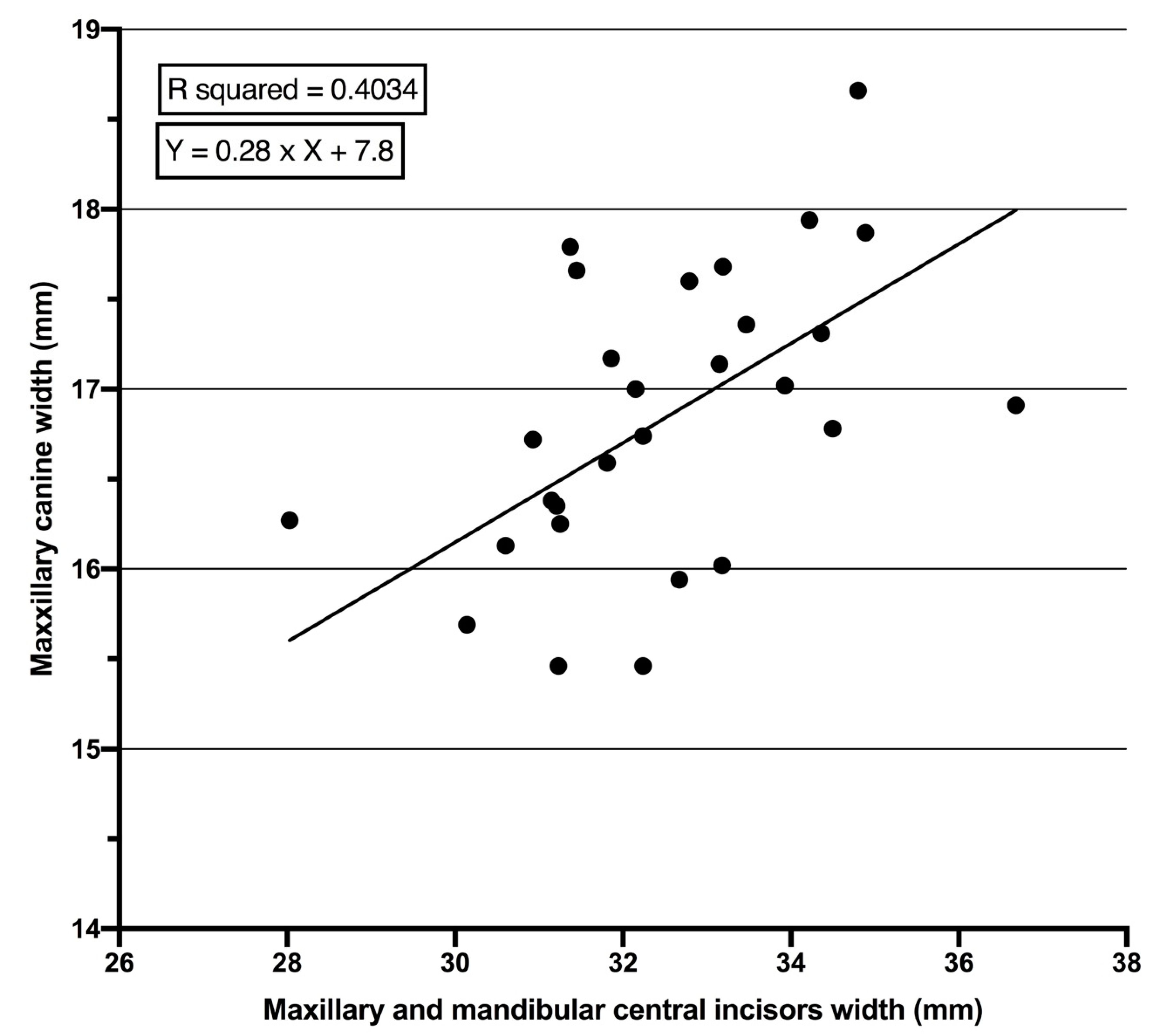
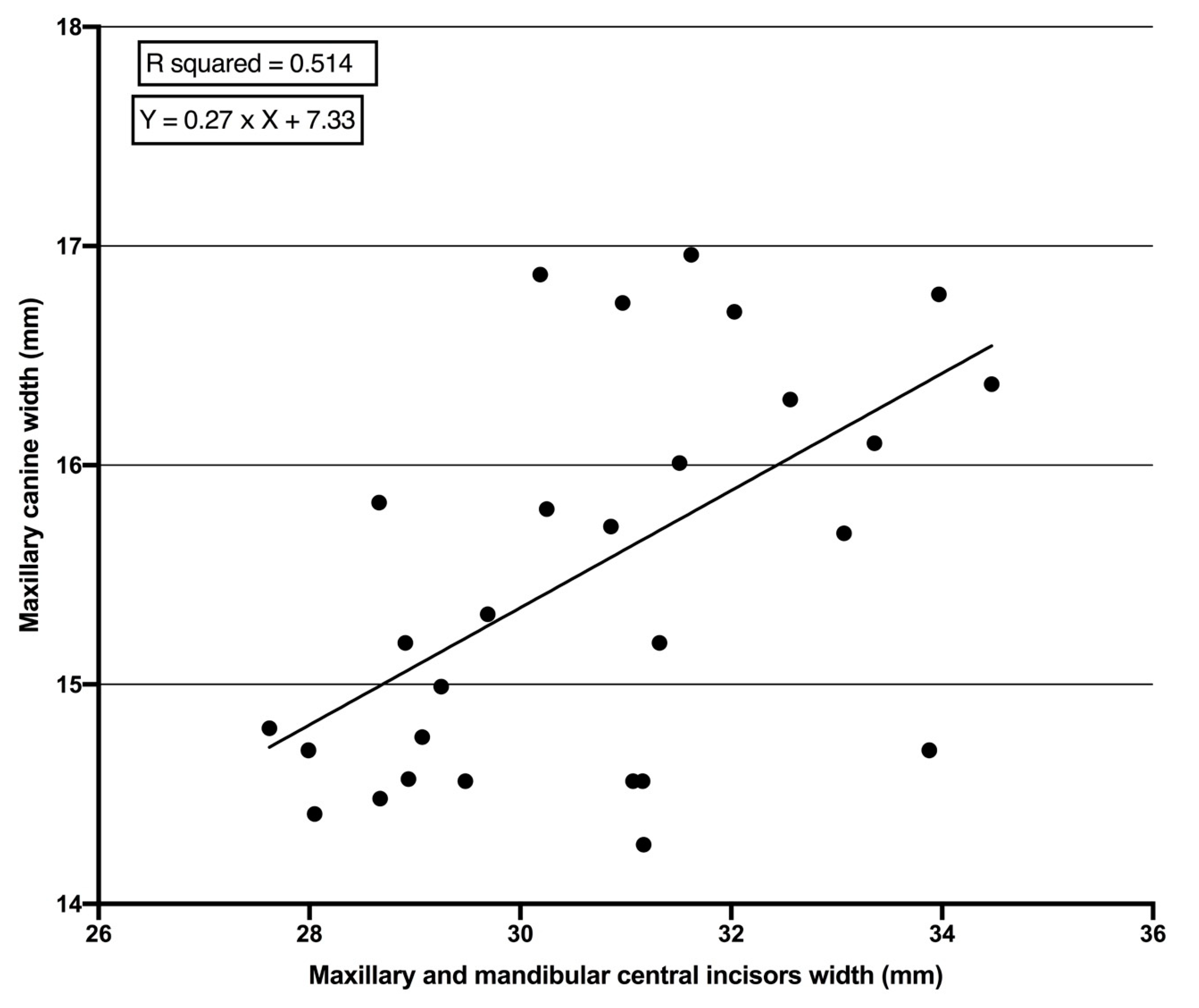
| Mesio-distal width of PMCs | Pearson Correlation | 0.633 | 0.717 |
| Sig. (2-tailed) | 0.0001 | 0.0001 | |
| Teeth Width | Width of Permanent Maxillary Canines | ||||||
|---|---|---|---|---|---|---|---|
| Gender | R | R Square | Std. Errors of the Estimate | 95% CI | t | p Value | |
| Permanent maxillary and mandibular central incisors | Male | 0.633 | 0.403 | 0.059 | 0.167–0.386 | 5.07 | 0.0001 |
| Female | 0.717 | 0.514 | 0.049 | 0.169–0.366 | 5.46 | 0.0001 | |
© 2020 by the author. Licensee MDPI, Basel, Switzerland. This article is an open access article distributed under the terms and conditions of the Creative Commons Attribution (CC BY) license (http://creativecommons.org/licenses/by/4.0/).
Share and Cite
Kareem, F.A. Permanent Maxillary and Mandibular Central Incisor Width as Predictor of Permanent Maxillary Canine Width in a Kurdish Population: A Pilot Study. Children 2020, 7, 92. https://doi.org/10.3390/children7080092
Kareem FA. Permanent Maxillary and Mandibular Central Incisor Width as Predictor of Permanent Maxillary Canine Width in a Kurdish Population: A Pilot Study. Children. 2020; 7(8):92. https://doi.org/10.3390/children7080092
Chicago/Turabian StyleKareem, Fadil Abdullah. 2020. "Permanent Maxillary and Mandibular Central Incisor Width as Predictor of Permanent Maxillary Canine Width in a Kurdish Population: A Pilot Study" Children 7, no. 8: 92. https://doi.org/10.3390/children7080092