


Children 2023, 10(11), 1779; https://doi.org/10.3390/children10111779 - 02 Nov 2023
Viewed by 861
Abstract
►
Show Figures
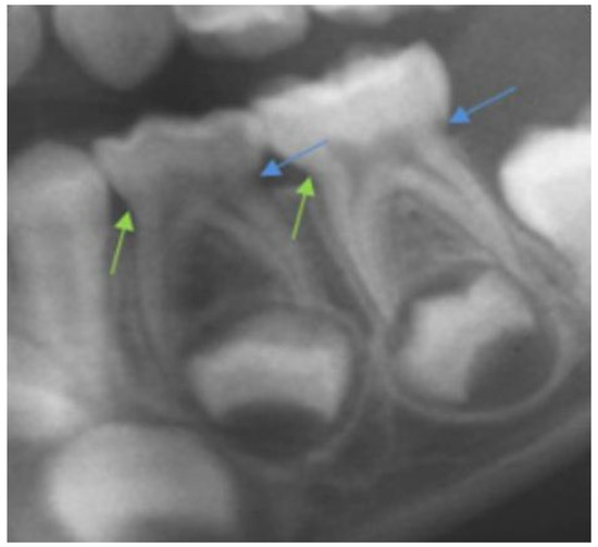
The role of diet in shaping oral microbiota and its potential contribution to the development of periodontal pathogens cannot be understated. This study aimed to explore the correlation between dietary habits and the prevalence of 11 periodontal pathogens among children and adolescents in
[...] Read more.
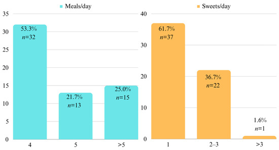
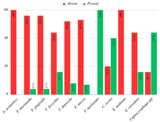
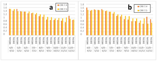
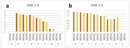
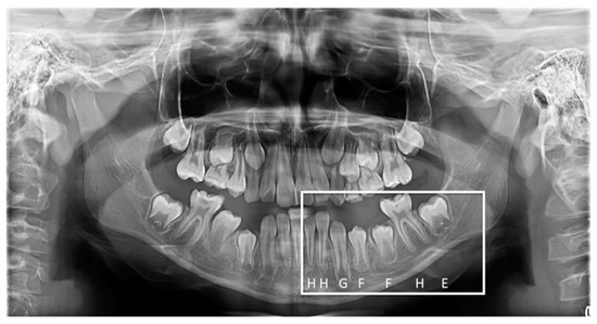
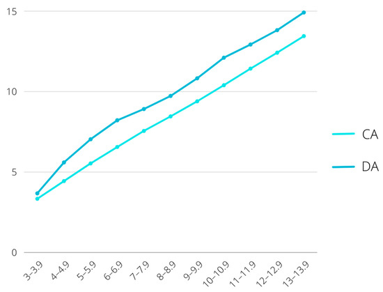
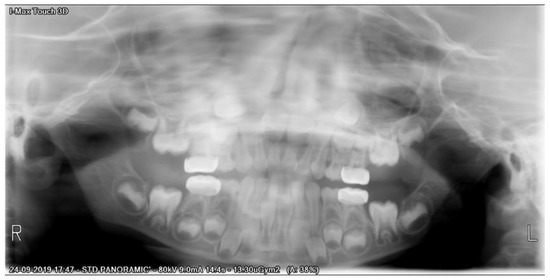
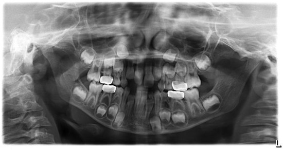
The role of diet in shaping oral microbiota and its potential contribution to the development of periodontal pathogens cannot be understated. This study aimed to explore the correlation between dietary habits and the prevalence of 11 periodontal pathogens among children and adolescents in Oradea, Romania. The identification of these pathogens was performed using the micro-IDent test kit, capable of detecting 11 specific periodontal pathogens. Bacterial sampling was conducted from the crevicular fluid in the morning, prior to brushing, followed by the completion of a brief questionnaire by parents. The questionnaire captured various aspects of the children’s eating habits, including meal frequency, consumption of sweets, and hydration levels. The collected samples were dispatched to the laboratory for analysis, which provided insights into the abundance of microorganisms. The study encompassed 60 participants aged between 2 and 18 years, with the majority reported by their parents to have regulated meal timings, frequent sugar intake, and adequate hydration. The findings revealed significant associations between certain dietary factors and the presence of specific periodontal pathogens. Notably, the absence of breastfeeding was linked with the detection of Tannerella forsythia and Campylobacter rectus. Furthermore, frequent consumption of sweets corresponded with the presence of Capnocytophaga spp., which was particularly observed in individuals consuming sweets 2–3 times a day. Insufficient age-appropriate hydration showed an association with the prevalence of T. forsythia, Peptostreptococcus micros, and Capnocytophaga spp. In this sample, it became evident that eating habits and diet influenced the presence of several periodontal pathogens. The lack of breastfeeding was predominantly associated with positive results for T. forsythia and C. rectus, while inadequate hydration correlated more frequently with the presence of T. forsythia and P. micros. Moreover, frequent consumption of sweets was linked to the presence of Capnocytophaga spp.
Full article
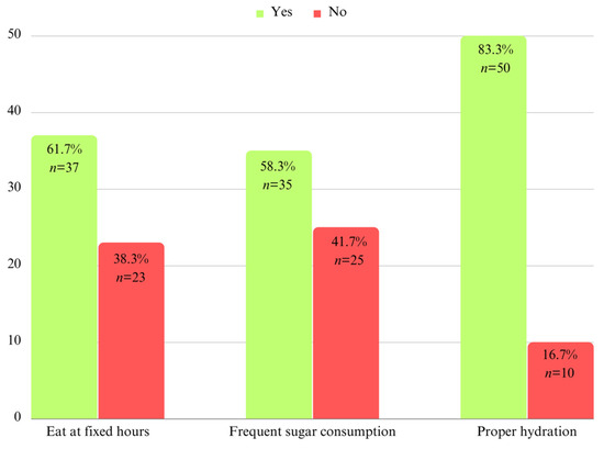
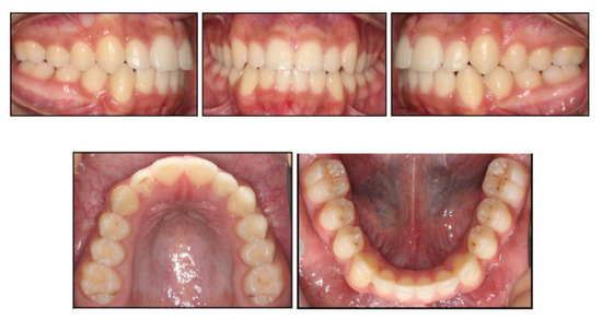
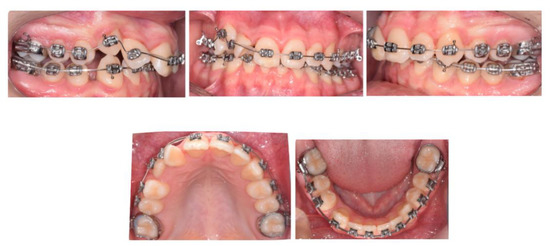
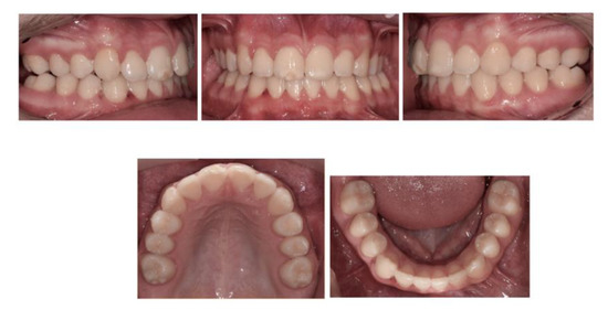
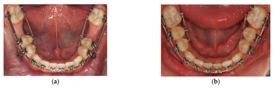
Figure 1


