Article

Elevated Urinary Hepcidin Level and Hypoferremia in Infants with Febrile Urinary Tract Infection: A Prospective Cohort Study

1
Department of Pediatrics, College of Medicine, Kaohsiung Chang Gung Memorial Hospital, Chang Gung University, Kaohsiung 83301, Taiwan
2
School of Medicine, College of Medicine, Chang Gung University, Linkou 33302, Taiwan
3
Antai Medical Care Corporation, Antai Tian-Sheng Memorial Hospital, Pingtung 92842, Taiwan
4
School of Traditional Chinese Medicine, College of Medicine, Chang Gung University, Linkou 33302, Taiwan
*
Author to whom correspondence should be addressed.
Children 2023, 10(5), 870; https://doi.org/10.3390/children10050870
Submission received: 16 April 2023 / Revised: 9 May 2023 / Accepted: 10 May 2023 / Published: 12 May 2023
(This article belongs to the Section Pediatric Neonatology)

Abstract

:
To evaluate the kinetics of serum and urinary hepcidin levels along with anemia-related parameters during the infection course of infants with febrile urinary tract infection (UTI), we enrolled febrile infants aged one to four months in this prospective study. Febrile patients with UTI were allocated into Escherichia coli (E. coli) or non-E. coli groups according to urine culture results. Septic workup, blood hepcidin, iron profile, urinalysis, and urinary hepcidin–creatinine ratio were collected upon admission and 3 days after antibiotic treatment. In total, 118 infants were included. On admission, the febrile UTI group showed a significant reduction in serum iron level and a significant elevation of urinary hepcidin–creatinine ratio compared to the febrile control counterpart. Moreover, urinary hepcidin–creatinine ratio had the highest odds ratio, 2.01, in logistics regression analysis. After 3 days of antibiotic treatment, hemoglobin and the urinary hepcidin–creatinine ratio were significantly decreased. Patients with an E. coli UTI had a significantly decreased urinary hepcidin–creatinine ratio after 3 days of antibiotics treatment, whereas the non-E. coli group showed insignificant changes. Our study suggested that the urinary hepcidin–creatinine ratio elevated during acute febrile urinary tract infection and significantly decreased after 3 days of antibiotics treatment, especially in E. coli UTI.

1. Introduction

Hepcidin, an antimicrobial peptide first discovered and isolated from urine [1], is produced majorly in the liver and plays a key role in the homeostasis of iron. Hepcidin inhibits iron release from senescent erythrocytes in macrophages, blocks duodenal iron absorption, and causes iron sequestration in the reticuloendothelial system [2]. During infection, hepcidin expression is upregulated by cytokines such as interleukin-6 (IL-6), leading to the characteristic hypoferremia and hyperferritinemia seen in anemia of inflammation. Although hepcidin is a principal regulator in the development of anemia associated with inflammation and infection, there are only a few clinical studies that investigate the kinetics of hepcidin and iron measurements in patients with bacterial infection [3].
Urinary tract infection (UTI) is one of the most common bacterial infectious diseases in young infants. The incidence of UTI in infants <3 months of age for girls is 7.5%, and for uncircumcised boys, 20.7% [4]. Diagnosis of urinary tract infection requires positive urinalysis and urine culture results, in combination with associated history and physical exam findings [5,6]. However, signs and symptoms of UTI in infants are nonspecific, such as fever, vomiting, lethargy, and irritability. Urine culture as the essential evidence for diagnosis of UTI, requires 2–3 days for detection of the responsible microorganism, and is not available in emergencies. In addition, prior studies have demonstrated that the absence of urine white blood cells (WBC) or leucocyte esterase is a common occurrence, observed in at least 10% of pediatric UTI cases [7]. This is particularly evident when the causative organism is not E. coli [8]. The conversion of nitrate to nitrite typically requires a four-hour incubation period, rendering nitrite an unreliable marker of UTI in infants, who frequently empty their bladders. Additionally, it is worth noting that not all urinary pathogens possess the ability to reduce nitrate to nitrite, further limiting the usefulness of this diagnostic tool [9].
The urinary tract provides host innate immune defenses through the restriction of iron availability [10,11] and antimicrobial peptides such as defensin, cathelicidin, Tamm-Horsfall protein, lactoferrin, and lipocalin [12]. Although hepcidin also exhibits antimicrobial activity as well and is freely filtered by the glomerulus, few clinical studies have focused on its role in the local innate defense mechanism in the urinary tract system. Recent studies have shed light on the clinical relevance of urinary hepcidin mitigating acute kidney injury by promoting iron sequestration and thereby limiting oxidative stress, free radical damage, and renal injury in cardiac surgery patients [13,14].
UTI is primarily caused by ascending infection rather than hematogenous spread in infants, with Escherichia coli (E. coli) accounting for approximately 80 percent of cases [15]. In animal studies, urinary hepcidin has been identified as an effective renal antibacterial defense against uropathogenic E. coli through the regulation of iron availability, triggering of urinary acidic pH, and direct bacteriostatic action to counter E. coli infection [16]. However, clinical studies on the role of urinary hepcidin in infants with E. coli UTI are scarce. Hence, the present study aimed to investigate the kinetic changes of serum and urinary hepcidin levels and their relationship with other anemia-associated parameters in infants with febrile UTI. Moreover, we sought to identify potential clinical clues concerning the role of urinary hepcidin in infants with E. coli UTI.

2. Materials and Methods

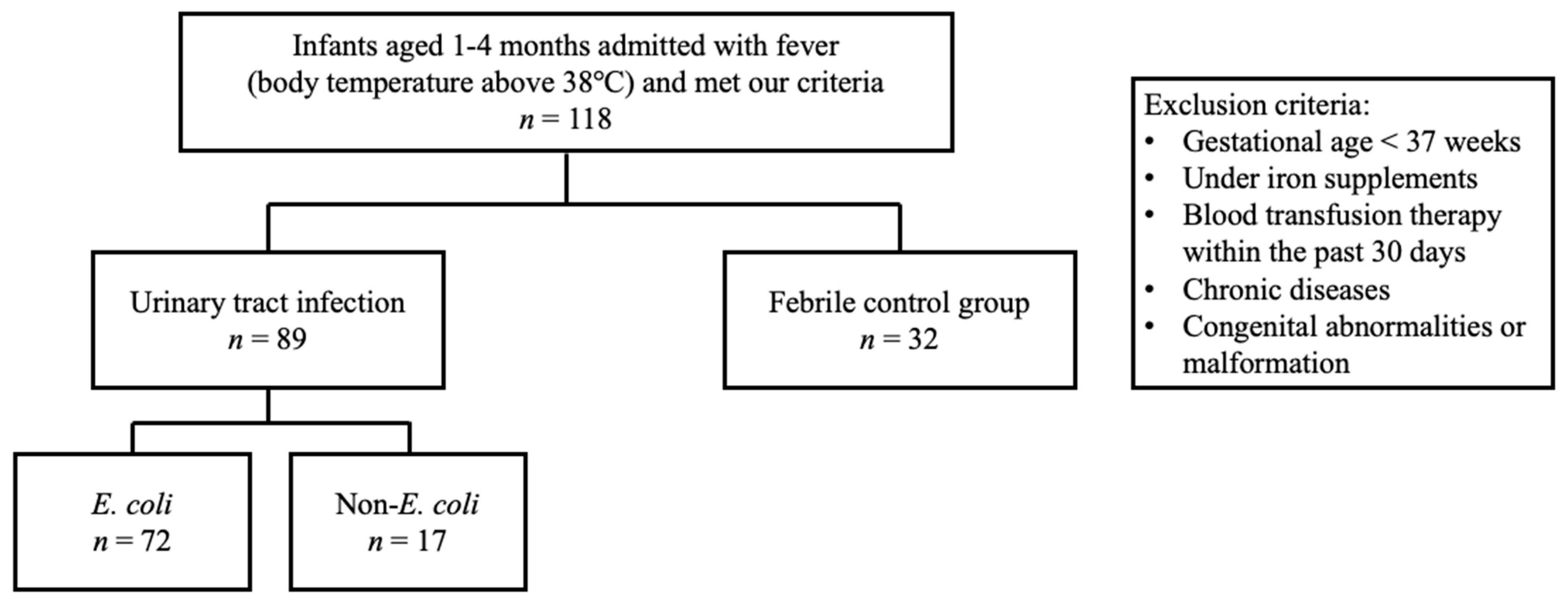
2.1. Patient Population

This prospective cohort study recruited febrile infants aged 1 to 4 months who were admitted through the pediatric emergency department of a level III hospital between December 2017 and February 2021. We specifically selected infants in this age group to ensure a relatively homogenous population, taking into consideration the known age-related difference in hepcidin [17,18]. In addition, we excluded neonates with UTI as they are often associated with congenital anomalies of the kidney and urinary tract (CAKUT) [19,20]. Furthermore, neonates were not included as changes in Hb levels in this group may be influenced by factors such as polycythemia, hemodilution with somatic growth, alloimmune hemolytic disease of the newborn, or nutritional anemia. The number of infants included was determined by G power software with effect size of 0.6 and power of 0.8. Infants born prematurely, receiving iron supplements, undergoing blood transfusion therapy within the past 30 days, and having documented chronic diseases or congenital abnormalities or malformation were excluded from this study. Based on urine culture results, the febrile UTI group was further categorized into Escherichia coli and non-E. coli subgroups. The flow chart of inclusion and exclusion criteria is shown in Figure 1.
Febrile infants with negative results on urine and blood culture samples were recruited as the febrile control group. Virus isolation results revealed etiology as parainfluenza, respiratory syncytial virus, adenovirus, or enterovirus infection in the febrile control group. Empirical antibiotics were given to all febrile infants for the first three days following admission before urine or blood culture identified or excluded bacterial infection. In addition, 10 healthy infants aged 1 month to 4 months old were recruited to evaluate the urinary hepcidin–creatinine ratio to increase the intensity of the results.

2.2. Laboratory Tests and Examinations

Patients’ baseline characteristics were recorded, including age at admission, sex, the occurrence of bacteremia, and preadmission fever duration. Blood analysis included complete blood count with white blood count (WBC) differential count (CBC/DC), serum C-reactive protein (CRP), procalcitonin (PCT), iron, ferritin, total iron binding capacity (TIBC), hepcidin, and blood culture. Urinalysis included measurement of the microscopic exam, urine culture, hepcidin, and creatinine. First blood and urine samples were collected for analysis in the emergency department before initiation of antibiotic treatment. Three days after antibiotic treatment, second blood and urine collection for CBC/DC, serum CRP, PCT, urinalysis, urine culture, hepcidin, and creatinine were performed. Serum and urine samples were stored at −80 °C before analysis, and hepcidin 25-isoforms were measured by using a commercial enzyme-linked immunosorbent assay kit (Catalog Number MBS2516119, Detection Range 1.56–100 ng/mL, MyBioSource, Inc., San Diego, CA, USA) according to the manufacturer’s instructions. The intra- and interassay coefficients of variability (CV) for hepcidin were less than 6.3% and 5.3% respectively. Considering the variation in urine dilution following intravenous fluid administration, urinary hepcidin concentrations were normalized to urinary creatinine. Due to parents’ concerns about the invasive nature of suprapubic aspiration and urinary catheterization, all urine specimens were obtained via a sterile catch-bag method. Urinary tract infection was diagnosed when 100,000 or more colony-forming units (CFU)/mL of a single bacterial strain were detected on the urine culture combined with a positive dipstick and microscopic urinalysis [21].

2.3. Statistical Analysis

All parameters were compared between febrile UTI and control groups and E. coli and non-E. coli groups. Categorical and continuous data were analyzed by Chi-square or Fisher’s exact test and Student’s t-test, respectively. In addition, the urinary hepcidin–creatinine ratios of 10 healthy, 10 febrile control, and 10 febrile UTI infants were compared by Kruskal–Wallis Test. Blood and urinary biomarkers on admission and 3 days after antibiotic treatment were compared by paired t-test in each group. Univariate and multivariate logistic regression, receiver operating characteristic (ROC) curve analysis, and Spearman’s rank correlation coefficient were used for further analysis of the variables. All statistical tests were performed using IBM SPSS 25 statistics software (IBM Corp., Armonk, NY, USA), and p < 0.05 was considered statistically significant.

3. Results

3.1. Characteristics of Study Population

We recruited a total of 118 febrile infants aged one to four months in this study. Eighty-six infants were diagnosed with febrile UTI, and thirty-two febrile infants with negative blood, urine, and stool culture results were included in the febrile control group. Among the febrile UTI patients, there were 69 infants with E. coli UTI, 17 with non-E. coli UTI, and 6 with bacteremia. The baseline demographic and lab data of the febrile UTI group and control group are shown in Table 1. No significant differences were found in age, sex, fever duration on admission, or incidence of bacteremia between the two groups.

3.2. Analysis between the Febrile UTI and Febrile Control Groups

The first analysis focused on laboratory data between the febrile UTI and febrile control groups (Table 2). On admission, the febrile UTI group had higher WBC, absolute neutrophil count (ANC), serum CRP, procalcitonin, urine WBC and red blood cell (RBC), and urinary hepcidin–creatinine ratio than the fever control group (p < 0.001, <0.001, <0.001, 0.008, <0.001, 0.001, and 0.016, respectively). However, a significant reduction in serum iron level was seen in the febrile UTI group compared to the febrile control counterpart (25.03 ± 9.72 vs. 48.55 ± 25.41 mcg/dL, p < 0.001). After 3 days of antibiotics treatment, WBC, ANC, RBC, hemoglobin (Hb), mean cell volume (MCV), serum CRP, and urinary hepcidin–creatinine ratio were all showing reduction in the febrile UTI group (p < 0.001, <0.001, 0.001, <0.001, <0.001, <0.001, and 0.009, respectively). In the fever control group, only ANC had a significant reduction after 3 days of antibiotic treatment. Insignificant differences in serum hepcidin levels before or after antibiotic treatment were observed in the febrile UTI and febrile control groups. We conducted univariate logistic regression analysis, and the urinary hepcidin–creatinine ratio had an odds ratio of 2.01 (95% confidence interval: 1.089–3.720). In multivariate analysis, there was no significant difference between WBC, CRP, and urine hepcidin before antibiotic treatment. ROC curve analysis demonstrated that the area under the curve for serum WBC, CRP, and urine hepcidin was 0.931, 0.894, and 0.894, respectively. Further ROC curve analysis did not show any significant difference in performance between these variables. Spearman’s correlation coefficient analysis showed a moderate correlation between urine hepcidin and serum WBC before antibiotic treatment (p = 0.037).

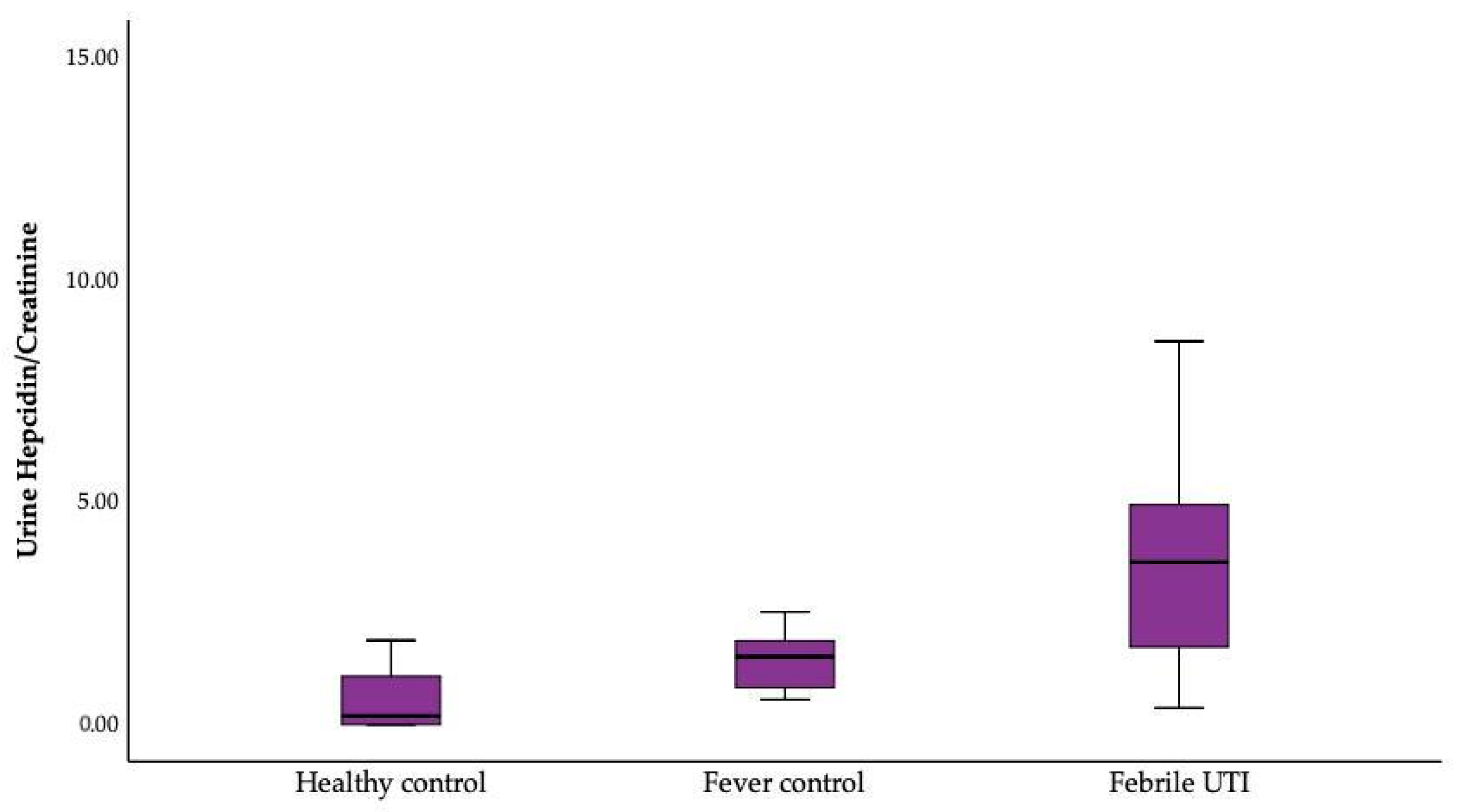
Analysis between the Healthy, Febrile Control, and Febrile UTI Groups

The urinary hepcidin–creatinine ratio was significantly different between the healthy, febrile control, and febrile UTI groups (p = 0.001). An increasing trend was observed from healthy to febrile UTI groups (Figure 2).

3.3. Analysis between the E. coli and Non-E. coli Groups

The relationship between urinary hepcidin–creatinine ratio and the microbial etiology in the febrile UTI group is shown in Table 3. Among febrile UTI patients, 69 yielded E. coli on urine culture and 17 non-E. coli infection, including Klebsiella pneumonia, Group B Streptococcus, and Enterococcus. On admission, parameters in blood and urine were not significantly different in the two groups, except hematuria was more significant in the E. coli group than non-E. coli groups. Hemoglobin in both E. coli and non-E. coli UTI groups significantly decreased after 3 days of antibiotic treatment (p < 0.001 and p = 0.040, respectively), and patients with E. coli UTI had a significant decrease in urinary hepcidin–creatinine ratio (p = 0.030) whereas the non-E. coli group showed insignificant change. A difference in serum hepcidin was not found between E. coli and non-E. coli groups.

4. Discussion

In this prospective observational study, clinical pediatric data on serum and urinary hepcidin before and after 3 days of antibiotic treatment and their association with anemia-related parameters in febrile UTI were analyzed. Focusing on young infants, patients with febrile UTI had elevated urinary hepcidin–creatinine ratios compared to the non-UTI group on admission which decreased 3 days after antibiotic treatment. Moreover, hypoferremia was noted upon emergency department admission and developed a decreasing trend of Hb level as early as 3 days after antibiotic treatment. Further analysis comparing E. coli and non-E. coli UTI showed a significant decreasing trend of urinary hepcidin–creatinine ratio 3 days after antibiotic treatment in the E. coli UTI group.
The primary aim of our study was to investigate the occurrence of anemia of inflammation and its association with hepcidin in the infant population. We found elevated serum hepcidin level in both the febrile UTI and febrile control group, but evidence of anemia of inflammation were only seen in febrile UTI patients, such as concurrent decreased Hb levels and hypoferremia. This may reflect the host’s iron sequestration strategy in response to the bacterial invasion. Iron is vital for many functional proteins involving oxygen transport and energy production and thus an essential component for survival among humans and nearly all organisms [22]. During acute infection, microbes have evolved strategies to obtain iron from the host. Meanwhile, human innate immunity has evolved defense mechanisms to restrict the accessibility of iron from pathogens such as decreased absorption of dietary iron and release of iron from macrophages, hepatocytes, and enterocytes by upregulating hepcidin, a disulfide-rich antimicrobial peptide [23]. The induction of hepcidin expression during inflammation can result in a rapid reduction in serum iron concentrations, and persistent high levels of hepcidin and hypoferremia may lead to anemia of inflammation [22]. However, we did not observe decreased Hb levels on admission in this study. According to previous research, estimated lifespan of neonatal erythrocytes was around 54.2 days [24]. If erythropoiesis is abated by hypoferremia, it would take approximately five days in theory to reduce Hb levels by 10%, a decrease to be considered clinically relevant and detectable. Because all patients in this study were admitted with fever less than one day, a difference in Hb levels was not detectable on admission or three days afterward. However, a decreasing trend in Hb levels was observed in the febrile UTI group three days after admission, which could potentially lead to anemia if we followed the hemogram further on. These results are consistent with previous studies by Kossiva et al. [3] and van Eijk et al. [25], both of which showed a decrease in Hb levels following hepcidin elevation during infection.
Despite observing concurrent elevated serum hepcidin, decreased serum iron level, and decreasing trend in Hb levels, our Spearman’s correlation coefficient analysis did not identify a significant correlation between serum hepcidin levels and serum iron levels or Hb levels in either the febrile UTI group or febrile control group. These results suggest that while hepcidin may be involved in the regulation of iron metabolism during infection in infancy, its relationship with serum iron and Hb levels may be complex and multifactorial. Indeed, previous studies have reported a hepcidin-independent mechanism of hypoferremia under inflammatory stimulation [26,27]. Both bacterial and viral infection would elevate serum hepcidin levels, while hypoferremia has only been seen in bacterial infection [3,28,29]. Consistent with previous studies, no significant difference in serum hepcidin but significant hypoferremia and associated anemia were found in febrile UTI in this study.
The production of hepcidin in hepatocytes is stimulated by various factors, including high extracellular iron level or infectious or inflammatory stimulus. Elevated serum hepcidin level in combination with Hb-for-age Z-score were found to be predictive for Kawasaki disease [30]. In patients with acute leukemia or undergoing hematopoietic cell transplantation who frequently received red blood cell concentrate transfusions, serum ferritin and hepcidin levels were upregulated at the same time by systemic iron overload [31]. Furthermore, higher levels of serum hepcidin level was noted in obese children and adolescents, which may suggest a low-grade inflammatory state and may reflect in increased iron content in these patient groups [32].
Our study demonstrated concurrent elevation of serum and urinary hepcidin on admission, but only urinary hepcidin showed significant decrease three days after antibiotic treatment, while serum hepcidin remained elevated. Similarly, serum WBC and CRP levels also had a significant decrease three days after antibiotic treatment, but only serum WBC before antibiotic treatment showed moderate correlation with urinary hepcidin (p = 0.037). No significant correlation was observed between the changes of CRP and changes of urinary hepcidin. Additionally, we explored the relationship of urinary hepcidin with anemia-related parameters. Despite a decreasing trend in Hb levels similar to that seen in urinary hepcidin levels and serum iron levels that decreased significantly on admission, we did not find a statistically significant correlation between urinary hepcidin and either Hb or serum iron. These findings were compatible with previous studies in Crohn’s disease and older persons with proinflammatory states, which reported no correlation between urinary hepcidin and CRP [33,34]. In addition, a study in anemic school children with Schistosoma haematobium demonstrated a significant correlation between urinary hepcidin and CRP but not Hb levels [35].
Hepcidin is a small protein, and after filtration across the glomerulus, it is almost completely reabsorbed in the proximal tubule. Its urinary faction excretion is estimated to be between 0% and 3% [36]. In addition, researchers found renal expression of hepcidin at the apical pole of the thick ascending limb and collecting tubules [37]. This indicates that hepcidin may be synthesized locally in the kidney and then released into the urine. Clinical studies on the relevance of urinary hepcidin are just emerging. In adult cardiac surgery cohorts, urinary hepcidin was evaluated as a diagnostic biomarker for acute kidney injury, and its elevation discriminated patients who did not develop acute kidney injury. In our study, an elevation of urinary hepcidin–creatinine ratio in the febrile UTI group on admission was found. We explored the ability of urine hepcidin in predicting UTI among febrile infants, and ROC curve analysis did not show significant difference in performance between urine hepcidin, serum WBC, and CRP. This suggests that urine hepcidin may not be inferior to the other two makers in predicting UTI.
Possible causes of the elevated urinary hepcidin–creatinine ratio in UTI may be due to decreased tubular reabsorption, or local synthesis, or both. Moreover, hematuria was also more prominent in the febrile UTI group. Red blood cells in the urine release Hb and heme-related products. Once taken up by tubular cells, Hb dissociates and releases the heme group, which induces oxidative stress, cell death, and inflammation. It is unknown whether local renal hepcidin exerts protective effects in face of the oxidative stress of hematuria in febrile UTI patients.
Hepcidin, as an antimicrobial peptide, has broad antimicrobial activity against Gram-positive bacteria (e.g., Staphylococcus aureus, Staphylococcus epidermidis, Enterococcus faecium), Gram-negative bacteria (e.g., Pseudomonas aeruginosa, Escherichia coli, Acinetobacter baumannii), and fungi (e.g., Candida albicans and Saccharomyces cerevisiate) without cytotoxicity in mammalian cells [38,39,40]. A previous animal study revealed that hepcidin could be an effective component of renal defense systems against uropathogenic E. coli infection via the mechanisms of urinary iron restriction, urine acidification, renal inflammatory response, and its antimicrobial activity [16]. Additionally, uropathogenic E. coli could repress renal hepcidin production 24 h post-E. coli infection as a strategy to evade the antibacterial activity of hepcidin [16], which could have clinical relevance. In our results, the urinary hepcidin–creatinine ratio decreased more in the E. coli UTI group 3 days after antibiotic treatment than that of the non-E. coli UTI group. We postulated that this was likely caused by E. coli locally repressing renal hepcidin synthesis as an escape strategy against human innate immunity. In contrast, urine cultures 3 days after antibiotics treatment all yielded negative results. This may indicate that decreased E. coli bacterial load leads to decreased urinary hepcidin–creatinine ratios. However, further animal studies will be required to clarify the relationship between bacterial load and urinary hepcidin level.
To our knowledge, no prior studies have examined the phenomenon of anemia of inflammation and its association with hepcidin levels in the infant population. Serum and urinary hepcidin levels and Hb were investigated simultaneously to detect whether there is an interrelationship between them. Pediatric studies defining reference ranges for serum and urinary hepcidin are limited. Previous studies showed age differences in hepcidin levels, and Aranda et al. found that the hepcidin level increased in healthy infants during the first year of life [18]. In our prospective study with serial samples, we used a febrile age-matched control group to minimize the potential impact of age on hepcidin levels. We recruited a healthy control group to evaluate the impact of infection on hepcidin levels as well. Nevertheless, the study has several limitations. One limitation is that the urine samples were collected via perineal bags, not via urine catheter or suprapubic puncture, and may be more easily contaminated. However, only the urine culture results with 100,000 or more CFU/mL of a single bacterial strain combined with pyuria in urinalysis were considered UTI in this study. Furthermore, compared with the procedure of perineal bags, suprapubic puncture is more invasive and, though rare, may lead to complications, e.g., bowel or large vessel puncture, leakage of urine into the tissue, and abscess formation, all which are great concerns to the parents and health care providers [41]. Moreover, invasive procedures may impose stress on children, with possible negative psychological consequences [42]. Thus, it is our policy to use perineal bag instead of suprapubic puncture to collect the urine. Furthermore, our study includes only febrile infants from a single tertiary hospital, and future studies conducted in a multicenter setting may be warranted.

5. Conclusions

The urinary hepcidin–creatinine ratio had the highest augmentation during the acute infection in febrile UTI patients, especially in E. coli UTI. Significant hypoferremia and a decreasing trend of Hb were also found in these patients. Thus, urinary hepcidin might play a pathological role in anemia of febrile UTI infants. We identified the occurrence of anemia and an additive value of urinary hepcidin in infants with UTI. Future research to investigate the diagnostic and prognostic values of urinary hepcidin will be necessary.

Author Contributions

Conceptualization, H.-C.H. (Hsin-Chun Huang) and I.-L.C.; Data curation, Y.-H.H. and I.-L.C.; Formal analysis, I.-L.C.; Funding acquisition, Y.-C.H., H.-C.H. (Hsin-Chun Huang) and I.-L.C.; Investigation, I.-L.C.; Methodology, H.-C.H. (Hui-Chen Huang) and I.-L.C.; Project administration, Y.-C.H., H.-C.H. (Hsin-Chun Huang) and I.-L.C.; Resources, Y.-C.H. and I.-L.C.; Software, I.-L.C.; Supervision, H.-C.H. (Hsin-Chun Huang) and I.-L.C.; Validation, K.-S.T., L.-T.S. and Y.-H.H.; Visualization, I.-L.C.; Writing—original draft, Y.-C.H.; Writing—review and editing, Y.-C.H. and H.-C.H. (Hsin-Chun Huang). All authors have read and agreed to the published version of the manuscript.

Funding

This research was funded by Chang Gung Memorial, grant number CMRPG8L1461.

Institutional Review Board Statement

The study was conducted in accordance with the Declaration of Helsinki and approved by the Institutional Review Board of Chang Gung Memorial Hospital (approval number 201701399A3 was approved on 7 November 2017, and 202101310B0A3 was approved on 27 August 2021).

Informed Consent Statement

Informed consent was obtained from all subjects involved in the study. Written informed consent has been obtained from the parents or legal guardians of all enrolled infants.

Data Availability Statement

Data are unavailable due to privacy or ethical restrictions.

Acknowledgments

We appreciate the Biostatistics Center, Kaohsiung Chang Gung Memorial Hospital, for the statistical work.

Conflicts of Interest

The authors declare no conflict of interest.

References

  1. Park, C.H.; Valore, E.V.; Waring, A.J.; Ganz, T. Hepcidin, a urinary antimicrobial peptide synthesized in the liver. J. Biol. Chem. 2001, 276, 7806–7810. [Google Scholar] [CrossRef]
  2. Ganz, T.; Nemeth, E. Iron homeostasis in host defence and inflammation. Nat. Rev. Immunol. 2015, 15, 500–510. [Google Scholar] [CrossRef]
  3. Kossiva, L.; Soldatou, A.; Gourgiotis, D.I.; Stamati, L.; Tsentidis, C. Serum hepcidin: Indication of its role as an “acute phase” marker in febrile children. Ital. J. Pediatr. 2013, 39, 25. [Google Scholar] [CrossRef]
  4. Robinson, J.L.; Finlay, J.C.; Lang, M.E.; Bortolussi, R. Urinary tract infections in infants and children: Diagnosis and management. Paediatr. Child. Health 2014, 19, 315–325. [Google Scholar] [CrossRef]
  5. Mattoo, T.K.; Shaikh, N.; Nelson, C.P. Contemporary Management of Urinary Tract Infection in Children. Pediatrics 2021, 147, e2020012138. [Google Scholar] [CrossRef]
  6. National Collaborating Centre for Women’s and Children’s Health. Urinary Tract Infection in Children: Diagnosis, Treatment and Long-Term Management; National Collaborating Centre for Women’s and Children’s Health: London, UK; RCOG Press: London, UK, 2007. [Google Scholar]
  7. Williams, G.J.; Macaskill, P.; Chan, S.F.; Turner, R.M.; Hodson, E.; Craig, J.C. Absolute and relative accuracy of rapid urine tests for urinary tract infection in children: A meta-analysis. Lancet Infect. Dis. 2010, 10, 240–250. [Google Scholar] [CrossRef]
  8. Shaikh, N.; Shope, T.R.; Hoberman, A.; Vigliotti, A.; Kurs-Lasky, M.; Martin, J.M. Association Between Uropathogen and Pyuria. Pediatrics 2016, 138, e20160087. [Google Scholar] [CrossRef]
  9. Chaudhari, P.P.; Monuteaux, M.C.; Bachur, R.G. Should the Absence of Urinary Nitrite Influence Empiric Antibiotics for Urinary Tract Infection in Young Children? Pediatr. Emerg. Care 2020, 36, 481–485. [Google Scholar] [CrossRef]
  10. Flo, T.H.; Smith, K.D.; Sato, S.; Rodriguez, D.J.; Holmes, M.A.; Strong, R.K.; Akira, S.; Aderem, A. Lipocalin 2 mediates an innate immune response to bacterial infection by sequestrating iron. Nature 2004, 432, 917–921. [Google Scholar] [CrossRef]
  11. Goetz, D.H.; Holmes, M.A.; Borregaard, N.; Bluhm, M.E.; Raymond, K.N.; Strong, R.K. The neutrophil lipocalin NGAL is a bacteriostatic agent that interferes with siderophore-mediated iron acquisition. Mol. Cell 2002, 10, 1033–1043. [Google Scholar] [CrossRef]
  12. Zasloff, M. Antimicrobial peptides, innate immunity, and the normally sterile urinary tract. J. Am. Soc. Nephrol. 2007, 18, 2810–2816. [Google Scholar] [CrossRef] [PubMed]
  13. Ho, J.; Lucy, M.; Krokhin, O.; Hayglass, K.; Pascoe, E.; Darroch, G.; Rush, D.; Nickerson, P.; Rigatto, C.; Reslerova, M. Mass spectrometry-based proteomic analysis of urine in acute kidney injury following cardiopulmonary bypass: A nested case-control study. Am. J. Kidney Dis. 2009, 53, 584–595. [Google Scholar] [CrossRef]
  14. Ho, J.; Reslerova, M.; Gali, B.; Gao, A.; Bestland, J.; Rush, D.N.; Nickerson, P.W.; Rigatto, C. Urinary hepcidin-25 and risk of acute kidney injury following cardiopulmonary bypass. Clin. J. Am. Soc. Nephrol. 2011, 6, 2340–2346. [Google Scholar] [CrossRef] [PubMed]
  15. Edlin, R.S.; Shapiro, D.J.; Hersh, A.L.; Copp, H.L. Antibiotic resistance patterns of outpatient pediatric urinary tract infections. J. Urol. 2013, 190, 222–227. [Google Scholar] [CrossRef]
  16. Houamel, D.; Ducrot, N.; Lefebvre, T.; Daher, R.; Moulouel, B.; Sari, M.A.; Letteron, P.; Lyoumi, S.; Millot, S.; Tourret, J.; et al. Hepcidin as a Major Component of Renal Antibacterial Defenses against Uropathogenic Escherichia coli. J. Am. Soc. Nephrol. 2016, 27, 835–846. [Google Scholar] [CrossRef]
  17. Sdogou, T.; Tsentidis, C.; Gourgiotis, D.; Marmarinos, A.; Gkourogianni, A.; Papassotiriou, I.; Anastasiou, T.; Kossiva, L. Immunoassay-based serum hepcidin reference range measurements in healthy children: Differences among age groups. J. Clin. Lab. Anal. 2015, 29, 10–14. [Google Scholar] [CrossRef]
  18. Aranda, N.; Bedmar, C.; Arija, V.; Jardí, C.; Jimenez-Feijoo, R.; Ferré, N.; Tous, M. Serum hepcidin levels, iron status, and HFE gene alterations during the first year of life in healthy Spanish infants. Ann. Hematol. 2018, 97, 1071–1080. [Google Scholar] [CrossRef]
  19. Bonadio, W.; Maida, G. Urinary tract infection in outpatient febrile infants younger than 30 days of age: A 10-year evaluation. Pediatr. Infect. Dis. J. 2014, 33, 342–344. [Google Scholar] [CrossRef]
  20. Goldman, M.; Lahat, E.; Strauss, S.; Reisler, G.; Livne, A.; Gordin, L.; Aladjem, M. Imaging after urinary tract infection in male neonates. Pediatrics 2000, 105, 1232–1235. [Google Scholar] [CrossRef] [PubMed]
  21. Roberts, K.B. Urinary tract infection: Clinical practice guideline for the diagnosis and management of the initial UTI in febrile infants and children 2 to 24 months. Pediatrics 2011, 128, 595–610. [Google Scholar] [CrossRef] [PubMed]
  22. Ganz, T. Anemia of Inflammation. N. Engl. J. Med. 2019, 381, 1148–1157. [Google Scholar] [CrossRef] [PubMed]
  23. Nemeth, E.; Rivera, S.; Gabayan, V.; Keller, C.; Taudorf, S.; Pedersen, B.K.; Ganz, T. IL-6 mediates hypoferremia of inflammation by inducing the synthesis of the iron regulatory hormone hepcidin. J. Clin. Investig. 2004, 113, 1271–1276. [Google Scholar] [CrossRef] [PubMed]
  24. Kuruvilla, D.J.; Widness, J.A.; Nalbant, D.; Schmidt, R.L.; Mock, D.M.; An, G.; Veng-Pedersen, P. Estimation of adult and neonatal RBC lifespans in anemic neonates using RBCs labeled at several discrete biotin densities. Pediatr. Res. 2017, 81, 905–910. [Google Scholar] [CrossRef] [PubMed]
  25. Van Eijk, L.T.; Kroot, J.J.; Tromp, M.; van der Hoeven, J.G.; Swinkels, D.W.; Pickkers, P. Inflammation-induced hepcidin-25 is associated with the development of anemia in septic patients: An observational study. Crit. Care 2011, 15, R9. [Google Scholar] [CrossRef] [PubMed]
  26. Guida, C.; Altamura, S.; Klein, F.A.; Galy, B.; Boutros, M.; Ulmer, A.J.; Hentze, M.W.; Muckenthaler, M.U. A novel inflammatory pathway mediating rapid hepcidin-independent hypoferremia. Blood 2015, 125, 2265–2275. [Google Scholar] [CrossRef]
  27. Kim, A.; Fung, E.; Parikh, S.G.; Valore, E.V.; Gabayan, V.; Nemeth, E.; Ganz, T. A mouse model of anemia of inflammation: Complex pathogenesis with partial dependence on hepcidin. Blood 2014, 123, 1129–1136. [Google Scholar] [CrossRef]
  28. Oppen, K.; Ueland, T.; Siljan, W.W.; Skadberg, Ø.; Brede, C.; Lauritzen, T.; Aukrust, P.; Steinsvik, T.; Husebye, E.; Michelsen, A.E.; et al. Hepcidin and Ferritin Predict Microbial Etiology in Community-Acquired Pneumonia. Open Forum Infect. Dis. 2021, 8, ofab082. [Google Scholar] [CrossRef]
  29. Kossiva, L.; Gourgiotis, D.I.; Tsentidis, C.; Anastasiou, T.; Marmarinos, A.; Vasilenko, H.; Sdogou, T.; Georgouli, H. Serum hepcidin and ferritin to iron ratio in evaluation of bacterial versus viral infections in children: A single-center study. Pediatr. Infect. Dis. J. 2012, 31, 795–798. [Google Scholar] [CrossRef]
  30. Yang, Y.L.; Kuo, H.C.; Chen, K.D.; Chu, C.H.; Kuo, K.C.; Guo, M.M.; Chang, L.S.; Huang, Y.H. Combination of Hemoglobin-for-Age Z-Score and Plasma Hepcidin Identified as a Novel Predictor for Kawasaki Disease. Children 2022, 9, 913. [Google Scholar] [CrossRef]
  31. Słomka, A.; Łęcka, M.; Styczyński, J. Hepcidin in Children and Adults with Acute Leukemia or Undergoing Hematopoietic Cell Transplantation: A Systematic Review. Cancers 2022, 14, 4936. [Google Scholar] [CrossRef]
  32. Gajewska, J.; Ambroszkiewicz, J.; Klemarczyk, W.; Głąb-Jabłońska, E.; Weker, H.; Chełchowska, M. Ferroportin-Hepcidin Axis in Prepubertal Obese Children with Sufficient Daily Iron Intake. Int. J. Environ. Res. Public Health 2018, 15, 2156. [Google Scholar] [CrossRef]
  33. Ferrucci, L.; Semba, R.D.; Guralnik, J.M.; Ershler, W.B.; Bandinelli, S.; Patel, K.V.; Sun, K.; Woodman, R.C.; Andrews, N.C.; Cotter, R.J.; et al. Proinflammatory state, hepcidin, and anemia in older persons. Blood 2010, 115, 3810–3816. [Google Scholar] [CrossRef]
  34. Semrin, G.; Fishman, D.S.; Bousvaros, A.; Zholudev, A.; Saunders, A.C.; Correia, C.E.; Nemeth, E.; Grand, R.J.; Weinstein, D.A. Impaired intestinal iron absorption in Crohn’s disease correlates with disease activity and markers of inflammation. Inflamm. Bowel Dis. 2006, 12, 1101–1106. [Google Scholar] [CrossRef] [PubMed]
  35. Ayoya, M.A.; Spiekermann-Brouwer, G.M.; Stoltzfus, R.J.; Nemeth, E.; Habicht, J.P.; Ganz, T.; Rawat, R.; Traoré, A.K.; Garza, C. Alpha 1-acid glycoprotein, hepcidin, C-reactive protein, and serum ferritin are correlated in anemic schoolchildren with Schistosoma haematobium. Am. J. Clin. Nutr. 2010, 91, 1784–1790. [Google Scholar] [CrossRef] [PubMed]
  36. Swinkels, D.W.; Girelli, D.; Laarakkers, C.; Kroot, J.; Campostrini, N.; Kemna, E.H.; Tjalsma, H. Advances in quantitative hepcidin measurements by time-of-flight mass spectrometry. PLoS ONE 2008, 3, e2706. [Google Scholar] [CrossRef]
  37. Kulaksiz, H.; Theilig, F.; Bachmann, S.; Gehrke, S.G.; Rost, D.; Janetzko, A.; Cetin, Y.; Stremmel, W. The iron-regulatory peptide hormone hepcidin: Expression and cellular localization in the mammalian kidney. J. Endocrinol. 2005, 184, 361–370. [Google Scholar] [CrossRef]
  38. Verga Falzacappa, M.V.; Muckenthaler, M.U. Hepcidin: Iron-hormone and anti-microbial peptide. Gene 2005, 364, 37–44. [Google Scholar] [CrossRef] [PubMed]
  39. Maisetta, G.; Petruzzelli, R.; Brancatisano, F.L.; Esin, S.; Vitali, A.; Campa, M.; Batoni, G. Antimicrobial activity of human hepcidin 20 and 25 against clinically relevant bacterial strains: Effect of copper and acidic pH. Peptides 2010, 31, 1995–2002. [Google Scholar] [CrossRef]
  40. Jiang, X.F.; Liu, Z.F.; Lin, A.F.; Xiang, L.X.; Shao, J.Z. Coordination of Bactericidal and Iron Regulatory Functions of Hepcidin in Innate Antimicrobial Immunity in a Zeb.brafish Model. Sci. Rep. 2017, 7, 4265. [Google Scholar] [CrossRef]
  41. Polnay, L.; Fraser, A.M.; Lewis, J.M. Complication of suprapubic bladder aspiration. Arch. Dis. Child. 1975, 50, 80–81. [Google Scholar] [CrossRef]
  42. Hägglöf, B. Psychological reaction by children of various ages to hospital care and invasive procedures. Acta Paediatr Suppl. 1999, 88, 72–78. [Google Scholar] [CrossRef] [PubMed]
Figure 1. The flowchart of study selection.
Children 10 00870 g001
Figure 2. Urinary hepcidin–creatinine ratio analyzed between the healthy, febrile control, and febrile UTI groups by Kruskal–Wallis Test.
Children 10 00870 g002
Table 1. The characteristics of patients with febrile urinary tract infection and fever control.
Febrile UTI
(n = 86)
Fever Control
(n = 32)
p Value
Age (day)75.03 ± 26.0776.31 ± 20.430.779
Sex (F/M)24/6513/190.113
Bacteremia (n, %)6, 7%0, 0%0.151
Fever duration at admission (day)0.84 ± 0.690.78 ± 0.520.676
Continuous variables were analyzed by Student’s t-test of variance and presented as mean ± standard deviation. Categorical variables were analyzed by the Chi-square test or Fisher’s exact test. F/M: female/male.
Table 2. The blood and urinary laboratory data before and 3 days after antibiotic treatment.
Febrile
UTI
(n = 86)
Fever
Control
(n = 32)
p ValueLogistic Regression
OR (95% C.I.)p Value
Blood Laboratory * p Value * p Value
WBC (1000/μL) <0.001 0.24
Before14.61 ± 4.808.21 ± 3.05<0.0011.46 (1.26–1.70)<0.001
After10.27 ± 3.668.96 ± 2.790.1031.14 (0.98–1.33)0.093
ANC (1000/μL) <0.001 <0.001
Before51.97 ± 13.1940.64 ± 16.50<0.0011.06 (1.03–1.09)<0.001
After23.64 ± 8.9421.65 ± 12.180.3731.02 (0.97–1.07)0.376
RBC (million/μL) 0.001 0.176
Before3.72 ± 0.573.70 ± 0.430.8331.02 (0.47–2.22)0.955
After3.64 ± 0.483.63 ± 0.460.9170.99 (0.37–2.62)0.986
Hb (g/dL) <0.001 0.083
Before10.48 ± 1.2310.53 ± 0.860.820.94 (0.66–1.33)0.717
After10.10 ± 0.9410.18 ± 0.910.7040.88 (0.54–1.44)0.619
MCV (fL) <0.001 0.13
Before85.66 ± 7.3085.96 ± 5.790.9180.99 (0.94–1.06)0.968
After83.16 ± 6.5784.80 ± 4.900.260.96 (0.89–1.04)0.282
CRP (mg/L) <0.001 0.12
Before42.55 ± 40.805.36 ± 6.45<0.0011.16 (1.08–1.25)<0.001
After12.63 ± 15.524.03 ± 4.28<0.0011.17 (1.04–1.31)0.007
Procalcitonin (ng/mL) 0.998 0.51
Before1.88 ± 4.810.13 ± 0.090.0083522.36 (10.73–1,156,353.46)0.006
After2.83 ± 10.910.12 ± 0.090.2353.65 (0.95–3028.83)0.053
Fe (ng/mL)25.03 ± 9.72 48.55 ± 25.41 <0.0010.88 (0.82–0.94)<0.001
Ferritin (ng/mL)165.38 ± 99.98233.70 ± 187.560.0940.99 (0.99–1.00)0.114
TIBC (ug/dL)294.58 ± 45.85295.10 ± 50.670.9681.00 (0.99–1.01)0.967
Hepcidin (ng/mL) 0.907 0.744
Before239.01 ± 41.23229.0 ± 35.660.511.00 (0.99–1.03)0.495
After242.65 ± 52.94239.49 ± 60.370.8931.00 (0.99–1.02)1.887
Urinary laboratory
WBC (/μL)314.57 ± 145.094.69 ± 6.81<0.0011.16 (1.063–1.261)0.001
RBC (/μL)87.12 ± 178.571.22 ± 2.730.0011.39 (1.174–1.652)<0.001
Creatinine (mg/dL) 0.257 0.698
Before11.06 ± 8.1812.92 ± 5.880.5770.97 (0.88–1.08)0.566
After11.61 ± 7.029.31 ± 6.210.4361.06 (0.91–1.24)0.43
Hepcidin/Creatinine 0.009 0.259
Before11.06 ± 6.934.22 ± 2.260.0162.01 (1.089–3.720)0.026
After7.49 ± 4.413.73 ± 2.770.0411.37 (0.997–1.886)0.052
All variables were analyzed by Student’s t-test between febrile urinary tract infection (UTI) and fever control groups, expressed as p value. Data before and after 3 days of antibiotic treatment were analyzed by paired t-test and expressed as * p value. The relationship of each variable with febrile UTI was analyzed by univariate logistic regression, expressed as odds ratio (OR) with 95% confidence interval (CI) and p value. All data are presented as mean ± standard deviation. ANC: absolute neutrophil count, C.I.: confidence interval, CRP: C-reactive protein, Hb: hemoglobin, MCV: mean cell volume, OR: odds ratio, RBC: red blood cell, TIBC: total iron binding capacity, UTI: urinary tract infection, WBC: white blood cell.
Table 3. The laboratory data before and after 3 days of antibiotic treatment in E. coli and non-E. coli urinary tract infection.
Urinary Tract Infectionp ValueLogistic Regression
E. coli
(n = 69)
Non-E. coli
(n = 17)
OR
(95% C.I.)
p Value
Blood laboratory * p value * p value
WBC (1000/μL) <0.001 0.007
Before14.96 ± 4.5013.09 ± 5.830.231.09 (0.97–1.22)0.147
After10.22 ± 3.619.66 ± 3.150.4641.07 (0.91–1.26)0.413
ANC (1000/μL) <0.001 0.001
Before52.11 ± 13.6951.42 ± 11.140.8461.00 (0.96–1.05)0.831
After24.27 ± 8.7521.79 ± 9.690.1161.06 (0.99–1.13)0.121
RBC (million/μL) 0.005 0.083
Before3.76 ± 0.553.55 ± 0.650.1751.98 (0.67–5.80)0.214
After3.69 ± 0.453.43 ± 0.550.0493.63 (0.919–14.347)0.066
Hb (g/dL) <0.001 0.04
Before10.54 ± 1.2010.22 ± 1.470.3391.23 (0.77–1.97)0.388
After10.18 ± 0.969.73 ± 0.770.0791.78 (0.90–3.53)0.1
MCV (fL) <0.001 <0.001
Before85.37 ± 7.0587.72 ± 7.790.2290.96 (0.88–1.03)0.253
After82.59 ± 6.2885.58 ± 7.390.1010.93 (0.84–1.02)0.114
CRP (mg/L) <0.001 0.001
Before42.14 ± 39.7944.25 ± 46.090.851.00 (0.99–1.01)0.797
After13.31 ± 15.999.72 ± 13.410.4081.02 (0.97–1.07)0.418
Procalcitonin (ng/mL) 0.996 0.961
Before2.11 ± 5.361.03 ± 1.310.4961.08 (0.87–1.34)0.5
After3.02 ± 11.701.61 ± 2.890.8151.02 (0.88–1.18)0.804
Fe (ng/mL)23.41 ±7.86 34.00 ± 15.12 0.2570.89 (0.79–1.00)0.068
Ferritin (ng/mL)142.86 ± 67.96289.23 ± 163.700.1710.99 (0.97–0.99)0.032
TIBC (ug/dL)287.18 ± 36.23335.25 ± 75.660.2950.98 (0.95–1.00)0.078
Hepcidin (ng/mL) 0.839 0.616
Before242.26 ± 43.34219.5 ± 23.330.4921.02 (0.97–1.06)0.463
After248.14 ± 54.80212.40 ± 37.900.4031.02 (0.98–1.06)0.377
Urinary laboratory
WBC (/μL)333.21 ± 180.24235.65 ± 208.500.0551.00 (1.00–1.00)0.065
RBC (/μL)103.11 ± 156.7419.41 ± 27.17<0.0011.02 (0.997–1.036)0.09
Creatinine (mg/dL) 0.838 0.493
Before11.15 ± 8.6910.52 ±5.100.891.01 (0.88–1.16)0.884
After10.90 ± 6.1215.68 ±11.250.2150.92 (0.80–1.05)0.224
Hepcidin/Creatinine 0.03 0.102
Before11.17 ± 7.2010.42 ± 5.890.8471.02 (0.86–1.20)0.84
After7.84 ± 4.665.46 ± 1.720.3291.17 (0.86–1.61)0.322
All variables were analyzed by Student’s t-test between E. coli urinary tract infection (UTI) and non-E. coli UTI groups, expressed as p value. Data before and after 3 days of antibiotic treatment were analyzed by paired t-test and expressed as * p value. The relationship of each variable with E. coli UTI was analyzed by univariate logistic regression, expressed as odds ratio (OR) with 95% confidence interval (CI) and p value. All data are presented as mean ± standard deviation. ANC: absolute neutrophil count, C.I.: confidence interval, CRP: C-reactive protein, Hb: hemoglobin, MCV: mean cell volume, OR: odds ratio. RBC: red blood cell, TIBC: total iron binding capacity, UTI: urinary tract infection, WBC: white blood cell.
Disclaimer/Publisher’s Note: The statements, opinions and data contained in all publications are solely those of the individual author(s) and contributor(s) and not of MDPI and/or the editor(s). MDPI and/or the editor(s) disclaim responsibility for any injury to people or property resulting from any ideas, methods, instructions or products referred to in the content.

