Article

Impaired Response to Mismatch Novelty in the Li2+-Pilocarpine Rat Model of TLE: Correlation with Hippocampal Monoaminergic Inputs

by
Carlos Nascimento
1,
Vasco Guerreiro-Pinto
2,
Seweryn Pawlak
2,
Ana Caulino-Rocha
2,
Laia Amat-Garcia
2 and
Diana Cunha-Reis
2,3,*
1
Unidade de Fisiologia Clínica e Translacional, Instituto de Medicina Molecular João Lobo Antunes, Faculdade de Medicina, Universidade de Lisboa, Av. Prof. Egas Moniz, 1649-028 Lisboa, Portugal
2
BioISI—Biosystems & Integrative Sciences Institute, Faculdade de Ciências, Universidade de Lisboa, Campo Grande, 1749-016 Lisboa, Portugal
3
Departamento de Biologia Vegetal, Faculdade de Ciências, Universidade de Lisboa, Campo Grande, 1749-016 Lisboa, Portugal
*
Author to whom correspondence should be addressed.
Biomedicines 2024, 12(3), 631; https://doi.org/10.3390/biomedicines12030631
Submission received: 8 February 2024 / Revised: 6 March 2024 / Accepted: 8 March 2024 / Published: 12 March 2024

Abstract

:
Novelty detection, crucial to episodic memory formation, is impaired in epileptic patients with mesial temporal lobe resection. Mismatch novelty detection, that activates the hippocampal CA1 area in humans and is vital for memory reformulation and reconsolidation, is also impaired in patients with hippocampal lesions. In this work, we investigated the response to mismatch novelty, as occurs with the new location of known objects in a familiar environment, in the Li2+-pilocarpine rat model of TLE and its correlation with hippocampal monoaminergic markers. Animals showing spontaneous recurrent seizures (SRSs) for at least 4 weeks at the time of behavioural testing showed impaired spatial learning in the radial arm maze, as described. Concurrently, SRS rats displayed impaired exploratory responses to mismatch novelty, yet novel object recognition was not significantly affected in SRS rats. While the levels of serotonin and dopamine transporters were mildly decreased in hippocampal membranes from SRS rats, the levels on the norepinephrine transporter, tyrosine hydroxylase and dopamine-β-hydroxylase were enhanced, hinting for an augmentation, rather than an impairment in noradrenergic function in SRS animals. Altogether, this reveals that mismatch novelty detection is particularly affected by hippocampal damage associated to the Li2+-pilocarpine model of epilepsy 4–8 weeks after the onset of SRSs and suggests that deficits in mismatch novelty detection may substantially contribute to cognitive impairment in MTLE. As such, behavioural tasks based on these aspects of mismatch novelty may prove useful in the development of cognitive therapy strategies aiming to rescue cognitive deficits observed in epilepsy.

Graphical Abstract

1. Introduction

Epilepsy, a complex disease portrayed by the occurrence of recurrent, unprovoked seizures, affects over 65 million people worldwide and is one of the most common, chronic, serious neurological diseases, and a major public health burden [1,2,3]. Epilepsy is frequently allied to neurological comorbidities, such as cognitive deficits, depression, anxiety, and psychiatric disturbances [1,3,4]. Mesial temporal lobe epilepsy, one of the most prevalent forms of epilepsy, is characterised by mesial temporal lobe symptoms affecting the hippocampus, parahippocampal gyrus, and amygdala, and is frequently (in 56% of cases) linked to hippocampal sclerosis (MTLE-HS) [5]. Impaired cognitive functioning including language, executive function, and declarative memory deficits [6] are a major hallmark of MTLE-HS. A few studies have also reported that MTLE patients present attention deficits and arousal abnormalities that can greatly affect novelty processing not only by the dorsal attention network but also through the mesial temporal lobe, affecting particularly hippocampal-dependent tasks [7,8,9,10]. Since only about 11–26% of patients with MTLE-HS achieve complete seizure control under pharmacological treatment with existing multiple antiseizure drugs (ASDs), finding new therapeutic strategies to prevent progressive cognitive decline in epilepsy patients is a main priority.
Novelty is a critical trigger for episodic memory establishment with different aspects of novelty distinctively impacting hippocampal-dependent learning and synaptic plasticity. In fact, hippocampal long-term potentiation (LTP) or long-term depression (LTD) of hippocampal synaptic transmission encode different aspects of novelty acquisition [11], LTD being enabled during the location of new items or known items in novel locations, a behavioural mismatch novelty paradigm, and LTP being favoured during exploration of a new environment, key to the formation of spatial maps and memory consolidation [11,12,13,14]. Memories, and associated synaptic plasticity processes, are also affected by previous learning events (either recent or remote) by means of metaplasticity [15]. Novelty is an important trigger for these metaplastic changes. Furthermore, while spatial novelty mainly increases retrieval of an earlier acquired memory [16], mismatch novelty paradigms instead favour inhibitory avoidance learning by influencing hippocampal LTD [13].
Interestingly, mismatch novelty detection, particularly important in memory reformulation and reconsolidation, was also linked with the activation of the human hippocampal CA1 area, and is impaired in patients with hippocampal lesions [17,18]. Furthermore, studies from our group showed that repeated exposure to mismatch novelty increases both LTP and LTD in the hippocampus [19], suggesting that behavioural tasks involving mismatch novelty features may reveal helpful in the development of cognitive therapy strategies aiming to reduce the LTP/LTD imbalance as found in aging or diseases like epilepsy or Down’s syndrome.
Several neurotransmitter systems have been implicated in the hippocampal processing and regulation of novelty stimuli. Among these, monoaminergic projections from the ventral tegmental area (VTA), locus coeruleus, and median raphe to the hippocampus have been implicated in regulating physiological arousal, attention, and motivation, and are thought to play an essential role in the efficiency of cognitive function [20,21,22,23] while playing a crucial role in recognition memory and novelty signalling [23]. Altered monoaminergic neurotransmission not only constitutes a risk factor for the development of epilepsy [24] but is either linked to the degeneration or upregulation of ascending projections to the hippocampus and cortex, altered neurotransmitter levels or altered monoamine receptor levels, and function has been reported in the hippocampus of MTLE patients [25]. Similar observations occurred in experimental models of epilepsy, including loss of limbic-projecting serotonergic neurons from the median raphe nucleus [26] and deterioration of dopaminergic projections from the VTA to the nucleus accumbens [27] coupled to decreased vesicular monoamine transporter 2 in the temporal cortex and hippocampus [28].
In this work, we investigated the exploratory response to mismatch novelty in the Li2+-pilocarpine rat model of TLE and studied the changes in hippocampal monoaminergic and synaptic markers that could underly alterations in mismatch novelty detection, as dopamine, serotonin, and noradrenaline are known be involved in attentional, motivational, and cognitive alterations during and following exposure to different aspects of novelty. In addition, we also probed other hippocampal-dependent learning and memory tasks known to be altered in animal models of TLE. Exploration of a known environment containing familiar objects presented in a new location was impaired in rats showing spontaneous recurrent seizures (SRSs) for at least 4 weeks, suggesting that deficits in mismatch novelty detection indeed contribute to cognitive impairment in MTLE. This was correlated with alterations in the hippocampal monoaminergic system that may contribute to the attention deficit-like traits previously observed in the Li2+-pilocarpine model of epilepsy. Preliminary accounts of the reported results as a preprint and published abstract were provided [29,30].

2. Materials and Methods

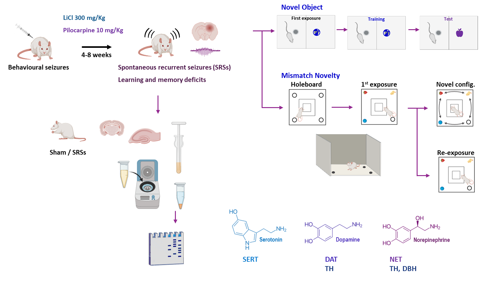
2.1. Animals and Induction of SRSs

Animals were housed in the local Animal House of the Institute of Physiology, Faculty of Medicine, University of Lisbon until usage, and were maintained under a 12:12 h light/dark cycle at a temperature of 22 °C, with food and water ad libitum. All procedures were in accordance with the standards established in the Guide for Care and Use of Laboratory Animals, the Portuguese and European law on animal welfare, and were approved by the Ethical Committee of the Faculty of Medicine, University of Lisbon.
Adult (12-week-old, 335–375 g, n = 52) male Wistar rats were handled twice a day for three days and status epilepticus (SE) was induced (n = 28 out of 52) by intraperitoneal pilocarpine administration (10 mg/kg) essentially as described [31]. Briefly, animals were pre-treated with LiCl (300 mg/kg, i.p.) and twenty-four hours later, they received methyl-scopolamine (1 mg/kg, i.p.) to block the peripheral cholinergic effects of pilocarpine. SE was induced 15 min later by administering pilocarpine (10 mg/kg) and, if necessary, additional lower doses (5 mg/kg) of pilocarpine were administered every 20 min until either SE is observed or a maximum of four doses was attained. Behavioural epileptiform seizures were monitored for 30 min, scored according to the scale of Racine modified by Lüttjohann et al. (2009) [32], and terminated by diazepam (i.p., 10 mg/kg) delivery. All animals here reported developed SE of distinct severities (scores 3–5 in Racine scale) following pilocarpine administration. Xilazine (i.m., 10 mg/kg) was administered immediately after seizure onset to prevent muscle exhaustion caused by convulsions. Seizure recurrence within the next 24–48 h was controlled, when required, with additional diazepam (i.p., 5–10 mg/kg, n = 16). This greatly increased animal survival and survival rate with this procedure was 86% (24/28). Sham animals (n = 24) were subjected to the same procedure of sequential drug administration except for the administration of pilocarpine that was replaced by an equivalent volume of saline (NaCl, 0.9%). Occurrence of spontaneous recurrent seizures (SRSs) was detected by 24 h video monitoring for 3–4 days within 4–10 weeks of pilocarpine administration and the frequency and severity of behavioural seizures were scored as described [32]. Behaviour was evaluated 4–12 weeks following the detection of the first unprovoked seizure (6–10 months of age). Animals presenting SRSs (n = 24) and respective Sham controls (n = 24) were subjected to a global evaluation of their motor capacity and anxiety levels using the elevated plus maze (EPM) test and the open-field (OF) test. Learning and memory impairment was evaluated in a subset of animals using the 8-arm radial arm maze (RAM, n = 8 Sham/SRSs) test for spatial memory or the novel object recognition (NOR, n = 8 Sham/SRSs) test for non-spatial memory. Evaluation of the mismatch novelty (MN, n = 16 Sham/SRSs) response using the holeboard with objects was performed as described with minor modifications [11] starting 24 h after the OF. All behavioural testing/training sessions were performed between 9:00 a.m. and 17:00 p.m. in a sound attenuated room. Each trial was video recorded and analysed using the video-tracking software ANY-maze 4.5 (Stoelting, Europe).

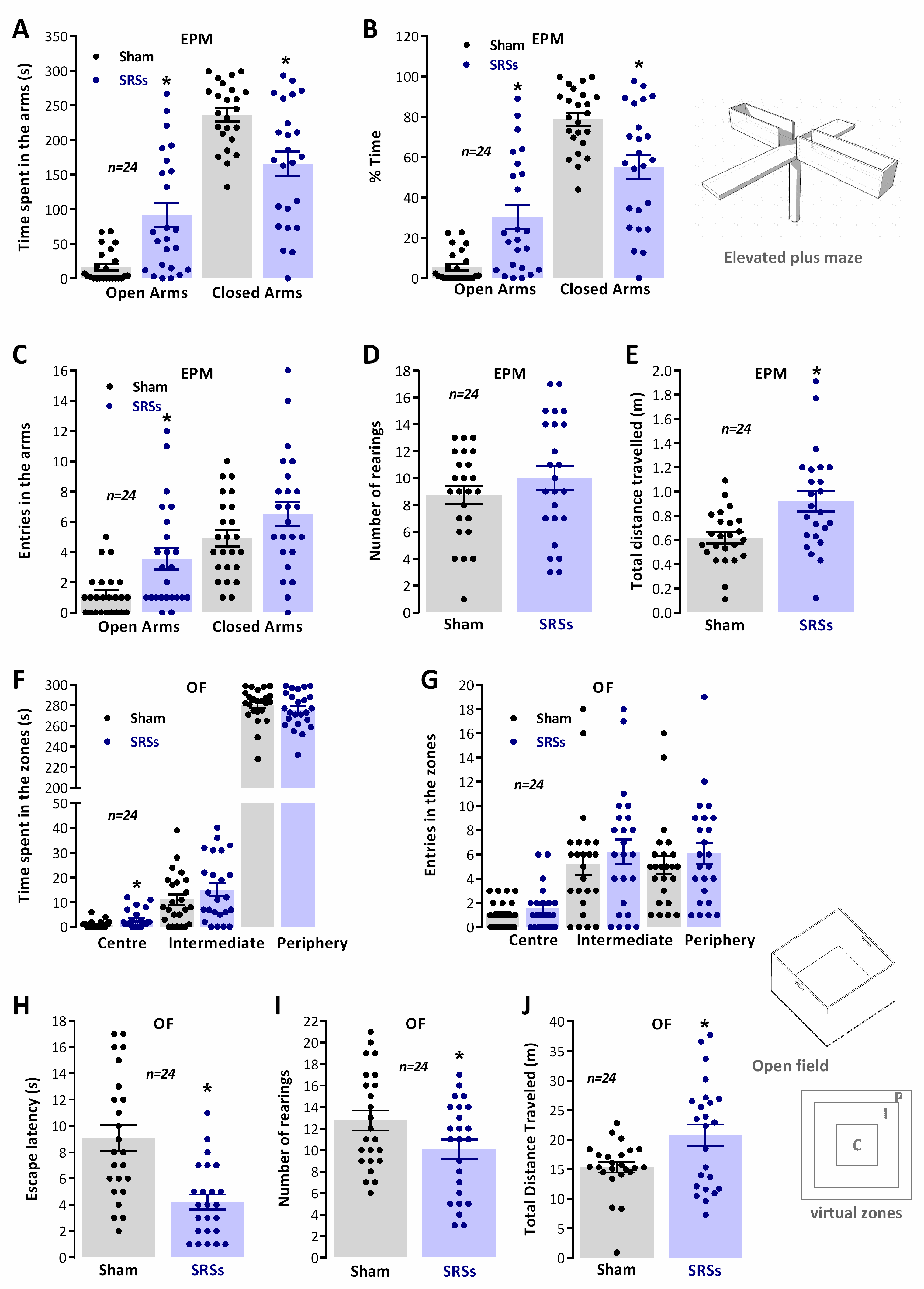
2.2. Evaluation of Anxiety and Locomotion Using the EPM and OF

Individual levels of anxiety and general locomotor behaviour were evaluated using the OF test [33] and the EPM test as adapted by Schneider [34] essentially as previously described [19].
The EPM comprised two open arms (50 cm × 10 cm) and two enclosed arms (50 cm × 10 cm × 40 cm), expanding from a central platform (10 cm × 10 cm) and elevated by 50 cm from floor level. Upon starting the EPM test, the rat was placed on the central platform, looking at the open arm, and left to explore the maze for 5 min. The maze was wiped with 70% ethanol between each trial. Animal activity was either observed and recorded manually by the experimenter or video documented and later examined using the Anymaze video-tracking software. Behaviour was quantified by evaluating the number of entries in open/closed arms, the time spent in open/closed arms, the time on the central platform, the distance travelled by the animal in the entire maze [34] and the number of rearings (considered when the rat was on its hind legs, touching or not with his front paws on the wall).
The OF test involved the exploration of a large square chamber (66 cm × 66 cm wide, 60 cm high walls), that lasted for 5 min. When initiating the test, animals were introduced directly in the centre of the apparatus. The movement of the animal in the arena for the duration of the test session was video recorded. Behaviour analysis relied on the division of space into three virtual zones (a central square 20 × 20 cm, an intermediate zone, and a peripheral zone 15 cm wide adjacent to the walls) and animal performance was assessed by the escape latency (s), the total distance travelled, the number of rearings, the number of entries, and the time spent in each virtual zone [33].

2.3. Evaluation of Cognitive Performance in the RAM

The cognitive performance of Sham versus SRS rats was evaluated using the radial arm maze (RAM) test. The RAM was first used to evaluate the ability of rats to memorise the location of baited arms upon one-week repeated exposure to the baited RAM. The RAM consisted of an octagonal centre platform 27 cm in diameter connected to eight equally spaced arms, each measuring 50 cm × 10 cm and 20 cm high, with a cylindrical food cup (3.5 cm ø, 0.5 cm depth) at the end of each arm. The maze was elevated 50 cm from the floor and was surrounded by several extra-maze cues. The parameters evaluated in the RAM were arm entries, counted if all four paws were placed on that arm [33]; arm latencies (maximum time to find the three available food rewards), and the number of rearings. Re-entries into baited arms were scored as errors of working memory and entries into non-baited arms were scored as reference memory errors.
Before each trial, animals were food-deprived (maintained at 85–90% of free feeding body weight) [33]. Rats were placed in the centre of the apparatus at the beginning of the test, surrounded by an opaque cylinder that was kept for 5 s before the animal was allowed to perform arms choices. Spatial cues were placed in the room walls to allow animals to locate the three food rewards, always located in the same set of three arms. The maze was rotated by 45° at the end of each session (between subsequent trials) in a way that intra-maze and extra-maze cues were dissociated, to avoid the use of within-maze cues in spatial learning. The test lasted for 10 min on the first trial (maze recognition) and took 5 min on the remaining trials (2 trials per day). Cognitive performance each day was quantified as the averaging performance in the two trials.

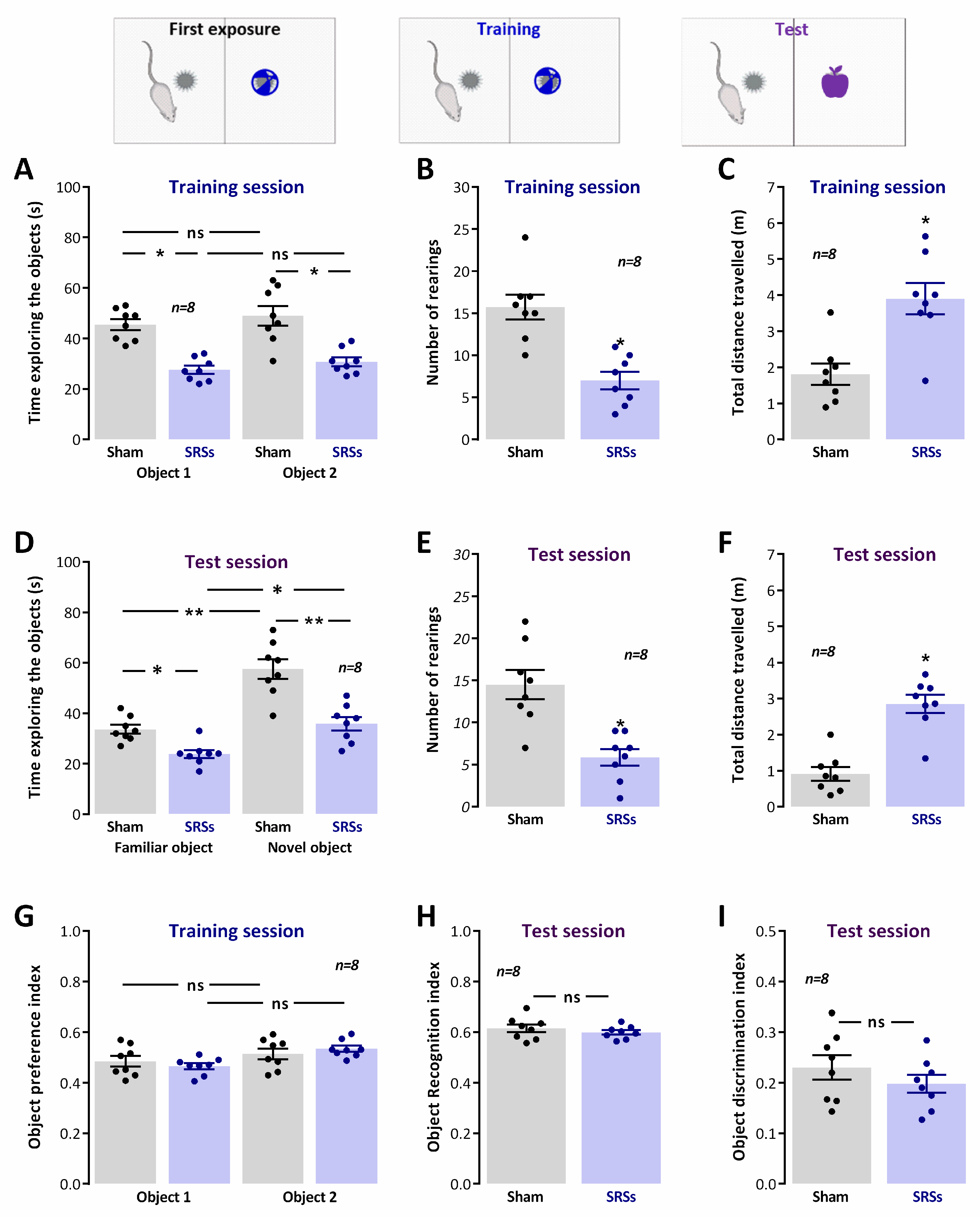
2.4. Novel Object Recognition (NOR)

The NOR test was performed in a square arena essentially as described [35] and was composed of three sessions: habituation, training, and test sessions [23]. The habituation session consisted of one session of free exploration in the arena (5 min). In the training session, two objects (similar in size and shape but different in texture, colour, and patterning) were added to the arena, and this location was kept for the persisting object in the subsequent test session. Animals were placed in the middle of the arena facing away from the objects and allowed to freely move for 5 min (Supplementary Figure S1). The test for novel object recognition was performed 24 h later by replacing one of the previously experienced objects (familiar objects F1 and F2) by a novel object (N). Exploration was scored when the animal touched an object with its forepaws or snout, bit, licked, or sniffed the object from no more than 1.5 cm. Animal movements and the time spent exploring each object were analysed. Animals spending less than 20 s exploring the objects were excluded from the study. Exploration of novel objects was scored using: (1) the object preference index—ratio between the time spent exploring one object over the total time spent exploring both objects [tF1 or tF2/(tF1 + tF2)]; (2) the object recognition index—ratio between the time spent exploring the novel object and the total time spent exploring both objects [tN/(tN + tF)], an index of memory retention; and (3) the object discrimination index—ratio between the difference between the time spent exploring the novel and the familiar object, and the total time spent exploring both objects [(tN − tF)/(tN + tF)], that allowed visualisation of data with no memory retention scored as zero.

2.5. Mismatch Novelty Test

The mismatch novelty (MN) test consisted in the exploration of the novel location of known objects in a familiar environment essentially as described (Supplementary Figure S1) [13]. This consisted of a holeboard composed by an arena (66 × 66 cm, 60 cm high walls) and containing one hole at each corner (four holes, 5.5 cm diameter, 4.5 cm deep). Animal habituation and testing was performed in 5 min sessions. One day prior to object exposure, all animals were exposed to the empty holeboard to get accustomed to the environment. Objects were introduced on the next day (1st exposure) in three of the four holes for all animals. On the second day, animals were either exposed to the same spatial distribution of objects (re-exposure) or to a new spatial configuration of the objects (novel configuration). Each trial was video monitored, recorded, and later analysed using automated video-tracking software (Anymaze software, Stoelting, Europe). The parameters scored were the travelled distance, the number of entries, and the time spent in each virtual zone of the apparatus (central, intermediate, and peripheral zones; essentially as defined for the OF). Exploration of objects and general exploratory activity were evaluated by the number of nose pokes and the number of rearings, respectively. Increased nose pokes vs. rearings were taken as a positive response to novelty.

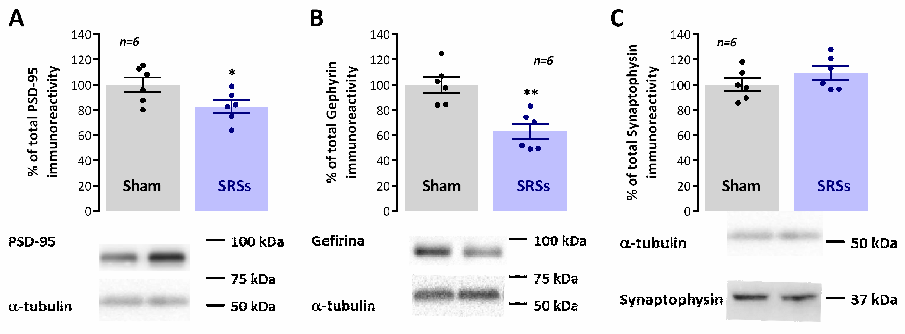
2.6. Western Blot Analysis of Monoaminergic Markers and Synaptic Proteins

For Western blot studies, total hippocampal membranes were isolated essentially as previously described [36]. Briefly, the hippocampi of Sham and SRS rats were dissected and collected in sucrose solution (320 mM Sucrose, 1 mg/mL BSA, 10 mM HEPES e 1 mM EDTA, pH 7.4) containing protease (complete, mini, EDTA-free Protease Inhibitor Cocktail) and phosphatase (1 mM PMSF, 2 mM Na3VO4, and 10 mM NaF) inhibitors, homogenised with a Potter-Elvejham apparatus and centrifuged at 1500× g for 10 min. The supernatant was collected and further centrifuged at 14,000× g for 12 min. The pellet was washed twice with modified aCSF (20 mM HEPES, 1 mM MgCl2, 1.2 mM NaH2PO4, 2.7 mM NaCl; 3 mM KCl, 1.2 mM CaCl2, 10 mM glucose, pH 7.4) also containing protease and phosphatase inhibitors and resuspended in modified aCSF to a concentration of 1 mg/mL protein. Aliquots of this suspension of hippocampal membranes were snap-frozen in liquid nitrogen and stored at −80 °C until use.
For Western blot, samples were incubated at 95 °C for 5 min with Laemmli buffer (125 mM Tris-BASE, 4% SDS, 50% glycerol, 0.02% Bromophenol Blue, 10% β-mercaptoethanol), were run on standard 10% sodium dodecyl sulphate polyacrylamide gel electrophoresis (SDS-PAGE) and transferred to PVDF membranes (Immobilon-P transfer membrane PVDF, pore size 0.45 μm). These were then blocked for 1 h with either 3% BSA or 5% milk solution in Tris-buffered saline containing 1% Tween (TBST) and incubated overnight at 4 °C with rabbit anti-dopamine transporter (DAT, 1:2000, Proteintech Cat# 22524-1-AP, RRID:AB_2879116), mouse monoclonal anti-norepinephrine transporter (NET, 1:500, Atlas Antibodies Cat# AMAb91116, RRID:AB_2665806), rabbit anti-serotonin transporter (SERT, 1:1000, Proteintech Cat# 19559-1-AP; AB_2878590), rabbit anti-tyrosine hydroxylase (TH, 1:1000, Abcam Cat# Ab112; RRID:AB_297840), mouse monoclonal anti-gephyrin (1:3000, Synaptic Systems #147011, RRID:AB_2810215), rabbit anti-PSD-95 (1:750, Cell Signalling Technology #2507, RRID:AB_561221), rabbit anti-GluA1 (1:4000, Millipore Cat# AB1504; RRID:AB_2113602), rabbit anti-GluA2 (1:1000, Proteintech Cat# 11994-1-AP; RRID: AB_2113725), mouse monoclonal anti-GluN1 (1:1000, Proteintech Cat# 67717-1-Ig; RRID: AB_2882906), rabbit anti-GluN2B (1:1000, Cell Signalling Technology Cat#4207; RRID: AB_1264223), rabbit anti-synaptophysin (1:7500, Synaptic Systems #101002, RRID:AB_887905), and either mouse monoclonal anti-β-actin (1:5000, Proteintech #60008-1-Ig, RRID: AB_2289225) or rabbit polyclonal anti-α-tubulin (1:4000, Proteintech #PT11224-1-AP, RRID: AB_ 2210206) primary antibodies. After washing 3× for 10 min with TBST, the membranes were incubated for 1 h with anti-rabbit IgG or anti-mouse IgG secondary antibody both conjugated with horseradish peroxidase (HRP) at room temperature. Excess bound secondary antibody was then removed by washing and HRP activity was visualised by enhanced chemiluminescence with Clarity ECL Western Blotting Detection System (Bio-Rad, HongKong, China). Band intensity was evaluated with the Image J software (1.52a) using either β-actin or α-tubulin band density as loading control. Immunostaining of the different targets was normalised to loading control band density, and differences in target protein expression in SRS animals were expressed as percentage change relative to Sham controls.

2.7. Statistics

Values of behavioural assessment parameters are presented as the mean ± S.E.M of 6–24 animals. In Western blot experiments, each n represents one experiment in a single animal. Significance of the differences between the Sham and SRS groups was calculated by Student’s t test with Welch correction for unequal variances. Differences were considered significant for p values of 0.05 or less. Statistical analysis was performed using GraphPad Prism 6.01 for Windows.

3. Results

3.1. Elevated Plus Maze (EPM) and Open-Field (OF) Tests

During rodent exploration of the EPM (Figure 1A–E), the time spent by Sham animals in the open arms (5.4 ± 1.6%, n = 24) was much lower than the time spent in the closed arms (78.8 ± 3.2%, n = 24). In the remaining time (15.8 ± 3.9%, n = 24), the animals were in the centre of the apparatus (the crossing of closed and open arms). In contrast, SRS animals spent more time in the open arms (30.5 ± 5.8%, n = 24) and less time in the closed arms (55.2 ± 6.0%, n = 24). Accordingly, Sham animals entered the open arms less (1.2 ± 0.3, n = 24) than SRS rats (3.5 ± 0.7%, n = 24), and the opposite was observed with the number of entries in the closed arms (4.9 ± 0.5%, n = 24 for Sham vs. 6.5 ± 0.8, n = 24 for SRS animals) from this central position. The total distance travelled in the EPM during the 5 min trial was larger for SRSs (0.92 ± 0.08 m, n = 24) than for Sham rats (0.62 ± 0.5 m, n = 24). Rearings were almost entirely performed within the closed arms and were slightly higher for SRS rats (10.0 ± 0.9, n = 24) when compared to Sham rats (8.8 ± 0.7, n = 24).
Behaviour in the OF during the 5 min trial (Figure 1F–J) was analysed through the definition of three virtual zones (peripheral, intermediate, and central zones). Sham animals showed a high thigmotaxis (Figure 1A) as evidenced by the minimum time spent in the centre (0.9 ± 0.3 s, n = 24) and intermediate (11.0 ± 2.1 s, n = 24) zones of the OF and preference for remaining in the periphery zone (286.4 ± 3.4 s, n = 24), as previously described for naïve animals of similar age. Comparatively, the time spent by SRS animals in the central (3.0 ± 0.8 s, n = 24) zone was higher than for Sham animals while the time spent in the intermediate (15.1 ± 2.7 s, n = 24) and periphery (280.9 ± 3.6 s, n = 24) zones did not substantially differentiate from Sham rats. The global locomotor activity, accessed by the total distance travelled in the open-field, was higher in SRS animals (20.8 ± 1.8 m, n = 24) as compared to the one covered by Sham rats (15.4 ± 0.9 m, n = 24). Sham and SRS animals did not present significant differences in the number of entries in any of the different zones. The total number of rearings was lower for SRS animals (10.1 ± 0.9, n = 24) as compared to Sham animals (12.8 ± 0.9, n = 24), and reflects rearings performed predominantly in the periphery zone.

3.2. Spatial Learning in the RAM

During exploration of the RAM, the latency to find the 1st baited arm (Supplementary Figure S2A) was not significantly different between Sham and SRS animals during the whole testing period. However, the latency to find the 2nd and 3rd baited arms was higher in SRS rats (Supplementary Figure S2B,C). The latency to task completion was significantly higher from the third day of testing (270.1 ± 19.9s, n = 8 for SRS vs. 181.0 ± 16.4 s, n = 8 for Sham on day 3) and SRS rats showed little improvement in their performance from the 1st to the 5th day of testing (Supplementary Figure S2C), and three of the SRS animals were unable to complete the task on day 5, evidencing a markedly decreased capacity to memorise the location of the baited arms. The distance travelled in the RAM (Supplementary Figure S2D) was higher on the first day for SRS rats (30.9 ± 2.8, n = 8 for SRS vs. 23.7 ± 1.4, n = 8 for Sham) but no significant differences between the two groups were encountered in the remaining days. The number of rearings performed during the RAM training and test sessions (Supplementary Figure S2E) was higher for SRS rats on the first day (33.9 ± 2.5, n = 8 for SRS vs. 27.3 ± 2.4, n = 8 for Sham) but decreased markedly throughout the sessions being much lower than the ones performed by Sham rats by the last day of the RAM test (11.5 ± 1.3, n = 8 for SRS vs. 19.0 ± 1.9, n = 8 for Sham).

3.3. Novel Object Recognition

The NOR task consisted of a training session when animals were exposed to previously unknown objects, and a test session delivered 24 h later, when one of these two now familiar objects and one novel object were presented. During the training session, we observed that Sham animals explored both objects equally (Figure 2A, Obj. 1: 51.2 ± 2.4%, Obj. 2: 48.8 ± 2.4%; n = 8), as given by the object preference index (Figure 2G), whereas during the test session, the animals explored the novel object to a significantly larger extent than the familiar object (Figure 2D, F Obj.: 61.5 ± 1.5%, N Obj.: 38.5 ± 1.5%; n = 8). This is a clear behavioural indication that Sham rats successfully formed a memory of the objects lasting for 24 h during the training phase. SRS animals showed a similar behaviour during both the training session (Figure 2A, Obj. 1: 48.4 ± 3.5%, Obj. 2: 51.6 ± 3.5%; n = 8, Figure 2G) or when a novel object was introduced 24 h later (Figure 2D, F Obj.: 59.9 ± 0.8%, N Obj.: 40.1 ± 0.8%; n = 8), suggesting that novel object recognition was not significantly affected in SRS rats. This was also evidenced by the absence of differences in the object recognition and discrimination indexes (Figure 2H and Figure 3I), despite the marked differences observed in general exploration between Sham and SRS animals, as given by the number of rearings during the training (7.0 ± 1.0, n = 8 for SRS vs. 15.8 ± 1.5, n = 8 for Sham, Figure 2B) and the test sessions (5.9 ± 1.0, n = 8 for SRS vs. 14.5 ± 1.7, n = 8 for Sham, Figure 2E), and by the total distance travelled in the training (3.9 ± 0.4 m, n = 8 for SRS vs. 1.8 ± 0.3 m, n = 8 for Sham, Figure 2C) and test session (2.9 ± 0.3 m, n = 8 for SRS vs. 0.9 ± 0.2 m, n = 8 for Sham, Figure 2F).

3.4. Mismatch Novelty Detection

The MN test consisted of the exploration of the novel location of known objects in a holeboard in a familiar environment essentially as described [11]. In the first session, animals were exposed to the empty holeboard, and in the following session, objects were introduced. In the third session, Sham and SRS animals were divided into two groups, one being exposed to the same object configuration and the other exposed to the novel object configuration (Figure 3A). The behavioural responses of Sham and SRS animals were monitored for 5 min.
Upon the first exposure to the holeboard, the general exploratory activity of Sham animals was lower than the one observed in the OF maze, as evidenced by the decrease in the number of rearings that was 6.0 ± 0.8 (n = 16) in holeboard exploration and 12.8 ± 0.9 (n = 24) in the OF test, and by the decrease in the total distance travelled on the holeboard 13.0 ± 1.4 m (n = 16) compared to the distance travelled in the OF 15.4 ± 0.9 m (n = 24). The animals spent slightly more time in the periphery zone of the holeboard (291.6 ± 2.5 s, n = 16), where the holes are located, than in the periphery of the open-field (286.4 ± 3.4 s, n = 24). Thus, animals, although likely now less fearful, spent more time in the periphery exploring the holes, as evidenced by the number of nose-pokes (9.0 ± 0.7, n = 16). SRS animals behaved similarly when exposed to the holeboard, since the total number of rearings (8.6 ± 1.0, n = 16) was only a bit higher for SRS animals than the one performed by Sham animals, while the number of nose-pokes was slightly lower (7.8 ± 1.2, n = 16), yet the total distance travelled by SRS animals (26.1 ± 2.7 m, n = 16) in the holeboard was double the one travelled by Sham animals (13.0 ± 1.4 m, n = 16), as occurring in virtually all other tests, a reflection of their anxious and attention deficit-like behaviour, as previously described [37,38].
The introduction of objects on the second day increased the number of nose-pokes for both Sham (15.3 ± 1.0, n = 16) and SRS (10.6 ± 1.0, n = 16) animals (Figure 3C) but this effect was more patent for Sham animals since now there was a significant difference between Sham and SRS animals’ behaviour. No noticeable differences were observed in the behaviour of Sham and SRS animals regarding the number of rearings (Figure 3B), time spent (Figure 3E), or number of entries (Figure 3I) in the different zones of the apparatus in the session when the objects were first presented. Total distance travelled was nevertheless higher (p < 0.05, t-test) for SRS (20.4 ± 2.1 m, n = 16) than for Sham animals (10.7 ± 1.5 m, n = 16).
For the subgroup of animals subjected to re-exposure to the same configuration of objects, no significant differences (p > 0.05) between Sham (n = 8) and SRS (n = 8) animals in any of the behavioural parameters were monitored (Figure 3B,C,F,J), including the total distance travelled (15.4 ± 1.4 m, n = 8 for Sham vs. 16.5 ± 3.3 m, n = 8 for SRSs). Conversely, for the subgroup exposed to a novel configuration of the objects, marked differences were observed between Sham and SRS animals in global exploratory activity as given by the number of rearings (10.3 ± 1.3 m, n = 8 for Sham vs. 6.8 ± 1.5 m, n = 8 for SRSs) and total distance travelled (13.7 ± 2.2 m, n = 8 for Sham vs. 28.6 ± 3.3 m, n = 8 for SRSs). In addition, the number of nose-pokes, a major indicator of a response to novelty, was also significantly higher for Sham (13.2 ± 1.4, n = 8, p < 0.05) than for SRS animals (8.4 ± 1.4, n = 8), and significantly higher (p < 0.05) than the one performed by Sham animals undergoing re-exposure to the same object arrangement (8.0 ± 1.2, n = 8). Altogether, this suggests an impairment in the response to mismatch novelty in SRS rats.

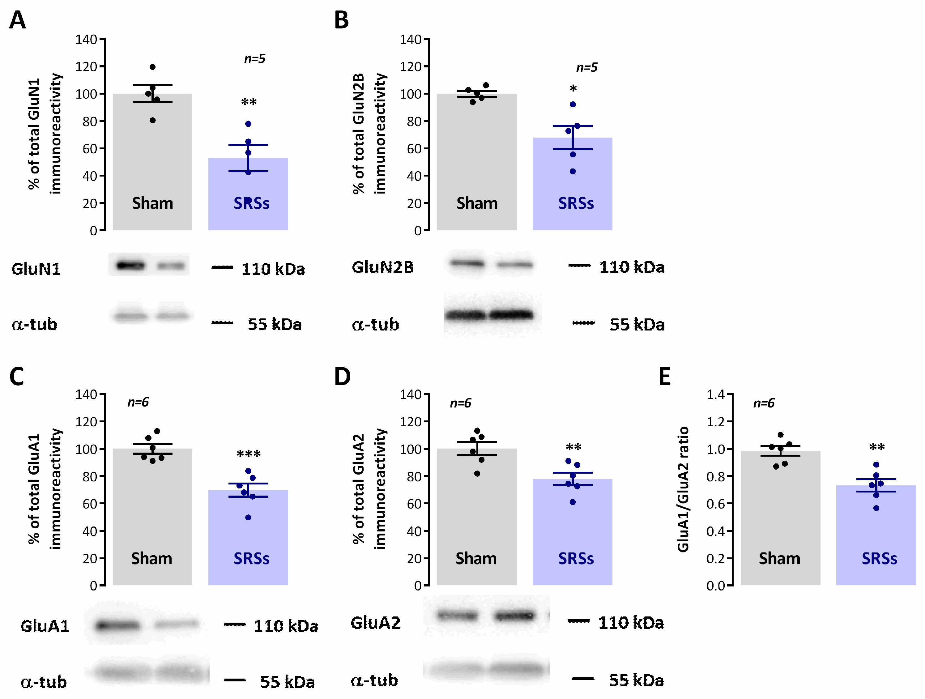
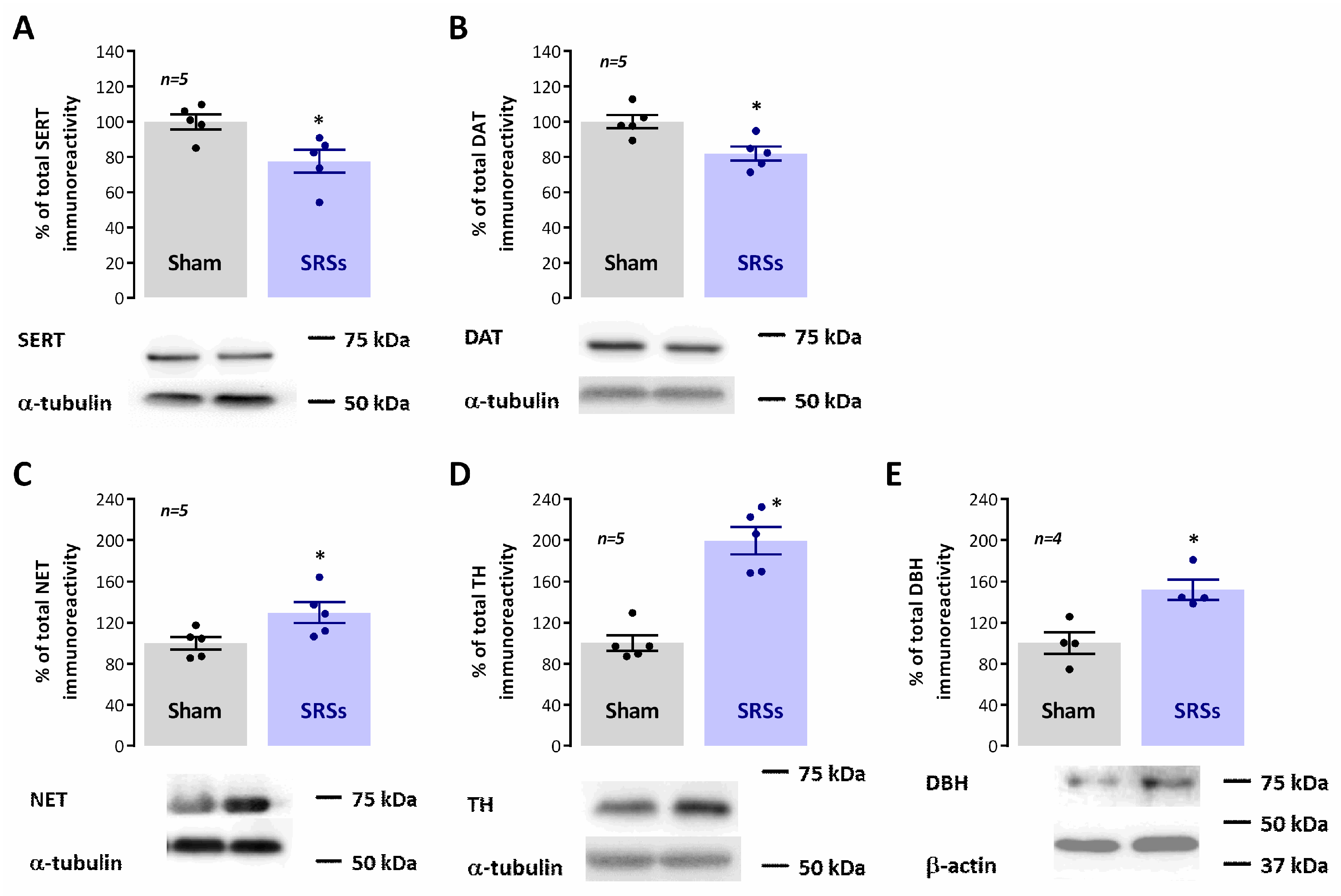
3.5. Evaluation of Hippocampal Levels of Synaptic and Monoaminergic Markers

Numerous studies have identified marked differences in synaptic structure and molecular composition in epileptic rodent models. However, findings are often contradictory between different rodent models or even distinct rat strains. As such, we briefly characterised the levels of a general synaptic marker (synaptophysin) and markers of both glutamatergic (PSD-95) and GABAergic synapses (gephyrin). The global levels of synaptophysin were not changed in hippocampal membranes of SRS rats as compared to Sham controls (n = 6, p < 0.05, Figure 4C). Yet, both PSD-95 and gephyrin were significantly reduced to 82.7 ± 5.0% (n = 6, p < 0.05, Figure 4A) and 63.0 ± 6.0% (n = 6, p < 0.05, Figure 4B) of the levels observed in Sham animals.
Impaired synaptic plasticity, as observed in animal models of epilepsy, is believed to be associated with cognitive deficits in epileptic patients. Changes in both NMDA and AMPA receptor subunit composition have also been demonstrated in animal models of epilepsy and are believed to contribute to altered cognitive ability human epilepsy. As such, we also verified in our model how AMPA GluA1 and GluA2 subunits as well as NMDA GluN1 and GluN2B subunits were altered in our model. Both GluN1 and GluN2B subunit levels were decreased in SRS animals to 52.8 ± 9.7% (n = 6, p < 0.05, Figure 5A) and 68.0 ± 8.5% (n = 6, p < 0.05, Figure 5B), respectively, of the total immunoreactivity detected in Sham controls. Likewise, GluA1 and GluA2 levels were similarly decreased in SRS animals vs. Sham controls, showing, respectively, only 69.8 ± 4.9% (n = 6, p < 0.05, Figure 5C) and 77.9 ± 4.5% (n = 6, p < 0.05, Figure 5D) of the total Sham immunoreactivity. As a consequence, the GluA1/GluA2 ratio was also smaller (p < 0.05, Figure 5E) in SRS (0.733 ± 0.045, n = 6) than in Sham rodents (0.985 ± 0.036, n = 6).
Differences in behavioural parameters, particularly impulsive behaviour, motivation, and depression, in the Li2+-pilocarpine model of epilepsy have often been attributed to changes in dopaminergic, noradrenergic, and serotonergic transmission, important modulators of arousal, motivation, and attention, all very relevant capacities for both the NOR and MN tasks. As such, we investigated the changes in the levels of enzymes and synaptic transporters associated with the catecholaminergic and serotonergic transmission and correlated them with the levels of constitutive synaptic proteins. In SRS animals, when compared to Sham controls, we observed a mild decrease in the hippocampal levels of the plasma membrane serotonin (SERT) and dopamine transporters (DAT) to 77.6 ± 6.5% (n = 5, p < 0.05, Figure 6A) and 81.9 ± 4.0% (n = 6, p < 0.05, Figure 6B) of the observed immunoreactivities in Sham animals, respectively. Conversely, the hippocampal levels of norepinephrine transporters (NET) were increased by 29.6 ± 10.2% (n = 5, p < 0.05, Figure 6C) in SRS vs. Sham rats. The levels of tyrosine hydroxylase, the enzyme catalysing the rate-limiting step in the synthesis of catecholamines, was also enhanced by 99.8 ± 13.3% (n = 6, p < 0.05, Figure 5D) in SRS vs. Sham animals, while the levels of dopamine-β-hydroxylase (DBH), fundamental to the synthesis of catecholamines, was increased by 51.9 ± 9.8% (n = 4, p < 0.05, Figure 5D).

4. Discussion

The main findings of the present work are that: (1) exploration of the novel location of known objects in a holeboard is impaired in the Li2+-pilocarpine rat model of SRSs; (2) novel object recognition was not significantly altered in SRS animals; (3) the levels of serine and dopamine nerve terminal transporters (SERT and DAT) were mildly decreased in SRS rat hippocampal membranes while (4) the levels of the nerve terminal norepinephrine transporter (NET), of tyrosine hydroxylase (TH), and dopamine-b-hydroxylase (DBH) were enhanced. We also confirmed deficits in spatial learning and alterations in AMPA and NMDA receptor composition and synaptic proteins as found in previous studies in rodent models of epilepsy. Altogether, these observations provide evidence for a disfunction of the novelty processing circuits in SRS animals while characterising monoaminergic transmission disfunction in the Li2+-pilocarpine model of SRSs and suggest similar changes that may also occur in human MTLE. As such, these could be relevant targets for future pharmacological, behavioural, or possibly combined therapies to mitigate cognitive decline in MTLE.
Cognitive deficits in animal models of epilepsy have extensively been studied, as have the roles of different monoamines in animal models of epilepsy, yet previous studies relating cognition and novelty detection impairment to hippocampal monoamines were performed individually, i.e., focusing on one or two neurotransmitters at a time, in distinct animal models of epilepsy, and at different time points following spontaneous recurrent seizure (SRS) onset, making it difficult to evaluate the relative contribution of each monoamine transmitter to hippocampal-dependent disfunction and its relation to ongoing cognitive disfunction and altered synaptic plasticity. As such, we set out to investigate this in the Li2+-pilocarpine model of SRSs.
As mentioned earlier, novelty is an important stimulus in episodic memory formation, and numerous studies have shown that different aspects of novelty have a distinct impact on hippocampal-dependent learning and synaptic plasticity. The fact that hippocampal long-term potentiation (LTP) or long-term depression (LTD) of synaptic transmission contribute to encode different aspects of novelty acquisition [11], with LTD being facilitated during the location of new objects or known objects in new locations, and LTP being favoured during exploration of a new environment, is by itself proof of a complex interplay of these two forms of synaptic plasticity in hippocampal-dependent cognitive processes. The balance of the two is not only crucial to the formation of a complete spatial map [11,12] but to the consolidation of spatial memory [13,14] and the reversal learning of recently acquired spatial memories [39]. The stability of the memories formed, and of the associated synaptic plasticity phenomena, can be shaped by previously learning experiences (either recent or remote) through metaplasticity [15]. In this respect, novelty, besides being an important trigger for memory acquisition, can also influence ongoing learning and synaptic plasticity events. Spatial novelty enhances retrieval of a previously acquired memory when appearing up to 2 h before retrieval through an NMDA-dependent mechanism [16], while known objects presented in new locations in a familiar environment enhance inhibitory avoidance learning in a process dependent on hippocampal LTD [13].
The new location of known objects in a familiar environment, a behavioural mismatch novelty paradigm, profoundly alters network activity in the CA1 area of the hippocampus in mice [40], and modulates rodent hippocampal synaptic plasticity in vivo through short-term metaplasticity [11]. Mismatch novelty detection, a novelty paradigm remarkably important in memory reformulation and reconsolidation, also triggers the activation of the hippocampal CA1 area in human studies, and is compromised in patients with hippocampal lesions [17,18]. Furthermore, studies from our group showed that repeated exposure to mismatch novelty has a long-term metaplastic effect on both LTP and LTD in the hippocampus [19], suggesting that behavioural tasks involving mismatch novelty may be of value in cognitive therapy strategies aiming to mitigate the LTP/LTD imbalance found in aging or diseases like epilepsy or Down’s syndrome.
In this paper, we demonstrate that mismatch novelty detection is specifically compromised in SRS rats as compared to other novelty paradigms like novel object recognition. Altogether, this suggests that recurrent seizures affect more prominently hippocampal neural pathways specifically associated with mismatch novelty detection and processing. Although exploratory responses to open-field exposure were also significantly distinct in SRS and Sham animals, this task, unlike the other two, involves facing an unfamiliar and potentially dangerous environment. As such, amygdala damage also observed in this model [41] may play a role in altered SRS animal performance in this test. Interestingly, the performance of SRS rats in the EPM, a test specifically designed to evaluate anxiety traits, revealed that SRS rats are particularly unaware of danger, as previously described [42]. It is believed that altered performance in this test is also related to attention deficits, as the Li2+-pilocarpine model of temporal lobe epilepsy has been advanced as a model of attention deficit hyperactivity disorder (ADHD) [38].
Several neurotransmitter systems have been implicated in the hippocampal detection and processing of novelty stimuli. Dopaminergic neurons originating from the VTA and locus coeruleus neurons co-releasing dopamine and norepinephrine innervate the ventral hippocampus, regulate physiological arousal, attention, and motivation, and are thought to play an essential role in the efficiency of cognitive function [20,21], playing a crucial role in recognition memory and novelty signalling [23]. Likewise, transmission by medium raphe serotonergic fibres and septal cholinergic and GABAergic projections, fundamental for the pacing, engagement, and suppression of hippocampal theta rhythm [43,44,45] and for hippocampal-dependent memory formation [46,47,48], was shown to be differentially modulated by novelty stimuli [49,50].
Furthermore, altered monoaminergic neurotransmission not only constitutes a risk factor for the development of epilepsy [24] but is either linked to the degeneration or upregulation of ascending projections to the hippocampus and cortex, altered hippocampal monoamine levels, or altered distribution and function of monoamine receptors in MTLE patients [25]. Similar observations occurred in experimental models of epilepsy, such as (1) selective loss of GABAergic septal projections to the limbic cortex [51], (2) enrichment in cholinergic neurons in the median septum together with proliferation of cholinergic boutons and fibre sprouting in the hippocampus [52], (3) loss of limbic-projecting median raphe serotonergic neurons [26], decreased serotonin levels [38], and (4) decline of VTA dopaminergic projections to the nucleus accumbens [27] coupled to decreased vesicular monoamine transporter 2 in the temporal cortex and hippocampus [28]. Our observations, that in the Li2+-pilocarpine model of SRS rats’ hippocampal SERT and DAT levels are decreased, are in agreement with these reports yet partially contradict data showing that SERT immunoreactivity is enhanced in the hippocampus of epileptic patients at risk of SUDEP [53]. Nevertheless, loss of serotonergic and dopaminergic signalling may play a role in the impairment of mismatch novelty processing in SRS rats, as novelty-sensitive dopaminergic neurons in the Human substantia nigra have been implicated in declarative memory formation [54]. Furthermore, both 5-HT and dopamine play a role in memory destabilization and reactivation by distinct novelty stimuli, that in turn trigger memory reconsolidation [49,55].
In our work, the levels of TH, DBH, and NET were, on the contrary, increased in SRS animals vs. Sham controls. This is overall conflicting with previous knowledge that noradrenaline levels are decreased in the Li2+-pilocarpine model, and that this is related to depression-like symptoms [38]. However, these findings are in line with several studies demonstrating that inactivation of NET ameliorates seizures in animal models of epilepsy [56], and with reports that TH and NET are upregulated following seizures in animal models [57,58]. Altogether, this suggests that the ADHD-like phenotype observed in this animal model may be related to an excessive, rather than impaired, noradrenergic signalling. Alternatively, the impairment in serotonergic signalling that is concomitantly observed [38,58] may be the determinant factor in this respect. This question should be further investigated but is currently beyond the scope of this paper.
In this study, we also observed a decline in gephyrin levels and PSD-95 levels, specific markers of GABAergic and glutamatergic synapses, that is suggestive of a global decline in both intrahippocampal and external hippocampal-projecting glutamatergic and GABAergic fibres or respective nerve terminals. Since this effect is much more pronounced for gephyrin than for PSD-95, it is to be considered that seizures affect GABAergic transmission more strongly than glutamatergic, at least at this time point after SRS onset. Although this may reflect in part the above-mentioned selective loss of GABAergic septal projections [51], it is also long known that hippocampal GABAergic interneurons involved in disinhibition are particularly affected by seizures and epileptic state [4,59,60,61,62], a factor that is determinant in gradually enhanced hippocampal excitability and progressive epileptogenesis. The decline in PSD-95 levels in SRS hippocampal membranes is consistent with previous observations in the kainic acid model of SRSs [63]. This study also describes a concomitant decrease in GluN2B subunits that is consistent with our current paper and was expected given the role of PSD-95 in membrane-anchoring of NMDA receptor subunits [64,65]. Interestingly, the decline in GluN1 subunits in SRS rats was much stronger, suggesting a major role for these receptors in hippocampal-dependent cognitive decline and epilepsy pathology, and in line with what was observed in previous studies [64,66,67]. Regardless of the multiple controversies generated by studies in different models of epilepsy, at multiple time points following SRS onset and, using multiple approaches to detect levels of synaptic proteins, the role of synaptic reshaping in temporal lobe epilepsy onset and progression is consensual. AMPA and NMDA receptor remodelling plays a crucial role in this process, and altered synaptic plasticity and transmission is a major hallmark of epilepsy animal models but it has been demonstrated also in the human brain [67,68,69]. Our work confirms in our model that GluA1 and GluA2 levels as well as the GluA1/GluA2 ratio are diminished in SRS animals, as previously described [42], a necessary confirmation given the conflicting results found in the literature.
This study describes alterations in the Li2+-pilocarpine model of TLE in the rat that put in evidence the plastic susceptibility of the monoaminergic system in epilepsy and its possible contribution to cognitive decline in MTLE. Although animal models of epilepsy are useful to study cellular alterations in epileptogenesis and ictiogenesis, these do not often reproduce accurately the human pathology. This model reproduces both the structural damages and subsequent development of spontaneous recurrent seizures resembling those of human complex partial seizures [39,70]. Responses to antiepileptic drugs (AEDs) that are effective against complex partial seizures in humans can also halt spontaneous seizures in this model that also replicates several neurochemical features and network alterations of human TLE, like the generation of interictal activity from the subiculum or cognitive and memory deficits commonly found in TLE patients [71]. Nevertheless, we cannot say at this point that the alterations here reported closely reproduce alterations in the monoaminergic system in MTLE patients, and this should be further investigated in this animal model and in the human condition. The observations in our study, using mostly Western blot studies, also do not allow for a more precise mapping of the hippocampal subarea and subcellular location of the observed changes. This would be paramount to further understand and infer the possible clinical implications of our observations. Nevertheless, this and other studies [25,49] suggest that the monoaminergic system could be targeted to rescue cognitive decline in MTLE, and that this should be taken into consideration while treating comorbid affections in MTLE, such as depression and anxiety, that often target monoamine synaptic availability.
In conclusion, we investigated the response to mismatch novelty in the Li2+-pilocarpine rat model of TLE and its correlation to hippocampal monoaminergic and synaptic markers and other hippocampal-dependent learning and memory tasks. We observed an impairment in the exploration of a known environment containing familiar objects presented in a new location in rats showing spontaneous recurrent seizures (SRSs) for at least 4 weeks, suggesting that deficits in mismatch novelty detection indeed contribute to cognitive impairment in MTLE. This was correlated with alterations in the hippocampal monoaminergic system that may contribute to the attention deficit-like profile previously observed in the Li2+-pilocarpine model of epilepsy.

Supplementary Materials

The following supporting information can be downloaded at: https://www.mdpi.com/article/10.3390/biomedicines12030631/s1, Figure S1: Experimental workflow for induction of SRSs with Li2+-pilocarpine in the rat (A) and for behavioural evaluation (B); Figure S2: Impaired learning in the radial arm maze in the Li2+-pilocarpine rat model of epilepsy.

Author Contributions

C.N.: formal analysis and methodology; V.G.-P.: formal analysis and methodology; S.P.: formal analysis and methodology; A.C.-R.: formal analysis and methodology; L.A.-G.: formal analysis and methodology; D.C.-R.: formal analysis and methodology, resources, supervision, funding acquisition, project administration, and writing—original draft, review, and editing. All authors have read and agreed to the published version of the manuscript.

Funding

This work was supported by national and international funds managed by Fundação para a Ciência e a Tecnologia (FCT, IP), Portugal. Grants: UIDB/04046/2020 (DOI: 10.54499/UIDB/04046/2020) and UIDP/04046/2020 (DOI: 10.54499/UIDP/04046/2020) Centre grants to BioISI, and research grants PTDC/SAU-NEU/103639/2008 and FCT/POCTI (PTDC/SAUPUB/28311/2017) EPIRaft grant to D.C.-R. Fellowships: SFRH/BPD/81358/2011 to D.C.-R. and Researcher contract: Norma Transitória—DL57/2016/CP1479/CT0044 to D.C.-R (DOI: 10.54499/DL57/2016/CP1479/CT0044). S.P. and L.A.-G. were in receipt of an Erasmus+ Mobility Fellowship from the Faculty of Biological Sciences, University of Wrocław and Faculty of Sciences, University of Girona, respectively. Funding sources made no contribution to the writing, research plan, and decision to publish this paper.

Institutional Review Board Statement

All protocols and procedures were performed according to ARRIVE guidelines for experimental design, analysis, and their reporting. Animal housing and handling was performed in accordance with the Portuguese law (DL 113/2013) and European Community guidelines (86/609/EEC and 63/2010/CE). The animal study protocol was approved by the Ethics Committee of the Faculty of Medicine, University of Lisbon (protocol code N/A and date of approval June 2013).

Informed Consent Statement

Not applicable.

Data Availability Statement

Dataset available on request from the authors.

Acknowledgments

The authors thank the Institute of Physiology, Faculty of Medicine, University of Lisbon for animal housing and Marta Bento and André Serpa for their technical contribution.

Conflicts of Interest

The authors have no conflicts of interests in publication of this paper.

References

  1. Devinsky, O.; Vezzani, A.; O’Brien, T.J.; Jette, N.; Scheffer, I.E.; De Curtis, M.; Perucca, P. Epilepsy. Nat. Rev. Dis. Prim. 2018, 4, 18024. [Google Scholar] [CrossRef]
  2. Moshé, S.L.; Perucca, E.; Ryvlin, P.; Tomson, T. Epilepsy: New Advances. Lancet 2015, 385, 884–898. [Google Scholar] [CrossRef]
  3. Vezzani, A.; Balosso, S.; Ravizza, T. Neuroinflammatory Pathways as Treatment Targets and Biomarkers in Epilepsy. Nat. Rev. Neurol. 2019, 15, 459–472. [Google Scholar] [CrossRef]
  4. Cunha-Reis, D.; Caulino-Rocha, A.; Correia-de-Sá, P. VIPergic Neuroprotection in Epileptogenesis: Challenges and Opportunities. Pharmacol. Res. 2021, 164, 105356. [Google Scholar] [CrossRef] [PubMed]
  5. Thom, M. Review: Hippocampal Sclerosis in Epilepsy: A Neuropathology Review. Neuropathol. Appl. Neurobiol. 2014, 40, 520–543. [Google Scholar] [CrossRef] [PubMed]
  6. Helmstaedter, C.; Kockelmann, E. Cognitive Outcomes in Patients with Chronic Temporal Lobe Epilepsy. Epilepsia 2006, 47 (Suppl. S2), 96–98. [Google Scholar] [CrossRef] [PubMed]
  7. Schomaker, J.; Grouls, M.M.E.; van der Linden, C.G.M.; Rau, E.M.; Hendriks, M.; Colon, A.; Meeter, M. Novelty Processing Depends on Medial Temporal Lobe Structures. Neurobiol. Learn. Mem. 2021, 183, 107464. [Google Scholar] [CrossRef] [PubMed]
  8. Celiker Uslu, S.; Yuksel, B.; Tekin, B.; Sariahmetoglu, H.; Atakli, D. Cognitive Impairment and Drug Responsiveness in Mesial Temporal Lobe Epilepsy. Epilepsy Behav. 2019, 90, 162–167. [Google Scholar] [CrossRef] [PubMed]
  9. Poprelka, K.; Patrikelis, P.; Takousi, M.; Messinis, L.; Fasilis, T.; Margariti, S.; Ntinopoulou, E.; Verentzioti, A.; Stefanatou, M.; Alexoudi, A.; et al. Arousal Deregulation in the Co-Shaping of Neuropsychological Dysfunction in Frontal and Mesial Temporal Lobe Epilepsy. Epilepsy Res. 2023, 194, 107189. [Google Scholar] [CrossRef] [PubMed]
  10. Zhang, Z.; Lu, G.; Zhong, Y.; Tan, Q.; Yang, Z.; Liao, W.; Chen, Z.; Shi, J.; Liu, Y. Impaired Attention Network in Temporal Lobe Epilepsy: A Resting FMRI Study. Neurosci. Lett. 2009, 458, 97–101. [Google Scholar] [CrossRef]
  11. Kemp, A.; Manahan-Vaughan, D. Hippocampal Long-Term Depression and Long-Term Potentiation Encode Different Aspects of Novelty Acquisition. Proc. Natl. Acad. Sci. USA 2004, 101, 8192–8197. [Google Scholar] [CrossRef] [PubMed]
  12. Kemp, A.; Manahan-Vaughan, D. Hippocampal Long-Term Depression: Master or Minion in Declarative Memory Processes? Trends Neurosci. 2007, 30, 111–118. [Google Scholar] [CrossRef]
  13. Dong, Z.; Gong, B.; Li, H.; Bai, Y.; Wu, X.; Huang, Y.; He, W.; Li, T.; Wang, Y.T. Mechanisms of Hippocampal Long-Term Depression Are Required for Memory Enhancement by Novelty Exploration. J. Neurosci. 2012, 32, 11980–11990. [Google Scholar] [CrossRef]
  14. Ge, Y.; Dong, Z.; Bagot, R.C.; Howland, J.G.; Phillips, A.G.; Wong, T.P.; Wang, Y.T. Hippocampal Long-Term Depression Is Required for the Consolidation of Spatial Memory. Proc. Natl. Acad. Sci. USA 2010, 107, 16697–16702. [Google Scholar] [CrossRef] [PubMed]
  15. Abraham, W.C.; Bear, M.F. Metaplasticity: The Plasticity of Synaptic Plasticity. Trends Neurosci. 1996, 19, 126–130. [Google Scholar] [CrossRef] [PubMed]
  16. Izquierdo, L.A.; Viola, H.; Barros, D.M.; Alonso, M.; Vianna, M.R.M.; Furman, M.; Levi De Stein, M.; Szapiro, G.; Rodrigues, C.; Choi, H.; et al. Novelty Enhances Retrieval: Molecular Mechanisms Involved in Rat Hippocampus. Eur. J. Neurosci. 2001, 13, 1464–1467. [Google Scholar] [CrossRef]
  17. Stepankova, K.; Fenton, A.A.; Pastalkova, E.; Kalina, M.; Bohbot, V.É.D. Object-Location Memory Impairment in Patients with Thermal Lesions to the Right or Left Hippocampus. Neuropsychologia 2004, 42, 1017–1028. [Google Scholar] [CrossRef]
  18. Bohbot, V.D.; Jech, R.; Růžička, E.; Nadel, L.; Kalina, M.; Stěpánková, K.; Bureš, J. Rat Spatial Memory Tasks Adapted for Humans: Characterization in Subjects with Intact Brain and Subjects with Medial Temporal Lobe Lesions. Physiol. Res. 2002, 51, 49–64. [Google Scholar] [CrossRef]
  19. Aidil-Carvalho, M.F.; Carmo, A.J.S.; Ribeiro, J.A.; Cunha-Reis, D. Mismatch Novelty Exploration Training Enhances Hippocampal Synaptic Plasticity: A Tool for Cognitive Stimulation? Neurobiol. Learn. Mem. 2017, 145, 240–250. [Google Scholar] [CrossRef]
  20. Kempadoo, K.A.; Mosharov, E.V.; Choi, S.J.; Sulzer, D.; Kandel, E.R. Dopamine Release from the Locus Coeruleus to the Dorsal Hippocampus Promotes Spatial Learning and Memory. Proc. Natl. Acad. Sci. USA 2016, 113, 14835–14840. [Google Scholar] [CrossRef]
  21. Takeuchi, T.; Duszkiewicz, A.J.; Sonneborn, A.; Spooner, P.A.; Yamasaki, M.; Watanabe, M.; Smith, C.C.; Fernández, G.; Deisseroth, K.; Greene, R.W.; et al. Locus Coeruleus and Dopaminergic Consolidation of Everyday Memory. Nature 2016, 537, 357–362. [Google Scholar] [CrossRef]
  22. Twarkowski, H.; Manahan-Vaughan, D. Loss of Catecholaminergic Neuromodulation of Persistent Forms of Hippocampal Synaptic Plasticity with Increasing Age. Front. Synaptic Neurosci. 2016, 8, 30. [Google Scholar] [CrossRef]
  23. Titulaer, J.; Björkholm, C.; Feltmann, K.; Malmlöf, T.; Mishra, D.; Bengtsson Gonzales, C.; Schilström, B.; Konradsson-Geuken, Å. The Importance of Ventral Hippocampal Dopamine and Norepinephrine in Recognition Memory. Front. Behav. Neurosci. 2021, 73, 667244. [Google Scholar] [CrossRef]
  24. Kurian, M.A.; Gissen, P.; Smith, M.; Heales, S.J.R.; Clayton, P.T. The Monoamine Neurotransmitter Disorders: An Expanding Range of Neurological Syndromes. Lancet Neurol. 2011, 10, 721–733. [Google Scholar] [CrossRef]
  25. Marinho, A.M.d.N.; Lobão-Soares, B.; Targino, H.A.S.; Vasconcellos, Y.J.F.; Guarnieri, R.; Carlotti, C.G. Decreased Hippocampal Serotonin 5HT1A Expression in Mesial Temporal Lobe of Epilepsy Patients. Epilepsy Behav. 2022, 129, 108574. [Google Scholar] [CrossRef] [PubMed]
  26. Maia, G.H.; Soares, J.I.; Andrade, P.A.; Leite, J.F.; Luz, L.L.; Andrade, J.P.; Lukoyanov, N.V. Altered Taste Preference and Loss of Limbic-Projecting Serotonergic Neurons in the Dorsal Raphe Nucleus of Chronically Epileptic Rats. Behav. Brain Res. 2016, 297, 28–36. [Google Scholar] [CrossRef] [PubMed]
  27. Medel-Matus, J.S.; Shin, D.; Sankar, R.; Mazarati, A. Inherent Vulnerabilities in Monoaminergic Pathways Predict the Emergence of Depressive Impairments in an Animal Model of Chronic Epilepsy. Epilepsia 2017, 58, e116–e121. [Google Scholar] [CrossRef] [PubMed]
  28. Jiang, G.; Cao, Q.; Li, J.; Zhang, Y.; Liu, X.; Wang, Z.; Guo, F.; Chen, Y.; Chen, Y.; Chen, G.; et al. Altered Expression of Vesicular Monoamine Transporter 2 in Epileptic Patients and Experimental Rats. Synapse 2013, 67, 415–426. [Google Scholar] [CrossRef] [PubMed]
  29. Nascimento, C.; Guerreiro-Pinto, V.; Pawlak, S.; Caulino-Rocha, A.; Amat-Garcia, L.; Cunha-Reis, D. Impaired Response to Mismatch Novelty in the Li2+-Pilocarpine Rat Model of TLE: Correlation with Hippocampal Monoaminergic Inputs. bioRxiv 2024, 2024.02.07.579299. [Google Scholar] [CrossRef]
  30. Caulino-Rocha, A.; Nascimento, C.; Amaro-Leal, A.; Serpa, A.; Cunha-Reis, D. Impaired Response to Mismatch Novelty in the Li2+-Pilocarpine Rat Model of TLE: Correlation with Synaptic Remodeling and Hippocampal Monoamines. Epilepsia 2023, 64, 227–228. [Google Scholar] [CrossRef]
  31. Glien, M.; Brandt, C.; Potschka, H.; Voigt, H.; Ebert, U.; Löscher, W. Repeated Low-Dose Treatment of Rats with Pilocarpine: Low Mortality but High Proportion of Rats Developing Epilepsy. Epilepsy Res. 2001, 46, 111–119. [Google Scholar] [CrossRef] [PubMed]
  32. Lüttjohann, A.; Fabene, P.F.; van Luijtelaar, G. A Revised Racine’s Scale for PTZ-Induced Seizures in Rats. Physiol. Behav. 2009, 98, 579–586. [Google Scholar] [CrossRef] [PubMed]
  33. Crusio, W.E.; Schwegler, H.; van Abeelen, J.H.F. Behavioral Responses to Novelty and Structural Variation of the Hippocampus in Mice. II. Multivariate Genetic Analysis. Behav. Brain Res. 1989, 32, 81–88. [Google Scholar] [CrossRef]
  34. Schneider, P.; Ho, Y.-J.; Spanagel, R.; Pawlak, C.R. A Novel Elevated Plus-Maze Procedure to Avoid the One-Trial Tolerance Problem. Front. Behav. Neurosci. 2011, 5, 43. [Google Scholar] [CrossRef]
  35. Goh, J.J.; Manahan-Vaughan, D. Spatial Object Recognition Enables Endogenous LTD That Curtails LTP in the Mouse Hippocampus. Cereb. Cortex 2013, 23, 1118–1125. [Google Scholar] [CrossRef]
  36. Rodrigues, N.C.; Silva-Cruz, A.; Caulino-Rocha, A.; Bento-Oliveira, A.; Alexandre Ribeiro, J.; Cunha-Reis, D. Hippocampal CA1 Theta Burst-Induced LTP from Weaning to Adulthood: Cellular and Molecular Mechanisms in Young Male Rats Revisited. Eur. J. Neurosci. 2021, 54, 5272–5292. [Google Scholar] [CrossRef] [PubMed]
  37. Ramos, F.O.; Carreiro, L.R.R.; Scorza, F.A.; Cysneiros, R.M. Impaired Executive Functions in Experimental Model of Temporal Lobe Epilepsy. Arq. Neuropsiquiatr. 2016, 74, 470–477. [Google Scholar] [CrossRef]
  38. Pineda, E.; Jentsch, J.D.; Shin, D.; Griesbach, G.; Sankar, R.; Mazarati, A. Behavioral Impairments in Rats with Chronic Epilepsy Suggest Comorbidity between Epilepsy and Attention Deficit/Hyperactivity Disorder. Epilepsy Behav. 2014, 31, 267–275. [Google Scholar] [CrossRef]
  39. Dong, Z.; Bai, Y.; Wu, X.; Li, H.; Gong, B.; Howland, J.G.; Huang, Y.; He, W.; Li, T.; Wang, Y.T. Hippocampal Long-Term Depression Mediates Spatial Reversal Learning in the Morris Water Maze. Neuropharmacology 2013, 64, 65–73. [Google Scholar] [CrossRef]
  40. Valenti, O.; Mikus, N.; Klausberger, T. The Cognitive Nuances of Surprising Events: Exposure to Unexpected Stimuli Elicits Firing Variations in Neurons of the Dorsal CA1 Hippocampus. Brain Struct. Funct. 2018, 223, 3183–3211. [Google Scholar] [CrossRef]
  41. Curia, G.; Longo, D.; Biagini, G.; Jones, R.S.G.; Avoli, M. The Pilocarpine Model of Temporal Lobe Epilepsy. J. Neurosci. Methods 2008, 172, 143–157. [Google Scholar] [CrossRef]
  42. Serpa, A.; Bento, M.; Caulino-Rocha, A.; Pawlak, S.; Cunha-Reis, D. Opposing Reduced VPAC1 and Enhanced VPAC2 VIP Receptors in the Hippocampus of the Li2+-Pilocarpine Rat Model of Temporal Lobe Epilepsy. Neurochem. Int. 2022, 158, 105383. [Google Scholar] [CrossRef]
  43. Vinogradova, O.S.; Kitchigina, V.F.; Kudina, T.A.; Zenchenko, K.I. Spontaneous Activity and Sensory Responses of Hippocampal Neurons during Persistent Theta-Rhythm Evoked by Median Raphe Nucleus Blockade in Rabbit. Neuroscience 1999, 94, 745–753. [Google Scholar] [CrossRef]
  44. Borhegyi, Z.; Varga, V.; Szilágyi, N.; Fabo, D.; Freund, T.F. Phase Segregation of Medial Septal GABAergic Neurons during Hippocampal Theta Activity. J. Neurosci. 2004, 24, 8470–8479. [Google Scholar] [CrossRef]
  45. Vandecasteele, M.; Varga, V.; Berényi, A.; Papp, E.; Barthó, P.; Venance, L.; Freund, T.F.; Buzsáki, G. Optogenetic Activation of Septal Cholinergic Neurons Suppresses Sharp Wave Ripples and Enhances Theta Oscillations in the Hippocampus. Proc. Natl. Acad. Sci. USA 2014, 111, 13535–13540. [Google Scholar] [CrossRef]
  46. Teixeira, C.M.; Rosen, Z.B.; Suri, D.; Sun, Q.; Hersh, M.; Sargin, D.; Dincheva, I.; Morgan, A.A.; Spivack, S.; Krok, A.C.; et al. Hippocampal 5-HT Input Regulates Memory Formation and Schaffer Collateral Excitation. Neuron 2018, 98, 992–1004.e4. [Google Scholar] [CrossRef] [PubMed]
  47. De Leo, G.; Gulino, R.; Coradazzi, M.; Leanza, G. Acetylcholine and Noradrenaline Differentially Regulate Hippocampus-Dependent Spatial Learning and Memory. Brain Commun. 2022, 5, fcac338. [Google Scholar] [CrossRef] [PubMed]
  48. Twarkowski, H.; Hagena, H.; Manahan-Vaughan, D. The 5-Hydroxytryptamine 4 Receptor Enables Differentiation of Informational Content and Encoding in the Hippocampus. Hippocampus 2016, 26, 875–891. [Google Scholar] [CrossRef] [PubMed]
  49. Nachtigall, E.G.; Furini, C.R.G.; Behling, J.A.K.; Farias, C.P.; Izquierdo, I.; Myskiw, J.d.C. Facilitation of Fear Extinction by Novelty Is Modulated by β-Adrenergic and 5-HT1A Serotoninergic Receptors in Hippocampus. Neurobiol. Learn. Mem. 2019, 166, 107101. [Google Scholar] [CrossRef] [PubMed]
  50. Pimpinella, D.; Mastrorilli, V.; Giorgi, C.; Coemans, S.; Lecca, S.; Lalive, A.L.; Ostermann, H.; Fuchs, E.C.; Monyer, H.; Mele, A.; et al. Septal Cholinergic Input to Ca2 Hippocampal Region Controls Social Novelty Discrimination via Nicotinic Receptor-Mediated Disinhibition. Elife 2021, 10, 65580. [Google Scholar] [CrossRef] [PubMed]
  51. Garrido Sanabria, E.R.; Castañeda, M.T.; Banuelos, C.; Perez-Cordova, M.G.; Hernandez, S.; Colom, L.V. Septal GABAergic Neurons Are Selectively Vulnerable to Pilocarpine-Induced Status Epilepticus and Chronic Spontaneous Seizures. Neuroscience 2006, 142, 871–883. [Google Scholar] [CrossRef]
  52. Soares, J.I.; Valente, M.C.; Andrade, P.A.; Maia, G.H.; Lukoyanov, N.V. Reorganization of the Septohippocampal Cholinergic Fiber System in Experimental Epilepsy. J. Comp. Neurol. 2017, 525, 2690–2705. [Google Scholar] [CrossRef]
  53. Patodia, S.; Somani, A.; Liu, J.; Cattaneo, A.; Paradiso, B.; Garcia, M.; Othman, M.; Diehl, B.; Devinsky, O.; Mills, J.D.; et al. Serotonin Transporter in the Temporal Lobe, Hippocampus and Amygdala in SUDEP. Brain Pathol. 2022, 32, e13074. [Google Scholar] [CrossRef]
  54. Kamiński, J.; Mamelak, A.N.; Birch, K.; Mosher, C.P.; Tagliati, M.; Rutishauser, U. Novelty-Sensitive Dopaminergic Neurons in the Human Substantia Nigra Predict Success of Declarative Memory Formation. Curr. Biol. 2018, 28, 1333–1343.e4. [Google Scholar] [CrossRef] [PubMed]
  55. Wideman, C.E.; Jardine, K.H.; Winters, B.D. Involvement of Classical Neurotransmitter Systems in Memory Reconsolidation: Focus on Destabilization. Neurobiol. Learn. Mem. 2018, 156, 68–79. [Google Scholar] [CrossRef] [PubMed]
  56. Ahern, T.H.; Javors, M.A.; Eagles, D.A.; Martillotti, J.; Mitchell, H.A.; Liles, L.C.; Weinshenker, D. The Effects of Chronic Norepinephrine Transporter Inactivation on Seizure Susceptibility in Mice. Neuropsychopharmacology 2005, 31, 730–738. [Google Scholar] [CrossRef] [PubMed]
  57. Bengzon, J.; Hansson, S.R.; Hoffman, B.J.; Lindvall, O. Regulation of Norepinephrine Transporter and Tyrosine Hydroxylase MRNAs after Kainic Acid-Induced Seizures. Brain Res. 1999, 842, 239–242. [Google Scholar] [CrossRef]
  58. Akyuz, E.; Doganyigit, Z.; Paudel, Y.N.; Koklu, B.; Kaymak, E.; Villa, C.; Arulsamy, A.; Shaikh, M.F.; Devinsky, O. Immunoreactivity of Muscarinic Acetylcholine M2 and Serotonin 5-HT2B Receptors, Norepinephrine Transporter and Kir Channels in a Model of Epilepsy. Life 2021, 11, 276. [Google Scholar] [CrossRef] [PubMed]
  59. Cossart, R.; Dinocourt, C.; Hirsch, J.C.; Merchan-Perez, A.; De Felipe, J.; Ben-Ari, Y.; Esclapez, M.; Bernard, C. Dendritic but Not Somatic GABAergic Inhibition Is Decreased in Experimental Epilepsy. Nat. Neurosci. 2001, 4, 52–62. [Google Scholar] [CrossRef]
  60. Wyeth, M.; Buckmaster, P.S. Lack of Hyperinhibition of Oriens Lacunosum-Moleculare Cells by Vasoactive Intestinal Peptide-Expressing Cells in a Model of Temporal Lobe Epilepsy. eNeuro 2021, 8, 0299-21.2021. [Google Scholar] [CrossRef]
  61. David, L.S.; Topolnik, L. Target-Specific Alterations in the VIP Inhibitory Drive to Hippocampal GABAergic Cells after Status Epilepticus. Exp. Neurol. 2017, 292, 102–112. [Google Scholar] [CrossRef]
  62. Rahimi, S.; Salami, P.; Matulewicz, P.; Schmuck, A.; Bukovac, A.; Ramos-Prats, A.; Tasan, R.O.; Drexel, M. The Role of Subicular VIP-Expressing Interneurons on Seizure Dynamics in the Intrahippocampal Kainic Acid Model of Temporal Lobe Epilepsy. Exp. Neurol. 2023, 370, 114580. [Google Scholar] [CrossRef]
  63. Sun, Q.-J.J.; Duan, R.-S.S.; Wang, A.-H.H.; Shang, W.; Zhang, T.; Zhang, X.-Q.Q.; Chi, Z.-F.F. Alterations of NR2B and PSD-95 Expression in Hippocampus of Kainic Acid-Exposed Rats with Behavioural Deficits. Behav. Brain Res. 2009, 201, 292–299. [Google Scholar] [CrossRef] [PubMed]
  64. Ren, E.; Curia, G. Synaptic Reshaping and Neuronal Outcomes in the Temporal Lobe Epilepsy. Int. J. Mol. Sci. 2021, 22, 3860. [Google Scholar] [CrossRef]
  65. Keith, D.; El-Husseini, A. Excitation Control: Balancing PSD-95 Function at the Synapse. Front. Mol. Neurosci. 2008, 1, 4. [Google Scholar] [CrossRef]
  66. Hamamoto, O.; Tirapelli, D.P.d.C.; Lizarte Neto, F.S.; Freitas-Lima, P.; Saggioro, F.P.; Cirino, M.L.d.A.; Assirati, J.A.; Serafini, L.N.; Velasco, T.R.; Sakamoto, A.C.; et al. Modulation of NMDA Receptor by MiR-219 in the Amygdala and Hippocampus of Patients with Mesial Temporal Lobe Epilepsy. J. Clin. Neurosci. 2020, 74, 180–186. [Google Scholar] [CrossRef] [PubMed]
  67. Hanada, T. Ionotropic Glutamate Receptors in Epilepsy: A Review Focusing on AMPA and NMDA Receptors. Biomolecules 2020, 10, 464. [Google Scholar] [CrossRef] [PubMed]
  68. Curia, G. Hebbian and Homeostatic Synaptic Plasticity of AMPA Receptors in Epileptogenesis. Cell Rep. Med. 2023, 4, 101047. [Google Scholar] [CrossRef]
  69. Issa, N.P.; Nunn, K.C.; Wu, S.; Haider, H.A.; Tao, J.X. Putative Roles for Homeostatic Plasticity in Epileptogenesis. Epilepsia 2023, 64, 539–552. [Google Scholar] [CrossRef]
  70. Wang, Y.; Wei, P.; Yan, F.; Luo, Y.; Zhao, G. Animal Models of Epilepsy: A Phenotype-Oriented Review. Aging Dis. 2022, 13, 215–231. [Google Scholar] [CrossRef]
  71. Kandratavicius, L.; Balista, P.A.; Lopes-Aguiar, C.; Ruggiero, R.N.; Umeoka, E.H.; Garcia-Cairasco, N.; Bueno-Junior, L.S.; Leite, J.P. Animal Models of Epilepsy: Use and Limitations. Neuropsychiatr. Dis. Treat. 2014, 10, 1693–1705. [Google Scholar] [CrossRef] [PubMed]
Figure 1. Enhanced risk-taking behaviour and locomotion in the Li2+-pilocarpine rat model of epilepsy. Perception of risk by SRS and Sham rats was evaluated in the EPM as given by the time spent in the open and closed arms (A,B) and number of entries in the arms (C). A schematic representation of the apparatus is depicted (top, right). The number of rearings performed during the trial (D) and the total distance travelled in the apparatus (E) were used to evaluate total EPM exploratory behaviour. Thigmotaxis in the OF was evaluated by measuring the time spent in the different virtual zones of the OF (F) and the number of interzone crossings (G), as defined in the schematic representation of the OF ((G), left). Escape latency (H), time for animals to begin locomotion and trial start, was taken as a measure of impulsive behaviour. Global exploratory activity in the OF was evaluated by the number of rearings (I) and the total distance travelled (J). Values are the mean ± S.E.M. Total trial duration was 5 min for each EPM or OF session. * p < 0.05, (One-way ANOVA) vs. Sham controls, followed by Sidak’s multiple comparison test for time spent and number of entries in the arms in RAM and time and number of entries in the zones of the OF. For the number of rearings, total distance travelled, and escape latencies, the Student’s t test was used instead.
Biomedicines 12 00631 g001
Figure 2. Capacity for novel object recognition is not significantly affected in the Li2+-pilocarpine rat model of epilepsy. Exploration of the objects during the training and test sessions was evaluated by accessing the total time exploring the objects (A,D) assessed by the time spent in close vicinity (sniffing/facing the object from <1.5 cm) or contacting the object with the forepaws or snout and biting or licking the object. Global exploratory activity in the arena was evaluated by the number of rearings (B,E) and total distance travelled (C,F) during each trial. Novel object exploration was evaluated by the object preference index (G) in training sessions and by the object recognition (H) and object discrimination (I) indexes in the test sessions. Total trial duration was 5 min for all sessions. Values are the mean ± S.E.M. * p < 0.05, ** p < 0.01 and ns—non-significant differences (Student’s t-test) vs. Sham animals.
Biomedicines 12 00631 g002
Figure 3. Mismatch novelty response is impaired in the Li2+-pilocarpine rat model of epilepsy. (A) Schematic representation of the holeboard apparatus and of the sequence of novelty test sessions. Rearings (B) and nose-pokes ((C), head dips) in each of the four holes of the holeboard apparatus for the duration of the testing procedure for (from left to right) holeboard exposure (n = 16), first exposure to the objects (n = 16), re-exposure to the same configuration of objects (n = 8), and exposure to a novel spatial configuration of the objects (n = 8). Total distance travelled in the different virtual zones of the holeboard apparatus is shown during holeboard exposure (D), first exposure to the objects (E), re-exposure to the same configuration of objects (F), and exposure to a novel spatial configuration of the objects (G). Number of entries in the different virtual zones of the holeboard apparatus is shown during holeboard exposure (H), first exposure to the objects (I), re-exposure to the same configuration of objects (J), and exposure to a novel spatial configuration of the objects (K). Total duration of each daily training session was 5 min. Values are the mean ± S.E.M. * p < 0.05, ** p < 0.01 and *** p < 0.001 (students t-test) as compared with Sham animals for the same testing procedure.
Biomedicines 12 00631 g003
Figure 4. Hippocampal synaptic composition is altered in the Li2+-pilocarpine rat model of epilepsy. Each panel shows at the bottom the Western blot immunodetection of PSD-95 (A), gephyrin (B), and synaptophysin-1 (C). obtained in one individual experiment. Western blot experiments were performed using total hippocampal membranes isolated from five individual animals for both Sham and SRS animals. Respective average change in PSD-95 (A), gephyrin (B), and synaptophysin-1 (C) immunoreactivities are also plotted at the top in each panel. Individual values and the mean ± S.E.M of 5 independent experiments are depicted. 100%—averaged target protein immunoreactivity in Sham controls. * p < 0.05 and ** p < 0.01 (Student’s t-test) as compared to Sham controls.
Biomedicines 12 00631 g004
Figure 5. Hippocampal synaptic AMPA and NMDA receptor subunit composition is altered in the Li2+-pilocarpine rat model of epilepsy. Each panel shows at the bottom the Western blot immunodetection of NMDA GluN1 (A) and GluN2B (B) subunits and AMPA GluA1 (C) and GluA2 (D) subunits obtained in one individual experiment. Western blot experiments were performed using total hippocampal membranes isolated from six individual animals for both Sham and SRSs. Respective average change NMDA GluN1 (A) and GluN2B (B) together with AMPA GluA1 (C) and GluA2 (D) subunit immunoreactivities are also plotted at the top in each panel. The GluA1/GluA2 (E) ratio is also depicted. Individual values and the mean ± S.E.M of 5–6 independent experiments are depicted. 100%—averaged target protein immunoreactivity in Sham controls. * p < 0.05, ** p < 0.01 and *** p < 0.001 (Student’s t-test) as compared to Sham.
Biomedicines 12 00631 g005
Figure 6. Hippocampal monoaminergic synaptic content is altered in the Li2+-pilocarpine rat model of epilepsy. Each panel shows at the bottom the Western blot immunodetection of SERT (A), DAT (B), NET (C), TH (D), and DBH (E) obtained in one individual experiment. Western blot experiments were performed using total hippocampal membranes isolated from 5–6 individual animals for both Sham and SRSs. Respective average change in SERT (A), DAT (B), NET (C), TH (D), and DBH (E) immunoreactivities are also plotted at the top in each panel. Individual values and the mean ± S.E.M of 4–6 independent experiments are depicted. 100%—averaged target protein immunoreactivity in Sham controls. * p < 0.05 (Student’s t-test) as compared to Sham.
Biomedicines 12 00631 g006
Disclaimer/Publisher’s Note: The statements, opinions and data contained in all publications are solely those of the individual author(s) and contributor(s) and not of MDPI and/or the editor(s). MDPI and/or the editor(s) disclaim responsibility for any injury to people or property resulting from any ideas, methods, instructions or products referred to in the content.

