
Application of Artificial Intelligence at All Stages of Bone Tissue Engineering
 , ,
, , 
Abstract
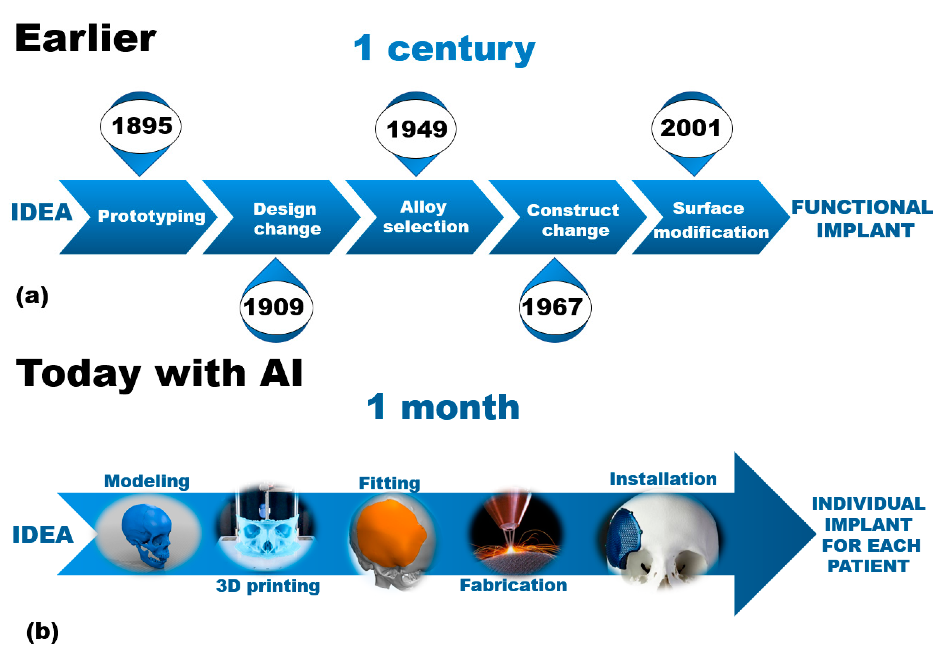
:1. Introduction
- At what stages of tissue engineering is artificial intelligence used?
- What AI tools do scientists and medical professionals use?
- How does artificial intelligence help reduce the material’s development time?
- Most importantly, how does AI improve a patient’s quality of life?
2. Prediction of Implantation Cases
3. Selection of Candidate’s Material
4. Shaping of Scaffold Construction
5. Visualization
6. Modeling of Biodegradation
7. Screening
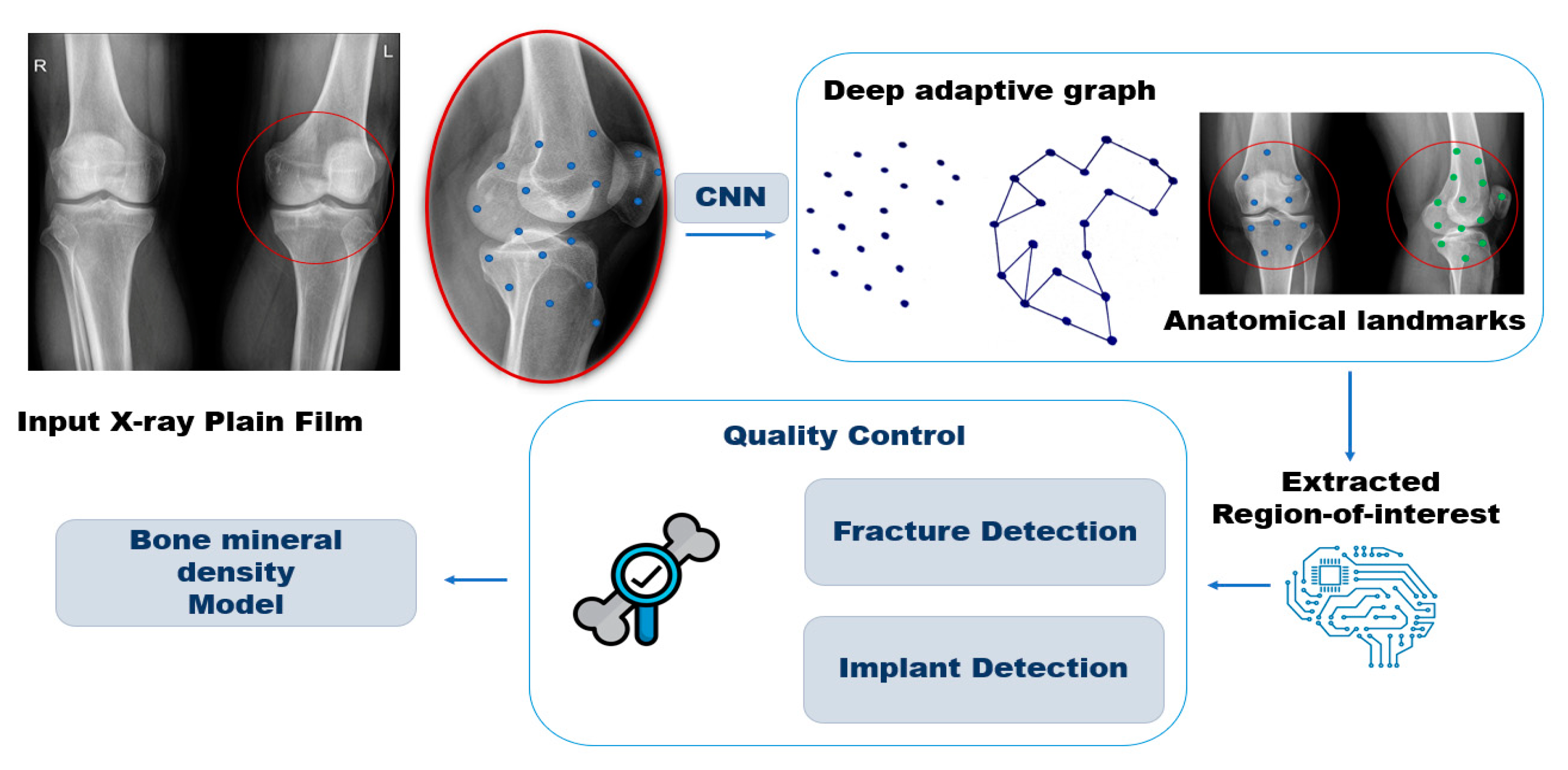
- CT scans—Also called CAT scans, computed tomography (CT) scans use special X-ray equipment to take images from different angles, which are then processed by a computer to show a cross-section of body tissues and organs.
- Fluoroscopy—A type of imaging that shows real-time, moving X-rays of the internal body structures.
- Mammogram—This involves several X-ray images of the breast.
- MRI—Magnetic resonance imaging (MRI) uses radio waves, a strong magnetic field, and a computer to generate detailed, cross-sectional images of the patient’s internal body part.
- Nuclear imaging—A diagnostic tool used to accurately visualize the flow and function of different organs of the body.
- Ultrasound imaging (or sonography)—A method of seeing inside the human body using high-frequency sound waves.
- X-rays—This type of imaging uses a minimal dose of ionizing radiation to produce pictures of the body’s internal structures.
8. Future Directions
9. Conclusions
Author Contributions
Funding
Conflicts of Interest
References
- Zhang, X.; Liu, T.; Li, Z.; Peng, W. Reconstruction with Callus Distraction for Nonunion with Bone Loss and Leg Shortening Caused by Suppurative Osteomyelitis of the Femur. J. Bone Jt. Surg. Br. 2007, 89, 1509–1514. [Google Scholar] [CrossRef] [PubMed]
- Griffin, K.S.; Davis, K.M.; McKinley, T.O.; Anglen, J.O.; Chu, T.-M.G.; Boerckel, J.D.; Kacena, M.A. Evolution of Bone Grafting: Bone Grafts and Tissue Engineering Strategies for Vascularized Bone Regeneration. Clin. Rev. Bone Miner. Metab. 2015, 13, 232–244. [Google Scholar] [CrossRef]
- Beck, R.T.; Illingworth, K.D.; Saleh, K.J. Review of Periprosthetic Osteolysis in Total Joint Arthroplasty: An Emphasis on Host Factors and Future Directions. J. Orthop. Res. 2012, 30, 541–546. [Google Scholar] [CrossRef] [PubMed]
- Burk, T.; Del Valle, J.; Finn, R.A.; Phillips, C. Maximum Quantity of Bone Available for Harvest from the Anterior Iliac Crest, Posterior Iliac Crest, and Proximal Tibia Using a Standardized Surgical Approach: A Cadaveric Study. J. Oral Maxillofac. Surg. 2016, 74, 2532–2548. [Google Scholar] [CrossRef] [PubMed]
- Collins, M.N.; Ren, G.; Young, K.; Pina, S.; Reis, R.L.; Oliveira, J.M. Scaffold Fabrication Technologies and Structure/Function Properties in Bone Tissue Engineering. Adv. Funct. Mater. 2021, 31, 2010609. [Google Scholar] [CrossRef]
- Barberi, J.; Spriano, S. Titanium and Protein Adsorption: An Overview of Mechanisms and Effects of Surface Features. Materials 2021, 14, 1590. [Google Scholar] [CrossRef]
- Ødegaard, K.S.; Torgersen, J.; Elverum, C.W. Structural and Biomedical Properties of Common Additively Manufactured Biomaterials: A Concise Review. Metals 2020, 10, 1677. [Google Scholar] [CrossRef]
- Barber, C.C.; Burnham, M.; Ojameruaye, O.; McKee, M.D. A Systematic Review of the Use of Titanium Versus Stainless Steel Implants for Fracture Fixation. OTA Int. Open Access J. Orthop. Trauma 2021, 4, e138. [Google Scholar] [CrossRef]
- Mani, G. Metallic Biomaterials: Cobalt-Chromium Alloys. In Handbook of Biomaterial Properties, 2nd ed.; Springer: New York, NY, USA, 2016; pp. 159–166. [Google Scholar] [CrossRef]
- Goodacre, C.J. Palladium-Silver Alloys: A Review of the Literature. J. Prosthet. Dent. 1989, 62, 34–37. [Google Scholar] [CrossRef]
- Sharma, A.; Waddell, J.N.; Li, K.C.; Sharma, L.A.; Prior, D.J.; Duncan, W.J. Is Titanium–Zirconium Alloy a Better Alternative to Pure Titanium for Oral Implant? Composition, Mechanical Properties, and Microstructure Analysis. Saudi Dent. J. 2021, 33, 546–553. [Google Scholar] [CrossRef]
- Salman, M.A.; Haleem, A.H.; Awad, S.H. Surface Modification of Zirconium Based Alloys for Bio Application by Micro-Arc Oxidation Process: A Review. J. Phys. Conf. Ser. 2021, 1973, 012110. [Google Scholar] [CrossRef]
- Muthaiah, V.M.S.; Indrakumar, S.; Suwas, S.; Chatterjee, K. Surface Engineering of Additively Manufactured Titanium Alloys for Enhanced Clinical Performance of Biomedical Implants: A Review of Recent Developments. Bioprinting 2022, 25, e00180. [Google Scholar] [CrossRef]
- Najafabadi, F.M.; Karbasi, S.; Benisi, S.Z.; Shojaei, S. Physical, Mechanical, and Biological Performance of Chitosan-Based Nanocomposite Coating Deposited on the Polycaprolactone-Based 3D Printed Scaffold: Potential Application in Bone Tissue Engineering. Int. J. Biol. Macromol. 2023, 243, 125218. [Google Scholar] [CrossRef] [PubMed]
- Choudhary, N.; Ghosh, C.; Sharma, V.; Roy, P.; Kumar, P. Investigations on Effect of Pore Architectures of Additively Manufactured Novel Hydroxyapatite Coated PLA/Al2O3 Composite Scaffold for Bone Tissue Engineering. Rapid Prototyp. J. 2023, 29, 1061–1079. [Google Scholar] [CrossRef]
- Park, S.; Choi, J.; Vo, T.M.T.; Mondal, S.; Vo, T.H.; Ko, N.; Kim, C.-s.; O, S.H.; Oh, J. In Vivo Mimicking Injectable Self-Setting Composite Bio-Cement: Scanning Acoustic Diagnosis and Biological Property Evaluation for Tissue Engineering Applications. Colloids Surf. B Biointerfaces 2022, 218, 112722. [Google Scholar] [CrossRef]
- Mahmoodiyan Najafabadi, F.; Karbasi, S.; Benisi, S.Z.; Shojaei, S.; Poursamar, S.A.; Nasr Azadani, R. Evaluation of the Effects of Alumina Nanowire on 3D Printed Polycaprolactone/Magnetic Mesoporous Bioactive Glass Scaffold for Bone Tissue Engineering Applications. Mater. Chem. Phys. 2023, 303, 127616. [Google Scholar] [CrossRef]
- Pina, S.; Kwon, I.K.; Reis, R.L.; Oliveira, J.M. Biocomposites and Bioceramics in Tissue Engineering: Beyond the Next Decade. In Springer Series in Biomaterials Science and Engineering; Springer Science and Business Media Deutschland GmbH: Berlin/Heidelberg, Germany, 2022; Volume 17, pp. 319–350. [Google Scholar]
- Ruys, A.J.; Cowdery, D.J.; Soh, E.K.L. Alumina: Implantable Bionics and Tissue Scaffolds. In Springer Series in Biomaterials Science and Engineering; Springer Science and Business Media Deutschland GmbH: Berlin/Heidelberg, Germany, 2022; Volume 17, pp. 281–318. [Google Scholar]
- Alam, M.; Manivannan, E.; Rizwan, M.; Gopan, G.; Mani, M.; Kannan, S. 3D Printed Polylactide-Based Zirconia-Toughened Alumina Composites: Fabrication, Mechanical, and In Vitro Evaluation. Int. J. Appl. Ceram. Technol. 2023. [Google Scholar] [CrossRef]
- Babu, M.M.; Bhemarajam, J.; Lakshmi, A.M.; Murimadugula, S.; Devi, T.I.; Sroda, M.; Özcan, M.; Rao, P.V.; Prasad, P.S. Exploring the Potential of Silica Mixed Zinc Phosphate Bioactive Glasses for Bone Regeneration: In Vitro Bioactivity and Antibacterial Activity Analysis. J. Non-Cryst. Solids 2023, 622, 122639. [Google Scholar] [CrossRef]
- Peluso, V.; D’Amora, U.; Prelipcean, A.M.; Scala, S.; Gargiulo, N.; Seciu-Grama, A.M.; Caputo, D.; De Santis, R.; Gloria, A.; Russo, T. Design of Silver Containing Mesoporous Bioactive Glass-Embedded Polycaprolactone Substrates with Antimicrobial and Bone Regenerative Properties. Mater. Today Commun. 2023, 37, 107509. [Google Scholar] [CrossRef]
- Koons, G.L.; Diba, M.; Mikos, A.G. Materials Design for Bone-Tissue Engineering. Nat. Rev. Mater. 2020, 5, 584–603. [Google Scholar] [CrossRef]
- Zhou, B.; Jiang, X.; Zhou, X.; Tan, W.; Luo, H.; Lei, S.; Yang, Y. GelMA-Based Bioactive Hydrogel Scaffolds with Multiple Bone Defect Repair Functions: Therapeutic Strategies and Recent Advances. Biomater. Res. 2023, 27, 86. [Google Scholar] [CrossRef] [PubMed]
- Khan, M.U.A.; Aslam, M.A.; Bin Abdullah, M.F.; Hasan, A.; Shah, S.A.; Stojanović, G.M. Recent Perspective of Polymeric Biomaterial in Tissue Engineering—A Review. Mater. Today Chem. 2023, 34, 101818. [Google Scholar] [CrossRef]
- Toloue, E.B.; Mohammadalipour, M.; Mukherjee, S.; Karbasi, S. Ultra-thin Electrospun Nanocomposite Scaffold of Poly (3-Hydroxybutyrate)-Chitosan/Magnetic Mesoporous Bioactive Glasses for Bone Tissue Engineering Applications. Int. J. Biol. Macromol. 2024, 254, 127860. [Google Scholar] [CrossRef] [PubMed]
- Sadreddini, S.; Jodati, H.; Evis, Z.; Keskin, D. Novel Barium-Doped-Baghdadite Incorporated PHBV-PCL Composite Fibrous Scaffolds for Bone Tissue Engineering. J. Mech. Behav. Biomed. Mater. 2023, 148, 106185. [Google Scholar] [CrossRef] [PubMed]
- Batchelar, D.L.; Davidson, M.T.; Dabrowski, W.; Cunningham, I.A. Bone-Composition Imaging Using Coherent-Scatter Computed Tomography: Assessing Bone Health Beyond Bone Mineral Density. Med. Phys. 2006, 33, 904–915. [Google Scholar] [CrossRef] [PubMed]
- Malmberg, P.; Nygren, H. Methods for the Analysis of the Composition of Bone Tissue, with a Focus on Imaging Mass Spectrometry (TOF-SIMS). Proteomics 2008, 8, 3755–3762. [Google Scholar] [CrossRef] [PubMed]
- Cross, L.M.; Thakur, A.; Jalili, N.A.; Detamore, M.; Gaharwar, A.K. Nanoengineered Biomaterials for Repair and Regeneration of Orthopedic Tissue Interfaces. Acta Biomater. 2016, 42, 2–17. [Google Scholar] [CrossRef]
- Habibovic, P.; Kruyt, M.C.; Juhl, M.V.; Clyens, S.; Martinetti, R.; Dolcini, L.; Theilgaard, N.; van Blitterswijk, C.A. Comparative In Vivo Study of Six Hydroxyapatite-Based Bone Graft Substitutes. J. Orthop. Res. 2008, 26, 1363–1370. [Google Scholar] [CrossRef]
- Kokubo, T.; Takadama, H. How Useful is SBF in Predicting In Vivo Bone Bioactivity? Biomaterials 2006, 27, 2907–2915. [Google Scholar] [CrossRef]
- Marini, E.; Ballanti, P.; Silvestrini, G.; Valdinucci, F.; Bonucci, E. The Presence of Different Growth Factors Does Not Influence Bone Response to Hydroxyapatite: Preliminary Results. J. Orthop. Traumatol. 2004, 5, 34–43. [Google Scholar] [CrossRef]
- O’Hare, P.; Meenan, B.J.; Burke, G.A.; Byrne, G.; Dowling, D.; Hunt, J.A. Biological Responses to Hydroxyapatite Surfaces Deposited Via a Co-Incident Microblasting Technique. Biomaterials 2010, 31, 515–522. [Google Scholar] [CrossRef] [PubMed]
- Lv, Q.; Nair, L.; Laurencin, C.T. Fabrication, Characterization, and In Vitro Evaluation of Poly(Lactic Acid Glycolic Acid)/Nano-Hydroxyapatite Composite Microsphere-Based Scaffolds for Bone Tissue Engineering in Rotating Bioreactors. J. Biomed. Mater. Res. A 2009, 91, 679–691. [Google Scholar] [CrossRef] [PubMed]
- Seol, Y.-J.; Kim, J.Y.; Park, E.K.; Kim, S.-Y.; Cho, D.-W. Fabrication of a Hydroxyapatite Scaffold for Bone Tissue Regeneration Using Microstereolithography and Molding Technology. Microelectron. Eng. 2009, 86, 1443–1446. [Google Scholar] [CrossRef]
- Hennink, W.E.; van Nostrum, C.F. Novel Crosslinking Methods to Design Hydrogels. Adv. Drug Deliv. Rev. 2002, 54, 13–36. [Google Scholar] [CrossRef]
- Dorozhkin, S.V. Nanosized and Nanocrystalline Calcium Orthophosphates. Acta Biomater. 2010, 6, 715–734. [Google Scholar] [CrossRef]
- Zhou, H.; Lee, J. Nanoscale Hydroxyapatite Particles for Bone Tissue Engineering. Acta Biomater. 2011, 7, 2769–2781. [Google Scholar] [CrossRef]
- Akiyama, N.; Patel, K.D.; Jang, E.J.; Shannon, M.R.; Patel, R.; Patel, M.; Perriman, A.W. Tubular Nanomaterials for Bone Tissue Engineering. J. Mater. Chem. B 2023, 11, 6225–6248. [Google Scholar] [CrossRef]
- González Rodríguez, O.A.; Ramírez Guerrero, N.C.; Casañas Pimentel, R.G.; Jaime Fonseca, M.R.; San Martín Martínez, E. Polycaprolactone, Polylactic Acid, and Nanohydroxyapatite Scaffolds Obtained by Electrospinning and 3D Printing for Tissue Engineering. Int. J. Polym. Mater. Polym. Biomater. 2023, 1–12. [Google Scholar] [CrossRef]
- Zhang, X.; Yang, Y.; Yang, Z.; Ma, R.; Aimaijiang, M.; Xu, J.; Zhang, Y.; Zhou, Y. Four-Dimensional Printing and Shape Memory Materials in Bone Tissue Engineering. Int. J. Mol. Sci. 2023, 24, 814. [Google Scholar] [CrossRef]
- Hench, L.L.; Polak, J.M. Third-Generation Biomedical Materials. Science 2002, 295, 1014–1017. [Google Scholar] [CrossRef]
- Dorj, B.; Won, J.E.; Purevdorj, O.; Patel, K.D.; Kim, J.H.; Lee, E.J.; Kim, H.W. A Novel Therapeutic Design of Microporous-Structured Biopolymer Scaffolds for Drug Loading and Delivery. Acta Biomater. 2014, 10, 1238–1250. [Google Scholar] [CrossRef] [PubMed]
- Swetha, M.; Sahithi, K.; Moorthi, A.; Srinivasan, N.; Ramasamy, K.; Selvamurugan, N. Biocomposites Containing Natural Polymers and Hydroxyapatite for Bone Tissue Engineering. Int. J. Biol. Macromol. 2010, 47, 1–4. [Google Scholar] [CrossRef] [PubMed]
- Tu, J.; Wang, H.; Li, H.; Dai, K.; Wang, J.; Zhang, X. The In Vivo Bone Formation by Mesenchymal Stem Cells in Zein Scaffolds. Biomaterials 2009, 30, 4369–4376. [Google Scholar] [CrossRef] [PubMed]
- Pais, A.I.; Belinha, J.; Alves, J.L. Advances in Computational Techniques for Bio-Inspired Cellular Materials in the Field of Biomechanics: Current Trends and Prospects. Materials 2023, 16, 3946. [Google Scholar] [CrossRef] [PubMed]
- Merayo, D.; Rodriguez-Prieto, A.; Camacho, A.M. Prediction of Mechanical Properties by Artificial Neural Networks to Characterize the Plastic Behavior of Aluminum Alloys. Materials 2020, 13, 5227. [Google Scholar] [CrossRef] [PubMed]
- Sun, C.; Dong, E.; Chen, J.; Zheng, J.; Kang, J.; Jin, Z.; Liu, C.; Wang, L.; Li, D. The Promotion of Mechanical Properties by Bone Ingrowth in Additive-Manufactured Titanium Scaffolds. J. Funct. Biomater. 2022, 13, 127. [Google Scholar] [CrossRef] [PubMed]
- Campoli, G.; Borleffs, M.S.; Amin Yavari, S.; Wauthle, R.; Weinans, H.; Zadpoor, A.A. Mechanical Properties of Open-Cell Metallic Biomaterials Manufactured Using Additive Manufacturing. Mater. Des. 2013, 49, 957–965. [Google Scholar] [CrossRef]
- Shelat, A.A.; Guy, R.K. Scaffold Composition and Biological Relevance of Screening Libraries. Nat. Chem. Biol. 2007, 3, 442–446. [Google Scholar] [CrossRef]
- Fitzgerald, S.H.; Sabat, M.; Geysen, H.M. Diversity Space and Its Application to Library Selection and Design. J. Chem. Inf. Model. 2006, 46, 1588–1597. [Google Scholar] [CrossRef]
- Kaoud, H.A.E.-S. Introductory Chapter: Concepts of Tissue Regeneration. In Tissue Regeneration; IntechOpen: London, UK, 2018. [Google Scholar] [CrossRef]
- Lucidi, G.A.; Grassi, A.; Agostinone, P.; Di Paolo, S.; Dal Fabbro, G.; D’Alberton, C.; Pizza, N.; Zaffagnini, S. Risk Factors Affecting the Survival Rate of Collagen Meniscal Implant for Partial Meniscal Deficiency: An Analysis of 156 Consecutive Cases at a Mean 10 Years of Follow-up. Am. J. Sports Med. 2022, 50, 2900–2908. [Google Scholar] [CrossRef]
- Reddy, S.; Fox, J.; Purohit, M.P. Artificial Intelligence-Enabled Healthcare Delivery. J. R. Soc. Med. 2019, 112, 22–28. [Google Scholar] [CrossRef] [PubMed]
- Chauhan, U.; Shah, A. Topic Modeling Using Latent Dirichlet allocation. ACM Comput. Surv. 2021, 54, 1–35. [Google Scholar] [CrossRef]
- Angelov, D. Top2Vec: Distributed Representations of Topics. arXiv 2020, arXiv:2008.09470. [Google Scholar]
- Ayyadevara, V.K. Pro Machine Learning Algorithms: A Hands-On Approach to Implementing Algorithms in Python and R; Apress: Berkeley, CA, USA, 2018. [Google Scholar]
- Wolber, G.; Seidel, T.; Bendix, F.; Langer, T. Molecule-Pharmacophore Superpositioning and Pattern Matching in Computational drug design. Drug Discov. Today 2008, 13, 23–29. [Google Scholar] [CrossRef]
- Grootendorst, M. BERTopic: Neural Topic Modeling with a Class-Based TF-IDF Procedure. arXiv 2022, arXiv:2203.05794. [Google Scholar]
- Leslie, W.D.; Morin, S.N. New Developments in Fracture Risk Assessment for Current Osteoporosis Reports. Curr. Osteoporos. Rep. 2020, 18, 115–129. [Google Scholar] [CrossRef] [PubMed]
- Compston, J.E.; McClung, M.R.; Leslie, W.D. Osteoporosis. Lancet 2019, 393, 364–376. [Google Scholar] [CrossRef]
- Black, D.M.; Cauley, J.A.; Wagman, R.; Ensrud, K.; Fink, H.A.; Hillier, T.A.; Lui, L.Y.; Cummings, S.R.; Schousboe, J.T.; Napoli, N. The Ability of a Single BMD and Fracture History Assessment to Predict Fracture Over 25 Years in Postmenopausal Women: The Study of Osteoporotic Fractures. J. Bone Miner. Res. 2018, 33, 389–395. [Google Scholar] [CrossRef]
- Nguyen, T.V. Individualized Fracture Risk Assessment: State-of-the-Art and Room for Improvement. Osteoporos. Sarcopenia 2018, 4, 2–10. [Google Scholar] [CrossRef]
- Beaudoin, C.; Moore, L.; Gagne, M.; Bessette, L.; Ste-Marie, L.G.; Brown, J.P.; Jean, S. Performance of Predictive Tools to Identify Individuals at Risk of Non-Traumatic Fracture: A Systematic Review, Meta-Analysis, and Meta-Regression. Osteoporos. Int. 2019, 30, 721–740. [Google Scholar] [CrossRef]
- El-Hajj Fuleihan, G.; Chakhtoura, M.; Cauley, J.A.; Chamoun, N. Worldwide Fracture Prediction. J. Clin. Densitom. 2017, 20, 397–424. [Google Scholar] [CrossRef] [PubMed]
- Holzinger, A.; Langs, G.; Denk, H.; Zatloukal, K.; Muller, H. Causability and Explainability of Artificial Intelligence in Medicine. Wiley Interdiscip. Rev. Data Min. Knowl. Discov. 2019, 9, e1312. [Google Scholar] [CrossRef] [PubMed]
- Alharbi, M.T.; Almutiq, M.M. Prediction of Dental Implants Using Machine Learning Algorithms. J. Healthc. Eng. 2022, 2022, 7307675. [Google Scholar] [CrossRef]
- Lyakhov, P.A.; Dolgalev, A.A.; Lyakhova, U.A.; Muraev, A.A.; Zolotayev, K.E.; Semerikov, D.Y. Neural Network System for Analyzing Statistical Factors of Patients for Predicting the Survival of Dental Implants. Front. Neuroinform 2022, 16, 1067040. [Google Scholar] [CrossRef] [PubMed]
- Kubinyi, H. Computer Applications in Pharmaceutical Research and Development; John Wiley & Sons: Hoboken, NJ, USA, 2006. [Google Scholar] [CrossRef]
- Bohm, H.J.; Flohr, A.; Stahl, M. Scaffold Hopping. Drug Discov. Today Technol. 2004, 1, 217–224. [Google Scholar] [CrossRef] [PubMed]
- Langdon, S.R.; Ertl, P.; Brown, N. Bioisosteric Replacement and Scaffold Hopping in Lead Generation and Optimization. Mol. Inform. 2010, 29, 366–385. [Google Scholar] [CrossRef]
- Mauser, H.; Guba, W. Recent Developments in De Novo Design and Scaffold Hopping. Curr. Opin. Drug Discov. Devel. 2008, 11, 365–374. [Google Scholar]
- Schneider, G.; Schneider, P.; Renner, S. Scaffold-Hopping: How Far Can You Jump? QSAR Comb. Sci. 2006, 25, 1162–1171. [Google Scholar] [CrossRef]
- Bandyopadhyay, A.; Mitra, I.; Bose, S. 3D Printing for Bone Regeneration. Curr. Osteoporos. Rep. 2020, 18, 505–514. [Google Scholar] [CrossRef]
- Coulter, F.B.; Schaffner, M.; Faber, J.A.; Rafsanjani, A.; Smith, R.; Appa, H.; Zilla, P.; Bezuidenhout, D.; Studart, A.R. Bioinspired Heart Valve Prosthesis Made by Silicone Additive Manufacturing. Matter 2019, 1, 266–279. [Google Scholar] [CrossRef]
- Ozan, S.; Munir, K.; Biesiekierski, A.; Ipek, R.; Li, Y.; Wen, C. 1.3.3A—Titanium Alloys, Including Nitinol. In Biomaterials Science, 4th ed.; Wagner, W.R., Sakiyama-Elbert, S.E., Zhang, G., Yaszemski, M.J., Eds.; Academic Press: Cambridge, MA, USA, 2020; pp. 229–247. [Google Scholar] [CrossRef]
- Wang, Z.; Wang, Y.; Yan, J.; Zhang, K.; Lin, F.; Xiang, L.; Deng, L.; Guan, Z.; Cui, W.; Zhang, H. Pharmaceutical Electrospinning and 3D Printing Scaffold Design for Bone Regeneration. Adv. Drug Deliv. Rev. 2021, 174, 504–534. [Google Scholar] [CrossRef]
- Derby, B. Printing and Prototyping of Tissues and Scaffolds. Science 2012, 338, 921–926. [Google Scholar] [CrossRef]
- Jones, J.R.; Lin, S.; Yue, S.; Lee, P.D.; Hanna, J.V.; Smith, M.E.; Newport, R.J. Bioactive Glass Scaffolds for Bone Regeneration and Their Hierarchical Characterisation. Proc. Inst. Mech. Eng. H. 2010, 224, 1373–1387. [Google Scholar] [CrossRef]
- Lenas, P.; Moos, M.; Luyten, F.P. Developmental Engineering: A New Paradigm for the Design and Manufacturing of Cell-Based Products. Part II: From Genes to Networks: Tissue Engineering from the Viewpoint of Systems Biology and Network Science. Tissue Eng. Part. B Rev. 2009, 15, 395–422. [Google Scholar] [CrossRef]
- Ingber, D.E.; Mow, V.C.; Butler, D.; Niklason, L.; Huard, J.; Mao, J.; Yannas, I.; Kaplan, D.; Vunjak-Novakovic, G. Tissue Engineering and Developmental Biology: Going Biomimetic. Tissue Eng. 2006, 12, 3265–3283. [Google Scholar] [CrossRef]
- Kaplan, D.L.; Moon, R.T.; Vunjak-Novakovic, G. It Takes a Village to Grow a Tissue. Nat. Biotechnol. 2005, 23, 1237–1239. [Google Scholar] [CrossRef]
- Aittokallio, T.; Schwikowski, B. Graph-Based Methods for Analysing Networks in Cell Biology. Brief. Bioinform. 2006, 7, 243–255. [Google Scholar] [CrossRef]
- Albert, R. Network Inference, Analysis, and Modeling in Systems Biology. Plant Cell 2007, 19, 3327–3338. [Google Scholar] [CrossRef]
- Lygeros, J.; Johansson, K.H.; Simic, S.N.; Jun, Z.; Sastry, S.S. Dynamical Properties of Hybrid Automata. IEEE Trans. Autom. Control 2003, 48, 2–17. [Google Scholar] [CrossRef]
- Tenazinha, N.; Vinga, S. A Survey on Methods for Modeling and Analyzing Integrated Biological Networks. IEEE/ACM Trans. Comput. Biol. Bioinform. 2011, 8, 943–958. [Google Scholar] [CrossRef]
- Rullmann, J.A.; Struemper, H.; Defranoux, N.A.; Ramanujan, S.; Meeuwisse, C.M.; van Elsas, A. Systems Biology for Battling Rheumatoid Arthritis: Application of the Entelos PhysioLab Platform. Syst. Biol. 2005, 152, 256–262. [Google Scholar] [CrossRef]
- Sansalone, V.; Naili, S.; Bousson, V.; Bergot, C.; Peyrin, F.; Zarka, J.; Laredo, J.D.; Haiat, G. Determination of the Heterogeneous Anisotropic Elastic Properties of Human Femoral Bone: From Nanoscopic to Organ Scale. J. Biomech. 2010, 43, 1857–1863. [Google Scholar] [CrossRef]
- Podshivalov, L.; Fischer, A.; Bar-Yoseph, P.Z. 3D Hierarchical Geometric Modeling and Multiscale FE Analysis as a Base for Individualized Medical Diagnosis of Bone Structure. Bone 2011, 48, 693–703. [Google Scholar] [CrossRef]
- Podshivalov, L.; Fischer, A.; Bar-Yoseph, P.Z. On the Road to Personalized Medicine: Multiscale Computational Modeling of Bone Tissue. Arch. Comput. Methods Eng. 2014, 21, 399–479. [Google Scholar] [CrossRef]
- Gao, X.; Fraulob, M.; Haiat, G. Biomechanical Behaviours of the Bone-Implant Interface: A Review. J. R. Soc. Interface 2019, 16, 20190259. [Google Scholar] [CrossRef]
- Korabi, R.; Shemtov-Yona, K.; Dorogoy, A.; Rittel, D. The Failure Envelope Concept Applied To The Bone-Dental Implant System. Sci. Rep. 2017, 7, 2051. [Google Scholar] [CrossRef]
- Rittel, D.; Dorogoy, A.; Shemtov-Yona, K. Modeling the Effect of Osseointegration on Dental Implant Pullout and Torque Removal Tests. Clin. Implant. Dent. Relat. Res. 2018, 20, 683–691. [Google Scholar] [CrossRef]
- Eser, A.; Akça, K.; Eckert, S.; Cehreli, M.C. Nonlinear Finite Element Analysis Versus Ex Vivo Strain Gauge Measurements on Immediately Loaded Implants. Int. J. Oral Maxillofac. Implant. 2009, 24, 439–446. [Google Scholar]
- Imran, R.; Al Rashid, A.; Koç, M. Review on Computational Modeling for the Property, Process, Product and Performance (PPPP) Characteristics of Additively Manufactured Porous Magnesium Implants. Bioprinting 2022, 28, e00236. [Google Scholar] [CrossRef]
- Guo, A.X.Y.; Cheng, L.; Zhan, S.; Zhang, S.; Xiong, W.; Wang, Z.; Wang, G.; Cao, S.C. Biomedical Applications of the Powder-Based 3D Printed Titanium Alloys: A Review. J. Mater. Sci. Technol. 2022, 125, 252–264. [Google Scholar] [CrossRef]
- Parthasarathy, J.; Parthiban, J.K. Rapid Prototyping in Custom Fabrication of Titanium Mesh Implants for Large Cranial Defects. In Proceedings of the RAPID 2008 Society of Manufacturing Engineers, Lake Buena Vista, FL, USA, 20–22 May 2008. [Google Scholar]
- Chen, J.-J.; Liu, W.; Li, M.-Z.; Wang, C.-T. Digital Manufacture of Titanium Prosthesis for Cranioplasty. Int. J. Adv. Manuf. Technol. 2005, 27, 1148–1152. [Google Scholar] [CrossRef]
- Peltola, S.M.; Melchels, F.P.; Grijpma, D.W.; Kellomaki, M. A Review of Rapid Prototyping Techniques for Tissue Engineering Purposes. Ann. Med. 2008, 40, 268–280. [Google Scholar] [CrossRef]
- Palmquist, A.; Snis, A.; Emanuelsson, L.; Browne, M.; Thomsen, P. Long-Term Biocompatibility and Osseointegration of Electron Beam Melted, Free-Form-Fabricated Solid and Porous Titanium Alloy: Experimental Studies in Sheep. J. Biomater. Appl. 2013, 27, 1003–1016. [Google Scholar] [CrossRef]
- Kumar, V.; Kaur, H.; Kumari, A.; Hooda, G.; Garg, V.; Dureja, H. Drug Delivery and Testing via 3D Printing. Bioprinting 2023, 36, e00298. [Google Scholar] [CrossRef]
- Choy, S.Y.; Sun, C.-N.; Sin, W.J.; Leong, K.F.; Su, P.-C.; Wei, J.; Wang, P. Superior Energy Absorption of Continuously Graded Microlattices by Electron Beam Additive Manufacturing. Virtual Phys. Prototyp. 2021, 16, 14–28. [Google Scholar] [CrossRef]
- Li, D.; Liao, W.; Dai, N.; Xie, Y.M. Anisotropic Design and Optimization of Conformal Gradient Lattice Structures. Comput.-Aided Des. 2020, 119, 102787. [Google Scholar] [CrossRef]
- Seharing, A.; Azman, A.H.; Abdullah, S. A Review on Integration of Lightweight Gradient Lattice Structures in Additive Manufacturing Parts. Adv. Mech. Eng. 2020, 12, 1687814020916951. [Google Scholar] [CrossRef]
- Sienkiewicz, J.; Płatek, P.; Jiang, F.; Sun, X.; Rusinek, A. Investigations on the Mechanical Response of Gradient Lattice Structures Manufactured via SLM. Metals 2020, 1, 213. [Google Scholar] [CrossRef]
- Culmone, C.; Smit, G.; Breedveld, P. Additive Manufacturing of Medical Instruments: A State-of-the-Art Review. Addit. Manuf. 2019, 27, 461–473. [Google Scholar] [CrossRef]
- Li, H.; Wang, M.; Lou, D.; Xia, W.; Fang, X. Microstructural Features of Biomedical Cobalt–Chromium–Molybdenum (CoCrMo) Alloy from Powder Bed Fusion to Aging Heat Treatment. J. Mater. Sci. Technol. 2020, 45, 146–156. [Google Scholar] [CrossRef]
- Mukherjee, P.; Cheng, K. 3D Printing and Virtual Surgical Planning in a Difficult Bonebridge Case. Virtual Phys. Prototyp. 2018, 14, 53–58. [Google Scholar] [CrossRef]
- Simoneau, C.; Terriault, P.; Jetté, B.; Dumas, M.; Brailovski, V. Development of a Porous Metallic Femoral Stem: Design, Manufacturing, Simulation and Mechanical Testing. Mater. Des. 2017, 114, 546–556. [Google Scholar] [CrossRef]
- Zhang, C.; Chen, F.; Huang, Z.; Jia, M.; Chen, G.; Ye, Y.; Lin, Y.; Liu, W.; Chen, B.; Shen, Q.; et al. Additive Manufacturing of Functionally Graded Materials: A Review. Mater. Sci. Eng. A 2019, 764, 138209. [Google Scholar] [CrossRef]
- Maconachie, T.; Leary, M.; Lozanovski, B.; Zhang, X.; Qian, M.; Faruque, O.; Brandt, M. SLM Lattice Structures: Properties, Performance, Applications and Challenges. Mater. Des. 2019, 183, 108137. [Google Scholar] [CrossRef]
- Tan, X.P.; Tan, Y.J.; Chow, C.S.L.; Tor, S.B.; Yeong, W.Y. Metallic Powder-Bed Based 3D Printing of Cellular Scaffolds for Orthopaedic Implants: A State-of-the-Art Review on Manufacturing, Topological Design, Mechanical Properties and Biocompatibility. Mater. Sci. Eng. C Mater. Biol. Appl. 2017, 76, 1328–1343. [Google Scholar] [CrossRef]
- Loh, G.H.; Pei, E.; Harrison, D.; Monzón, M.D. An Overview of Functionally Graded Additive Manufacturing. Addit. Manuf. 2018, 23, 34–44. [Google Scholar] [CrossRef]
- Wang, D.; He, G.; Tian, Y.; Ren, N.; Ni, J.; Liu, W.; Zhang, X. Evaluation of Channel-Like Porous-Structured Titanium in Mechanical Properties and Osseointegration. J. Mater. Sci. Technol. 2020, 44, 160–170. [Google Scholar] [CrossRef]
- Zhang, X.Y.; Fang, G.; Leeflang, S.; Zadpoor, A.A.; Zhou, J. Topological Design, Permeability and Mechanical Behavior of Additively Manufactured Functionally Graded Porous Metallic Biomaterials. Acta Biomater. 2019, 84, 437–452. [Google Scholar] [CrossRef] [PubMed]
- Wu, Y.C.; Kuo, C.N.; Shie, M.Y.; Su, Y.L.; Wei, L.J.; Chen, S.Y.; Huang, J.C. Structural Design and Mechanical Response of Gradient Porous Ti-6Al-4V Fabricated by Electron Beam Additive Manufacturing. Mater. Des. 2018, 158, 256–265. [Google Scholar] [CrossRef]
- Wu, Y.C.; Kuo, C.N.; Wu, T.H.; Liu, T.Y.; Chen, Y.W.; Guo, X.H.; Huang, J.C. Empirical Rule for Predicting Mechanical Properties of Ti-6Al-4V Bone Implants with Radial-Gradient Porosity Bionic Structures. Mater. Today Commun. 2021, 27, 102346. [Google Scholar] [CrossRef]
- Parthasarathy, J. 3D Modeling, Custom Implants and Its Future Perspectives in Craniofacial Surgery. Ann. Maxillofac. Surg. 2014, 4, 9–18. [Google Scholar] [CrossRef] [PubMed]
- Khosravani, M.R.; Reinicke, T. Correction to: On the Use of X-ray Computed Tomography in Assessment of 3D-Printed Components. J. Nondestruct. Eval. 2021, 40, 97. [Google Scholar] [CrossRef]
- Szeliski, R. Computer Vision; Springer: Berlin/Heidelberg, Germany, 2011. [Google Scholar] [CrossRef]
- Blum, S.M.; Lee, M.S.; Mgboji, G.E.; Funk, V.L.; Beabout, K.; Harbaugh, S.V.; Roth, P.A.; Liem, A.T.; Miklos, A.E.; Emanuel, P.A.; et al. Impact of Porous Matrices and Concentration by Lyophilization on Cell-Free Expression. ACS Synth. Biol. 2021, 10, 1116–1131. [Google Scholar] [CrossRef] [PubMed]
- Komlev, V.S.; Peyrin, F.; Mastrogiacomo, M.; Cedola, A.; Papadimitropoulos, A.; Rustichelli, F.; Cancedda, R. Kinetics of In Vivo Bone Deposition by Bone Marrow Stromal Cells into Porous Calcium Phosphate Scaffolds: An X-ray Computed Microtomography Study. Tissue Eng. 2006, 12, 3449–3458. [Google Scholar] [CrossRef] [PubMed]
- Cedola, A.; Mastrogiacomo, M.; Burghammer, M.; Komlev, V.; Giannoni, P.; Favia, A.; Cancedda, R.; Rustichelli, F.; Lagomarsino, S. Engineered Bone from Bone Marrow Stromal Cells: A Structural Study by an Advanced X-ray Microdiffraction Technique. Phys. Med. Biol. 2006, 51, N109–N116. [Google Scholar] [CrossRef] [PubMed]
- Cedola, A.; Campi, G.; Pelliccia, D.; Bukreeva, I.; Fratini, M.; Burghammer, M.; Rigon, L.; Arfelli, F.; Chang Chen, R.; Dreossi, D.; et al. Three Dimensional Visualization of Engineered Bone and Soft Tissue by Combined X-ray Micro-Diffraction and Phase Contrast Tomography. Phys. Med. Biol. 2014, 59, 189–201. [Google Scholar] [CrossRef] [PubMed]
- Eliaz, N. Degradation of Implant Materials; Springer: New York, NY, USA; Heidelberg, Germany; Dordrecht, The Netherlands; London, UK, 2012. [Google Scholar] [CrossRef]
- Barzegari, M.; Mei, D.; Lamaka, S.V.; Geris, L. Computational Modeling of Degradation Process of Biodegradable Magnesium Biomaterials. Corros. Sci. 2021, 190, 109674. [Google Scholar] [CrossRef]
- Gastaldi, D.; Sassi, V.; Petrini, L.; Vedani, M.; Trasatti, S.; Migliavacca, F. Continuum Damage Model for Bioresorbable Magnesium Alloy Devices—Application to Coronary Stents. J. Mech. Behav. Biomed. Mater. 2011, 4, 352–365. [Google Scholar] [CrossRef]
- Bajger, P.; Ashbourn, J.M.A.; Manhas, V.; Guyot, Y.; Lietaert, K.; Geris, L. Mathematical Modelling of the Degradation Behaviour of Biodegradable Metals. Biomech. Model. Mechanobiol. 2017, 16, 227–238. [Google Scholar] [CrossRef]
- da Costa-Mattos, H.S.; Bastos, I.N.; Gomes, J.A.C.P. A Simple Model for Slow Strain Rate and Constant Load Corrosion Tests of Austenitic Stainless Steel in Acid Aqueous Solution Containing Sodium Chloride. Corros. Sci. 2008, 50, 2858–2866. [Google Scholar] [CrossRef]
- Uhthoff, H.K.; Poitras, P.; Backman, D.S. Internal Plate Fixation of Fractures: Short History and Recent Developments. J. Orthop. Sci. 2006, 11, 118–126. [Google Scholar] [CrossRef] [PubMed]
- Chen, Y.; Xu, Z.; Smith, C.; Sankar, J. Recent Advances on the Development of Magnesium Alloys for Biodegradable Implants. Acta Biomater. 2014, 10, 4561–4573. [Google Scholar] [CrossRef] [PubMed]
- Guizzardi, S.; Colangelo, M.T.; Mirandola, P.; Galli, C. Modeling New Trends in Bone Regeneration, Using the BERTopic Approach. Regen. Med. 2023, 18, 719–734. [Google Scholar] [CrossRef] [PubMed]
- Hsieh, C.I.; Zheng, K.; Lin, C.; Mei, L.; Lu, L.; Li, W.; Chen, F.P.; Wang, Y.; Zhou, X.; Wang, F.; et al. Automated Bone Mineral Density Prediction and Fracture Risk Assessment Using Plain Radiographs via Deep Learning. Nat. Commun. 2021, 12, 5472. [Google Scholar] [CrossRef]
- Lundberg, S.M.; Erion, G.; Chen, H.; DeGrave, A.; Prutkin, J.M.; Nair, B.; Katz, R.; Himmelfarb, J.; Bansal, N.; Lee, S.I. From Local Explanations to Global Understanding with Explainable AI for Trees. Nat. Mach. Intell. 2020, 2, 56–67. [Google Scholar] [CrossRef] [PubMed]
- Lee, H.; Yune, S.; Mansouri, M.; Kim, M.; Tajmir, S.H.; Guerrier, C.E.; Ebert, S.A.; Pomerantz, S.R.; Romero, J.M.; Kamalian, S.; et al. An Explainable Deep-Learning Algorithm for the Detection of Acute Intracranial Haemorrhage from Small Datasets. Nat. Biomed. Eng. 2019, 3, 173–182. [Google Scholar] [CrossRef] [PubMed]
- Lamy, J.B.; Sekar, B.; Guezennec, G.; Bouaud, J.; Seroussi, B. Explainable Artificial Intelligence for Breast Cancer: A Visual Case-Based Reasoning Approach. Artif. Intell. Med. 2019, 94, 42–53. [Google Scholar] [CrossRef] [PubMed]
- Biran, O.; Cotton, C. Explanation and Justification in Machine Learning: A Survey. In Proceedings of the IJCAI-17 Workshop on Explainable AI (XAI), Melbourne, Australia, 20 August 2017. [Google Scholar]
- Lea, W.W.; Hong, S.J.; Nam, H.K.; Kang, W.Y.; Yang, Z.P.; Noh, E.J. External Validation of Deep Learning-Based Bone-Age Software: A Preliminary Study with Real World Data. Sci. Rep. 2022, 12, 1232. [Google Scholar] [CrossRef]
- Farshidfar, N.; Assar, S.; Amiri, M.A.; Sahmeddini, S.; Hamedani, S.; Zarei, M.; Tayebi, L. The Feasible Application of Microfluidic Tissue/Organ-on-a-Chip as an Impersonator of Oral Tissues and Organs: A Direction for Future Research. Bio-Des. Manufact. 2023, 6, 478–506. [Google Scholar] [CrossRef]
- Syahruddin, M.H.; Anggraeni, R.; Ana, I.D.; Ana, I.D. A Microfluidic organ-on-a-chip: Into the Next Decade of Bone Tissue Engineering Applied in Dentistry. Futur. Sci. OA 2023, 9, FSO902. [Google Scholar] [CrossRef]
- Aazmi, A.; Zhang, D.; Mazzaglia, C.; Yu, M.; Wang, Z.; Yang, H.; Huang, Y.Y.S.; Ma, L. Biofabrication Methods for Reconstructing Extracellular Matrix Mimetics. Bioact. Mater. 2024, 31, 475–496. [Google Scholar] [CrossRef] [PubMed]
- Dasgupta, Q.; Jiang, A.; Wen, A.M.; Mannix, R.J.; Man, Y.; Hall, S.; Javorsky, E.; Ingber, D.E. A Human Lung Alveolus-on-a-Chip Model of Acute Radiation-Induced Lung Injury. Nat. Commun. 2023, 14, 6506. [Google Scholar] [CrossRef] [PubMed]
- Gabbin, B.; Meraviglia, V.; Angenent, M.L.; Ward-van Oostwaard, D.; Sol, W.; Mummery, C.L.; Rabelink, T.J.; van Meer, B.J.; van den Berg, C.W.; Bellin, M. Heart and Kidney Organoids Maintain Organ-Specific Function in a Microfluidic System. Mater. Today Bio. 2023, 23, 100818. [Google Scholar] [CrossRef]
- Wiriyakulsit, N.; Keawsomnuk, P.; Thongin, S.; Ketsawatsomkron, P.; Muta, K. A model of Hepatic Steatosis with Declined Viability and Function in a Liver-Organ-on-a-Chip. Sci. Rep. 2023, 13, 17019. [Google Scholar] [CrossRef] [PubMed]
- Paek, K.; Kim, S.; Tak, S.; Kim, M.K.; Park, J.; Chung, S.; Park, T.H.; Kim, J.A. A high-Throughput Biomimetic Bone-on-a-Chip Platform with Artificial Intelligence-Assisted Image Analysis for Osteoporosis Drug Testing. Bioeng. Transl. Med. 2023, 8, e10313. [Google Scholar] [CrossRef]
- Vis, M.A.M.; Zhao, F.; Bodelier, E.S.R.; Bood, C.M.; Bulsink, J.; van Doeselaar, M.; Amirabadi, H.E.; Ito, K.; Hofmann, S. Osteogenesis and osteoclastogenesis on a chip: Engineering a self-assembling 3D coculture. Bone 2023, 173, 116812. [Google Scholar] [CrossRef] [PubMed]
- Kim, M.K.; Paek, K.; Woo, S.M.; Kim, J.A. Bone-on-a-Chip: Biomimetic Models Based on Microfluidic Technologies for Biomedical Applications. ACS Biomater. Sci. Eng. 2023, 9, 3058–3073. [Google Scholar] [CrossRef]
- Suwardi, A.; Wang, F.; Xue, K.; Han, M.Y.; Teo, P.; Wang, P.; Wang, S.; Liu, Y.; Ye, E.; Li, Z.; et al. Machine Learning-Driven Biomaterials Evolution. Adv. Mater. 2022, 34, e2102703. [Google Scholar] [CrossRef]
- Sakhakarmi, S.; Arteaga, C.; Park, J.; Cho, C. Automated Scaffolding Safety Analysis: Strain Feature Investigation Using Support Vector Machines. Can. J. Civil. Eng. 2020, 47, 921–928. [Google Scholar] [CrossRef]
- Javaid, S.; Gorji, H.T.; Soulami, K.B.; Kaabouch, N. Identification and Ranking Biomaterials for Bone Scaffolds Using Machine Learning and Promethee. Res. Biomed. Eng. 2023, 39, 129–138. [Google Scholar] [CrossRef]
- Shaikhina, T.; Khovanova, N.; Mallick, K. Artificial Neural Networks in Hard Tissue Engineering: Another Look at Age-Dependence of Trabecular Bone Properties in Osteoarthritis. In Proceedings of the IEEE-EMBS International Conference on Biomedical and Health Informatics (BHI), Valencia, Spain, 1–4 June 2014. [Google Scholar] [CrossRef]
- Jirik, M.; Gruber, I.; Moulisova, V.; Schindler, C.; Cervenkova, L.; Palek, R.; Rosendorf, J.; Arlt, J.; Bolek, L.; Dejmek, J.; et al. Semantic Segmentation of Intralobular and Extralobular Tissue from Liver Scaffold H&E Images. Sensors 2020, 20, 7063. [Google Scholar] [CrossRef] [PubMed]
- Blatt, J.; Kirkendoll, J.; Mandava, P.K.; Preston, Z.; Joyce, R.; Salary, R. An Image-Based Convolutional Neural Network Platform for the Prediction of the Porosity of Composite Bone Scaffolds, Fabricated Using Material Extrusion Additive Manufacturing. In Proceedings of the ASME 2022 International Mechanical Engineering Congress and Exposition, Columbus, OH, USA, 30 October–3 November 2022. [Google Scholar]
- Yu, C.; Jiang, J. A Perspective on Using Machine Learning in 3D Bioprinting. Int. J. Bioprint 2020, 6, 253. [Google Scholar] [CrossRef] [PubMed]
- Bermejillo Barrera, M.D.; Franco-Martinez, F.; Diaz Lantada, A. Artificial Intelligence Aided Design of Tissue Engineering Scaffolds Employing Virtual Tomography and 3D Convolutional Neural Networks. Materials 2021, 14, 5278. [Google Scholar] [CrossRef] [PubMed]
- Zhang, H.; Yang, L.; Li, C.; Wu, B.; Wang, W. ScaffoldGAN: Synthesis of Scaffold Materials based on Generative Adversarial Networks. Comput.-Aided Des. 2021, 138, 103041. [Google Scholar] [CrossRef]
- Li, C.; Yamanishi, Y. SpotGAN: A Reverse-Transformer GAN Generates Scaffold-Constrained Molecules with Property Optimization. In Proceedings of the Joint European Conference on Machine Learning and Knowledge Discovery in Databases, Turin, Italy, 18–22 September 2023; pp. 323–338. [Google Scholar] [CrossRef]
- Vallat, G.; Wang, J.; Maddux, A.; Kamgarpour, M.; Parascho, S. Reinforcement Learning for Scaffold-Free Construction of Spanning Structures. In Proceedings of the 8th ACM Symposium on Computational Fabrication, New York, NY, USA, 8–10 October 2023; p. Article 12. [Google Scholar]
- Liu, X.; Ye, K.; van Vlijmen, H.W.T.; AP, I.J.; van Westen, G.J.P. DrugEx v3: Scaffold-Constrained Drug Design with Graph Transformer-Based Reinforcement Learning. J. Cheminform. 2023, 15, 24. [Google Scholar] [CrossRef]


| AI Model | Application | Model Key Parameters | Advantages | Disadvantages |
|---|---|---|---|---|
| Machine Learning Models | ||||
| Logistic Regression | Material biocompatibility, bone health assessment [149] | Weights, bias, regularization, solver | Efficiently models relationships, controls overfitting, and ensures convergence, thereby aiding in the identification of key factors | Assumes linearity, requires tuning for optimal regularization, features solver sensitivity, has potential suboptimal convergence |
| Support Vector Machines (SVMs) | Material identification and classification [150,151] | Kernel functions, regularization parameters | Usable for real-time assessment of scaffolding structures, particularly in high-dimensional spaces | Limited scalability to large datasets, sensitivity to the choice of kernel and hyperparameters |
| Decision Trees | Cell classification, Bone health sssessment [151] | Hierarchical decision structure | Easy to interpret, handles nonlinear relationships | Prone to overfitting, sensitive to small changes in data |
| Random Forests | Comprehensive parameter evaluation [68,149,151] | Decision trees, number of estimators, max depth | Simultaneous evaluation of multiple scaffold parameters, robust against overfitting | Prone to overfitting with noisy or imbalanced datasets, potential issues with interpretability |
| Gradient Boosting | Predicting biomaterial properties [151] | Learning rate, number of trees, depth of trees, loss function | Improves model accuracy by combining weak learners, captures intricate relationships, provides feature importance insights | Sensitive to noisy data, potential overfitting with limited data, computationally intensive, requires careful hyperparameter tuning |
| Deep Learning Models | ||||
| Artificial neural networks (ANNs) | Prediction of material properties, biocompatibility assessment of implants [69,151,152] | Nonlinear modeling, intricate parameter interactions such as cell proliferation, differentiation, material properties | Adaptability, data-driven learning, accurate prediction of complex patterns in bone tissue engineering | Large dataset requirements, potential overfitting, complex neural network structures, challenging interpretation |
| Convolutional Neural Networks (CNNs) | Image analysis for screening, bone mineral density prediction, and fracture risk assessment [134,153,154] | Convolutional layers, filter sizes, pooling operations | Robust feature extraction, object detection, semantic and instance segmentation | Computational intensity, reliance on large datasets, potential overfitting in certain scenarios |
| 3D Deep Convolutional Neural Networks (DCNNs) | 3D image analysis for scaffold design, screening [155,156] | Architecture, filters, voxel size | Captures intricate spatial features in 3D medical images; enhances screening accuracy | Computationally intensive for large datasets, requires substantial computational resources, potential overfitting with limited data |
| Generative Adversarial Networks (GANs) | Scaffold design and optimization [155,157,158] | Latent space representation, generator and discriminator architectures | Enables the synthesis of realistic scaffold structures, thereby facilitating optimization processes | Susceptible to mode collapse; potential challenges in generating clinically viable structures |
| Reinforcement Learning (RL) | Scaffold fabrication process optimization [159,160] | State space, action space, reward functions | Optimizes scaffold production processes through iterative learning | Limited applicability in highly dynamic or complex environments; challenges in defining reward functions |
| Explainable AI (XAI) | Interpretable models for bone tissue screening [135,136,137,138,139] | Local and global explanations, interpretable models, feature importance, model-specific parameters, and human-understandable representations | Provides insights into model decisions; enhances trust in screening outcomes | Trade-off between accuracy and interpretability; potential complexity in explaining deep learning models |
Disclaimer/Publisher’s Note: The statements, opinions and data contained in all publications are solely those of the individual author(s) and contributor(s) and not of MDPI and/or the editor(s). MDPI and/or the editor(s) disclaim responsibility for any injury to people or property resulting from any ideas, methods, instructions or products referred to in the content. |
© 2023 by the authors. Licensee MDPI, Basel, Switzerland. This article is an open access article distributed under the terms and conditions of the Creative Commons Attribution (CC BY) license (https://creativecommons.org/licenses/by/4.0/).
Share and Cite
Kolomenskaya, E.; Butova, V.; Poltavskiy, A.; Soldatov, A.; Butakova, M. Application of Artificial Intelligence at All Stages of Bone Tissue Engineering. Biomedicines 2024, 12, 76. https://doi.org/10.3390/biomedicines12010076
Kolomenskaya E, Butova V, Poltavskiy A, Soldatov A, Butakova M. Application of Artificial Intelligence at All Stages of Bone Tissue Engineering. Biomedicines. 2024; 12(1):76. https://doi.org/10.3390/biomedicines12010076
Chicago/Turabian StyleKolomenskaya, Ekaterina, Vera Butova, Artem Poltavskiy, Alexander Soldatov, and Maria Butakova. 2024. "Application of Artificial Intelligence at All Stages of Bone Tissue Engineering" Biomedicines 12, no. 1: 76. https://doi.org/10.3390/biomedicines12010076