Article

The Human Soluble NKG2D Ligand Differentially Impacts Tumorigenicity and Progression in Temporal and Model-Dependent Modes

1
Department of Hematology/Oncology, Feinberg School of Medicine, Northwestern University, Chicago, IL 60611, USA
2
Department of Urology, Feinberg School of Medicine, Northwestern University, Chicago, IL 60611, USA
3
Department of Microbiology and Immunology, Feinberg School of Medicine, Northwestern University, Chicago, IL 60611, USA
*
Author to whom correspondence should be addressed.
These authors contributed equally to this work.
Current address: Genentech, One DNA Way, South San Francisco, CA 94080, USA.
§
Current address: Bristol Myers Squibb, 1500 Owens St Suite 500, San Francisco, CA 94158, USA.
Biomedicines 2024, 12(1), 196; https://doi.org/10.3390/biomedicines12010196
Submission received: 8 November 2023 / Revised: 25 December 2023 / Accepted: 12 January 2024 / Published: 16 January 2024

Abstract

:
NKG2D is an activating receptor expressed by all human NK cells and CD8 T cells. Harnessing the NKG2D/NKG2D ligand axis has emerged as a viable avenue for cancer immunotherapy. However, there is a long-standing controversy over whether soluble NKG2D ligands are immunosuppressive or immunostimulatory, originating from conflicting data generated from different scopes of pre-clinical investigations. Using multiple pre-clinical tumor models, we demonstrated that the impact of the most characterized human solid tumor-associated soluble NKG2D ligand, the soluble MHC I chain-related molecule (sMIC), on tumorigenesis depended on the tumor model being studied and whether the tumor cells possessed stemness-like properties. We demonstrated that the potential of tumor formation or establishment depended upon tumor cell stem-like properties irrespective of tumor cells secreting the soluble NKG2D ligand sMIC. Specifically, tumor formation was delayed or failed if sMIC-expressing tumor cells expressed low stem-cell markers; tumor formation was rapid if sMIC-expressing tumor cells expressed high stem-like cell markers. However, once tumors were formed, overexpression of sMIC unequivocally suppressed tumoral NK and CD8 T cell immunity and facilitated tumor growth. Our study distinguished the differential impacts of soluble NKG2D ligands in tumor formation and tumor progression, cleared the outstanding controversy over soluble NKG2D ligands in modulating tumor immunity, and re-enforced the viability of targeting soluble NKG2D ligands for cancer immunotherapy for established tumors.

1. Introduction

NKG2D, an activating receptor expressed by all human NK cells, also defined as a co-stimulatory receptor for human NKT, CD8T and γδT cells [1,2,3,4,5,6]. In mice, NKG2D is expressed by all NK cells, but is only expressed by activated CD8 T cells [1,2,3,4,5,6]. The function and major signaling pathways of NKG2D in human and mouse NK cells are largely conserved. However, identified natural NKG2D ligands in human and mice are diverse. In humans, identified NKG2D ligands include the MICA/B and HCMV UL-16 binding proteins ULBP1-6 [7,8,9]. These ligands are often expressed by tumor cells and viral infected cells in response to genotoxic insults or viral infection but are generally absent in normal tissues except in gut epithelium at a threshold level [5,10,11,12,13]. The MICA/B molecules are found in almost all human solid tumors, whereas the expression of ULBPs in human tumors are sporadic [7,8,9]. Homologs of human ULBPs were found to be expressed by mice; however, a homolog of human MICA or MICB was not identified in mice. It was demonstrated that mouse or human tumors with forced expression of membrane-bound NKG2D ligands can be rejected in vivo through activation of NK cells and sometimes CD8 T cells, heightening the significance of NKG2D stimulating in anti-tumor immunity [14,15]. However, the majority of human solid tumors express high levels of NKG2D ligands, in particular MICA and MICB molecules, suggesting that tumors developed mechanisms to evade NKG2DL-NKG2D mediated anti-tumor immunity.
Advanced human tumors release soluble NKG2D ligands (sNKG2D-L) via proteolytic pathways, and in some instances via exosome pathways [7,8,9,16]. It has been shown at large that human sNKG2D-L is immune suppressive. Clinical correlative studies have shown elevated serum levels of sMICA and sMICB correlated with disease progression, metastasis, less responsiveness to therapy, and ultimately poor clinical outcome in a broad spectrum of cancer patients with solid tumors [17,18,19,20,21,22,23,24,25]. In preclinical models where human MIC was overexpressed in mouse tumors, eliminating the effect of sMIC by an antibody-clearing serum sMIC or by an antibody-inhibiting MIC shedding, stimulated NK cell and/or CD8T cell anti-tumor immunity and significantly inhibited tumor progression or growth [26,27,28,29,30,31]. These clinical and pre-clinical studies support the notion that soluble human NKG2D ligands, at least sMICA and sMICB, are immune suppressive and that sMIC (A/B) is a cancer therapeutic target. In contrast to these studies, the soluble mouse NKG2D ligand, sMULT-1, was shown to stimulate antitumor immunity and prevent B16F10 melanoma tumor formation [32]. These inconsistencies in different experimental settings raised the question as to whether sNKG2D-L is immune stimulatory or inhibitory in the context of tumor and raised the concern as to whether soluble human NKG2D ligand is a cancer therapeutic target.
Harnessing NKG2D and the NKG2D ligand axis is an emerging avenue in cancer immune therapeutics [8,9,33,34]. To address the outstanding controversy and clear the pathway for therapeutic development to empower the NKG2D/NKG2D-L pathway for cancer immunotherapy, herein we utilized three mouse models to address the impact of the human sNKG2D-L, sMIC, on tumorigenesis and tumor progression. With three syngeneic tumor models that express human sMICB, we interrogated the differential impact of sMIC on tumor establishment and progression of established tumors with multiple disease models.

2. Material and Methods

2.1. Mice and Cell Lines

Cell lines of mouse melanoma B16F10, Lewis lung carcinoma LLC1, and the prostate tumor TRAMP-C2 were used in this study. All three cell lines were purchased from ATCC. The derivative cell lines, TRAMP-C2-sMICB, LLC-sMICB, and B16F10-sMICB cell lines, expressing the soluble human NKG2D ligand sMICB, were generated by transduction with an IRES-GFP retroviral vector containing the construct for recombinant soluble MICB, as described previously [35]. sMICB+ cells were selected by puromycin and further by flow cytometry sorting for GFP-positive cells and further validated by ELISA for sMICB in the culture supernatant (Supplement Figure S1). All cells were cultured in RPMI 1640 complete media with supplement of 10% FBS.

2.2. In Vivo Experiments

Mice were bred and housed under specific pathogen-free conditions in the animal facility at the Northwestern University in accordance with institutional guidelines for the approved Institutional Animal Care and Use Committee (IACUC) protocols. All mice used in this study were male rPB-MICB transgenic mice on the B6 background as previously described [36]. The rationale of using mice to be a sMIC-tumor host was previously described [36]. Briefly, the male rPB-MICB mice have the transgene of full-length human MICB in the prostate whose expression is controlled by the male hormone sensitive promoter rat probasin (rPB). As we have previously described, these mice have the identical phenotype as the wild-type B6 mice but are more tolerant to tumor cell lines expressing human sMIC, due to “endogenous” expression of MIC in the prostate [28,36]. To ensure that all tumor cells grew in the same background as the host, the rPB-MICB male mice were used for tumors. Tumor cells were implanted subcutaneously (s.c.) in the right flank region of cohorts of syngeneic rPB-MICB male mice with cell numbers indicated in each specific experiment. Mice were monitored for tumor incidence and tumor size once tumors were formed, three times per week. The tumor volume (calculated by L × W2/2) of 1800 mm3 was defined as the survival endpoint, unless otherwise specified.

2.3. Flow Cytometry Analysis

For analyzing tumoral NK and CD8 T cells, tumors were minced, gently meshed with syringe tips, and filtered through 70 μm mesh. Single-cell suspensions were first incubated for 20 min on ice with a viability dye and for 10 min with the anti-CD16/32 Fc-blocker solution, then stained with a combination of antibodies specific to cell surface markers for identification of lymphocyte subsets. These antibodies are: anti-CD45-AF700, anti-CD3-APC/Cy7 (clone 145-2C11), anti-NK1.1-PE (clone PK136), anti-CD8α-FITC (clone 53-6.7). To assess the capacity of IFN-γ production, tumor cell suspension was stimulated at 37 °C for 4 h with 50 ng/mL phorbol myristate acetate (PMA) and 500 ng/mL ionomycin with addition of 1 μM Golgi Plug during the last 2 h stimulation. Following stimulation, cells were stained with surface markers followed by fixation and permeabilization with BD Perm/Fix kits and an antibody specific to intracellular molecules IFN-γ. In some experiments, tumor cell suspension was incubated in complete RPMI 1640 media in the presence of 1 μM Golgi Plug for 2 h before being stained intracellularly for IFN-γ and granzyme B. All antibodies were mouse specific and were from BD Biosciences. Cells were analyzed using the BD Fortessa. Data were analyzed using the FlowJo v8 software (Tree Star).
For cell surface stem cell marker profiling, after incubation with the anti-CD16/32 Fc-blocker solution, tumor cells were stained with the following fluorochrome conjugated antibodies, respectively: anti-CD44-AF700 and anti-CD166-PE in combination with anti-SCA1-PE/Cy7 or anti-CD133-PE/Cy7. Each fluorochrome-conjugated isotype antibody was used as staining controls. PE-conjugated anti-mouse I-A/I-E antibody was used as a positive staining control and to examine tumor cell surface MHC I expression. For flow sorting to separate tumor cells with high and low stem cell markers, cells were stained with anti-CD44-AF700 and anti-CD166-PE for the separation of the CD44HiCD166Hi population from the CD44LoCD166Lo population. SCA-1 or CD133 expression of the sorted cell populations was examined by fluorochrome-conjugated respective antibodies.

2.4. Statistics

Wherever applicable, statistical data were expressed as mean ± standard error of the mean (SEM). Differences between means of populations were compared by an unpaired t-test. Tumor incidence was determined via a Kaplan–Meier analysis with “tumor incidence” as an occurring event. Mantel–Cox Log-rank test was used to analyze the significance level of tumor incidence among the two groups. A p-value of 0.05 or less was considered significant. GraphPad Prism software was used for all analyses.

3. Results

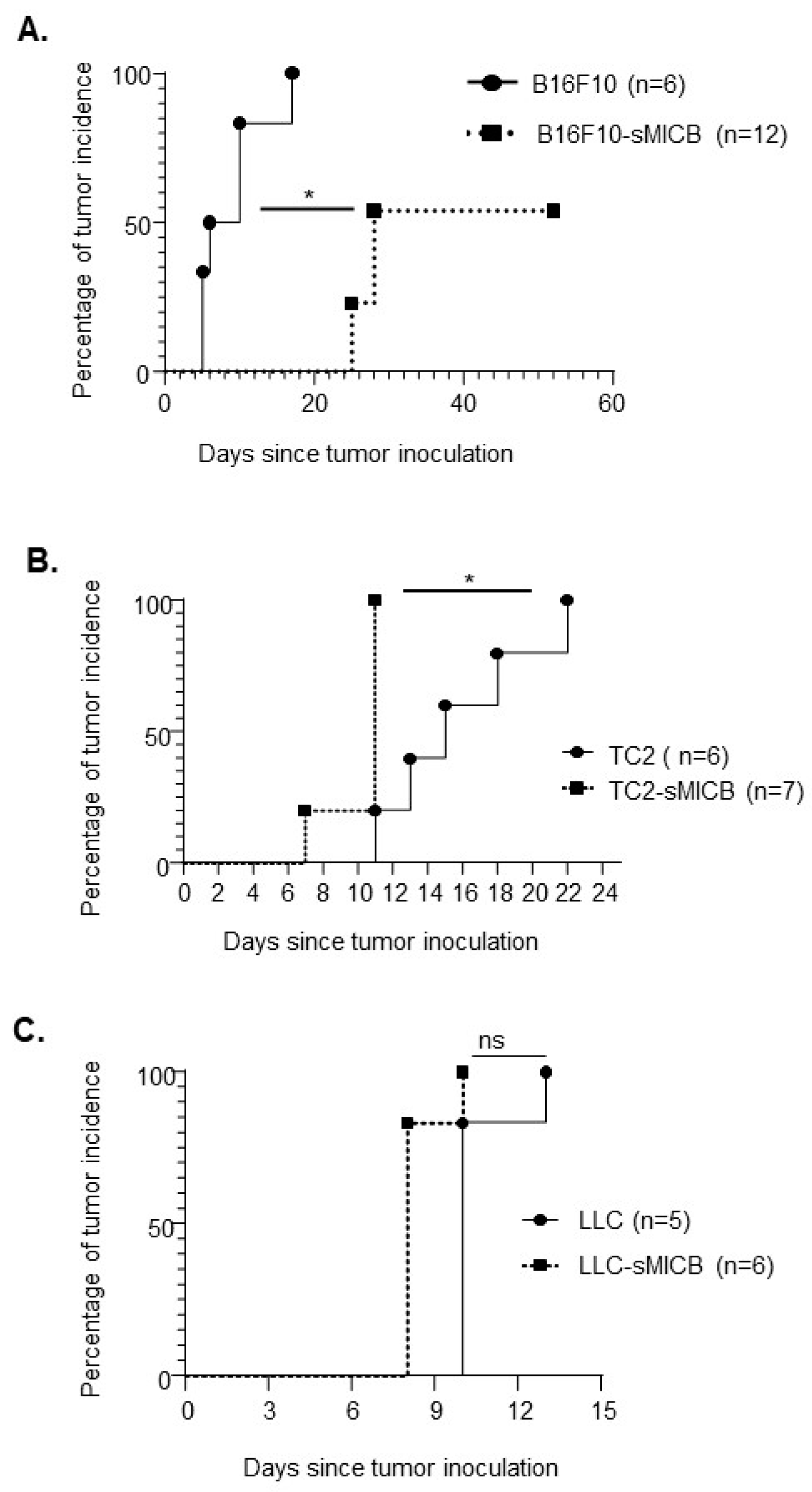
3.1. Model-Dependent Impact of Soluble Human NKG2D Ligand on Tumor Establishment

While most of the studies with human NKG2D ligands demonstrated that soluble NKG2D ligands suppress tumor immunity [29,37,38,39], controversy arose from findings that the overexpressing of soluble mouse NKG2D ligand, sMULT-1, in B16F10 tumor cells resulted in no tumor formation [32], suggesting that the role of soluble NKG2D ligands in regulating tumor immunity could be more complicated than what has been understood to date. To address the discrepancies between these study findings, we used three different syngeneic tumor models, the melanoma B16F10, the prostate tumor TRAMP-C2 (TC2), and the lung carcinoma LLC1, to compare tumor incidence between tumor cells engineered to overexpress the soluble human NKG2D ligand sMICB and the parental tumor cell lines. Secretion of sMICB was confirmed by ELISA in all three sMIC-overexpressing cell lines (Supplement Figure S1). Noteworthy, while mice do not express homologs of human NKG2D ligands MICA or MICB, human MICB or sMICB has been demonstrated to serve as a surrogate ligand for mouse NKG2D [30,31,35,36,40]. We subcutaneously implanted sMICB-expressing B16F10-sMICB, TC2-sMICB, and LLC1-sMICB and their parental counterparts B16F10, TC2, and LLC1 into syngeneic male mice that were engineered to express MICB specifically in the prostate under the androgen-sensitive promoter rat probasin (rPB, hence referred as rPB-MICB mice). Tumor incidence, time to tumor formation, and the growth dynamics of established tumors were monitored (Figure 1). The rationale for using rPB-MICB male mice as the host rather than wild type B6 mice for sMICB-expressing tumors was described previously [28,36]. To briefly reiterate, the rPB-MICB mice develop less immunity against implanted tumor cells overexpressing human MIC due to “endogenous” expression of MICB in the prostate upon puberty.
The B16F10-sMIC tumors demonstrated a significant delay in tumor establishment as compared to the parental B16F10 tumors (Figure 1A). On average, B16F10-sMICB tumors arose on day 23 post-inoculation, whereas the parental B16F10 tumors arose on average on day 6 post tumor inoculation (p < 0.05, Figure 1A). By day 26 post-tumor injection, tumors were established in 50% of mice inoculated with B16F10-sMICB tumors. These observations demonstrated that overexpression of the soluble human NKG2D ligand sMICB in B16F10 tumors resulted in a delay in tumor establishment, not the complete tumor rejection as being reported [32].
In contrast to the B16F10-sMICB melanoma model, the prostate TRAMP-C2-sMICB (TC2-sMICB) cells presented a significantly more rapid tumor onset than the parental TRAMP-C2 (TC2) cells after inoculation (p < 0.05, Figure 1B). Specifically, TC2-sMICB tumors arose as early as day 7 post inoculation and reached 100% tumor penetration by day 11, whereas parental TC2 tumor onset did not occur until day 11 and did not reach 100% tumor penetration until day 22 post inoculation (p < 0.05, Figure 1B). Interestingly, different from the B16F10-sMICB or the TC2-sMICB models, no significant difference in tumor establishment or tumor onset was observed between LLC-sMICB and the parental LLC tumors. LLC-sMICB and the parental LLC tumors displayed a similar timeline in tumor establishment, both of which reached 100% tumor penetration within 12 days post tumor inoculation (Figure 1C). Together, these data demonstrated a model-dependent effect of sMICB on tumorigenicity or tumor initiation.

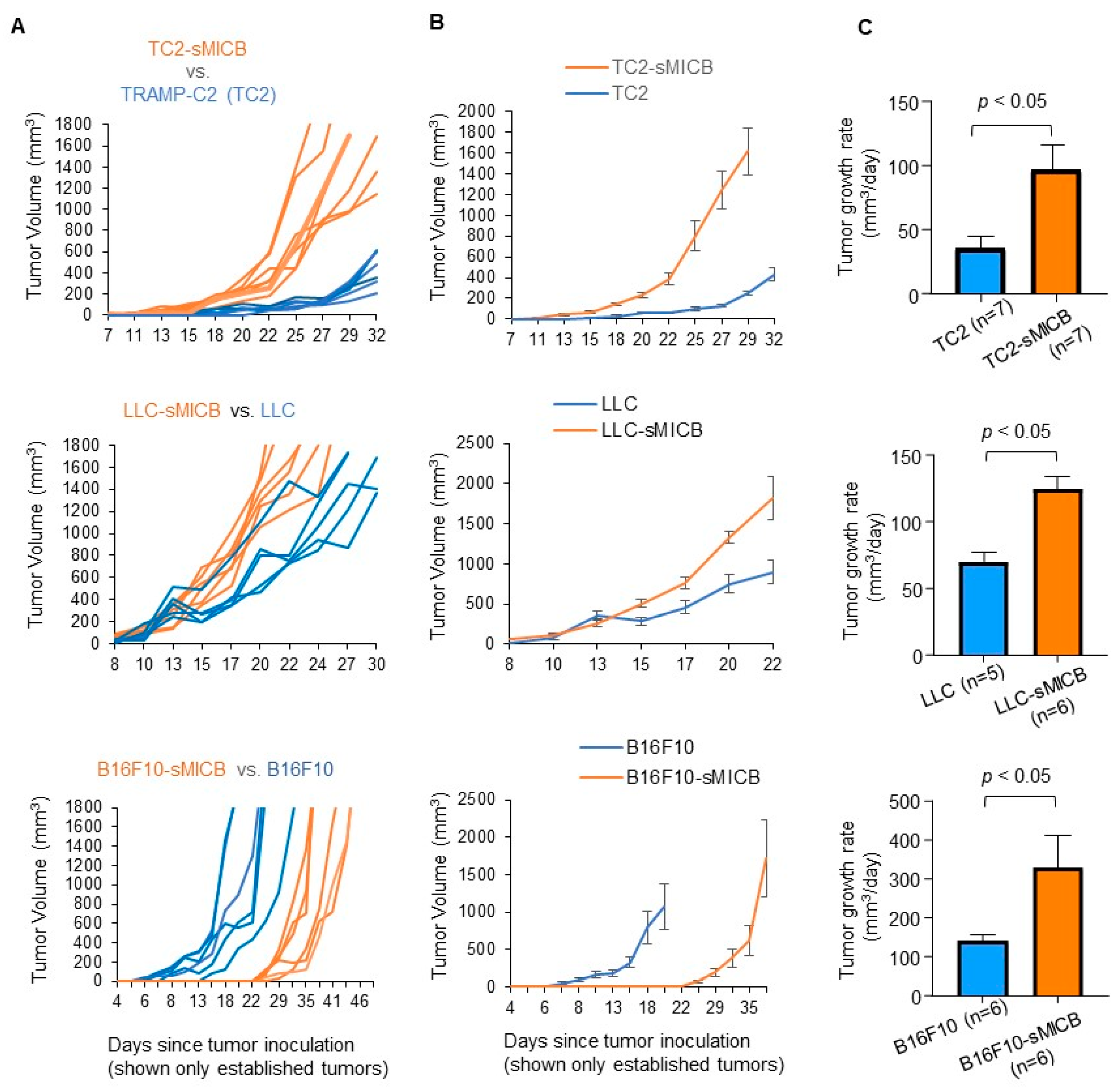
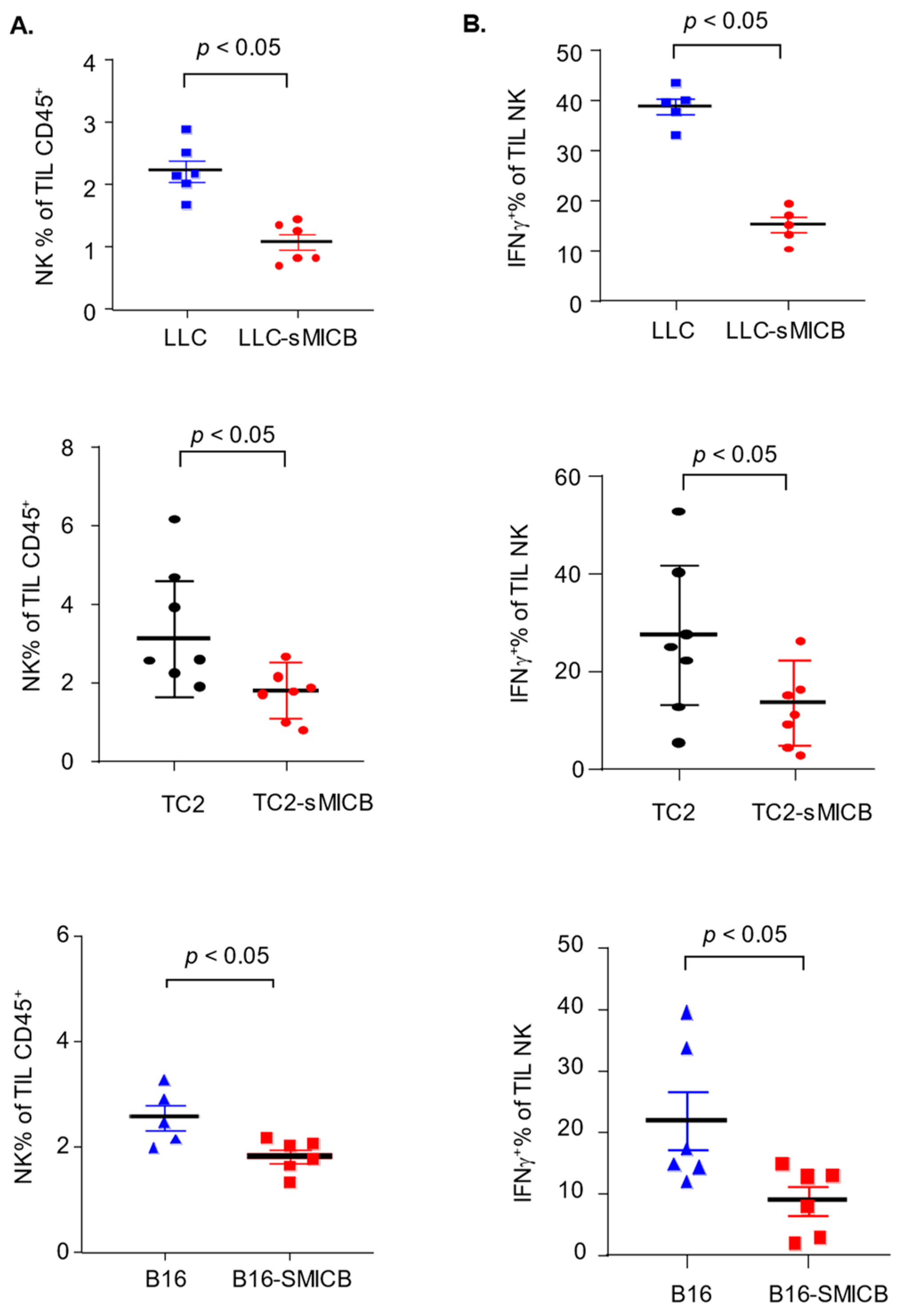
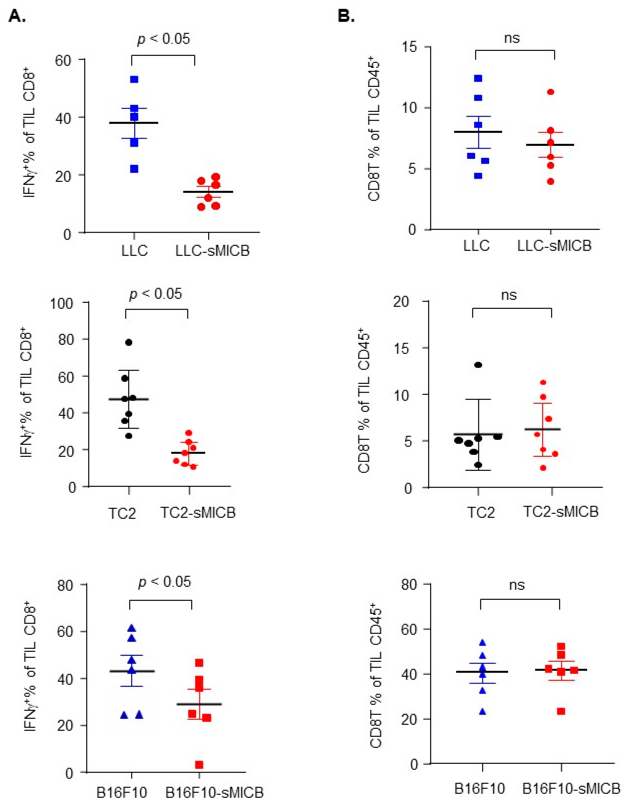
3.2. Model-Independent Impact of sMIC on the Growth of Established Tumors and on the Function of Tumoral Effector Cells

Irrespective of tumor incidence or the time of tumor onset, once tumors were initiated or formed, in all three disease models, sMIC-overexpressing tumors grew at a significantly more aggressive rate than the parental sMIC-negative tumors (Figure 2). The accelerated tumor growth with sMIC overexpression is in agreement with previous studies by multiple investigators in various experimental settings demonstrating that tumor produced sMIC can significantly comprise NK and CD8 T cell function [16,20,22,24,26,27,30,31,36,41]. To validate that the more aggressive tumor growth of sMICB-expressing tumors in current experimental settings is associated with impairment of NK and CD8 T cell immunity, we assayed the function of tumor-infiltrated NK and CD8 T cells (Supplement Figure S2), both of which express the receptor NKG2D for sMICB and are critical anti-tumor effector cells. In all three models, there was a significant reduction in NK cell content in tumor infiltrates when tumor cells expressed sMICB (Figure 3A, p < 0.05 for all models), which is consistent with the previous finding in the TRAMP/MICB autochthonous tumor model that tumor-produced sMIC impairs the NK cell’s homeostatic self-renewal ability [36]. NK cells in sMICB-expressing tumors had a significantly reduced response to PMA/I stimulation as measured by IFNγ expression (Figure 3B, p < 0.05 for all models), one of the key effector molecules for NK cell mediating anti-tumor immunity. sMIC expression also significantly impaired tumoral CD8 T cell response to PMA/I stimulation as measured by IFNγ expression (Figure 4A, p < 0.05 in all models); however, CD8 T cell content in tumor infiltrates was not significantly impacted by sMICB (Figure 4B). The impaired response to external PMA/I stimulation ultimately signifies the comprised ability of these cells to respond to tumor-specific stimulations in vivo. To support this concept, we conducted representative analyses of TRAMP-C2 and TRAMP-C2-sMICB tumoral NK and CD8 T cell IFNγ and granzyme B expression in an ex vivo setting without PMA/I stimulation. Corroboratively, the ability of tumoral NK and CD8 T cells to produce IFNγ and granzyme B without PMA/I stimulation was shown to be significantly compromised in TRAMP-C2-sMICB tumors as compared to TRAMP-C2 tumors (Supplement Figure S3). Collectively, these data re-reinforce the concept that soluble human NKG2D ligands negatively regulate anti-tumor NK and CD8 T immunity in established tumors with more profound impact on NK cells.

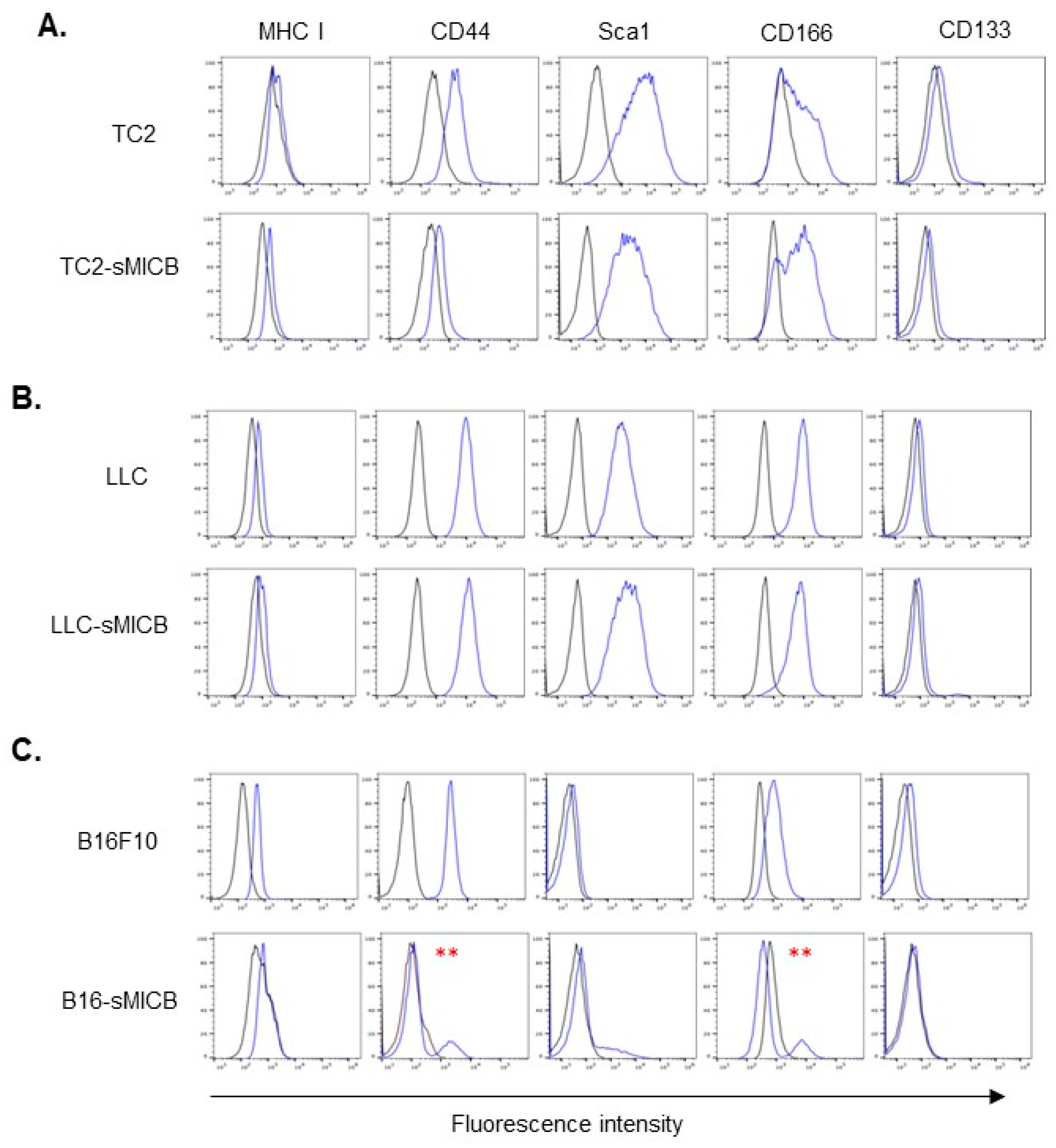
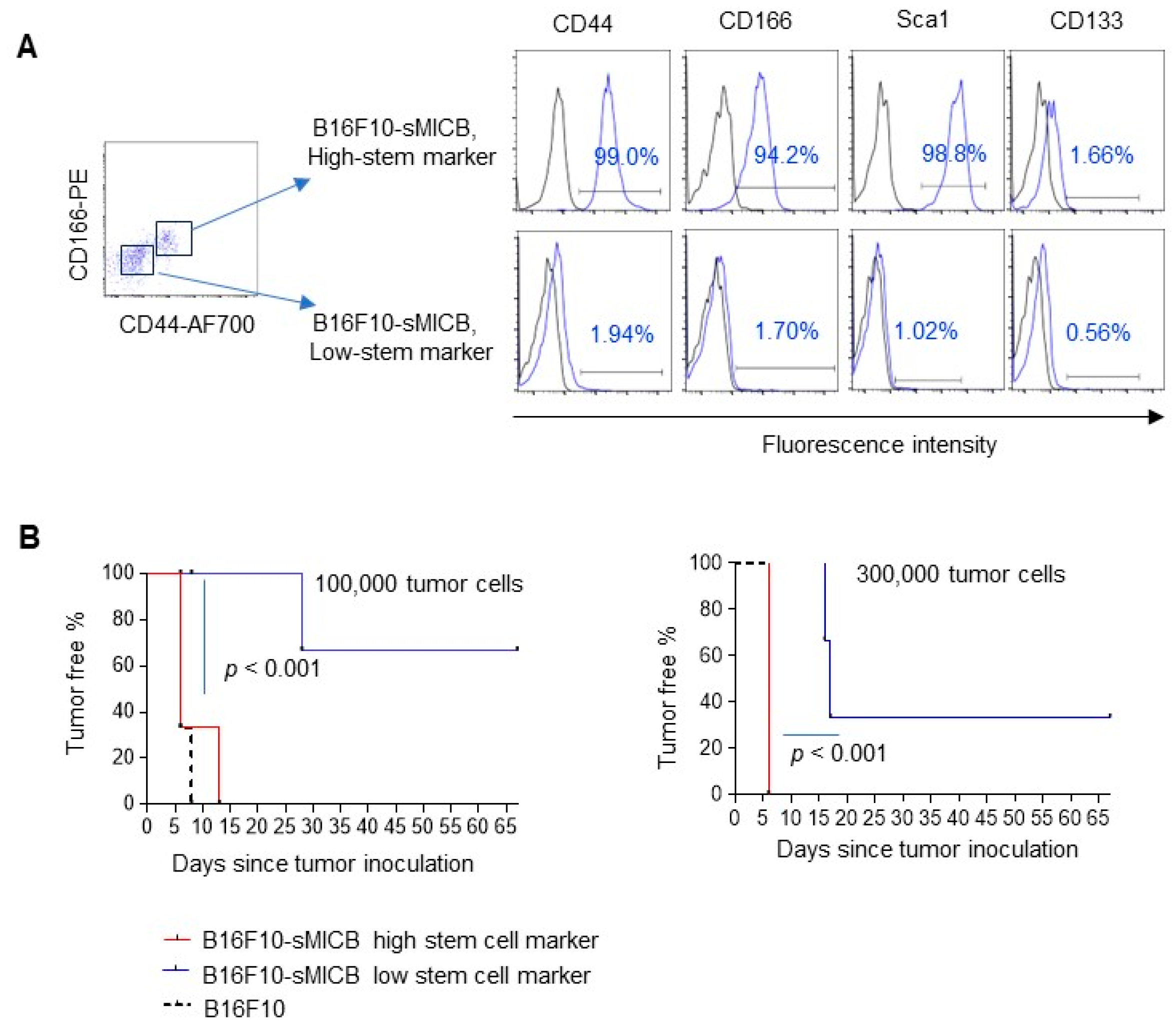
3.3. Tumor Cell Stem-like Property Rather Than sMIC Expression Determines the Ability of In Vivo Tumor Establishment

Our data in three different models demonstrated that sMIC facilitates growth or progression once tumors are formed; however, the impact of sMIC on the ability of tumor formation or time required for tumor formation appeared to be model-dependent. We further sought to understand the discretionary impact of sMIC on established tumor growth versus the potential of tumor formation. Given that stem-like features could profoundly enhance the ability of tumor cells to colonize and form tumors in vivo [42,43,44], we thus examined the common cancer stem cell surface markers in the three sMIC-expressing cell lines and their parental cell lines with a flow cytometry assay. Noteworthy, different tumor types possess different stem cell makers [45]; we thus selected the most common markers, CD44, SCA-1, CD166, and CD133 [45,46,47], in our analyses. Interestingly, there is a general correlation of expression of stem cell markers with the ability of tumor formation or the time required for tumor onset, irrespective of sMIC expression (Figure 5). Specifically, TC2-sMICB cells expressed higher levels of CD166 than the parental TC2 cells (Figure 5A), corresponding with higher tumorigenicity of TC2-sMICB cells than TC2 cells. There was no difference in the expression of stem cell markers between LLC-sMICB cells and the parental LLC cells (Figure 5B), correlating with no significant differences in the incidence of tumor formation or the time required for tumor formation between the two cell lines. Surprisingly, the parental B16F10 cells express high levels of stem cell markers, CD44, CD166, whereas the B16F10-sMICB cells presented a heterogenic population in the expression of CD44 and CD166 (Figure 5C), which potentially reflected the heterogenic tumor incidence in vivo as presented in Figure 1.
To further confirm that the ability of tumor formation of the sMIC-expressing tumor cell was determined by tumor cell stem-like property rather than sMIC-associated immune editing, we separated the CD166HiCD44Hi B16F10-sMICB cell population (termed Hi-stem marker) from the CD166LoCD44Lo cell population (termed Lo-stem marker) of B16F10-sMICB cells by flow cytometry sorting (Figure 6A). Interestingly, the sorted CD166HiCD44Hi B16F10-sMICB cells also express a higher level of SCA-1 as compared to the sorted CD166LoCD44Lo B16F10-sMICB cells (Figure 6A). Both populations express either no or an extremely low level of CD133 (Figure 6A). After sorting, we further confirmed that both cell lines secreted a comparable amount of sMICB in the culture (Supplement Figure S4). We inoculated the isolated B16F10-sMICB High-stem marker (CD166HiCD44Hi) and the B16F10-sMICB Low-stem marker cells (CD166LoCD44Lo), respectively into rPB-MICB mice. We inoculated the parental B10F10 (Hi-stem cell mark) CD44HiCD166Hi into rPB-MICB mice as the comparison. As shown in Figure 6B, at both cell doses, 100,000 cells/mouse and 300,000 cells/mouse, the Lo-stem marker B16-sMICB cells presented a much lower tumor formation rate as compared to the Hi-stem marker B16-sMICB cells (p < 0.001). These data suggest that tumor cell stem-like property rather than sMIC-mediated immune editing determines the potential of tumor formation of sMIC-expressing tumor cells.

4. Discussion

The objective of this study is to understand and to resolve the discrepancies or controversies in the literature with regard to whether soluble NKG2D ligands are immunosuppressive or immunostimulatory in anti-tumor responses. Utilizing three different animal models expressing the soluble human NKG2D ligand sMICB, we clearly distinguished the differential impact of soluble human NKG2D ligands on the potential of initial tumor formation versus the progression of established tumors. Our data demonstrated that the temporal requirement for tumor establishment was dominantly determined by tumor cell stem-like features, irrespective of sMICB expression; however, in established tumors, soluble NKG2D ligands facilitated more aggressive growth and induced the impaired function of NK and CD8 T cells irrespective of tumor models. Thus, the discrepancies in the literature are largely context-dependent, whether the question was set to address tumor formation or incidence versus growth rate of established tumors.
We demonstrate that the impact of sMICB on tumor formation was highly model-dependent. Tumor cell characterization showed that the temporal-dependent tumor establishment or ability to form tumors by sMICB-expressing tumor cells (as compared with their respective parental tumor cells) was strongly associated with the tumor cell expression of stem cell-like markers, represented by the levels of surface CD44 and CD166 expression. In both TRAMP-C2 and LLC tumor models, the correlation of the ability of tumor formation and cancer cell stem-like feature was evident, irrespective of sMICB expression. In the melanoma model, we showed a heterogenic pattern of tumor formation with B16F10-sMICB tumor cells as compared to the parental B16F10 tumor cells. The heterogeneric pattern of tumor formation was associated with the heterogenic expression level of tumor stem cell markers. With the separation of B16F10-sMICB cells expressing high stem-like markers, such as CD44 and CD166, and B16F10-sMICB cells with low or no expression of these stem-like cell markers, we demonstrated that B16F10-sMICB cells expressing high stem-cell markers had the comparable ability to form tumors with the parental B16F10 cells; whereas B16F10-sMICB cells with low or no expression of cell surface stem-like markers had a significantly impaired ability to form tumors, presented as a failure to form tumors or a delay in tumor formation, as compared to B16F10 tumor cells or B16F10-sMICB cells expressing high levels of stem-like markers.
It is noteworthy that CD166 is a recently described cancer stem marker that has been shown to dictate tumor cell self-renewal independent of CD133. Expression of CD166 can control tumor cell migration and metastasis and is correlated with poor disease outcome in a range of cancer types [48,49,50,51]. It was shown that knockdown of CD166 in tumor cells caused a delay in myeloma arising in mice [52], confirming the significance of CD166 in controlling tumor establishment.
It is unclear in this study how expression of sMICB modulates the B16F10 cell stem-like feature to differentiate a subset of cell populations into a less stem-like state. Studies have shown that certain tumor types can express NKG2D and that autonomous NKG2D signaling in tumor cells can modulate their plasticity [53,54,55]. It was demonstrated that NKG2D+ ovarian cancer cell populations harbor substantially greater capacities for self-renewing in vitro and in vivo tumor initiation in immunodeficient NSG mice than NKG2D- cell population controls. However, we did not detect NKG2D expression on B16F10 tumor cells, suggesting that this is not the potential pathway.
Our data presented a discrepancy with the study by Deng et al. in that expression of the mouse soluble NKG2D ligand, sMULT-1, inhibited tumor establishment [32]. There are several experimental differences between our study and the study by Deng et al. First, in the study by Deng et. al, tumor incidence was followed only up to 19 days post-tumor inoculation; the outcome with a longer-period of follow-up was unclear. We have followed the tumor incidence for a much longer period of time. Second, B16F10 was the only model being used in the study by Deng et al., and whether sMULT-1 would have the same impact in models beyond B16F10 was unknown [32]. Lastly, it is also possible that the human NKG2D ligand and the mouse NKG2D ligand act differently due to different affinity in binding to NKG2D. Nonetheless, our studies presented a model-dependent impact of human soluble NKG2D ligand on tumor establishment via impact on tumor cell stem-like property.
Consistent with the concept that a soluble NKG2D ligand was immunosuppressive, we show that once a tumor is established, sMIC-expression facilitated a more aggressive tumor growth in all three tumor models. Corroboratively, this study demonstrated that tumoral content of NK cells and the ability of tumoral NK and CD8 T cells to produce IFNγ were significantly reduced in sMICB-expressing tumors. In recent studies of multiple pre-clinical therapeutic settings, using antibodies to block human MIC shedding or to clear sMIC demonstrated a benefit in reducing tumoral sMIC and enhancing anti-tumor immunity [26,27]. Prior to these studies, it was demonstrated in the bi-transgenic TRAMP/MICB mouse model that an antibody-clearing tumor-shed, sMIC, resulted in de-bulking of tumors and eliminating metastasis through reinvigorating NK and CD8 T cell function [29,30,31,36]. It was also reported that patients with high levels of circulating sMIC presented a poor clinical response to immune checkpoint inhibitor therapy [24,41]. In retrospect, before the era of immunotherapy, clinical studies by Jinushi et al. demonstrated that cancer patients who developed anti-sMICA antibodies to clear sMIC during treatment with cancer vaccines or anti-CTLA-4 treatment, had better clinical responses and enhanced NK cell anti-tumor activity [21,56]. Collectively, this study reiterates the immunosuppressive nature of soluble human NKG2D ligands in suppressing anti-tumor immunity in established tumors.
NK cell activation is controlled by the amplified signals from its activating receptors over its inhibitory receptors. Sufficient and proper ligand stimulation of a given NK cell activating receptor can lead to acute NK cell activation [57,58]. However, prolonged stimulation of NK activating receptors could lead to NK cell desensitization or functional polarization. It was reported that chronic NKG2D stimulation by its ligand can lead to impaired NK cell function in both murine and human systems [59,60]. Uncontrolled or chronic NKG2D stimulation by its soluble ligands can skew NK cell to a proinflammatory phenotype with no or limited cytotoxicity [40,61]. Together, these studies corroboratively support the biology that consistent stimulation with sMIC produced by established tumor cells will lead to impaired NK and CD8 T cell function and thus allow tumors to grow more aggressively.
In conclusion, we differentiated the impact of soluble NKG2D on controlling tumor initiation versus progression of established tumors. We clearly demonstrated that soluble NKG2D ligands had no or little impact on tumor initiation and that tumor cell stem-like property dictated the time of tumor initiation in vivo. Our data further underscores the impact of soluble NKG2D ligands, at least human soluble NKG2D ligand sMIC, on the progression of established tumors. Importantly, our study clarified the discrepancies or controversies regarding whether soluble NKG2D ligands suppress or stimulate anti-tumor immunity. Given that sMIC was mostly found elevated in patients with metastatic diseases and associated with poor survival in a broad range of malignancies [62,63,64] and that sMICB is significantly elevated in patients with metastasis and poor response to immune checkpoint blockade therapy [24,41,64], our study reinforced the translational potential of targeting the soluble NKG2D ligand, sMIC, for metastatic diseases.

Supplementary Materials

The following supporting information can be downloaded at: https://www.mdpi.com/article/10.3390/biomedicines12010196/s1. Figure S1: ELISA detection of secreted sMICB in the culture media of sMICB-overexpressing cell lines; Figure S2: Step-wise gating strategy for analyses of tumoral NK and CD8 T cells; Figure S3: IFNγ and granzyme B expression in unstimulated tumoral NK and CD8 T cells; Figure S4: ELISA detection of secreted sMICB in the culture media of sMICB-overexpressing cell lines.

Author Contributions

Conceptualization, J.D.W.; methodology, P.S.-L.L., P.D., S.L. and S.J.; formal analysis, P.S.-L.L., P.D., S.L. and M.M.M.; investigation, P.S.-L.L., P.D., S.L. and S.J.; resources, J.D.W.; data curation, P.S.-L.L., P.D., S.L. and S.J.; writing—original draft preparation, A.V.S.; writing—review and editing, J.D.W., P.D., M.M.M. and Q.C.; supervision, J.D.W.; funding acquisition, J.D.W. All authors have read and agreed to the published version of the manuscript.

Funding

This work was supported by NIH/NCI grant R01CA208246, R01CA204021, R01CA212409, and P50CA180995.

Institutional Review Board Statement

There is no human subject involved in this study. All animal studies were approved by the Institutional Animal Care and Use Committee (IACUC) committee of Northwestern University.

Informed Consent Statement

There is no human subject in this study and thus no informed consent.

Data Availability Statement

Research data can be shared upon request.

Conflicts of Interest

The authors declare no conflicts of interest.

References

  1. Bauer, S.; Groh, V.; Wu, J.; Steinle, A.; Phillips, J.H.; Lanier, L.L.; Spies, T. Activation of NK cells and T cells by NKG2D, a receptor for stress-inducible MICA. Science 1999, 285, 727–729. [Google Scholar] [CrossRef] [PubMed]
  2. Jamieson, A.M.; Diefenbach, A.; McMahon, C.W.; Xiong, N.; Carlyle, J.R.; Raulet, D.H. The role of the NKG2D immunoreceptor in immune cell activation and natural killing. Immunity 2002, 17, 19–29. [Google Scholar] [CrossRef] [PubMed]
  3. Raulet, D.H. Roles of the NKG2D immunoreceptor and its ligands. Nat. Rev. Immunol. 2003, 3, 781–790. [Google Scholar] [CrossRef]
  4. Roberts, A.I.; Lee, L.; Schwarz, E.; Groh, V.; Spies, T.; Ebert, E.C.; Jabri, B. NKG2D receptors induced by IL-15 costimulate CD28-negative effector CTL in the tissue microenvironment. J. Immunol. 2001, 167, 5527–5530. [Google Scholar] [CrossRef] [PubMed]
  5. Zhang, J.; Basher, F.; Wu, J.D. NKG2D Ligands in Tumor Immunity: Two Sides of a Coin. Front. Immunol. 2015, 6, 97. [Google Scholar] [CrossRef]
  6. Groh, V.; Rhinehart, R.; Randolph-Habecker, J.; Topp, M.S.; Riddell, S.R.; Spies, T. Costimulation of CD8alphabeta T cells by NKG2D via engagement by MIC induced on virus-infected cells. Nat. Immunol. 2001, 2, 255–260. [Google Scholar] [CrossRef]
  7. Dhar, P.; Wu, J.D. NKG2D and its ligands in cancer. Curr. Opin. Immunol. 2018, 51, 55–61. [Google Scholar] [CrossRef]
  8. Frazao, A.; Rethacker, L.; Messaoudene, M.; Avril, M.F.; Toubert, A.; Dulphy, N.; Caignard, A. NKG2D/NKG2-Ligand Pathway Offers New Opportunities in Cancer Treatment. Front. Immunol. 2019, 10, 661. [Google Scholar] [CrossRef]
  9. Fuertes, M.B.; Domaica, C.I.; Zwirner, N.W. Leveraging NKG2D Ligands in Immuno-Oncology. Front. Immunol. 2021, 12, 713158. [Google Scholar] [CrossRef]
  10. Groh, V.; Rhinehart, R.; Secrist, H.; Bauer, S.; Grabstein, K.H.; Spies, T. Broad tumor-associated expression and recognition by tumor-derived gamma delta T cells of MICA and MICB. Proc. Natl. Acad. Sci. USA 1999, 96, 6879–6884. [Google Scholar] [CrossRef]
  11. Groh, V.; Steinle, A.; Bauer, S.; Spies, T. Recognition of stress-induced MHC molecules by intestinal epithelial Gammadelta T cells. Science 1998, 279, 1737–1740. [Google Scholar] [CrossRef]
  12. Lopez-Soto, A.; Huergo-Zapico, L.; Acebes-Huerta, A.; Villa-Alvarez, M.; Gonzalez, S. NKG2D signaling in cancer immunosurveillance. Int. J. Cancer 2015, 136, 1741–1750. [Google Scholar] [CrossRef] [PubMed]
  13. Ullrich, E.; Koch, J.; Cerwenka, A.; Steinle, A. New prospects on the NKG2D/NKG2DL system for oncology. Oncoimmunology 2013, 2, e26097. [Google Scholar] [CrossRef] [PubMed]
  14. Cerwenka, A.; Baron, J.L.; Lanier, L.L. Ectopic expression of retinoic acid early inducible-1 gene (RAE-1) permits natural killer cell-mediated rejection of a MHC class I-bearing tumor in vivo. Proc. Natl. Acad. Sci. USA 2001, 98, 11521–11526. [Google Scholar] [CrossRef] [PubMed]
  15. Diefenbach, A.; Jensen, E.R.; Jamieson, A.M.; Raulet, D.H. Rae1 and H60 ligands of the NKG2D receptor stimulate tumour immunity. Nature 2001, 413, 165–171. [Google Scholar] [CrossRef]
  16. Groh, V.; Wu, J.; Yee, C.; Spies, T. Tumour-derived soluble MIC ligands impair expression of NKG2D and T-cell activation. Nature 2002, 419, 734–738. [Google Scholar] [CrossRef]
  17. Chen, J.; Xu, H.; Zhu, X.X. Abnormal expression levels of sMICA and NKG2D are correlated with poor prognosis in pancreatic cancer. Ther. Clin. Risk Manag. 2016, 12, 11–18. [Google Scholar] [CrossRef]
  18. Arai, J.; Otoyama, Y.; Fujita, K.I.; Goto, K.; Tojo, M.; Katagiri, A.; Nozawa, H.; Kubota, Y.; Takahashi, T.; Ishida, H.; et al. Baseline soluble MICA levels act as a predictive biomarker for the efficacy of regorafenib treatment in colorectal cancer. BMC Cancer 2022, 22, 428. [Google Scholar] [CrossRef]
  19. Zingoni, A.; Vulpis, E.; Cecere, F.; Amendola, M.G.; Fuerst, D.; Saribekyan, T.; Achour, A.; Sandalova, T.; Nardone, I.; Peri, A.; et al. MICA-129 Dimorphism and Soluble MICA Are Associated with the Progression of Multiple Myeloma. Front. Immunol. 2018, 9, 926. [Google Scholar] [CrossRef]
  20. Rebmann, V.; Schutt, P.; Brandhorst, D.; Opalka, B.; Moritz, T.; Nowrousian, M.R.; Grosse-Wilde, H. Soluble MICA as an independent prognostic factor for the overall survival and progression-free survival of multiple myeloma patients. Clin. Immunol. 2007, 123, 114–120. [Google Scholar] [CrossRef]
  21. Jinushi, M.; Vanneman, M.; Munshi, N.C.; Tai, Y.T.; Prabhala, R.H.; Ritz, J.; Neuberg, D.; Anderson, K.C.; Carrasco, D.R.; Dranoff, G. MHC class I chain-related protein A antibodies and shedding are associated with the progression of multiple myeloma. Proc. Natl. Acad. Sci. USA 2008, 105, 1285–1290. [Google Scholar] [CrossRef]
  22. Wu, B.J.; Li, W.P.; Qian, C.; Ding, W.; Zhou, Z.W.; Jiang, H. Serum soluble MICB (sMICB) correlates with disease progression and survival in melanoma patients. Tumour Biol. 2013, 34, 565–569. [Google Scholar] [CrossRef] [PubMed]
  23. Nuckel, H.; Switala, M.; Sellmann, L.; Horn, P.A.; Durig, J.; Duhrsen, U.; Kuppers, R.; Grosse-Wilde, H.; Rebmann, V. The prognostic significance of soluble NKG2D ligands in B-cell chronic lymphocytic leukemia. Leukemia 2010, 24, 1152–1159. [Google Scholar] [CrossRef] [PubMed]
  24. Maccalli, C.; Giannarelli, D.; Chiarucci, C.; Cutaia, O.; Giacobini, G.; Hendrickx, W.; Amato, G.; Annesi, D.; Bedognetti, D.; Altomonte, M.; et al. Soluble NKG2D ligands are biomarkers associated with the clinical outcome to immune checkpoint blockade therapy of metastatic melanoma patients. Oncoimmunology 2017, 6, e1323618. [Google Scholar] [CrossRef] [PubMed]
  25. Kumar, V.; Yi Lo, P.H.; Sawai, H.; Kato, N.; Takahashi, A.; Deng, Z.; Urabe, Y.; Mbarek, H.; Tokunaga, K.; Tanaka, Y.; et al. Soluble MICA and a MICA variation as possible prognostic biomarkers for HBV-induced hepatocellular carcinoma. PLoS ONE 2012, 7, e44743. [Google Scholar] [CrossRef] [PubMed]
  26. Ferrari de Andrade, L.; Kumar, S.; Luoma, A.M.; Ito, Y.; Alves da Silva, P.H.; Pan, D.; Pyrdol, J.W.; Yoon, C.H.; Wucherpfennig, K.W. Inhibition of MICA and MICB Shedding Elicits NK-Cell-Mediated Immunity against Tumors Resistant to Cytotoxic T Cells. Cancer Immunol. Res. 2020, 8, 769–780. [Google Scholar] [CrossRef]
  27. Ferrari de Andrade, L.; Tay, R.E.; Pan, D.; Luoma, A.M.; Ito, Y.; Badrinath, S.; Tsoucas, D.; Franz, B.; May, K.F., Jr.; Harvey, C.J.; et al. Antibody-mediated inhibition of MICA and MICB shedding promotes NK cell-driven tumor immunity. Science 2018, 359, 1537–1542. [Google Scholar] [CrossRef]
  28. Basher, F.; Dhar, P.; Wang, X.; Wainwright, D.A.; Zhang, B.; Sosman, J.; Ji, Z.; Wu, J.D. Antibody targeting tumor-derived soluble NKG2D ligand sMIC reprograms NK cell homeostatic survival and function and enhances melanoma response to PDL1 blockade therapy. J. Hematol. Oncol. 2020, 13, 74. [Google Scholar] [CrossRef]
  29. Lu, S.; Zhang, J.; Liu, D.; Li, G.; Staveley-O’Carroll, K.F.; Li, Z.; Wu, J.D. Nonblocking Monoclonal Antibody Targeting Soluble MIC Revamps Endogenous Innate and Adaptive Antitumor Responses and Eliminates Primary and Metastatic Tumors. Clin. Cancer Res. 2015, 21, 4819–4830. [Google Scholar] [CrossRef]
  30. Zhang, J.; Larrocha, P.S.; Zhang, B.; Wainwright, D.; Dhar, P.; Wu, J.D. Antibody targeting tumor-derived soluble NKG2D ligand sMIC provides dual co-stimulation of CD8 T cells and enables sMIC+ tumors respond to PD1/PD-L1 blockade therapy. J. Immunother. Cancer 2019, 7, 223. [Google Scholar] [CrossRef]
  31. Zhang, J.; Liu, D.; Li, G.; Staveley-O’Carroll, K.F.; Graff, J.N.; Li, Z.; Wu, J.D. Antibody-mediated neutralization of soluble MIC significantly enhances CTLA4 blockade therapy. Sci. Adv. 2017, 3, e1602133. [Google Scholar] [CrossRef] [PubMed]
  32. Deng, W.; Gowen, B.G.; Zhang, L.; Wang, L.; Lau, S.; Iannello, A.; Xu, J.; Rovis, T.L.; Xiong, N.; Raulet, D.H. A shed NKG2D ligand that promotes natural killer cell activation and tumor rejection. Science 2015, 348, 136–139. [Google Scholar] [CrossRef] [PubMed]
  33. Lazarova, M.; Steinle, A. The NKG2D axis: An emerging target in cancer immunotherapy. Expert Opin. Ther. Targets 2019, 23, 281–294. [Google Scholar] [CrossRef] [PubMed]
  34. Schmiedel, D.; Mandelboim, O. NKG2D Ligands-Critical Targets for Cancer Immune Escape and Therapy. Front. Immunol. 2018, 9, 2040. [Google Scholar] [CrossRef] [PubMed]
  35. Wu, J.D.; Atteridge, C.L.; Wang, X.; Seya, T.; Plymate, S.R. Obstructing shedding of the immunostimulatory MHC class I chain-related gene B prevents tumor formation. Clin. Cancer Res. 2009, 15, 632–640. [Google Scholar] [CrossRef]
  36. Liu, G.; Lu, S.; Wang, X.; Page, S.T.; Higano, C.S.; Plymate, S.R.; Greenberg, N.M.; Sun, S.; Li, Z.; Wu, J.D. Perturbation of NK cell peripheral homeostasis accelerates prostate carcinoma metastasis. J. Clin. Investig. 2013, 123, 4410–4422. [Google Scholar] [CrossRef]
  37. Zingoni, A.; Molfetta, R.; Fionda, C.; Soriani, A.; Paolini, R.; Cippitelli, M.; Cerboni, C.; Santoni, A. NKG2D and Its Ligands: “One for All, All for One”. Front. Immunol. 2018, 9, 476. [Google Scholar] [CrossRef]
  38. Nausch, N.; Cerwenka, A. NKG2D ligands in tumor immunity. Oncogene 2008, 27, 5944–5958. [Google Scholar] [CrossRef]
  39. Berkley, A.M.; Toy, E.; Cook, R.; Ye, Z.; Grogan, J.; Schartner, J.; Kim, J. Immunosuppressive effects of sMIC abrogate immunotherapy efficacy in a mouse model of breast cancer. J. Immunol. 2018, 200 (Suppl. S1), 57.42. [Google Scholar] [CrossRef]
  40. Dhar, P.; Basher, F.; Ji, Z.; Huang, L.; Qin, S.; Wainwright, D.A.; Robinson, J.; Hagler, S.; Zhou, J.; MacKay, S.; et al. Tumor-derived NKG2D ligand sMIC reprograms NK cells to an inflammatory phenotype through CBM signalosome activation. Commun. Biol. 2021, 4, 905. [Google Scholar] [CrossRef]
  41. Lopez-Soto, A.; Gonzalez, S.; Galluzzi, L. Soluble NKG2D ligands limit the efficacy of immune checkpoint blockade. Oncoimmunology 2017, 6, e1346766. [Google Scholar] [CrossRef]
  42. Bajaj, J.; Diaz, E.; Reya, T. Stem cells in cancer initiation and progression. J. Cell Biol. 2020, 219, e201911053. [Google Scholar] [CrossRef] [PubMed]
  43. Massague, J.; Ganesh, K. Metastasis-Initiating Cells and Ecosystems. Cancer Discov. 2021, 11, 971–994. [Google Scholar] [CrossRef] [PubMed]
  44. Peitzsch, C.; Tyutyunnykova, A.; Pantel, K.; Dubrovska, A. Cancer stem cells: The root of tumor recurrence and metastases. Semin. Cancer Biol. 2017, 44, 10–24. [Google Scholar] [CrossRef] [PubMed]
  45. Walcher, L.; Kistenmacher, A.K.; Suo, H.; Kitte, R.; Dluczek, S.; Strauss, A.; Blaudszun, A.R.; Yevsa, T.; Fricke, S.; Kossatz-Boehlert, U. Cancer Stem Cells-Origins and Biomarkers: Perspectives for Targeted Personalized Therapies. Front. Immunol. 2020, 11, 1280. [Google Scholar] [CrossRef]
  46. Chen, X.; Liang, R.; Lin, H.; Chen, K.; Chen, L.; Tian, G.; Zhu, X. CD166 promotes cancer stem cell-like phenotype via the EGFR/ERK1/2 pathway in the nasopharyngeal carcinoma cell line CNE-2R. Life Sci. 2021, 267, 118983. [Google Scholar] [CrossRef] [PubMed]
  47. Yan, M.; Yang, X.; Wang, L.; Clark, D.; Zuo, H.; Ye, D.; Chen, W.; Zhang, P. Plasma membrane proteomics of tumor spheres identify CD166 as a novel marker for cancer stem-like cells in head and neck squamous cell carcinoma. Mol. Cell Proteom. 2013, 12, 3271–3284. [Google Scholar] [CrossRef]
  48. Zhang, W.C.; Shyh-Chang, N.; Yang, H.; Rai, A.; Umashankar, S.; Ma, S.; Soh, B.S.; Sun, L.L.; Tai, B.C.; Nga, M.E.; et al. Glycine decarboxylase activity drives non-small cell lung cancer tumor-initiating cells and tumorigenesis. Cell 2012, 148, 259–272. [Google Scholar] [CrossRef]
  49. Swart, G.W.; Lunter, P.C.; Kilsdonk, J.W.; Kempen, L.C. Activated leukocyte cell adhesion molecule (ALCAM/CD166): Signaling at the divide of melanoma cell clustering and cell migration? Cancer Metastasis Rev. 2005, 24, 223–236. [Google Scholar] [CrossRef]
  50. Darvishi, B.; Boroumandieh, S.; Majidzadeh, A.K.; Salehi, M.; Jafari, F.; Farahmand, L. The role of activated leukocyte cell adhesion molecule (ALCAM) in cancer progression, invasion, metastasis and recurrence: A novel cancer stem cell marker and tumor-specific prognostic marker. Exp. Mol. Pathol. 2020, 115, 104443. [Google Scholar] [CrossRef]
  51. Ferragut, F.; Vachetta, V.S.; Troncoso, M.F.; Rabinovich, G.A.; Elola, M.T. ALCAM/CD166: A pleiotropic mediator of cell adhesion, stemness and cancer progression. Cytokine Growth Factor Rev. 2021, 61, 27–37. [Google Scholar] [CrossRef] [PubMed]
  52. Xu, L.; Mohammad, K.S.; Wu, H.; Crean, C.; Poteat, B.; Cheng, Y.; Cardoso, A.A.; Machal, C.; Hanenberg, H.; Abonour, R.; et al. Cell Adhesion Molecule CD166 Drives Malignant Progression and Osteolytic Disease in Multiple Myeloma. Cancer Res. 2016, 76, 6901–6910. [Google Scholar] [CrossRef] [PubMed]
  53. Cai, X.; Dai, Z.; Reeves, R.S.; Caballero-Benitez, A.; Duran, K.L.; Delrow, J.J.; Porter, P.L.; Spies, T.; Groh, V. Autonomous stimulation of cancer cell plasticity by the human NKG2D lymphocyte receptor coexpressed with its ligands on cancer cells. PLoS ONE 2014, 9, e108942. [Google Scholar] [CrossRef] [PubMed]
  54. Benitez, A.C.; Dai, Z.; Mann, H.H.; Reeves, R.S.; Margineantu, D.H.; Gooley, T.A.; Groh, V.; Spies, T. Expression, signaling proficiency, and stimulatory function of the NKG2D lymphocyte receptor in human cancer cells. Proc. Natl. Acad. Sci. USA 2011, 108, 4081–4086. [Google Scholar] [CrossRef] [PubMed]
  55. Cai, X.; Caballero-Benitez, A.; Gewe, M.M.; Jenkins, I.C.; Drescher, C.W.; Strong, R.K.; Spies, T.; Groh, V. Control of Tumor Initiation by NKG2D Naturally Expressed on Ovarian Cancer Cells. Neoplasia 2017, 19, 471–482. [Google Scholar] [CrossRef]
  56. Jinushi, M.; Hodi, F.S.; Dranoff, G. Therapy-induced antibodies to MHC class I chain-related protein A antagonize immune suppression and stimulate antitumor cytotoxicity. Proc. Natl. Acad. Sci. USA 2006, 103, 9190–9195. [Google Scholar] [CrossRef]
  57. Barrow, A.D.; Edeling, M.A.; Trifonov, V.; Luo, J.; Goyal, P.; Bohl, B.; Bando, J.K.; Kim, A.H.; Walker, J.; Andahazy, M.; et al. Natural Killer Cells Control Tumor Growth by Sensing a Growth Factor. Cell 2018, 172, 534–548.e19. [Google Scholar] [CrossRef]
  58. El Costa, H.; Casemayou, A.; Aguerre-Girr, M.; Rabot, M.; Berrebi, A.; Parant, O.; Clouet-Delannoy, M.; Lombardelli, L.; Jabrane-Ferrat, N.; Rukavina, D.; et al. Critical and differential roles of NKp46- and NKp30-activating receptors expressed by uterine NK cells in early pregnancy. J. Immunol. 2008, 181, 3009–3017. [Google Scholar] [CrossRef]
  59. Coudert, J.D.; Zimmer, J.; Tomasello, E.; Cebecauer, M.; Colonna, M.; Vivier, E.; Held, W. Altered NKG2D function in NK cells induced by chronic exposure to NKG2D ligand-expressing tumor cells. Blood 2005, 106, 1711–1717. [Google Scholar] [CrossRef]
  60. Hanaoka, N.; Jabri, B.; Dai, Z.; Ciszewski, C.; Stevens, A.M.; Yee, C.; Nakakuma, H.; Spies, T.; Groh, V. NKG2D initiates caspase-mediated CD3zeta degradation and lymphocyte receptor impairments associated with human cancer and autoimmune disease. J. Immunol. 2010, 185, 5732–5742. [Google Scholar] [CrossRef]
  61. Rajasekaran, K.; Kumar, P.; Schuldt, K.M.; Peterson, E.J.; Vanhaesebroeck, B.; Dixit, V.; Thakar, M.S.; Malarkannan, S. Signaling by Fyn-ADAP via the Carma1-Bcl-10-MAP3K7 signalosome exclusively regulates inflammatory cytokine production in NK cells. Nat. Immunol. 2013, 14, 1127–1136. [Google Scholar] [CrossRef] [PubMed]
  62. Zhao, Y.; Chen, N.; Yu, Y.; Zhou, L.; Niu, C.; Liu, Y.; Tian, H.; Lv, Z.; Han, F.; Cui, J. Prognostic value of MICA/B in cancers: A systematic review and meta-analysis. Oncotarget 2017, 8, 96384–96395. [Google Scholar] [CrossRef] [PubMed]
  63. Holdenrieder, S.; Stieber, P.; Peterfi, A.; Nagel, D.; Steinle, A.; Salih, H.R. Soluble MICA in malignant diseases. Int. J. Cancer 2006, 118, 684–687. [Google Scholar] [CrossRef] [PubMed]
  64. Holdenrieder, S.; Stieber, P.; Peterfi, A.; Nagel, D.; Steinle, A.; Salih, H.R. Soluble MICB in malignant diseases: Analysis of diagnostic significance and correlation with soluble MICA. Cancer Immunol. Immunother. 2006, 55, 1584–1589. [Google Scholar] [CrossRef]
Figure 1. Overexpression of the soluble human NKG2D ligand sMICB differentially impacts tumor incidence and the time of tumor onset in different tumor models. Comparison of tumor incidence between parental tumors and sMICB overexpressing tumors. The percentage of mice that remained tumor free over time was compared. Tumors were subcutaneously inoculated with three disease models: (A) melanoma model B16F10 vs. B16F10-sMICB with 5 × 10⁵ cells/mouse being inoculated; (B) prostate tumor model TRAMP-C2 (TC2) vs. TRAMP-C2-sMICB (TC2-sMICB) with 1 × 10⁶ cells/mouse being inoculated; and (C) lung tumor model LLC vs. LLC-sMICB with 5 × 10⁵ cells/mouse being inoculated. Note that the number of cells being inoculated with each model was based on the published literature. Tumor incidence was accounted when the tumors were palpable. Tumor incidence was determined via Kaplan–Meier analysis with “tumor incidence” as an occurring event. The Mantel-Cox Log-rank test was used to analyze the significance level of tumor incidence among two groups (ns, not significant). * p < 0.05.
Biomedicines 12 00196 g001
Figure 2. sMICB facilitates more aggressive growth of established tumors in all disease models. (A) Individual tumor growth curves (A) in three tumor models of parental tumor cells and sMICB-overexpressing tumor cells. Each line represents one individual mouse. (B) Average tumor growth curve in mice where a tumor arose. Of note, due to tumors in individual animals reaching the IACUC approved maximum size at very early time points, the average tumor growth data presented in (B) is before any given animal reached the maximum tumor volume and thus presented a shorter time period than individual mouse tumor growth curves. (C) Comparison of established tumor growth rate between parental tumor cell line and corresponding sMICB-expressing cell lines. Tumor growth rate was calculated by linear regression using Prism software. Statistical significance in growth rate comparison was determined by unpaired t-test. Noteworthy, only established tumors were taken into consideration for growth curves and growth rates.
Biomedicines 12 00196 g002
Figure 3. Impaired NK number and function in established sMIC-positive tumors across models. (A) Significantly reduced tumoral NK cell content in all sMIC-expressing tumor models as compared to their parental lines (p < 0.05). (B) Significantly (p < 0.05) reduced ability of IFNγ production of tumoral NK cells from sMICB-expressing tumors as compared to parental sMIC-negative tumors. Statistical significance was determined by unpaired t-test.
Biomedicines 12 00196 g003
Figure 4. Impaired CD8 T cell function but not CD8 T cell content in established sMIC-positive tumors across models. (A) Significantly (p < 0.05) reduced ability of IFNγ production of tumoral CD8 T cells from sMICB-expressing tumors as compared to parental sMIC-negative tumors. (B) No significant difference in tumor CD8 T cell content between sMIC-expressing tumor models as compared to their parental models (p < 0.05), ns: no significance. Statistical significance was determined by unpaired t-test.
Biomedicines 12 00196 g004
Figure 5. Stem cell marker comparison of TRAMP-C2 (TC2)-sMICB (A), LLC-sMICB (B), and B16-sMICB (C) with respective parental counterparts. The established mouse tumor cell stem cell markers SCA1, CD166, and CD133 were characterized by flow cytometry assay. Black profile represents control IgG staining. Blue profile represents specific marker staining. Surface MHCI was also characterized as a positive staining control. MFI, Mean Fluorescence Intensity. ** indicates differences in expression levels with the respective comparing counterpart.
Biomedicines 12 00196 g005
Figure 6. Tumor cell stemness impacts the tumorigenic potential of B16-sMICB tumors. (A) Flow-cytometry sorted B16-sMICB cells with low expression of surface stem cell markers (CD44LoCD166Lo) and high expression of surface stem cell markers (CD44HiCD166Hi). (B) Tumorigenic ability of B16F10-sMICB tumors with Hi-stem cell markers and Lo-stem cell markers that are inoculated with 300,000 cells and 100,000 cells, respectively. N = 6, six mice per group. Tumor incidence was determined via Kaplan–Meier analysis with “tumor incidence” as an occurring event. Mantel-Cox Log-rank test was used to analyze the significance level of tumor incidence among two groups.
Biomedicines 12 00196 g006
Disclaimer/Publisher’s Note: The statements, opinions and data contained in all publications are solely those of the individual author(s) and contributor(s) and not of MDPI and/or the editor(s). MDPI and/or the editor(s) disclaim responsibility for any injury to people or property resulting from any ideas, methods, instructions or products referred to in the content.

