Review

The Unfolded Protein Response and Its Implications for Novel Therapeutic Strategies in Inflammatory Bowel Disease

by
Noel Verjan Garcia
1,
Kyung U. Hong
1,2 and
Nobuyuki Matoba
1,2,3,*
1
UofL Health—Brown Cancer Center, University of Louisville School of Medicine, Louisville, KY 40202, USA
2
Department of Pharmacology and Toxicology, University of Louisville School of Medicine, Louisville, KY 40202, USA
3
Center for Predictive Medicine, University of Louisville School of Medicine, Louisville, KY 40202, USA
*
Author to whom correspondence should be addressed.
Biomedicines 2023, 11(7), 2066; https://doi.org/10.3390/biomedicines11072066
Submission received: 24 June 2023 / Accepted: 20 July 2023 / Published: 23 July 2023

Abstract

:
The endoplasmic reticulum (ER) is a multifunctional organelle playing a vital role in maintaining cell homeostasis, and disruptions to its functions can have detrimental effects on cells. Dysregulated ER stress and the unfolded protein response (UPR) have been linked to various human diseases. For example, ER stress and the activation of the UPR signaling pathways in intestinal epithelial cells can either exacerbate or alleviate the severity of inflammatory bowel disease (IBD), contingent on the degree and conditions of activation. Our recent studies have shown that EPICERTIN, a recombinant variant of the cholera toxin B subunit containing an ER retention motif, can induce a protective UPR in colon epithelial cells, subsequently promoting epithelial restitution and mucosal healing in IBD models. These findings support the idea that compounds modulating UPR may be promising pharmaceutical candidates for the treatment of the disease. In this review, we summarize our current understanding of the ER stress and UPR in IBD, focusing on their roles in maintaining cell homeostasis, dysregulation, and disease pathogenesis. Additionally, we discuss therapeutic strategies that promote the cytoprotection of colon epithelial cells and reduce inflammation via pharmacological manipulation of the UPR.

Graphical Abstract

1. Endoplasmic Reticulum Stress (ER Stress)

The endoplasmic reticulum (ER) is a multifunctional organelle consisting of the nuclear envelope and the rough and smooth ER [1]. It performs a variety of cellular processes, including sterol and lipid biosynthesis, Ca2+ storage, and folding of newly synthetized proteins. Disruption of these processes negatively impacts the ER homeostasis, leading to the accumulation of misfolded/unfolded proteins (proteotoxicity) in the ER lumen, a condition called ER stress [2,3,4]. Upon sensing ER stress, the cell activates signaling pathways known as the unfolded protein response (UPR) to restore ER homeostasis [5].
The ER plays a key role in lipid membrane biogenesis. This involves the synthesis of enzymes required for the production of neutral lipids, such as triglycerides, cholesterol esters, and sphingolipids, that are incorporated into lipid membranes [6]. ER homeostasis is disturbed by factors such as the accumulation of exogenous saturated fatty acids, deficiency of desaturase enzymes, or diet-induced lipid depletion. These disruptions can cause lipotoxicity, followed by misfolded protein accumulation in the ER, and consequently, ER stress [7,8]. In addition, changes in the lipid composition of the ER membrane (saturated fatty acyl chains) increase the stiffness of the ER membrane, causing lipid bilayer stress [9]. This can directly activate the UPR without the involvement of unfolded proteins [10].
The ER serves as a reservoir for Ca2+ and regulates Ca2+ signaling through inositol 1,4,5-triphosphate (IP3) receptors (IP3R) and ryanodine receptors. These receptors are located in regions enriched with signaling proteins that are in contact with mitochondria, called mitochondria-associated ER-membrane (MAM) [3,4,11,12]. A constant level of Ca2+ in the ER lumen is essential for keeping Ca2+ receptors in a sensitive state [4] and supporting protein folding through Ca2+-dependent chaperones, such as calnexin (CANX), calreticulin (CALR), and heat shock protein family A member 5 (HSPA5), also known as immunoglobulin heavy chain-binding protein (BiP/GRP-78) [3]. Hence, a decrease in ER luminal Ca2+ may result in the accumulation of misfolded proteins, inducing ER stress.
Folding and assembly of newly synthesized proteins take place in the ER. This process ensures that only properly folded polypeptides proceed through the secretory pathway to their final cellular destinations [13]. In contrast, incompletely folded polypeptides are transported back to the cytosol for subsequent ubiquitylation and degradation by the 26S proteasome [14,15]. In conditions where there is an increased demand for protein synthesis, whether due to physiological needs or pathological conditions, the ER may accumulate misfolded proteins within its lumen, leading to ER stress [16].
Interruption of protein transport between organelles can also cause the accumulation of misfolded proteins in the ER, thus inducing ER stress. An example of this can be observed in brefeldin A-mediated inhibition of guanine nucleotide-exchange factors and vesicle trafficking [17,18]. Vesicle trafficking between the ER and Golgi is primarily regulated by seven transmembrane KDEL receptors (KDELRs 1–3) [19]. ER chaperones possesses a carboxyl-terminal Lys-Asp-Glu-Leu (KDEL) retrieval signal that binds to KDELRs in intermediate compartments and cis-Golgi, enabling their return to the ER via coat protein complex I (COPI)-coated vesicles [20,21]. Upon binding to chaperones and other KDEL-containing proteins in the Golgi, KDELRs become activated. This leads to the activation of heterotrimeric G proteins such as Gαq, which targets phospholipase C (PLC) for the generation of IP3 and diacylglycerol, and Gαs, which stimulates adenylate cyclase [22]. This process activates protein kinase A and Src family tyrosine kinases that mediate the phosphorylation of transport proteins to maintain homeostasis of the membrane transport apparatus [23,24,25]. Additionally, KDELR signaling activates cAMP response element binding protein 1, a transcription factor that upregulates genes involved in vesicle transport [26]. Through these mechanisms, KDELR activation helps maintain cell homeostasis by integrating transduction cascades with membrane trafficking, cytoskeleton reorganization, invadopodia (actin-based structures that facilitate extracellular matrix degradation and cancer cell invasion) formation, and remodeling of the extracellular matrix [25,27]. Conversely, impaired KDELR-mediated recycling of chaperones can cause their secretion into the extracellular medium and subsequent shortage in the ER [28]. This imbalance can result in increased accumulation of misfolded proteins in the ER, aggravating the ER stress and the UPR, which can have detrimental effects. For example, cells stably expressing a non-functional transport mutant KDELR (D193N) restricted reverse transport of COPI from the Golgi to the ER and became sensitive to ER stress. Transgenic mice expressing this mutant KDELR developed myocardial cell death and cardiac hypertrophy, and ultimately died due to heart failure [29]. These findings illustrate the consequences of disturbing the recycling of proteins between the ER and the Golgi complex, leading to the accumulation of misfolded protein and ER stress. Similarly, gene deletion or homozygous mutation of chaperone genes (HSPA5, CALR) affected heart physiology and were lethal [30,31,32].
In summary, disturbance in ER homeostasis, such as reduced calcium levels, increased misfolded proteins, or stiffness in the ER membrane, cause ER stress. If left unmitigated, this stress can have detrimental effects on cell physiology, leading to various pathological conditions and diseases.

2. The Unfolded Protein Response (UPR)

The UPR serves as a safeguard mechanism designed to adapt to physiological or pathological demands in protein synthesis [33] via the generation of effector molecules that control gene transcription, mRNA translation, and degradation of misfolded proteins [34]. The UPR is initiated by three highly conserved signal transduction machineries held within the ER membrane upon sensing ER stress. They include two type I transmembrane kinases—the ER transmembrane inositol-requiring enzyme 1α and 1β (IRE1α and IRE1β) [35] and the eukaryotic translation initiation factor 2 alpha kinase 3 (EIF2AK3/PERK) [36]—as well as one unprocessed transcription factor: activating transcription factor 6 (ATF6) [37].

2.1. IRE1α/β

IRE1α and IRE1β are encoded by ERN1 and ERN2 genes, respectively. IRE1α is ubiquitously expressed, whereas IRE1β is predominantly found in the mucosal epithelium [38]. IRE1 has an endoribonuclease (RNase) domain and a serine/threonine kinase domain, both of which participate in the UPR. The RNase activity of IRE1 mediates the splicing of a 26-basepair intron from the mRNA encoding X-box-binding protein 1 (XBP1), generating the spliced form (XBP1s). XBP1s activates the transcription of genes required for energy expenditure, metabolism, ER function (e.g., chaperones and KDELRs) [28], cell survival, and differentiation in a cell type specific manner [39]. Importantly, XBP1s mitigates ER stress and promotes ER function by controlling the transcription of ER factors required for protein folding (e.g., protein disulfide isomerase, PDI), secretion, and factors involved in degradation of misfolded proteins, which is known as the ER-associated degradation (ERAD) machinery. XBP1s controls cell differentiation, adaptation, survival, and cell identity of highly secretory cells such as hepatocytes, pancreatic acinar and β-cells, and intestinal goblet cells [36,39,40], and the increased levels of protein synthesis and secretion make these cell types vulnerable to ER stress.
IRE1-mediated degradation of mRNAs, known as regulated IRE1-dependent decay, helps reduce ER stress by decreasing the abundance of mRNAs and synthesized proteins arriving to the ER for protein folding, including degradation of transcripts that may promote apoptosis such as death receptor 5 [41,42]. However, during unmitigated ER stress, the phosphorylated IRE1α appears to favor a switch from homodimers to higher oligomers, increasing the affinity of its RNase domain to additional RNA substrates. This leads to depletion of ER protein folding components, which further exacerbates the ER stress [43]. Under conditions of persistent UPR activation, IRE1α RNase also degrades microRNAs (miRs-17, -24a, -96, and -125b) that normally repress translation of caspase 2 mRNA, promoting caspase 2 activation and cell death [44].
Activation of IRE1 is not restricted to ER stress and the UPR. Through its cytoplasmic domain, IRE1 located at MAMs can be activated by docking signaling competent factors, independently of ER stress. This activation helps in regulating the redistribution of IP3Rs and the local transfer of Ca2+ from the ER to the mitochondria matrix [11]. Subcellular distribution of IRE1 and EIF2AK3/PERK at MAMs has been suggested to optimize Ca2+ signaling and the crosstalk between these organelles [12]. In addition, the physical interaction of IRE1 with TNFα receptor-associated factor (TRAF2) activates NF-κB and c-Jun N-terminal kinase (JNK), thereby inducing inflammatory mediators. Through ER stress, inflammatory stimuli, or engagement of pattern recognition receptors (PRRs), IRE1-XBP1s signaling in myeloid cells controls eicosanoid metabolism, biosynthesis of prostaglandins (e.g., PGE2), and the resultant pain from tissue injury [45]. Finally, IRE1 can physically interact with proapoptotic proteins such as Bcl-2-associated X protein (BAX/BCL2L4) and Bcl-2-antagonist/killer 1 [46]. These interactions may alter the ER-mitochondria Ca2+ balance and subsequently induce mitochondrial-dependent cell death [3,46]

2.2. PERK

Upon ER stress, the EIF2AK3/PERK phosphorylates the eukaryotic translation initiation factor 2 alpha subunit (eIF2α) at serine 51, inhibiting protein synthesis. This action mitigates the ER stress while maintaining the translation of mRNA molecules that favor the UPR [15,47]. In this manner, the EIF2AK3/PERK-eIF2α pathway upregulates activating transcription factor 4 (ATF4), which appears to have dual functions. On one hand, it increases the biosynthesis of amino acids, chaperones, foldases, and components of the ERAD machinery to enhance ER function, mitigate the ER stress, and maintain cellular homeostasis [47]. ATF4 also upregulates protein phosphatase 1 (PP1) and the growth arrest and DNA damage-inducible protein (GADD34), which dephosphorylate and activate eIF2α, restoring protein synthesis [12]. On the other hand, EIF2AK3/PERK-eIF2α-ATF4 induces the transcription of CHOP (CCAAT/enhancer-binding protein homologous protein), leading to apoptosis during prolonged or unmitigated UPR. Of note, under established ER stress, restoration of mRNA translation by GADD34, or the expression of death receptor 5 by CHOP [42] can aggravate the dysfunctional UPR, leading to apoptosis [10,48,49]. EIF2AK3/PERK is found at MAMs, where it regulates reactive oxygen species (ROS) propagation under ER stress [11], supporting the idea that persistent EIF2AK3/PERK activation and Ca2+ release from the ER promotes mitochondrial damage and cell apoptosis [50].
CHOP enhances the expression of the ER oxidase 1α, which induces ROS-mediated oxidative damage and Ca2+ release from the ER by activating IP3Rs. Ca2+ released from the ER-storage proteins into the cytosol reaches the mitochondrial membrane, promoting oxidative damage and resulting in the release of c-cytochrome and the assembly of the apoptosome [4]. Additionally, ROS, apart from activating Ca2+ release from the ER, are also considered to act as signaling molecules by regulating the activity of protein kinases and protein phosphatases. For instance, ROS can activate NF-κB and JNK and subsequently promote inflammatory and apoptotic signaling in the UPR [4]. CHOP activates proapoptotic proteins Bim (BCL2L11), telomere repeat binding factor 3, and death receptors, while inhibiting the prosurvival factor, Bcl-2 [51]. The promotion of apoptosis by CHOP aligns with the low level of both apoptosis and inflammation in the colon of dextran sulfate sodium (DSS)-treated CHOP−/− mice [52].
EIF2AK3/PERK also phosphorylates the nuclear factor-erythroid-2-related factor 2 (NRF2) [53], which, together with ATF4, controls the expression of antioxidant proteins (e.g., oxidoreductases, glutathione-S-transferase, and phenolic sulfotransferases). These proteins counteract the effects of ROS and promote cell survival [53,54].
Moreover, eIF2α can also be phosphorylated by eukaryotic translation initiation factor 2 alpha kinase 2 (EIF2AK2), also known as double-stranded RNA-activated protein kinase (PKR) [55]. This kinase is usually activated by viral infection and pathogen-associated molecular patterns (PAMPs), such as lipopolysaccharide (LPS) and inflammatory cytokines (e.g., TNFα). A role of EIF2AK2 in activating the adaptive UPR, prosurvival signaling, and proliferation of intestinal epithelial cells was reported in DSS-colitic EIF2AK2/PKR−/− mice [56]. However, EIF2AK2/PKR-mediated activation of eIF2α was also linked to apoptosis [57,58]. Thus, moderate activation of the EIF2AK3/PERK branch of the UPR can be protective, whereas prolonged activation may promote apoptosis [34].

2.3. ATF6α

ATF6α, a type II transmembrane protein within the ER membrane, undergoes proteolytic processing to generate the active bZIP transcription factor ATF6αp50. Under normal conditions, ATF6α stably binds to HSPA5/BiP. However, ER stress stimulates the ATPase activity of HSPA5/BiP, leading to its dissociation from ATF6α [59]. This liberates ATF6α monomers, which then relocate to the Golgi apparatus to be cleaved by site-1 and site-2-proteases in the luminal and transmembrane domains, respectively [37,60,61]. This processing results in the generation of the ATF6αp50 transcription factor.
ATF6αp50 binds to the CCAAT consensus sequence known as the cis-acting ER stress response element in the DNA, initiating the transcription of ER and ERAD-associated genes to expand the ER organelle and its protein folding capacity, including the expression of XBP1, CHOP, and ER chaperones [62]. ATF6αp50 can form heterodimers with XBP1s and synergistically enhance the UPR response [63], favoring the synthesis of proteins essential for folding and degradation, which, in turn, confers cytoprotection.
Both XBP1s and ATF6p50 regulate the transcription of genes encoding ER chaperones (e.g., HSPA5/BiP, HSP90B1/GRP94 and DNAJC3/p58IPK), foldases such as PDI, growth factors including mesencephalic astrocyte-derived neurotrophic factor, and enzymes vital for lipid membrane biogenesis, as well as the modification, translocation, and secretion of proteins [12]. Taken together, ATF6α enacts various effector mechanisms essential for cytoprotection, membrane biogenesis, proper protein folding, and protein secretion to maintain ER-homeostasis.

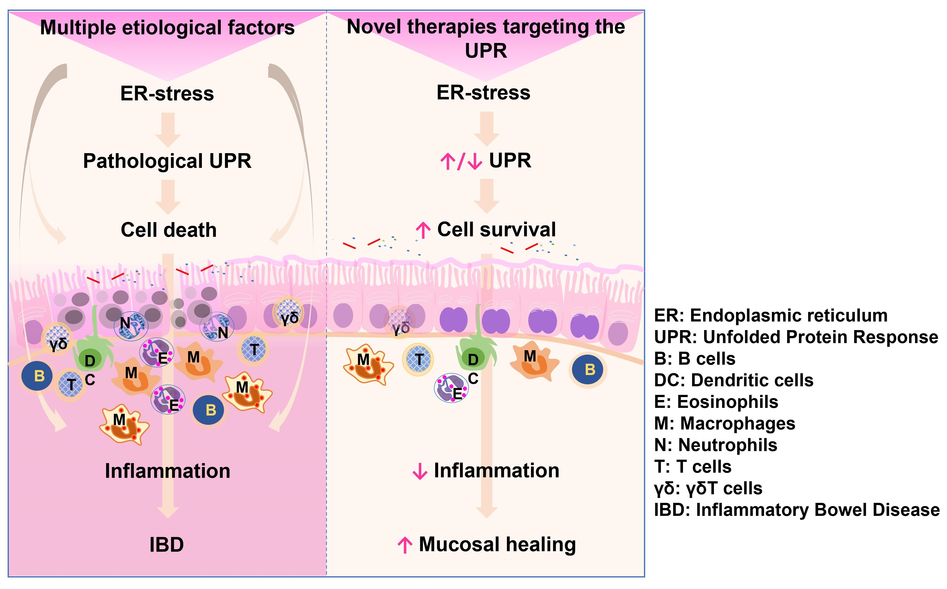
3. ER Stress and UPR in Inflammatory Bowel Disease (IBD)

3.1. Homeostasis of the Intestinal Epithelial Barrier

The intestinal mucosa constitutes a barrier with various components, including a thick layer of secreted mucins. The glycocalyx, composed of glycoproteins, glycolipids, and sulfated polysaccharides, resides beneath this mucus layer and above epithelial cells [64]. The barrier further incorporates a single layer of diverse columnar epithelial cells expressing transmembrane mucins and underpinned by a collagen-rich basement membrane. Additionally, leukocytes constantly patrol the epithelium, sampling intestinal luminal content and coordinating immune responses in the lamina propria. The intestinal epithelium integrates signals from the gut microbiota and from resident leukocytes to maintain intestinal homeostasis.
Intestinal epithelial cells (IECs) include at least four types of epithelial cells in both the small and large intestine: intestinal stem cells, tuft cells, absorptive epithelial cells (enterocytes), and a subgroup of secretory cells (including goblet, Paneth, and enteroendocrine cells). Disruption of the intestinal epithelial barrier can precipitate the onset of inflammatory bowel disease (IBD) [65,66]. Such defects in the epithelial barrier may originate from changes in the commensal microbiota composition (particularly dysbiosis favoring protease-producing mucolytic bacteria) [67], dysregulated immune responses, and genetic mutations (e.g., deficiency in α-defensins) [65,66].
IECs are continuously exposed to potential ER stress-inducing factors, including nutrient deprivation (such as glucose and lipid deprivation), alterations to different steps in protein synthesis (folding, glycosylation, and lipidation), and microbial toxins (e.g., LPS) that trigger inflammatory signaling. These factors induce basal levels of ER stress and activate the UPR, which can lead to either adaptation and survival or induction of epithelial cell death via apoptosis [68]. Persistent ER stress in IECs can overwhelm the capacity of the ER to synthesize the proteins required for major cell and organ functions, ultimately triggering a pathological UPR.
Besides nutrient and water absorption, the main functions of colon epithelial cells, indicated by the large number of goblet cells in this tissue, involve mucin production. Maintenance of a functional mucus layer is crucial for three main reasons: (1) forming a physical barrier to prevent commensal bacteria from infiltrating the lining epithelium while permitting the passage of low-molecular-weight soluble luminal antigens and nutritional factors; (2) maintaining lubrication for the colonic mucosa to facilitate the mechanical transport of feces; and (3) providing a first-line innate immunity lattice/matrix where secreted proteins such as immunoglobulins [69,70], antimicrobial peptides (AMP), and enzymes (e.g., lysozyme, defensins, cathelicidins, lipocalins, and C-type lectins) can be anchored to mediate neutralization effects [65]. In sum, certain levels of signals originating from the gut microbiota, the intestinal epithelium, and the innate immunity cells in the lamina propria are sensed as ER stress by IECs and immune cells, resulting in UPR activation to maintains intestinal homeostasis.

3.2. Pathogenesis of IBD

IBD, which encompasses ulcerative colitis (UC) and Crohn’s disease (CD), stems from multiple etiologies [65,68]. UC is mainly characterized by mucosal inflammation in the colon and rectum, whereas CD shows transmural inflammation in any region of the small and large intestine [71]. At least three pathogenic mechanisms have been proposed for IBD: (1) induction of IEC apoptosis; (2) disruption of mucosal barrier function; and (3) induction of proinflammatory responses in the gut [38]. In addition, abnormalities in the ER stress response have been proposed as an alternative mechanism for IBD pathogenesis [72,73].

3.2.1. Apoptosis of IECs

Apoptosis is a physiological catabolic process that is crucial for maintaining the homeostasis of the intestinal mucosa [66]. Increased apoptosis of colon epithelial cells has been observed in UC patients [74]. Apoptosis can be activated via various mechanisms, including signaling through death receptors, such as FAS cell surface death receptor and tumor necrosis factor receptor (TNFR) family members, and via mitochondrial damage and caspase activation [66]. In addition, unmitigated ER stress and UPR may lead to apoptosis [38]. Expression of Raf kinase inhibitor protein (RKIP) and p53-upregulated modulator of apoptosis (PUMA) is increased in human colon tissue and correlates positively with the severity of IBD in humans and colitis in mice [75,76]. Colon tissue from UC patients also shows increased levels of caspase recruitment domain (CARD9, CARD14, and CARD15) family members, mediators of apoptosis [77]. Furthermore, increased frequencies of FasL- and perforin-positive cells in the lamina propria have been observed in colon tissue from IBD patients [74]. Finally, apoptosis in IBD may involve mechanisms beyond the activation of canonical apoptosis pathways. For example, high-mobility group box 1 (HMGB1) and the human antigen R (HuR, ELAVL1) proteins have been found to mediate apoptosis in UC [78].

3.2.2. Disruption of the Mucosal Barrier

ER stress in IECs appears as a result of overwhelming stimulation to maintain barrier function (for instance, AMP production by Paneth cells and mucin production by goblet cells); this may lead to cell death and disruption of the mucosal barrier [38]. Mucins, which are large and highly O-glycosylated proteins that are produced abundantly, require complex posttranslational modifications, including glycosylation, disulfide bond formation, and oligomerization. For this reason, they have an increased likelihood of being misfolded during biosynthesis [79]. IRE1β regulates the rate of mucin biosynthesis, folding, and secretion by controlling MUC2 mRNA transcript levels. IRE1β deficiency causes ER stress and increases susceptibility to colitis [80], as does the misfolding of mucins in Muc2−/− mice [81].

3.2.3. Excessive Inflammation

IBD may arises from excessive inflammation, marked by the overproduction of inflammatory mediators and increased apoptosis of epithelial cells [66,75,77,82]. UC patients show increased expression and constitutive activation of toll-like receptor 4 and nuclear factor kappa B (TLR4/NF-κB) in colon epithelial cells that may progress into colitis associated cancer [83,84,85]. ER stress in IECs amplifies their response to TLR5 ligand Flagellin by producing IL-8 cytokine that impacts the activation of dendritic cells and promotes inflammation [86]. Moreover, activated leukocytes in the lamina propria of UC patients release proinflammatory cytokines and increase the production of ROS, exacerbating the apoptosis of epithelial, immune and bystander cells [66]. Under these conditions, the anti-inflammatory effects of IL-10 are attenuated, which typically upregulate genes required for ER stress mitigation, ERAD-mediated misfolded protein degradation, and enhanced MUC2 folding and secretion [87].
Sentinel goblet cells located at the entrance of colonic crypts express TLRs and Nucleotide binding oligomerization domain (NOD)-like receptors capable of sensing PAMPs. These cells activate the NLRP6 inflammasome to secrete mucins [88]. This response in the colon is similar to that in Paneth cells in the small intestine, which respond to TLR ligands by secreting AMP and defensins [89]. In the absence of those receptors or the MyD88 adaptor protein, mice display increased susceptibility to bacterial colonization and DSS colitis [65,90]. This supports the notion that tonic microbial signals induce the secretion of mucins and defensins to maintain intestinal homeostasis. However, persistent stimulation of PRRs can lead to chronic inflammation [65].

3.2.4. Aberrant UPR Activation

ER stress in IECs, including goblet cells, may also stem from genetic defects leading to aberrant UPR activation. The ileum and colon tissues of CD and UC patients show elevated levels of UPR activation compared to the normal tissues of healthy individuals, associated with increased expression of a chaperone, HSPA5/BiP, and a transcription factor, XBP1s [72]. Genetic defects in mice—for instance, those deficient in IRE1β or those with MUC2 mutations (Winnie and Eeyore mice) caused by carcinogen (N-ethyl-N-nitrosourea) exposure—result in aberrant MUC2 biosynthesis. This, in turn, induces ER stress and UPR, developing a UC-like phenotype [80]. Goblet cells in these mice accumulate misfolded MUC2 protein and its precursor, triggering ER stress, which leads to reduced MUC2 secretion and changes in the viscoelastic properties and composition of the mucus barrier. This favors inflammation, with enhanced local production of inflammatory cytokines (e.g., IL-1β, TNFα, and IFNγ) [81]. These proinflammatory cytokines also induce ER stress in intestinal epithelial cells to augment the expression and secretion of mucins [70], which not only exacerbates the ER stress-mediated mucin depletion but also leads to the disassembly of tight junction and adherence junction proteins, accelerating the epithelial layer leakiness [91,92]. Together, the epithelial barrier defects and inflammation progress to dysregulated UPR and persistent inflammation, both common features of IBD [79,81].
UC patients show a progressive loss/dysfunction of goblet cells [93]. MUC2, produced by goblet cells, plays a prominent role in colon homeostasis by maintaining appropriate levels of cell proliferation and apoptosis, regulating cell migration along the crypt, and suppressing inflammation and colorectal cancer [94]. Accordingly, Muc2−/− mice develop aberrant crypt morphology and exhibit altered maturation and migration of epithelial cells, which eventually transform into adenomas and adenocarcinomas due to an increased rate of cell proliferation over apoptosis [94]. A similar phenotype is observed in mice lacking the protein complexes required for post-translational processing of MUC2, such as glycosylation [95], sulfonation [96], disulfide bond formation [97], and lipidation/palmitoylation [98]. Consequently, proper MUC2 folding and secretion are required for colon crypt homeostasis. An increased demand in mucin secretion may cause misfolded MUC2, which, in turn, induces ER stress and UPR, contributing to the pathogenesis of UC.
Nonetheless, the pathogenesis of IBD is not completely understood, and there may be overlooked factors involved in intestinal homeostasis, whose presence or disruption could cause ER stress, thereby exacerbating the manifestation of IBD. For example, nutrient deprivation (glucose, lipids, or amino acids) induces ER stress, which subsequently activates autophagy. This mechanism generates increased levels of ROS that stimulate mucus secretion by intestinal goblet cells and contribute to the elimination of severely damaged cells to maintain colon homeostasis [99,100]. However, excessive production of ROS may cause cell death.

4. Pharmacologic Intervention of the UPR as a Potential Therapeutic Strategy in IBD Treatment

Given the protective roles of the intestinal epithelium often dysregulated in IBDs, such as secretion of the mucin-rich mucous layer and AMPs, interaction and sensing of the normal gut microbiota, and transmission or amplification of those signals to intraepithelial lymphocytes and lamina propria resident leukocytes, it would be beneficial to develop novel therapeutics targeting the restitution and functionality of epithelial cells for IBD treatment. Thus, new therapies could focus on (1) enhancing colon crypt goblet cell function (e.g., mucin folding and secretion); (2) promoting survival and proliferation of colon crypt stem cells and other proliferation-competent crypt cells such as transit-amplifying cells; and (3) integrating both strategies, namely, the survival/proliferation of colon crypt stem cells and their differentiation into goblet and other terminally differentiated epithelial cells.
The ER stress and activation of the UPR in eukaryotic cells is a mechanism that initially promotes cell homeostasis, even though chronic activation can lead to cell death. In the context of IBD, this notion is supported by findings that IRE1β−/− mice exhibited increased susceptibility to experimental colitis, indicative of a protective role of IRE1 of the UPR in the intestinal epithelium [72,73]. Similarly, Xbp1 knockout mice displayed increased ER stress in small intestine and colon epithelial cells, associated with the spontaneous inflammation and dysfunctional secretion of AMP by Paneth cells and mucin by goblet cells [72]. The heightened colon inflammation and susceptibility to DSS-induced colitis in mice deficient in ATF6α or chaperone genes highlight the role of this UPR transcription factor and chaperones during colitis [101]. ATF6α expression is induced by IFN-γ, with IFN-γ-ATF6/CEBPB signaling being necessary to initiate autophagy to combat bacterial infections, a situation exacerbated in ATF6α−/− mice [102].
Consequently, UPR components are promising targets for therapeutic intervention in ER stress- and UPR-associated pathologies. Several inhibitors targeting the IRE1 kinase activity or dephosphorylation of eIF2α have been developed and tested in in vitro studies of ER stress and UPR modulation [103,104,105]. Table 1 summarizes the strategies targeting the UPR for IBD treatment. Targeting the UPR elements, including the KDELRs, has been proposed either to activate and induce ER stress response or to inhibit and suppress ER stress sensors that favor cell survival/proliferation (organ preservation) [15,23,33]. Alternatively, a strategy that overwhelms the UPR by using proteasome inhibitors has been employed to induce apoptosis of malignant cells [106,107]. A range of natural and synthetic molecules, some of which impact the UPR, have been explored in IBD treatment and reviewed recently [108]. However, limited research has been conducted on recombinant pharmaceutical proteins designed to modulate the UPR towards the induction of cell survival/proliferation to restore cell homeostasis. Hereafter, we summarize those compounds found to modulate the UPR in IBD.

4.1. Targeting the UPR Sensors

A synthetic XBP1 agonist, HLJ2, was generated based on a monomeric compound purified from species of Coptis and Corydalis herbaceous plants. HLJ2 reduced inflammatory cytokines (e.g., TNFα, IL-1β, and IL-6) in DSS colitis mice. Additionally, HLJ2 increased the expression of tight junction proteins ZO-1 and claudin-1 in colon tissue, alleviated intestinal dysbiosis, and protected the intestinal mucosa [114]. Thus, HLJ2 is postulated to reinforce the colon epithelial barrier and reduce inflammation.
Salubrinal, a small molecule identified to block the phosphatases mediating eIF2α dephosphorylation, was found to enhance cell survival/cytoprotection in cells under ER stress induced by tunicamycin or viral infection, by maintaining levels of eIF2α and protein synthesis [105]. In DSS-induced colitis, salubrinal showed protective effects by suppressing the expression of proinflammatory cytokines and myeloperoxidase activity, while elevating ATF4 and HSPA5/BiP, which ameliorated ER stress and improved histological scores of colitis [110].

4.2. Non-Specific Targeting of the UPR

The ER stress and the UPR in colon epithelial cells and colitis have been non-specifically targeted with single amino acids (e.g., glutamine) [109], hormones (e.g., estrogen) [113], and chemical chaperones such as tauroursodeoxycholic acid (TUDCA) and 4-phenylburyrate (PBA) [101,108]. The PBA and TUDCA chaperones reduce ER stress in IECs and mitigate features of acute and chronic colitis induced by DSS or generated by deficiency in the anti-inflammatory IL-10 cytokine (IL10−/− mice) [101]. The estrogen receptor agonist, G-1 (1-[4-6-bromobenzol [1,3] dioxol-5yl]-3a,4,5,9b-tetrahydro-3H-cyclopenta[c]quinoloin-8-yl]-ethanone), acting through the G-protein-coupled estrogen receptor (GPER) at the plasma membrane, reduced the expression of HSPA5/BiP and CHOP, and attenuated all three arms of the UPR in DSS-colitis mice. In addition, G-1 reduced the level of cleaved caspase-3 and increased the number of Ki-67+ proliferating colon crypt epithelial cells [113].
Artesunate, a hemisuccinate derivative of artemisinin, isolated from Artemisia annua herbs, has demonstrated protective effects in colitis [111]. Artesunate inhibited ER stress signaling pathways involving PERK-eIF2α-ATF4-CHOP and IRE1α-XBP1 during DSS-induced colitis. Artesunate inhibited NF-κB and proinflammatory cytokines, improved clinical and histopathological scores, and maintained the levels of claudin-1 and MUC2 in the colonic mucosa [112].
Finally, ghrelin, a nutrient sensor expressed in epithelial and immune cells, acts through the growth hormone secretagogue receptor (GHS-R1a) and represents another potential UPR modulator in the gastrointestinal tract. Ghrelin was found to modulate the UPR by reducing the expression of proapoptotic factors including CHOP, caspase 3, and BAX while increasing Bcl-2, which provides survival signals. Additionally, ghrelin inhibited TNFα-induced apoptosis in Caco2 cells and protected colon epithelial cells from DSS and TNBS-induced colitis [82].

4.3. EPICERTIN, a KDELR Ligand Modulator of the UPR

Recombinant KDEL-tagged fusion proteins have been engineered for therapeutic intervention in cancer and for vaccine development against virus-induced tumors [117,118] and also as potential subunit vaccines against Vibrio cholerae [119]. EPICERTIN consists of the nontoxic, recombinant cholera toxin B-subunit (CTB) modified with a KDEL motif in its C-terminal region, exhibiting wound healing properties in the colon [115]. The potential of EPICERTIN as a therapeutic agent for IBD has been investigated in mouse models of colitis, where the KDEL motif has been found to be crucial for its wound healing activity [116]. EPICERTIN bears two structural modifications distinguishing it from CTB: (a) Asn4 → Ser mutation to avoid N-glycosylation when expressed in eukaryotic cells; and (b) a C-terminal hexapeptide containing the KDEL ER retrieval sequence [119]. EPICERTIN retains GM1-binding affinity, molecular stability, and vaccine efficacy, as demonstrated by the induction of anti-toxin IgG and IgA antibodies upon oral immunization in mice. The conformational structure model of EPT closely resembles that of CTB [120] (Figure 1A).
EPICERTIN binds to GM1-ganglioside at the plasma membrane of colon epithelial cells, undergoes endocytosis, and is retrogradely transported to trans Golgi network and the ER via a lipid-based sorting pathway [121,122] or by interacting with KDELRs at the plasma membrane [123]. Once in the ER, EPICERTIN displaces sensor-bound chaperones to induce the UPR (Figure 1B). EPICERTIN, unlike CTB or the C-terminal leucine-truncated EPICERTIN variant (“CTB-KDE”), possesses unique mucosal wound healing activity by increasing TGFβ1 and TGFβ2 levels, a cytokine and growth factor known to induce IEC migration and restitution [124] and found to facilitates Caco2 cell migration and wound healing in scratch assays [116]. In in vitro cultures of colon tissue explants from IBD patients, EPICERTIN promoted colon crypt survival and upregulated wound healing pathway genes, including TGFB1, CDH1, and WNT5A [116]. The wound healing effects of EPICERTIN were corroborated in in vivo mouse models of acute and chronic colitis induced by single and multiple DSS exposure cycles [125], respectively. EPICERTIN decreased histopathological scores and reduced inflammatory cytokine levels, while increasing innate immune cell populations, including dendritic cells, natural killer cells, and macrophages (both M1 and M2) in the colon [115]. In addition, EPICERTIN, orally administered once every two weeks (four doses), significantly reduced tumor development in an azoxymethane/DSS model of colitis-associated cancer.
In mice, EPICERTIN remained detectable in the colon epithelium 24 h after oral or intrarectal administration, particularly in the base region of colon crypts. This suggests a role for colonic crypt base stem cells in EPICERTIN-induced epithelial regeneration [126]. Mechanistically, the IRE1/XBP1s pathway of the UPR, which is known to maintain ER homeostasis by regulating cell survival/apoptosis balance [33,127], appears to mediate the wound healing activity of EPICERTIN, as the IRE1 inhibitor 4µ8C or siRNA knockdown of Xbp1 abolished EPICERTIN’s wound healing effect in Caco-2 cells [116]. Collectively, these findings suggest that EPICERTIN activates the IRE1/XBP1 pathway, which, in turn, appears to promote epithelial cell restitution via secretion of TGFβ and other wound healing-related growth factors. Although the link between IRE1 activation and mucosal epithelial repair is not fully understood, these studies have demonstrated the potential therapeutic application of EPICERTIN, a KDELR ligand and an UPR-modulating molecule, in colon epithelial cells. It remains to be investigated whether TGFβ is the sole signaling mechanism mediating the healing effects of EPICERTIN in both acute and chronic colon inflammation. In addition, the effects of EPICERTIN on leukocytes in the colon lamina propria [115] and their potential contribution to mucosal healing remain to be disclosed.
Figure 1. EPICERTIN: A KDELR ligand and a modulator of the unfolded protein response that promotes cell survival and proliferation. (A) Structural models of CTB and EPICERTIN (EPT) monomers generated by the Protein Homology/analogy Recognition Engine V 2.0 (Phyre2) software created by the Structural Bioinformatics Group, Imperial college, London and updated on January 25, 2021 [120] based on a crystal structure of CTB (Protein Data Bank ID: 3CHB). The C-terminal KDEL amino acid sequence extension of EPT is highlighted in red at the top of the image. (B) A proposed model of EPICERTIN-KDELR signaling and modulation of the UPR. 1. Both CTB and EPT bind to GM1 at the plasma membrane, but with differing outcomes. EPT may also bind to KDELR on the plasma membrane. 2. CTB and EPT are endocytosed in early endosomes, and eventually, the pentamer disintegrates into monomers within late endosomes. 3. EPT and CTB are retrogradely transported to the trans-Golgi network (TGN) and finally to the ER, following a lipid-based sorting pathway. Only EPT interacts with and activates KDELR signaling, promoting vesicular transport and the recycling of ER chaperones. Activation of KDELR by EPT may also occur early on the endosome membrane and in the TGN. 4. In the ER, both CTB and EPT may displace chaperones from ER stress sensors (PERK, ATF6α, IRE1α/β) and induce the unfolded protein response (UPR), although only EPT is known to activate IRE-XBP1s. 5. Activation of the ER stress sensors leads to the generation of the transcription factors phosphorylated eIF2α, ATF6αp50, and XBP1s, which transactivate genes in the nucleus that are associated with ER homeostasis. The increased endocytosis of CTB may trigger apoptosis by various mechanisms, including a chronic UPR or activation of death receptors [128]. In contrast, EPT–KDELR–chaperone complexes are allowed to recycle between ER and TGN, alleviating the ER stress by replenishing chaperones back into the ER, modulating the intensity and amplitude of UPR. Alternatively, the surplus of endocytosed CTB eventually overwhelms the endosome-to-TGN trafficking, which is known to activate the NLRP3 inflammasome to induce apoptosis [129]. EPT–KDELR interaction might activate signaling pathways regulating and improving vesicular trafficking and cell survival which are currently unknown; however, preliminary data suggest the involvement of MAPK kinases (unpublished observations). C: Cytoplasm; TGN: Tans-Golgi Network; ER: Endoplasmic Reticulum; N: Nuclei; L: Lysosome; LE: Late Endolysosome.
Biomedicines 11 02066 g001

5. Concluding Remarks

Our growing understanding of ER stress and the specific regulation of UPR that occurs in different types of eukaryotic cells is advancing towards a point where therapeutic manipulation of the UPR may be viable to address human pathologies associated with ER perturbation, such as IBD. Although results derived from various IBD models that demonstrate therapeutic effects through UPR manipulation are currently limited, there is potential for properly reversing ER-related pathological conditions associated with the disease. EPICERTIN emerges in this context as a promising UPR modulator that promotes cell survival, migration, and the proliferation of epithelial cells, ultimately leading to epithelial restitution, a therapeutic aim not yet addressed by current IBD treatments. EPICERTIN induces an adaptive UPR and stimulates the production and release of TGF-β, which, in turn, promotes epithelialization. Further investigation into EPICERTIN’s mode of action will illuminate the protective role of UPR in epithelial wound repair and pave the way for a potential new therapy for IBD.

Funding

This work was supported by a U.S. National Institute of Health grant (R01 DK123712).

Institutional Review Board Statement

No applicable.

Informed Consent Statement

Not applicable.

Data Availability Statement

Not applicable.

Conflicts of Interest

The authors declare no conflict of interest.

References

  1. Voeltz, G.K.; Rolls, M.M.; Rapoport, T.A. Structural organization of the endoplasmic reticulum. EMBO Rep. 2002, 3, 944–950. [Google Scholar] [CrossRef] [PubMed] [Green Version]
  2. Kumar, V.; Maity, S. ER Stress-Sensor Proteins and ER-Mitochondrial Crosstalk-Signaling Beyond (ER) Stress Response. Biomolecules 2021, 11, 173. [Google Scholar] [CrossRef] [PubMed]
  3. Berridge, M.J. The endoplasmic reticulum: A multifunctional signaling organelle. Cell Calcium 2002, 32, 235–249. [Google Scholar] [CrossRef] [PubMed]
  4. Schroder, M. Endoplasmic reticulum stress responses. Cell. Mol. Life Sci. 2008, 65, 862–894. [Google Scholar] [CrossRef]
  5. Cao, S.S. Epithelial ER Stress in Crohn’s Disease and Ulcerative Colitis. Inflamm. Bowel Dis. 2016, 22, 984–993. [Google Scholar] [CrossRef]
  6. Wilfling, F.; Haas, J.T.; Walther, T.C.; Farese, R.V., Jr. Lipid droplet biogenesis. Curr. Opin. Cell Biol. 2014, 29, 39–45. [Google Scholar] [CrossRef] [Green Version]
  7. Pineau, L.; Colas, J.; Dupont, S.; Beney, L.; Fleurat-Lessard, P.; Berjeaud, J.M.; Berges, T.; Ferreira, T. Lipid-induced ER stress: Synergistic effects of sterols and saturated fatty acids. Traffic 2009, 10, 673–690. [Google Scholar] [CrossRef]
  8. Han, J.; Kaufman, R.J. The role of ER stress in lipid metabolism and lipotoxicity. J. Lipid Res. 2016, 57, 1329–1338. [Google Scholar] [CrossRef] [Green Version]
  9. Hou, N.S.; Gutschmidt, A.; Choi, D.Y.; Pather, K.; Shi, X.; Watts, J.L.; Hoppe, T.; Taubert, S. Activation of the endoplasmic reticulum unfolded protein response by lipid disequilibrium without disturbed proteostasis in vivo. Proc. Natl. Acad. Sci. USA 2014, 111, E2271–E2280. [Google Scholar] [CrossRef]
  10. Radanovic, T.; Ernst, R. The Unfolded Protein Response as a Guardian of the Secretory Pathway. Cells 2021, 10, 2965. [Google Scholar] [CrossRef]
  11. Carreras-Sureda, A.; Jana, F.; Urra, H.; Durand, S.; Mortenson, D.E.; Sagredo, A.; Bustos, G.; Hazari, Y.; Ramos-Fernandez, E.; Sassano, M.L.; et al. Non-canonical function of IRE1alpha determines mitochondria-associated endoplasmic reticulum composition to control calcium transfer and bioenergetics. Nat. Cell Biol. 2019, 21, 755–767. [Google Scholar] [CrossRef]
  12. Hetz, C.; Zhang, K.; Kaufman, R.J. Mechanisms, regulation and functions of the unfolded protein response. Nat. Rev. Mol. Cell. Biol. 2020, 21, 421–438. [Google Scholar] [CrossRef]
  13. Oikonomou, C.; Hendershot, L.M. Disposing of misfolded ER proteins: A troubled substrate’s way out of the ER. Mol. Cell. Endocrinol. 2020, 500, 110630. [Google Scholar] [CrossRef]
  14. Ellgaard, L.; Helenius, A. Quality control in the endoplasmic reticulum. Nat. Rev. Mol. Cell. Biol. 2003, 4, 181–191. [Google Scholar] [CrossRef]
  15. Oakes, S.A.; Papa, F.R. The role of endoplasmic reticulum stress in human pathology. Annu. Rev. Pathol. 2015, 10, 173–194. [Google Scholar] [CrossRef] [Green Version]
  16. Zhang, K.; Kaufman, R.J. From endoplasmic-reticulum stress to the inflammatory response. Nature 2008, 454, 455–462. [Google Scholar] [CrossRef] [Green Version]
  17. Klausner, R.D.; Donaldson, J.G.; Lippincott-Schwartz, J. Brefeldin A: Insights into the control of membrane traffic and organelle structure. J. Cell Biol. 1992, 116, 1071–1080. [Google Scholar] [CrossRef]
  18. Citterio, C.; Vichi, A.; Pacheco-Rodriguez, G.; Aponte, A.M.; Moss, J.; Vaughan, M. Unfolded protein response and cell death after depletion of brefeldin A-inhibited guanine nucleotide-exchange protein GBF1. Proc. Natl. Acad. Sci. USA 2008, 105, 2877–2882. [Google Scholar] [CrossRef]
  19. Blum, A.; Khalifa, S.; Nordstrom, K.; Simon, M.; Schulz, M.H.; Schmitt, M.J. Transcriptomics of a KDELR1 knockout cell line reveals modulated cell adhesion properties. Sci. Rep. 2019, 9, 10611. [Google Scholar] [CrossRef] [Green Version]
  20. Lewis, M.J.; Pelham, H.R. Ligand-induced redistribution of a human KDEL receptor from the Golgi complex to the endoplasmic reticulum. Cell 1992, 68, 353–364. [Google Scholar] [CrossRef]
  21. Jin, H.; Komita, M.; Aoe, T. The Role of BiP Retrieval by the KDEL Receptor in the Early Secretory Pathway and its Effect on Protein Quality Control and Neurodegeneration. Front. Mol. Neurosci. 2017, 10, 222. [Google Scholar] [CrossRef] [PubMed] [Green Version]
  22. Sadana, R.; Dessauer, C.W. Physiological roles for G protein-regulated adenylyl cyclase isoforms: Insights from knockout and overexpression studies. Neurosignals 2009, 17, 5–22. [Google Scholar] [CrossRef] [PubMed]
  23. Cela, I.; Dufrusine, B.; Rossi, C.; Luini, A.; De Laurenzi, V.; Federici, L.; Sallese, M. KDEL Receptors: Pathophysiological Functions, Therapeutic Options, and Biotechnological Opportunities. Biomedicines 2022, 10, 1234. [Google Scholar] [CrossRef] [PubMed]
  24. Giannotta, M.; Ruggiero, C.; Grossi, M.; Cancino, J.; Capitani, M.; Pulvirenti, T.; Consoli, G.M.; Geraci, C.; Fanelli, F.; Luini, A.; et al. The KDEL receptor couples to Galphaq/11 to activate Src kinases and regulate transport through the Golgi. EMBO J. 2012, 31, 2869–2881. [Google Scholar] [CrossRef] [PubMed] [Green Version]
  25. Ruggiero, C.; Grossi, M.; Fragassi, G.; Di Campli, A.; Di Ilio, C.; Luini, A.; Sallese, M. The KDEL receptor signalling cascade targets focal adhesion kinase on focal adhesions and invadopodia. Oncotarget 2018, 9, 10228–10246. [Google Scholar] [CrossRef] [PubMed] [Green Version]
  26. Cancino, J.; Capalbo, A.; Di Campli, A.; Giannotta, M.; Rizzo, R.; Jung, J.E.; Di Martino, R.; Persico, M.; Heinklein, P.; Sallese, M.; et al. Control systems of membrane transport at the interface between the endoplasmic reticulum and the Golgi. Dev. Cell 2014, 30, 280–294. [Google Scholar] [CrossRef] [Green Version]
  27. Ruggiero, C.; Fragassi, G.; Grossi, M.; Picciani, B.; Di Martino, R.; Capitani, M.; Buccione, R.; Luini, A.; Sallese, M. A Golgi-based KDELR-dependent signalling pathway controls extracellular matrix degradation. Oncotarget 2015, 6, 3375–3393. [Google Scholar] [CrossRef] [Green Version]
  28. Trychta, K.A.; Back, S.; Henderson, M.J.; Harvey, B.K. KDEL Receptors Are Differentially Regulated to Maintain the ER Proteome under Calcium Deficiency. Cell Rep. 2018, 25, 1829–1840.e6. [Google Scholar] [CrossRef]
  29. Hamada, H.; Suzuki, M.; Yuasa, S.; Mimura, N.; Shinozuka, N.; Takada, Y.; Suzuki, M.; Nishino, T.; Nakaya, H.; Koseki, H.; et al. Dilated cardiomyopathy caused by aberrant endoplasmic reticulum quality control in mutant KDEL receptor transgenic mice. Mol. Cell. Biol. 2004, 24, 8007–8017. [Google Scholar] [CrossRef] [Green Version]
  30. Mimura, N.; Hamada, H.; Kashio, M.; Jin, H.; Toyama, Y.; Kimura, K.; Iida, M.; Goto, S.; Saisho, H.; Toshimori, K.; et al. Aberrant quality control in the endoplasmic reticulum impairs the biosynthesis of pulmonary surfactant in mice expressing mutant BiP. Cell Death Differ. 2007, 14, 1475–1485. [Google Scholar] [CrossRef]
  31. Mesaeli, N.; Nakamura, K.; Zvaritch, E.; Dickie, P.; Dziak, E.; Krause, K.H.; Opas, M.; MacLennan, D.H.; Michalak, M. Calreticulin is essential for cardiac development. J. Cell Biol. 1999, 144, 857–868. [Google Scholar] [CrossRef]
  32. Luo, S.; Mao, C.; Lee, B.; Lee, A.S. GRP78/BiP is required for cell proliferation and protecting the inner cell mass from apoptosis during early mouse embryonic development. Mol. Cell. Biol. 2006, 26, 5688–5697. [Google Scholar] [CrossRef] [Green Version]
  33. Lin, J.H.; Li, H.; Yasumura, D.; Cohen, H.R.; Zhang, C.; Panning, B.; Shokat, K.M.; Lavail, M.M.; Walter, P. IRE1 signaling affects cell fate during the unfolded protein response. Science 2007, 318, 944–949. [Google Scholar] [CrossRef] [Green Version]
  34. Walter, P.; Ron, D. The unfolded protein response: From stress pathway to homeostatic regulation. Science 2011, 334, 1081–1086. [Google Scholar] [CrossRef] [Green Version]
  35. Yoshida, H.; Matsui, T.; Yamamoto, A.; Okada, T.; Mori, K. XBP1 mRNA is induced by ATF6 and spliced by IRE1 in response to ER stress to produce a highly active transcription factor. Cell 2001, 107, 881–891. [Google Scholar] [CrossRef] [Green Version]
  36. Reimold, A.M.; Etkin, A.; Clauss, I.; Perkins, A.; Friend, D.S.; Zhang, J.; Horton, H.F.; Scott, A.; Orkin, S.H.; Byrne, M.C.; et al. An essential role in liver development for transcription factor XBP-1. Genes Dev. 2000, 14, 152–157. [Google Scholar] [CrossRef]
  37. Haze, K.; Yoshida, H.; Yanagi, H.; Yura, T.; Mori, K. Mammalian transcription factor ATF6 is synthesized as a transmembrane protein and activated by proteolysis in response to endoplasmic reticulum stress. Mol. Biol. Cell 1999, 10, 3787–3799. [Google Scholar] [CrossRef] [Green Version]
  38. Luo, K.; Cao, S.S. Endoplasmic reticulum stress in intestinal epithelial cell function and inflammatory bowel disease. Gastroenterol. Res. Pract. 2015, 2015, 328791. [Google Scholar] [CrossRef] [Green Version]
  39. Acosta-Alvear, D.; Zhou, Y.; Blais, A.; Tsikitis, M.; Lents, N.H.; Arias, C.; Lennon, C.J.; Kluger, Y.; Dynlacht, B.D. XBP1 controls diverse cell type- and condition-specific transcriptional regulatory networks. Mol. Cell 2007, 27, 53–66. [Google Scholar] [CrossRef]
  40. Lee, A.H.; Chu, G.C.; Iwakoshi, N.N.; Glimcher, L.H. XBP-1 is required for biogenesis of cellular secretory machinery of exocrine glands. EMBO J. 2005, 24, 4368–4380. [Google Scholar] [CrossRef] [Green Version]
  41. Hollien, J.; Lin, J.H.; Li, H.; Stevens, N.; Walter, P.; Weissman, J.S. Regulated Ire1-dependent decay of messenger RNAs in mammalian cells. J. Cell Biol. 2009, 186, 323–331. [Google Scholar] [CrossRef]
  42. Lu, M.; Lawrence, D.A.; Marsters, S.; Acosta-Alvear, D.; Kimmig, P.; Mendez, A.S.; Paton, A.W.; Paton, J.C.; Walter, P.; Ashkenazi, A. Opposing unfolded-protein-response signals converge on death receptor 5 to control apoptosis. Science 2014, 345, 98–101. [Google Scholar] [CrossRef] [PubMed] [Green Version]
  43. Han, D.; Lerner, A.G.; Vande Walle, L.; Upton, J.P.; Xu, W.; Hagen, A.; Backes, B.J.; Oakes, S.A.; Papa, F.R. IRE1alpha kinase activation modes control alternate endoribonuclease outputs to determine divergent cell fates. Cell 2009, 138, 562–575. [Google Scholar] [CrossRef] [PubMed] [Green Version]
  44. Upton, J.P.; Wang, L.; Han, D.; Wang, E.S.; Huskey, N.E.; Lim, L.; Truitt, M.; McManus, M.T.; Ruggero, D.; Goga, A.; et al. IRE1alpha cleaves select microRNAs during ER stress to derepress translation of proapoptotic Caspase-2. Science 2012, 338, 818–822. [Google Scholar] [CrossRef] [PubMed] [Green Version]
  45. Chopra, S.; Giovanelli, P.; Alvarado-Vazquez, P.A.; Alonso, S.; Song, M.; Sandoval, T.A.; Chae, C.S.; Tan, C.; Fonseca, M.M.; Gutierrez, S.; et al. IRE1alpha-XBP1 signaling in leukocytes controls prostaglandin biosynthesis and pain. Science 2019, 365, eaau6499. [Google Scholar] [CrossRef]
  46. Hetz, C.; Bernasconi, P.; Fisher, J.; Lee, A.H.; Bassik, M.C.; Antonsson, B.; Brandt, G.S.; Iwakoshi, N.N.; Schinzel, A.; Glimcher, L.H.; et al. Proapoptotic BAX and BAK modulate the unfolded protein response by a direct interaction with IRE1alpha. Science 2006, 312, 572–576. [Google Scholar] [CrossRef] [Green Version]
  47. Teske, B.F.; Wek, S.A.; Bunpo, P.; Cundiff, J.K.; McClintick, J.N.; Anthony, T.G.; Wek, R.C. The eIF2 kinase PERK and the integrated stress response facilitate activation of ATF6 during endoplasmic reticulum stress. Mol. Biol. Cell 2011, 22, 4390–4405. [Google Scholar] [CrossRef]
  48. Han, J.; Back, S.H.; Hur, J.; Lin, Y.H.; Gildersleeve, R.; Shan, J.; Yuan, C.L.; Krokowski, D.; Wang, S.; Hatzoglou, M.; et al. ER-stress-induced transcriptional regulation increases protein synthesis leading to cell death. Nat. Cell Biol. 2013, 15, 481–490. [Google Scholar] [CrossRef] [Green Version]
  49. Marciniak, S.J.; Yun, C.Y.; Oyadomari, S.; Novoa, I.; Zhang, Y.; Jungreis, R.; Nagata, K.; Harding, H.P.; Ron, D. CHOP induces death by promoting protein synthesis and oxidation in the stressed endoplasmic reticulum. Genes Dev. 2004, 18, 3066–3077. [Google Scholar] [CrossRef] [Green Version]
  50. Verfaillie, T.; Rubio, N.; Garg, A.D.; Bultynck, G.; Rizzuto, R.; Decuypere, J.P.; Piette, J.; Linehan, C.; Gupta, S.; Samali, A.; et al. PERK is required at the ER-mitochondrial contact sites to convey apoptosis after ROS-based ER stress. Cell Death Differ. 2012, 19, 1880–1891. [Google Scholar] [CrossRef] [Green Version]
  51. McCullough, K.D.; Martindale, J.L.; Klotz, L.O.; Aw, T.Y.; Holbrook, N.J. Gadd153 sensitizes cells to endoplasmic reticulum stress by down-regulating Bcl2 and perturbing the cellular redox state. Mol. Cell. Biol. 2001, 21, 1249–1259. [Google Scholar] [CrossRef] [PubMed] [Green Version]
  52. Namba, T.; Tanaka, K.; Ito, Y.; Ishihara, T.; Hoshino, T.; Gotoh, T.; Endo, M.; Sato, K.; Mizushima, T. Positive role of CCAAT/enhancer-binding protein homologous protein, a transcription factor involved in the endoplasmic reticulum stress response in the development of colitis. Am. J. Pathol. 2009, 174, 1786–1798. [Google Scholar] [CrossRef] [PubMed] [Green Version]
  53. Cullinan, S.B.; Zhang, D.; Hannink, M.; Arvisais, E.; Kaufman, R.J.; Diehl, J.A. Nrf2 is a direct PERK substrate and effector of PERK-dependent cell survival. Mol. Cell. Biol. 2003, 23, 7198–7209. [Google Scholar] [CrossRef] [PubMed] [Green Version]
  54. Chen, W.; Sun, Z.; Wang, X.J.; Jiang, T.; Huang, Z.; Fang, D.; Zhang, D.D. Direct interaction between Nrf2 and p21(Cip1/WAF1) upregulates the Nrf2-mediated antioxidant response. Mol. Cell 2009, 34, 663–673. [Google Scholar] [CrossRef] [PubMed] [Green Version]
  55. Lu, P.D.; Jousse, C.; Marciniak, S.J.; Zhang, Y.; Novoa, I.; Scheuner, D.; Kaufman, R.J.; Ron, D.; Harding, H.P. Cytoprotection by pre-emptive conditional phosphorylation of translation initiation factor 2. EMBO J. 2004, 23, 169–179. [Google Scholar] [CrossRef] [Green Version]
  56. Cao, S.S.; Song, B.; Kaufman, R.J. PKR protects colonic epithelium against colitis through the unfolded protein response and prosurvival signaling. Inflamm. Bowel Dis. 2012, 18, 1735–1742. [Google Scholar] [CrossRef] [Green Version]
  57. Srivastava, S.P.; Kumar, K.U.; Kaufman, R.J. Phosphorylation of eukaryotic translation initiation factor 2 mediates apoptosis in response to activation of the double-stranded RNA-dependent protein kinase. J. Biol. Chem. 1998, 273, 2416–2423. [Google Scholar] [CrossRef] [Green Version]
  58. Balachandran, S.; Kim, C.N.; Yeh, W.C.; Mak, T.W.; Bhalla, K.; Barber, G.N. Activation of the dsRNA-dependent protein kinase, PKR, induces apoptosis through FADD-mediated death signaling. EMBO J. 1998, 17, 6888–6902. [Google Scholar] [CrossRef] [Green Version]
  59. Shen, J.; Snapp, E.L.; Lippincott-Schwartz, J.; Prywes, R. Stable binding of ATF6 to BiP in the endoplasmic reticulum stress response. Mol. Cell. Biol. 2005, 25, 921–932. [Google Scholar] [CrossRef] [Green Version]
  60. Shen, J.; Chen, X.; Hendershot, L.; Prywes, R. ER stress regulation of ATF6 localization by dissociation of BiP/GRP78 binding and unmasking of Golgi localization signals. Dev. Cell 2002, 3, 99–111. [Google Scholar] [CrossRef] [Green Version]
  61. Nadanaka, S.; Okada, T.; Yoshida, H.; Mori, K. Role of disulfide bridges formed in the luminal domain of ATF6 in sensing endoplasmic reticulum stress. Mol. Cell. Biol. 2007, 27, 1027–1043. [Google Scholar] [CrossRef] [Green Version]
  62. Yoshida, H.; Okada, T.; Haze, K.; Yanagi, H.; Yura, T.; Negishi, M.; Mori, K. ATF6 activated by proteolysis binds in the presence of NF-Y (CBF) directly to the cis-acting element responsible for the mammalian unfolded protein response. Mol. Cell. Biol. 2000, 20, 6755–6767. [Google Scholar] [CrossRef] [Green Version]
  63. Yamamoto, K.; Sato, T.; Matsui, T.; Sato, M.; Okada, T.; Yoshida, H.; Harada, A.; Mori, K. Transcriptional induction of mammalian ER quality control proteins is mediated by single or combined action of ATF6alpha and XBP1. Dev. Cell 2007, 13, 365–376. [Google Scholar] [CrossRef] [Green Version]
  64. Chevalier, L.; Selim, J.; Genty, D.; Baste, J.M.; Piton, N.; Boukhalfa, I.; Hamzaoui, M.; Pareige, P.; Richard, V. Electron microscopy approach for the visualization of the epithelial and endothelial glycocalyx. Morphologie 2017, 101, 55–63. [Google Scholar] [CrossRef]
  65. Maloy, K.J.; Powrie, F. Intestinal homeostasis and its breakdown in inflammatory bowel disease. Nature 2011, 474, 298–306. [Google Scholar] [CrossRef]
  66. Wan, Y.; Yang, L.; Jiang, S.; Qian, D.; Duan, J. Excessive Apoptosis in Ulcerative Colitis: Crosstalk Between Apoptosis, ROS, ER Stress, and Intestinal Homeostasis. Inflamm. Bowel Dis. 2022, 28, 639–648. [Google Scholar] [CrossRef]
  67. Nishida, A.; Inoue, R.; Inatomi, O.; Bamba, S.; Naito, Y.; Andoh, A. Gut microbiota in the pathogenesis of inflammatory bowel disease. Clin. J. Gastroenterol. 2018, 11, 1–10. [Google Scholar] [CrossRef] [Green Version]
  68. Coleman, O.I.; Haller, D. ER Stress and the UPR in Shaping Intestinal Tissue Homeostasis and Immunity. Front. Immunol. 2019, 10, 2825. [Google Scholar] [CrossRef]
  69. Okai, S.; Usui, F.; Yokota, S.; Hori, I.Y.; Hasegawa, M.; Nakamura, T.; Kurosawa, M.; Okada, S.; Yamamoto, K.; Nishiyama, E.; et al. High-affinity monoclonal IgA regulates gut microbiota and prevents colitis in mice. Nat. Microbiol. 2016, 1, 16103. [Google Scholar] [CrossRef]
  70. Cornick, S.; Tawiah, A.; Chadee, K. Roles and regulation of the mucus barrier in the gut. Tissue Barriers 2015, 3, e982426. [Google Scholar] [CrossRef] [Green Version]
  71. Zhang, Y.Z.; Li, Y.Y. Inflammatory bowel disease: Pathogenesis. World J. Gastroenterol. 2014, 20, 91–99. [Google Scholar] [CrossRef] [PubMed]
  72. Kaser, A.; Lee, A.H.; Franke, A.; Glickman, J.N.; Zeissig, S.; Tilg, H.; Nieuwenhuis, E.E.; Higgins, D.E.; Schreiber, S.; Glimcher, L.H.; et al. XBP1 links ER stress to intestinal inflammation and confers genetic risk for human inflammatory bowel disease. Cell 2008, 134, 743–756. [Google Scholar] [CrossRef] [Green Version]
  73. Bertolotti, A.; Wang, X.; Novoa, I.; Jungreis, R.; Schlessinger, K.; Cho, J.H.; West, A.B.; Ron, D. Increased sensitivity to dextran sodium sulfate colitis in IRE1beta-deficient mice. J. Clin. Investig. 2001, 107, 585–593. [Google Scholar] [CrossRef] [PubMed] [Green Version]
  74. Souza, H.S.; Tortori, C.J.; Castelo-Branco, M.T.; Carvalho, A.T.; Margallo, V.S.; Delgado, C.F.; Dines, I.; Elia, C.C. Apoptosis in the intestinal mucosa of patients with inflammatory bowel disease: Evidence of altered expression of FasL and perforin cytotoxic pathways. Int. J. Color. Dis. 2005, 20, 277–286. [Google Scholar] [CrossRef] [PubMed]
  75. Lin, W.; Ma, C.; Su, F.; Jiang, Y.; Lai, R.; Zhang, T.; Sun, K.; Fan, L.; Cai, Z.; Li, Z.; et al. Raf kinase inhibitor protein mediates intestinal epithelial cell apoptosis and promotes IBDs in humans and mice. Gut 2017, 66, 597–610. [Google Scholar] [CrossRef]
  76. Qiu, W.; Wu, B.; Wang, X.; Buchanan, M.E.; Regueiro, M.D.; Hartman, D.J.; Schoen, R.E.; Yu, J.; Zhang, L. PUMA-mediated intestinal epithelial apoptosis contributes to ulcerative colitis in humans and mice. J. Clin. Investig. 2011, 121, 1722–1732. [Google Scholar] [CrossRef] [Green Version]
  77. Yamamoto-Furusho, J.K.; Fonseca-Camarillo, G.; Furuzawa-Carballeda, J.; Sarmiento-Aguilar, A.; Barreto-Zuniga, R.; Martinez-Benitez, B.; Lara-Velazquez, M.A. Caspase recruitment domain (CARD) family (CARD9, CARD10, CARD11, CARD14 and CARD15) are increased during active inflammation in patients with inflammatory bowel disease. J. Inflamm. 2018, 15, 13. [Google Scholar] [CrossRef] [Green Version]
  78. Guo, H.; Zhang, L.; Wang, Y.; He, X. Mechanisms of HuR in regulation of epithelial cell apoptosis in rat ulcerative colitis. Cell. Signal. 2021, 82, 109957. [Google Scholar] [CrossRef]
  79. McGuckin, M.A.; Eri, R.D.; Das, I.; Lourie, R.; Florin, T.H. Intestinal secretory cell ER stress and inflammation. Biochem. Soc. Trans. 2011, 39, 1081–1085. [Google Scholar] [CrossRef]
  80. Tsuru, A.; Fujimoto, N.; Takahashi, S.; Saito, M.; Nakamura, D.; Iwano, M.; Iwawaki, T.; Kadokura, H.; Ron, D.; Kohno, K. Negative feedback by IRE1beta optimizes mucin production in goblet cells. Proc. Natl. Acad. Sci. USA 2013, 110, 2864–2869. [Google Scholar] [CrossRef]
  81. Heazlewood, C.K.; Cook, M.C.; Eri, R.; Price, G.R.; Tauro, S.B.; Taupin, D.; Thornton, D.J.; Png, C.W.; Crockford, T.L.; Cornall, R.J.; et al. Aberrant mucin assembly in mice causes endoplasmic reticulum stress and spontaneous inflammation resembling ulcerative colitis. PLoS Med. 2008, 5, e54. [Google Scholar] [CrossRef] [Green Version]
  82. Zhang, L.; Cheng, J.; Shen, J.; Wang, S.; Guo, C.; Fan, X. Ghrelin Inhibits Intestinal Epithelial Cell Apoptosis Through the Unfolded Protein Response Pathway in Ulcerative Colitis. Front. Pharmacol. 2021, 12, 661853. [Google Scholar] [CrossRef]
  83. Fukata, M.; Chen, A.; Vamadevan, A.S.; Cohen, J.; Breglio, K.; Krishnareddy, S.; Hsu, D.; Xu, R.; Harpaz, N.; Dannenberg, A.J.; et al. Toll-like receptor-4 promotes the development of colitis-associated colorectal tumors. Gastroenterology 2007, 133, 1869–1869.E14. [Google Scholar] [CrossRef] [Green Version]
  84. Fukata, M.; Shang, L.; Santaolalla, R.; Sotolongo, J.; Pastorini, C.; Espana, C.; Ungaro, R.; Harpaz, N.; Cooper, H.S.; Elson, G.; et al. Constitutive activation of epithelial TLR4 augments inflammatory responses to mucosal injury and drives colitis-associated tumorigenesis. Inflamm. Bowel Dis. 2011, 17, 1464–1473. [Google Scholar] [CrossRef]
  85. Guo, J.; Liao, M.; Wang, J. TLR4 signaling in the development of colitis-associated cancer and its possible interplay with microRNA-155. Cell Commun. Signal. 2021, 19, 90. [Google Scholar] [CrossRef]
  86. Rees, W.D.; Stahl, M.; Jacobson, K.; Bressler, B.; Sly, L.M.; Vallance, B.A.; Steiner, T.S. Enteroids Derived From Inflammatory Bowel Disease Patients Display Dysregulated Endoplasmic Reticulum Stress Pathways, Leading to Differential Inflammatory Responses and Dendritic Cell Maturation. J. Crohn’s Colitis 2020, 14, 948–961. [Google Scholar] [CrossRef]
  87. Hasnain, S.Z.; Tauro, S.; Das, I.; Tong, H.; Chen, A.C.; Jeffery, P.L.; McDonald, V.; Florin, T.H.; McGuckin, M.A. IL-10 promotes production of intestinal mucus by suppressing protein misfolding and endoplasmic reticulum stress in goblet cells. Gastroenterology 2013, 144, 357–368.e9. [Google Scholar] [CrossRef]
  88. Birchenough, G.M.; Nystrom, E.E.; Johansson, M.E.; Hansson, G.C. A sentinel goblet cell guards the colonic crypt by triggering Nlrp6-dependent Muc2 secretion. Science 2016, 352, 1535–1542. [Google Scholar] [CrossRef] [Green Version]
  89. Vaishnava, S.; Behrendt, C.L.; Ismail, A.S.; Eckmann, L.; Hooper, L.V. Paneth cells directly sense gut commensals and maintain homeostasis at the intestinal host-microbial interface. Proc. Natl. Acad. Sci. USA 2008, 105, 20858–20863. [Google Scholar] [CrossRef]
  90. Wlodarska, M.; Thaiss, C.A.; Nowarski, R.; Henao-Mejia, J.; Zhang, J.P.; Brown, E.M.; Frankel, G.; Levy, M.; Katz, M.N.; Philbrick, W.M.; et al. NLRP6 inflammasome orchestrates the colonic host-microbial interface by regulating goblet cell mucus secretion. Cell 2014, 156, 1045–1059. [Google Scholar] [CrossRef] [Green Version]
  91. Andrews, C.; McLean, M.H.; Durum, S.K. Cytokine Tuning of Intestinal Epithelial Function. Front. Immunol. 2018, 9, 1270. [Google Scholar] [CrossRef] [PubMed] [Green Version]
  92. Lechuga, S.; Braga-Neto, M.B.; Naydenov, N.G.; Rieder, F.; Ivanov, A.I. Understanding disruption of the gut barrier during inflammation: Should we abandon traditional epithelial cell lines and switch to intestinal organoids? Front. Immunol. 2023, 14, 1108289. [Google Scholar] [CrossRef] [PubMed]
  93. Okamoto, R.; Tsuchiya, K.; Nemoto, Y.; Akiyama, J.; Nakamura, T.; Kanai, T.; Watanabe, M. Requirement of Notch activation during regeneration of the intestinal epithelia. Am. J. Physiol. Gastrointest. Liver Physiol. 2009, 296, G23–G35. [Google Scholar] [CrossRef] [PubMed] [Green Version]
  94. Velcich, A.; Yang, W.; Heyer, J.; Fragale, A.; Nicholas, C.; Viani, S.; Kucherlapati, R.; Lipkin, M.; Yang, K.; Augenlicht, L. Colorectal cancer in mice genetically deficient in the mucin Muc2. Science 2002, 295, 1726–1729. [Google Scholar] [CrossRef]
  95. An, G.; Wei, B.; Xia, B.; McDaniel, J.M.; Ju, T.; Cummings, R.D.; Braun, J.; Xia, L. Increased susceptibility to colitis and colorectal tumors in mice lacking core 3-derived O-glycans. J. Exp. Med. 2007, 204, 1417–1429. [Google Scholar] [CrossRef]
  96. Dawson, P.A.; Huxley, S.; Gardiner, B.; Tran, T.; McAuley, J.L.; Grimmond, S.; McGuckin, M.A.; Markovich, D. Reduced mucin sulfonation and impaired intestinal barrier function in the hyposulfataemic NaS1 null mouse. Gut 2009, 58, 910–919. [Google Scholar] [CrossRef]
  97. Park, S.W.; Zhen, G.; Verhaeghe, C.; Nakagami, Y.; Nguyenvu, L.T.; Barczak, A.J.; Killeen, N.; Erle, D.J. The protein disulfide isomerase AGR2 is essential for production of intestinal mucus. Proc. Natl. Acad. Sci. USA 2009, 106, 6950–6955. [Google Scholar] [CrossRef]
  98. Wei, X.; Yang, Z.; Rey, F.E.; Ridaura, V.K.; Davidson, N.O.; Gordon, J.I.; Semenkovich, C.F. Fatty acid synthase modulates intestinal barrier function through palmitoylation of mucin 2. Cell Host Microbe. 2012, 11, 140–152. [Google Scholar] [CrossRef] [Green Version]
  99. Patel, K.K.; Miyoshi, H.; Beatty, W.L.; Head, R.D.; Malvin, N.P.; Cadwell, K.; Guan, J.L.; Saitoh, T.; Akira, S.; Seglen, P.O.; et al. Autophagy proteins control goblet cell function by potentiating reactive oxygen species production. EMBO J. 2013, 32, 3130–3144. [Google Scholar] [CrossRef] [Green Version]
  100. Tsujimoto, Y.; Shimizu, S. Another way to die: Autophagic programmed cell death. Cell Death Differ. 2005, 12 (Suppl. 2), 1528–1534. [Google Scholar] [CrossRef] [Green Version]
  101. Cao, S.S.; Zimmermann, E.M.; Chuang, B.M.; Song, B.; Nwokoye, A.; Wilkinson, J.E.; Eaton, K.A.; Kaufman, R.J. The unfolded protein response and chemical chaperones reduce protein misfolding and colitis in mice. Gastroenterology 2013, 144, 989–1000.e6. [Google Scholar] [CrossRef] [Green Version]
  102. Kalvakolanu, D.V.; Gade, P. IFNG and autophagy: A critical role for the ER-stress mediator ATF6 in controlling bacterial infections. Autophagy 2012, 8, 1673–1674. [Google Scholar] [CrossRef] [Green Version]
  103. Han, D.; Upton, J.P.; Hagen, A.; Callahan, J.; Oakes, S.A.; Papa, F.R. A kinase inhibitor activates the IRE1alpha RNase to confer cytoprotection against ER stress. Biochem. Biophys. Res. Commun. 2008, 365, 777–783. [Google Scholar] [CrossRef]
  104. Wang, L.; Perera, B.G.; Hari, S.B.; Bhhatarai, B.; Backes, B.J.; Seeliger, M.A.; Schurer, S.C.; Oakes, S.A.; Papa, F.R.; Maly, D.J. Divergent allosteric control of the IRE1alpha endoribonuclease using kinase inhibitors. Nat. Chem. Biol. 2012, 8, 982–989. [Google Scholar] [CrossRef]
  105. Boyce, M.; Bryant, K.F.; Jousse, C.; Long, K.; Harding, H.P.; Scheuner, D.; Kaufman, R.J.; Ma, D.; Coen, D.M.; Ron, D.; et al. A selective inhibitor of eIF2alpha dephosphorylation protects cells from ER stress. Science 2005, 307, 935–939. [Google Scholar] [CrossRef]
  106. Lee, A.H.; Iwakoshi, N.N.; Anderson, K.C.; Glimcher, L.H. Proteasome inhibitors disrupt the unfolded protein response in myeloma cells. Proc. Natl. Acad. Sci. USA 2003, 100, 9946–9951. [Google Scholar] [CrossRef]
  107. Voorhees, P.M.; Orlowski, R.Z. The proteasome and proteasome inhibitors in cancer therapy. Annu. Rev. Pharmacol. Toxicol. 2006, 46, 189–213. [Google Scholar] [CrossRef]
  108. Deka, D.; D’Inca, R.; Sturniolo, G.C.; Das, A.; Pathak, S.; Banerjee, A. Role of ER Stress Mediated Unfolded Protein Responses and ER Stress Inhibitors in the Pathogenesis of Inflammatory Bowel Disease. Dig. Dis. Sci. 2022, 67, 5392–5406. [Google Scholar] [CrossRef]
  109. Crespo, I.; San-Miguel, B.; Prause, C.; Marroni, N.; Cuevas, M.J.; Gonzalez-Gallego, J.; Tunon, M.J. Glutamine treatment attenuates endoplasmic reticulum stress and apoptosis in TNBS-induced colitis. PLoS ONE 2012, 7, e50407. [Google Scholar] [CrossRef]
  110. Okazaki, T.; Nishio, A.; Takeo, M.; Sakaguchi, Y.; Fukui, T.; Uchida, K.; Okazaki, K. Inhibition of the dephosphorylation of eukaryotic initiation factor 2alpha ameliorates murine experimental colitis. Digestion 2014, 90, 167–178. [Google Scholar] [CrossRef]
  111. Yin, S.; Yang, H.; Tao, Y.; Wei, S.; Li, L.; Liu, M.; Li, J. Artesunate ameliorates DSS-induced ulcerative colitis by protecting intestinal barrier and inhibiting inflammatory response. Inflammation 2020, 43, 765–776. [Google Scholar] [CrossRef] [PubMed]
  112. Yin, S.; Li, L.; Tao, Y.; Yu, J.; Wei, S.; Liu, M.; Li, J. The Inhibitory Effect of Artesunate on Excessive Endoplasmic Reticulum Stress Alleviates Experimental Colitis in Mice. Front. Pharmacol. 2021, 12, 629798. [Google Scholar] [CrossRef] [PubMed]
  113. Wang, Q.; Li, Z.; Liu, K.; Liu, J.; Chai, S.; Chen, G.; Wen, S.; Ming, T.; Wang, J.; Ma, Y.; et al. Activation of the G Protein-Coupled Estrogen Receptor Prevented the Development of Acute Colitis by Protecting the Crypt Cell. J. Pharmacol. Exp. Ther. 2021, 376, 281–293. [Google Scholar] [CrossRef] [PubMed]
  114. Zhang, H.; Zhang, Z.; Song, G.; Tang, X.; Song, H.; Deng, A.; Wang, W.; Wu, L.; Qin, H. Development of an XBP1 agonist, HLJ2, as a potential therapeutic agent for ulcerative colitis. Eur. J. Pharm. Sci. 2017, 109, 56–64. [Google Scholar] [CrossRef] [PubMed]
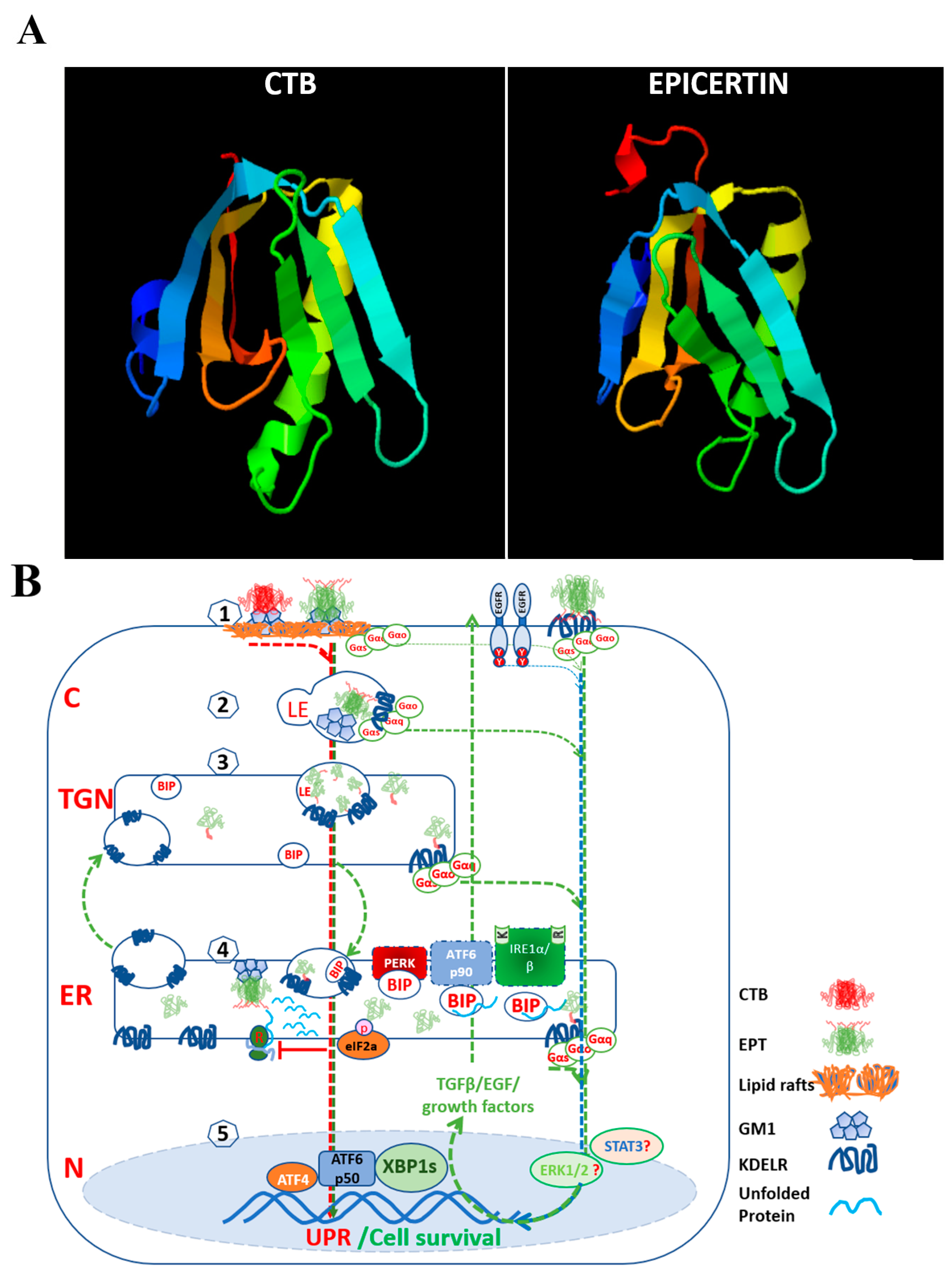
  115. Baldauf, K.J.; Royal, J.M.; Kouokam, J.C.; Haribabu, B.; Jala, V.R.; Yaddanapudi, K.; Hamorsky, K.T.; Dryden, G.W.; Matoba, N. Oral administration of a recombinant cholera toxin B subunit promotes mucosal healing in the colon. Mucosal Immunol. 2017, 10, 887–900. [Google Scholar] [CrossRef] [Green Version]
  116. Royal, J.M.; Oh, Y.J.; Grey, M.J.; Lencer, W.I.; Ronquillo, N.; Galandiuk, S.; Matoba, N. A modified cholera toxin B subunit containing an ER retention motif enhances colon epithelial repair via an unfolded protein response. FASEB J. 2019, 33, 13527–13545. [Google Scholar] [CrossRef] [Green Version]
  117. Koutsoumpli, G.; Ip, P.P.; Schepel, I.; Hoogeboom, B.N.; Boerma, A.; Daemen, T. Alphavirus-based hepatitis C virus therapeutic vaccines: Can universal helper epitopes enhance HCV-specific cytotoxic T lymphocyte responses? Ther. Adv. Vaccines Immunother. 2019, 7, 1–7. [Google Scholar] [CrossRef] [Green Version]
  118. Martinez-Puente, D.H.; Perez-Trujillo, J.J.; Gutierrez-Puente, Y.; Rodriguez-Rocha, H.; Garcia-Garcia, A.; Saucedo-Cardenas, O.; Montes-de-Oca-Luna, R.; Loera-Arias, M.J. Targeting HPV-16 antigens to the endoplasmic reticulum induces an endoplasmic reticulum stress response. Cell Stress Chaperones 2019, 24, 149–158. [Google Scholar] [CrossRef]
  119. Hamorsky, K.T.; Kouokam, J.C.; Bennett, L.J.; Baldauf, K.J.; Kajiura, H.; Fujiyama, K.; Matoba, N. Rapid and scalable plant-based production of a cholera toxin B subunit variant to aid in mass vaccination against cholera outbreaks. PLoS Neglected Trop Dis. 2013, 7, e2046. [Google Scholar] [CrossRef]
  120. Kelley, L.A.; Mezulis, S.; Yates, C.M.; Wass, M.N.; Sternberg, M.J. The Phyre2 web portal for protein modeling, prediction and analysis. Nat. Protoc. 2015, 10, 845–858. [Google Scholar] [CrossRef] [Green Version]
  121. Wolf, A.A.; Jobling, M.G.; Saslowsky, D.E.; Kern, E.; Drake, K.R.; Kenworthy, A.K.; Holmes, R.K.; Lencer, W.I. Attenuated endocytosis and toxicity of a mutant cholera toxin with decreased ability to cluster ganglioside GM1 molecules. Infect. Immun. 2008, 76, 1476–1484. [Google Scholar] [CrossRef] [Green Version]
  122. Jobling, M.G.; Yang, Z.; Kam, W.R.; Lencer, W.I.; Holmes, R.K. A single native ganglioside GM1-binding site is sufficient for cholera toxin to bind to cells and complete the intoxication pathway. mBio 2012, 3, e00401-12. [Google Scholar] [CrossRef] [Green Version]
  123. Jia, J.; Yue, X.; Zhu, L.; Jing, S.; Wang, Y.; Gim, B.; Qian, Y.; Lee, I. KDEL receptor is a cell surface receptor that cycles between the plasma membrane and the Golgi via clathrin-mediated transport carriers. Cell. Mol. Life Sci. 2021, 78, 1085–1100. [Google Scholar] [CrossRef]
  124. Beck, P.L.; Rosenberg, I.M.; Xavier, R.J.; Koh, T.; Wong, J.F.; Podolsky, D.K. Transforming growth factor-beta mediates intestinal healing and susceptibility to injury in vitro and in vivo through epithelial cells. Am. J. Pathol. 2003, 162, 597–608. [Google Scholar] [CrossRef]
  125. Royal, J.M.; Reeves, M.A.; Matoba, N. Repeated Oral Administration of a KDEL-tagged Recombinant Cholera Toxin B Subunit Effectively Mitigates DSS Colitis Despite a Robust Immunogenic Response. Toxins 2019, 11, 678. [Google Scholar] [CrossRef] [Green Version]
  126. Verjan Garcia, N.; Santisteban Celis, I.C.; Dent, M.; Matoba, N. Characterization and utility of two monoclonal antibodies to cholera toxin B subunit. Sci. Rep. 2023, 13, 4305. [Google Scholar] [CrossRef]
  127. Yang, J.; Liu, H.; Li, L.; Liu, H.; Shi, W.; Yuan, X.; Wu, L. Structural Insights into IRE1 Functions in the Unfolded Protein Response. Curr. Med. Chem. 2016, 23, 4706–4716. [Google Scholar] [CrossRef]
  128. Lam, M.; Marsters, S.A.; Ashkenazi, A.; Walter, P. Misfolded proteins bind and activate death receptor 5 to trigger apoptosis during unresolved endoplasmic reticulum stress. Elife 2020, 9, e52291. [Google Scholar] [CrossRef]
  129. Zhang, Z.; Venditti, R.; Ran, L.; Liu, Z.; Vivot, K.; Schurmann, A.; Bonifacino, J.S.; De Matteis, M.A.; Ricci, R. Distinct changes in endosomal composition promote NLRP3 inflammasome activation. Nat. Immunol. 2023, 24, 30–41. [Google Scholar] [CrossRef]
Table 1. Therapeutic strategies in inflammatory bowel disease targeting the unfolded protein response.
In Vivo/In Vitro ModelER Stress InducerTargeted UPR SensorDrug CandidateMode of ActionReferences
Mouse model of DSS-induced colitis. P58 (IPK−/−), ATF6a−/−, IL10−/− mice, and IEC-6 cellsDSS-induced colitis/cytokine cocktail (TNF-α, MCP-1, IL-1β)Induction of ATF6α- and chaperone P58; reduce ER stressPhenyl butyric acid (PBA), Taurine-conjugated ursodeoxycholic acid (TUDCA)Promote protein folding; reduce features of acute and chronic colitis; reduce BiP, peIF2a, CHOP, and cleaved caspase 3/12.Cao et al., 2013 [101]
Rat model of TNBS-induced colitis; Caco2 cellsTNBS-induced colitis. Brefeldin A and Tunicamyin-induced ER stressPERK-CHOP, ATF4, ATF6, XBP1sGlutamineReduce CHOP, BiP, Caspases 12, 9, 8, 3. calpain-1, and pJNK.Crespo et al., 2012 [109]
Mouse model of DSS-induced colitisDSS-induced colitisPhosphatase inhibitor Increase eIF2α phosphorylationSalubrinalIncrease BiP, ATF4, and HSP70; reduce CHOP; suppress MPO, TNF-α, and IL-1β.Okazaki et al., 2014 [110]
Mouse model of DSS-induced colitisDSS-induced colitis. ER stressPrevents activation of PERK-eIF2α-ATF4-CHOP and IRE1α-XBP1 signaling pathwaysArtesunateIncrease Bcl-2/Bax ratio; suppress NF-κB p65 and IκBα, IL-1β, IL-6, and TNF-α; increase IL-10; inhibit expression of cleaved-caspase 3.Yin et al., 2020, 2021 [111,112]
Mouse model of DSS-induced colitis; CCD841 cell lineDSS induced colon ER stress/ThapsigarginInhibited PERK, IRE1α, ATF6, reduced BiP, CHOPG-1Inhibit apoptosis, increased Ki-67+, and BrdU+ crypt cells; reverse the decrease in cyclin D1 and B1.Wang et al., 2021 [113]
Mouse model of DSS colitis modelDSS-induced colitis. ER stressXBP1 agonist, Coptisine-derivativeHLJ2Decrease MPO, TNFα, IL-1β, and IL-6; increase the expression of ZO-1 and claudin-1.Zhang et al., 2017 [114]
Mouse model of DSS-induced acute and chronic colitis. Caco2 cells scratch assayDSS-induced ER stressInduce IRE1-XBP1s axis.EPICERTINBinds to KDELRs and may promote vesicle trafficking; promotes mucosal wound healing mediated by TGFβ, CDH1, and WNT5.Baldauf et al., 2017 [115]; Royal et al., 2019 [116]
Disclaimer/Publisher’s Note: The statements, opinions and data contained in all publications are solely those of the individual author(s) and contributor(s) and not of MDPI and/or the editor(s). MDPI and/or the editor(s) disclaim responsibility for any injury to people or property resulting from any ideas, methods, instructions or products referred to in the content.

