Article

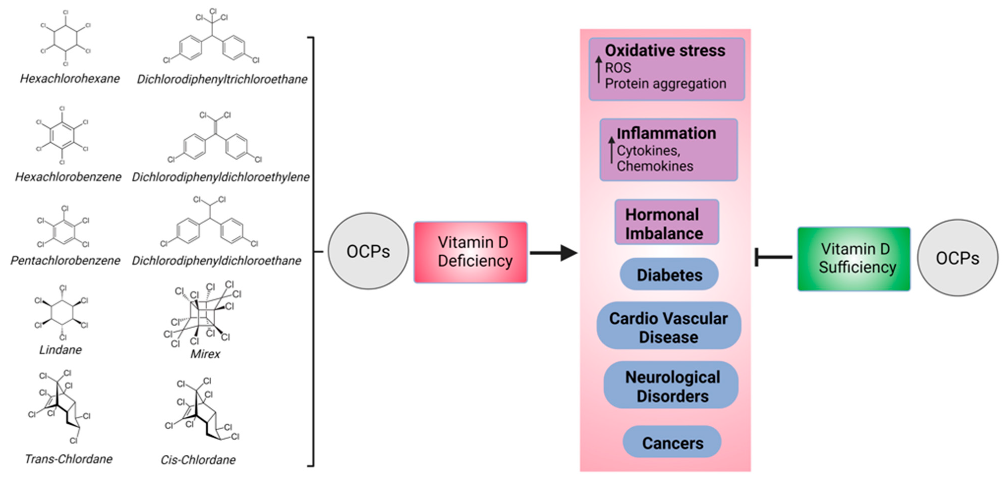
Association between Organochlorine Pesticides and Vitamin D in Female Subjects

by
Edwina Brennan
1,*,
Alexandra E. Butler
1,
Manjula Nandakumar
1,
Daniel S. Drage
2,3,
Thozhukat Sathyapalan
4 and
Stephen L. Atkin
1
1
School of Medicine, Royal College of Surgeons in Ireland-Medical University of Bahrain, Busaiteen 15503, Bahrain
2
School of Geography, Earth and Environmental Sciences, University of Birmingham, Edgbaston, Birmingham B15 2TT, UK
3
Queensland Alliance for Environmental Health Sciences, The University of Queensland, 39 Kessels Road, Brisbane, QLD 4108, Australia
4
Hull York Medical School, University of Hull, Hull HU6 7RX, UK
*
Author to whom correspondence should be addressed.
Biomedicines 2023, 11(5), 1451; https://doi.org/10.3390/biomedicines11051451
Submission received: 23 April 2023 / Revised: 4 May 2023 / Accepted: 10 May 2023 / Published: 15 May 2023
(This article belongs to the Special Issue Vitamin D in Health and Disease (2nd Edition))

Abstract

:
In human population studies, organochlorine pesticides (OCPs) have been linked to vitamin D deficiency. Therefore, this study examined the association between OCPs, vitamin D3 (cholecalciferol, 25(OH)D3), and the active metabolite 1,25-dihydrovitamin D3 (1,25(OH)2D3) in a cohort of non-obese women. The serum samples of 58 female participants (age—31.9 ± 4.6 years; body mass index (BMI)—25.7 ± 3.7 kg/m2) were screened for 10 indicator OCPs. 25(OH)D3 and 1,25(OH)2D3 levels were determined via isotope dilution liquid chromatography tandem mass spectrometry. In this cohort, the 25(OH)D3 and 1,25(OH)2D3 levels were 22.9 ± 11.2 ng/mL and 0.05 ± 0.02 ng/mL, respectively, with 28 participants classified as 25(OH)D3-deficient (<50 nmol/L). In the study cohort, no correlations were found between individual or total OCPs (ƩOCPs) and 25(OH)D3. p,p′-dichlorodiphenyldichloroethylene (DDE) and ƩOCPs correlated positively with 1,25(OH)2D3, with the latter being negatively correlated with estimated glomerular filtration rate (eGFR). In women with sufficient 25(OH)D3 levels, p,p′-dichlorodiphenyltrichloroethan (DDT) was positively correlated with 1,25(OH)2D3, whilst in the deficient group, hexachlorobenzene (HCB) and p,p′-(DDE) were positively correlated with 1,25(OH)2D3, β-Hexachlorocyclohexane (HCH) was positively correlated with 25(OH)D3, and none of the OCPs were associated with measures of renal function. Overall, OCPs and ƩOCPs were not associated with 25(OH)D3, suggesting that they are unrelated to vitamin D deficiency, but p,p′-DDE and ƩOCPs correlated positively with active 1,25(OH)2D3, while ƩOCPs correlated negatively with eGFR, suggesting a possible renal effect. Analysis of vitamin D deficiency revealed an association between β-HCH and 25(OH)D3, and between HCB and p,p′-DDE and 1,25(OH)2D3, suggesting that OCP effects may be enhanced in cases of vitamin D deficiency.

Graphical Abstract

1. Introduction

Organochlorine pesticides (OCPs) are a group of chlorinated hydrocarbons that were once used extensively in agriculture and mosquito control and have since been termed persistent organic pollutants (POPs) due to their high toxicity, slow degradation, and bioaccumulation [1]. OCPs are highly persistent in the environment and can accumulate in the fatty tissues of animals and humans [2,3]. They can also be transported over great distances in the air and water, leading to the contamination of soil and water sources far from the OCPs’ original application site [4]. Due to the potential health risks associated with OCPs, many countries have banned or severely restricted their use in recent years [5]. However, these chemicals can persist in the environment for decades [4] and may still be found in some food and water sources.
Exposure to OCPs has been linked to a variety of health issues. OCPs have been linked to an increased risk of breast [6] and prostate [7] cancer, and lymphoma [8]. OCPs have been linked to reproductive problems in both men and women, including decreased fertility [9,10] and miscarriages, particularly with respect to hexachlorocyclohexane (HCH) [11], and prenatal exposure to α-HCH, β-HCH, ο,p′-dichlorodiphenyldichloroethane (DDD), and p,p′-DDD has been associated with an increased risk for neural tube birth defects [12]. OCPs have been linked to neurological problems, which they are thought to contribute to via their accumulation in the body over time, potentially leading to Parkinson’s disease (HCH) [13] and Alzheimer’s disease (p,p′-dichlorodiphenyldichloroethylene (DDE)) [14]. OCPs have been linked to immune system disorders, with dichlorodiphenyltrichloroethane (DDT) causing the dysregulation of interleukin 6 (IL-6), leading to chronic inflammation [15], and having been related to oxidative stress and immune suppression [16], thus increasing susceptibility to infections and other diseases. OCPs have been classed as endocrine disruptors that can cause developmental problems in children (DDT) [17] and reproductive problems in adults [9]. In addition, low doses of OCPs have been strongly linked to various chronic diseases including diabetes [18] and cardiovascular diseases [19].
OCPs may have several mechanisms of action that contribute to their reported diverse effects. OCPs function by binding to specific target sites in the body, including binding to and blocking the GABAA receptors of sodium channels in the brain [20]. By binding to these sites, they disrupt the normal functioning of the nervous system, leading to the overstimulation or inhibition of nerve impulses. OCPs can also interfere with the normal function of the endocrine system by disrupting the production, release, transport, and metabolism of hormones at several sites of action such as the hypothalamus (increasing gonadotropin-releasing hormone mRNA levels), pituitary glands (affecting luteinizing hormone release), and ovaries (decreased sex steroid hormone production) [20].
Vitamin D deficiency is associated with various health conditions such as osteoporosis, cancer, cardiovascular disease, autoimmune diseases, and increased mortality [21,22]. In addition, vitamin D deficiency is associated with an increased incidence of type 2 diabetes [23,24,25] and microvascular and macrovascular diabetes-related complications [26]. Vitamin D3 (cholecalciferol, 25(OH)D3) is endogenously produced via the UV-B irradiation of 7-dehydrocholesterol and is hydroxylated to 25(OH)D3 by multiple 25-hydroxylases in the liver [27]. 25(OH)D3 is transported to the kidneys and converted to active 1,25-dihydrovitamin D3 (1,25(OH)2D3) by 1 alpha hydroxylase present in the renal tubular cells [28]. It has recently been reported that extrarenal tissues may also convert 25(OH)D3 to 1,25(OH)2D3, although activation in renal and non-kidney tissues is regulated differently via the macrophage production of 1,25(OH)2D3 through the type 2 interferon (IFN) response [29].
There is some evidence suggesting that exposure to OCPs may be associated with lower levels of vitamin D in the body [19,30]. Population studies have suggested that OCPs, including p,p′-DDT, p,p′-DDE, and β-HCH, can cause vitamin D deficiency [31]. In one such study, Inuit people who were heavily exposed to these pesticides through their traditional diet of fish and marine mammals, causing accumulation of these pesticides, showed higher levels of OCPs in their blood and had lower levels of vitamin D [32]; however, chronobiological factors within this population are likely to play a role.
OCPs may contribute to vitamin D deficiency through their effects on vitamin D metabolism in the liver, the disruption of the conversion of 25(OH)D3 to 1,25(OH)2D3, interference with the function of vitamin D receptors in the body, or the impairment of calcium homeostasis [33]. It is important to note that the evidence linking OCPs and vitamin D levels is still limited, and more research is needed to fully understand the relationship between these two factors. However, the evidence still highlights the potential health risks associated with exposure to these persistent environmental pollutants. Therefore, this study was undertaken to examine the association between OCPs (pentachlorobenzene (PeCB), hexachlorobenzene (HCB), HCH, lindane, trans-chlordane, cis-chlordane, DDE, DDD, DDT, and dodecachlorooctahydro-1,3,4-metheno-1H-cyclobuta[cd]pentalene (Mirex)) and 25(OH)D3 and its active metabolite 1,25(OH)2D3 in a group of non-obese healthy women prior to undergoing in vitro fertilization (IVF). In addition, we sought to determine if such an association may be precipitated by the impairment of calcium homeostasis, specifically with respect to its association with calcium/calmodulin-dependent (CaM) kinases (CaMK), CaMK kinase alpha (CaMKKα), and CaM phosphodiesterase (PDE) proteins.

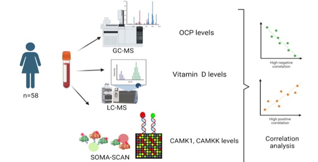
2. Materials and Methods

2.1. Patient Recruitment

Subjects were recruited from the Hull IVF Unit, UK, following ethical approval from The Yorkshire and The Humber NRES ethical committee, UK (approval number 02/03/043). Venesection was performed on 58 fasting, non-obese Caucasian women; samples were taken at 09:00 on day 21 of the menstrual cycle prior to IVF downregulation. Venous blood samples were centrifuged at 3500× g for 15 min at 4 °C, and serum was stored at −80 °C within 1 h of collection for further analysis. No participant was on any prescribed or over-the-counter medication. All participants provided written informed consent.

2.2. OCP Measurement

Samples were screened for 10 indicator OCPs (PeCB, HCB, HCH, lindane, trans-chlordane, cis-chlordane, DDE, DDD, DDT, and Mirex). A sum-of-OCPs (ƩOCPs) variable was calculated by adding the molar concentrations of each OCP analyzed. After vortexing the samples for 1 min, 6 mL of acetonitrile, 3 mL of milliQ water, 5 g of anhydrous MgSO4, and 1 g of NaCl were added along with a ceramic homogenizer. Samples were manually shaken for 1 min before being centrifuged at 4700 RPM for 8 min at 10 °C. The supernatant layer (approximately 6 mL of acetonitrile) was collected and transferred to 12 mL glass vials. An aliquot of 200 µL was collected and cleaned for non-targeted analysis (not presented here). The remainder of the extract was transferred to a 15 mL glass tube, evaporated under a gentle stream of nitrogen to near-dryness, and reconstituted in approximately 1 mL of hexane. Approximately 1 mL of >98% concentrated sulfuric acid was added, and the sample was vortexed for at least 30 s. The aqueous and hexane layers were left to separate overnight at <4 °C. The supernatant (hexane) layer was transferred directly onto a silica solid-phase extraction (SPE) cartridge (Supelco LC-Si, 3 mL/500 mg) (preconditioned with 6 mL dichloromethane (DCM) followed by 6 mL of hexane). Target compounds were eluted into a 15 mL glass collection tube using 6 mL of hexane followed by 8 mL of DCM. Samples were evaporated to near-dryness and reconstituted in 50 µL of methanol containing 2.5 ng 13C12-PCB-141 as a recovery standard. Samples were transferred to autosampler vial inserts prior to analysis.

2.3. Instrumental Analysis

OCPs were identified in serum samples via high-resolution gas chromatography coupled with high-resolution mass spectrometry (HRGC/HRMS). A Thermofisher TRACE 1300 gas chromatograph was coupled to a Thermofisher DFS mass spectrometer. The injector was operated in splitless mode with separation achieved on an Agilent DB-5ms column (30 m length × 0.25 mm in diameter × 0.25 µm film thickness). Experiments were conducted in multiple ion detection (MID) mode at a resolution level of 10,000 (10% valley definition). The inlet, transfer line, and source were maintained at 250 °C, 280 °C, and 280 °C, respectively. The flow rate was maintained at 1.0 mL/min. In the GC oven, an initial temperature of 80 °C was held for two minutes before being ramped up to 180 °C at 20 °C/min and held for 0.5 min. The temperature was then increased to 300 °C at 10 °C/min and held for 5 min.

2.4. Quality Assurance/Quality Control

The limit of reporting (LOR) was determined according to the quality of the peak shape and using a minimum signal-to-noise ratio of 10:1. Contamination was assessed via inclusion of procedural blanks (milli-Q water, n = 6) extracted in parallel with unknown samples. If a target compound was detected in a blank at below 5% of a sample, then no correction occurred. When blank concentrations were 5–25% of a respective sample, the sample was blank corrected. Linearity was established with a seven-point calibration across the range of 0.1 to 25 ng/mL for all target analytes. R2 values > 0.99 were achieved for all compounds. Analytes were identified based on retention time (<2% RSD from standard) and confirmed with reference to a second ion (HRGC/HRMS). Quantification was performed using isotope dilution mass spectrometry. All OCPs were quantified against 13C12-HCB. All data were validated by spiking 5 aliquots of bovine serum with 300 pg of target analytes. The concentrations of the spiked samples were measured, and the recoveries (measured concentration/spiked concentration × 100) were calculated. Average recoveries were 80–120% with a relative standard deviation of <15%, thus conforming to the OECD guidelines of good laboratory practice (Supplementary Table S1).

2.5. Vitamin D3 and Biochemical Parameters

Biochemical and hormonal parameters were measured as per a previously detailed method [34]. Briefly, serum 25(OH)D3 and 1,25(OH)2D3 levels were quantified using isotope dilution liquid chromatography tandem mass spectrometry (LC-MS/MS). Vitamin D metabolites and labelled internal standards were extracted from 250 µL of serum using supportive liquid–liquid extraction and Diels–Alder derivatization prior to LC-MS/MS analysis. Chromatographic separations were achieved using Hypersil Gold C18 column (150 × 2.1 mm; 1.9 µm) at a flow rate of 0.2 mL/min and operated in Electrospray Ionization (ESI) positive mode and analyzed using the multiple-reaction-monitoring (MRM) method. The limit of quantification (LOQ) for 1,25(OH)2D3 was 10 pg/mL and 25(OH)D3 1.2 nmol/L (0.5 ng/mL) [35]. Vitamin D deficiency was defined as a cutoff of 20 ng/mL (50 nmol/L) as per the clinical practice guidelines of the Endocrine Society Task Force on Vitamin D [36].

2.6. CaM Proteomic Measurement

Circulating levels of CaM-associated proteins (PDE1A, CaMK2A, CaMK2B, CaMK1D, CaMK2D, CaMK1, and CaMKKα) were determined via Slow Off-rate Modified Aptamer (SOMA)-scan plasma protein measurement (Somalogic, Boulder, CO, USA), the details of which have been previously reported [37]. Briefly, EDTA plasma samples were diluted, and the following assay steps were performed: (a) Binding: analytes and primer bead–SOMAmers (fully synthetic fluorophore-labelled SOMAmer coupled to a biotin moiety through a photocleavable linker) are equilibrated. (b) Catch I: all analyte–SOMAmer complexes are immobilized on a streptavidin-substituted support. Washing steps: proteins not stably bound to primer bead–SOMAmers are removed, and bound protein is biotinylated. (c) Cleave: long-wave ultraviolet light is applied to release analyte SOMAmer complexes into the solution. (d) Catch II: analyte–SOMAmer complexes are selectively immobilized on streptavidin support via the introduced analyte-borne biotinylation. Further washing is performed to select against non-specific analyte–SOMAmer complexes. (e) Elution: denaturation is induced to disrupt analyte–SOMAmer complexes. Released SOMAmers serve as surrogates for quantification of analyte concentrations. (f) Quantification: SOMAmer complementary oligonucleotides are hybridized to custom arrays. Normalization of raw intensities, hybridization, median signal, and calibration signal were performed based on the standard samples included on each plate, as previously described [38].

2.7. Statistics

No previous studies were available to enable the performance of a power analysis; therefore, this pilot study was designed according to the research conducted by Birkett and Day [39]. Descriptive data are presented as mean ± standard deviation (SD) for continuous data. Potential correlations between OCPs, 25(OH)D3), and 1,25(OH)2D3 were examined using exploratory Spearman’s rank order correlations. A p-value of <0.05 was considered to indicate statistical significance. Statistical analysis was carried out using Jamovi (version 2.0.0).

3. Results

3.1. Whole-Cohort Analysis

Demographic and biochemical data for this cohort are shown in Table 1. The participants had a mean age of 31.9 ± 4.6 years and a mean body mass index (BMI) of 25.7 ± 3.7 kg/m2. Thyroid function and C-reactive protein (CRP) levels, constituting measures of underlying inflammation, were normal. The mean levels of 25(OH)D3 and 1,25(OH)2D3 were 22.9 ± 11.2 ng/mL and 0.05 ± 0.02 ng/mL, respectively. Of the 58 women recruited, 28 had a 25(OH)D3 level less than 20 ng/mL (50 nmol/L). The obtained levels of OCPs, 25(OH)D3, and 1,25(OH)2D3 are shown in Table 1.

3.2. OCP Levels

The detection frequency of individual OCPs ranged from 100–12%. p,p′-DDE was detected in all serum samples, making it the most abundant OCP, followed by HCB (95%), p,p′-DDT (74%), β-HCH (67%), cis-chlordane (48%), Mirex (41%), PeCB (38%), trans-chlordane (24%), o,p′-DDE (22%), and lindane (12%). The OCPs α-HCH, o,p′-DDD, and p,p′-DDD+o,p′-DDT were not detected.

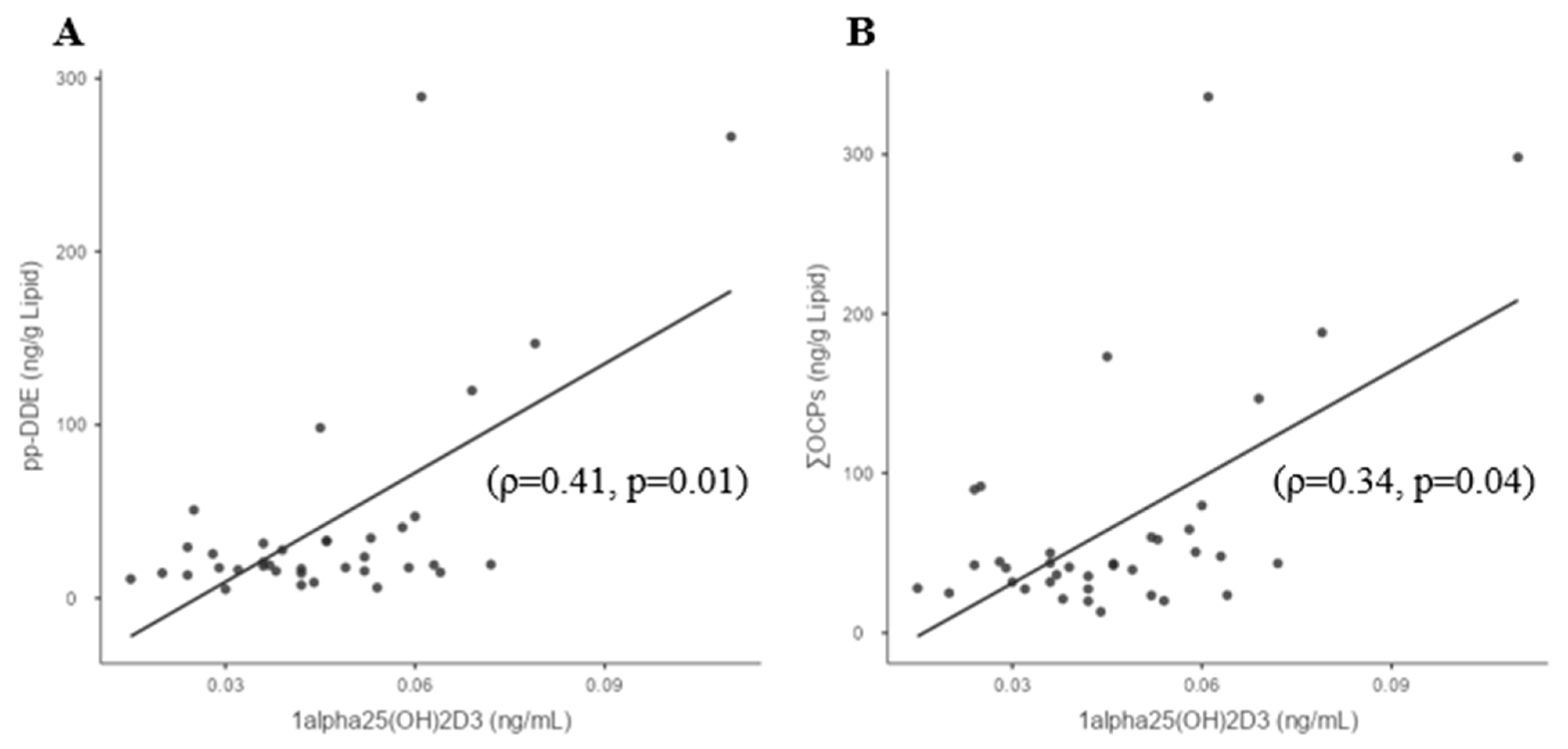
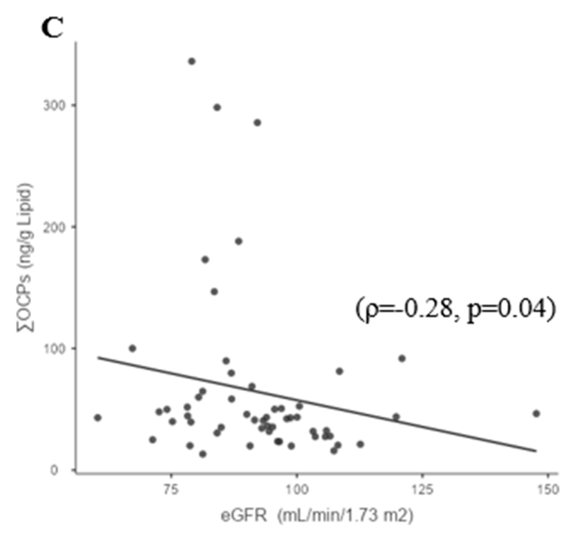
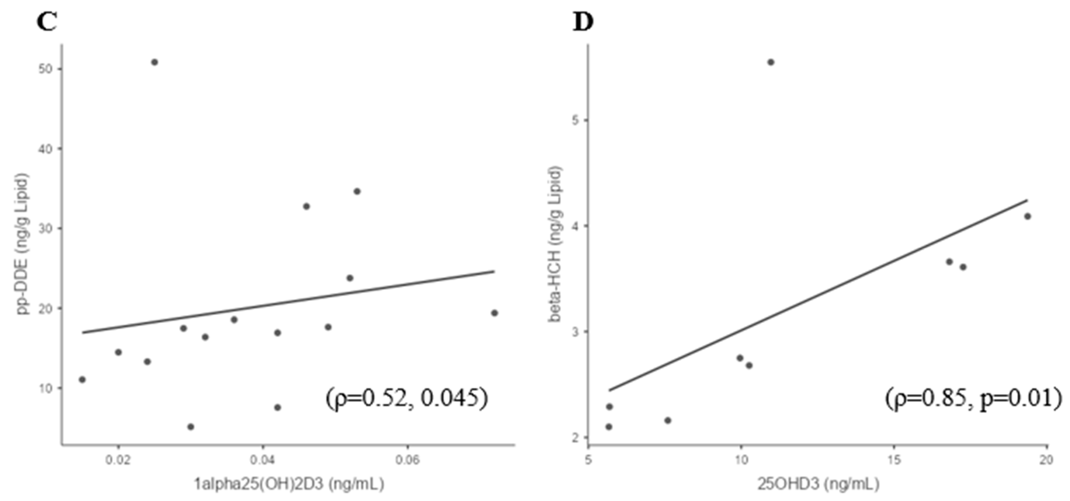
3.3. Whole-Group Correlations

In the study cohort, no correlations were found between ƩOCPs or individual OCPs and 25(OH)D3. The OCP p,p′-DDE and the ƩOCPs positively correlated with 1,25(OH)2D3, (ρ = 0.41; p = 0.01) and (ρ = 0.34; p = 0.04), respectively (Figure 1A,B), but no other correlations were found between individual OCPs and 1,25(OH)2D3 (Table 2). The ƩOCPs variable negatively correlated with eGFR (ρ = −0.28; p = 0.04) (Figure 1C). There were no correlations between p,p′-DDE or ƩOCPs with urea and creatinine.

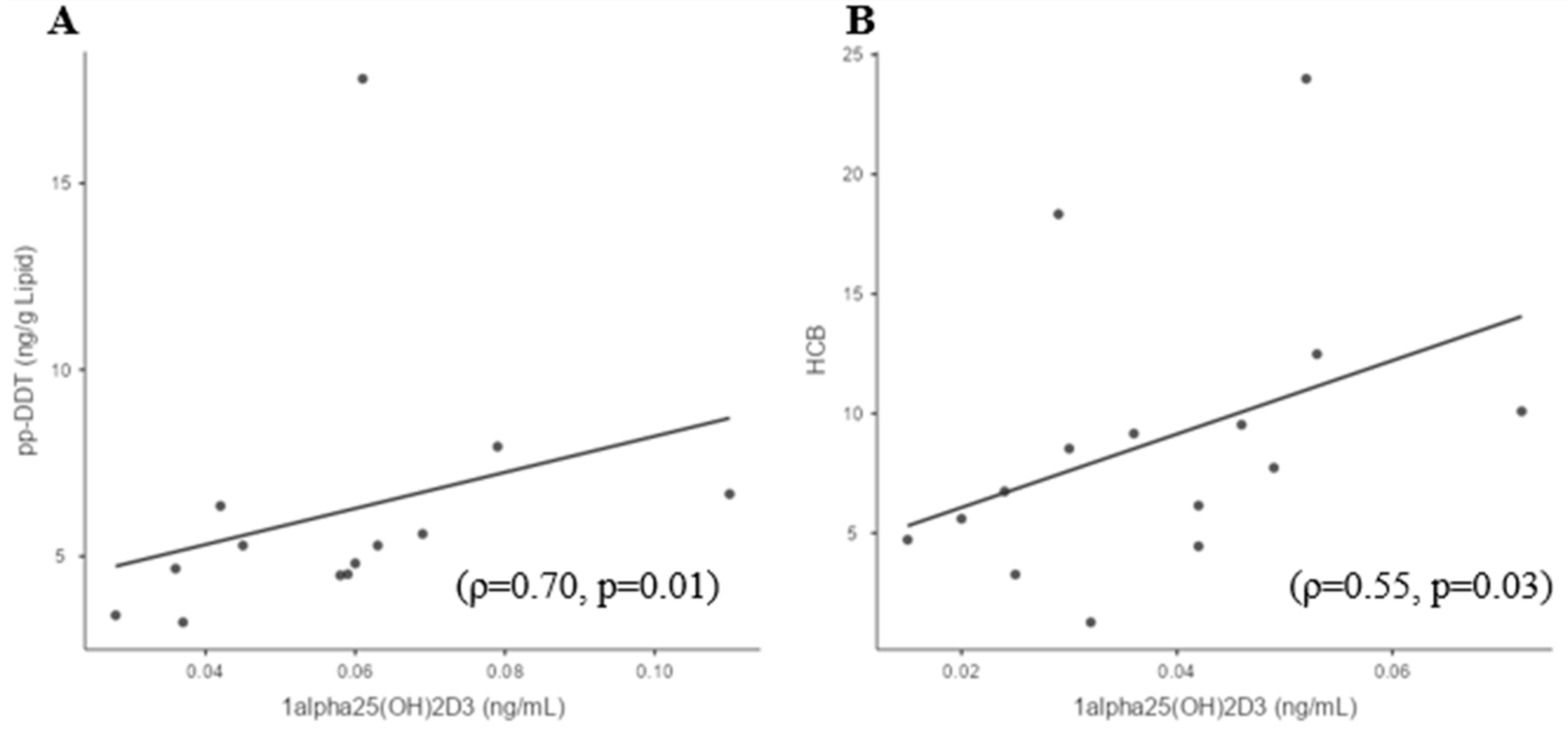
3.4. Sufficient and Deficient 25(OH)D3 Subgroup Correlations

The study cohort was divided into two subgroups based on the participants’ vitamin D levels: sufficient 25(OH)D3 (>20 ng/mL; >50 nmol/L) women and deficient 25(OH)D3 (<20 ng/mL; <50 nmol/L) women. In the sufficient 25(OH)D3 women, p,p′-DDT strongly positively correlated with 1,25(OH)2D3 (ρ = 0.70; p = 0.01) (Figure 2A). There were no correlations found between OCPs and 25(OH)D3 in the sufficient 25(OH)D3 women subgroup. In deficient 25(OH)D3 women, HCB and p,p′-DDE positively correlated with 1,25(OH)2D3 ((ρ = 0.55; p = 0.03) and (ρ = 0.52; p = 0.045), respectively (Figure 2B,C)), while β-HCH strongly positively correlated with 25(OH)D3 (ρ = 0.85; p = 0.01) (Figure 2D). Calcium levels did not differ between vitamin D sufficiency (2.3 ± 0.07 mmol/L) and vitamin D deficiency groups (2.3 ± 0.06 mmol/L).
We have found that 1,25(OH)2D3 levels may be modulated through CaMK1 by polybrominated diethyl ether 153 (unpublished results); therefore, the CaM-associated proteins (PDE1A, CaMK2A, CaMK2B, CaMK1D, CaMK2D, CaMK1, and CaMKKα) (Table 1) that effect the rapid action of 1,25(OH)2D3 at the cell membrane leading to increasing intracellular calcium levels were investigated, but no associations with the individual or total OCPs, or in vitamin-D-sufficient or deficient states, were found.

4. Discussion

These data show that the 10 indicator OCPs and ƩOCPs analyzed do not appear to be associated with 25(OH)D3 but that the OCP p,p′-DDE and ƩOCPs correlated positively with active 1,25(OH)2D3. Overall, the ƩOCPs were negatively associated with a reduced eGFR level, as noted by others [40], indicating that their action may be exerted on the kidneys, as suggested by the reported association of DDT burden with chronic kidney disease [41], but this was an observational study with no mechanistic insights. An inverse relationship of OCPs, including p,p′-DDT, p,p′-DDE, and β-HCH, with vitamin D has been reported [31], but this was not seen in the data presented herein; however, this difference is potentially due to the differing study populations, as those associations were seen amongst subjects of an older age, who were Caucasian, or who suffered from chronic diseases, whereas the population analyzed in this study was young and healthy.
When the study cohort was divided into two subgroups, namely, sufficient and deficient groups based on their vitamin D levels, in the vitamin-D-sufficient 25(OH)D3 women, p,p′-DDT strongly positively correlated with 1,25(OH)2D3. There were no correlations found between OCPs and 25(OH)D3 in the vitamin-D-sufficient 25(OH)D3 subgroup of women. In the vitamin-D-deficient 25(OH)D3 women, both p,p′-DDE and HCB positively correlated with 1,25(OH)2D3. In the vitamin-D-deficient group, β-HCH strongly positively correlated with 25(OH)D3, suggesting that a reduction in vitamin D may signal a potential effect of both HCB and HCH, but it is unclear what those specific effects may be. The mechanism(s) behind the positive associations between OCPs and 1,25(OH)2D3 is unclear, but it has been reported that in cells exposed to p,p′-DDT and p,p′-DDE, this led to an increase in the vitamin D binding protein [42] that binds to 1,25(OH)2D3 for its activity, perhaps leading to an increase in 1,25(OH)2D3 levels.
1,25(OH)2D3 increases calcium absorption via vitamin D receptor (VDR) binding and exerts rapid action at the cell membrane, increasing intracellular calcium levels by activating protein kinase cascades that stimulate VDR-mediated transcription-enhancing steroid receptor coactivator (SRC) activity, with the overall effect of enhancing 1,25(OH)2D3-dependent interactions between VDR and SRC coactivators [43]. To investigate a possible mechanism by which OCPs may affect 1,25(OH)2D3 action, the determination of the plasma levels of CaM proteins (CaMK, CaMKK, and PDE), which may be indicative of 1,25(OH)2D3 action, was undertaken. No correlations with total or individual OCPs were found, suggesting that the OCPs impacting 1,25(OH)2D3 are not acting at the membrane level through calmodulin-associated proteins. Therefore, it is clear that 25(OH)D3 is converted to its biologically active form 1,25(OH)2D3 in the presence of OCPs without affecting serum calcium levels, as has been described in other animal species [44], and it is unclear how OCPs may then impair the vitamin D biological response. That the OCPs appeared to be associated with vitamin D and vitamin D sufficiency or deficiency suggests that OCPs’ effects may be mediated through vitamin D. Of particular concern, OCPs may potentiate vitamin D deficiency, which is a condition that affects over 50% of the global population [45]. Moreover, the diseases that are associated with vitamin D deficiency, such as diabetes and cardiovascular disease [21,22], are the same as those attributed to OCP exposure [19,30], suggesting that one may represent an epiphenomenon of the other. Another potential explanation is that the association between OCPs and vitamin D may be caused by indirect mechanisms. In vitro, OCPs induce inflammation and oxidative stress, with β-HCH, DDE, and Dieldrin causing the upregulation of pro-inflammatory cytokines such as tumor necrosis factor (TNF)-α, IL-1β, IL-6, nuclear factor kappa B (NF-kB), and cyclooxygenase (COX)-2 expression in OCP-treated human ovary surface epithelial cells [46]. These findings are in line with observations that DDT causes the dysregulation of IL-6 leading to chronic inflammation [15] and that DDT is related to oxidative stress and immune suppression [15,16]. This is also reflected in in vivo findings, where, in a systematic review, exposure to OCPs was positively associated with CRP, IL-1β, IL-2, and IL-10, which is indicative of dysregulated inflammatory responses; however, the underlying mechanisms remain unclear [47]. Conversely, vitamin D prevents oxidative stress, while vitamin D deficiency induces the disruption of mitochondrial function [48] and is also associated with markers of inflammation [49]. Consequently, in vitamin D sufficiency, this may generally mitigate the inflammatory and oxidative stress effects of the OCPs; however, in vitamin D deficiency, these effects have already been uncovered and indeed may be exacerbated (Figure 3), as seen with results reporting that vitamin D deficiency promotes colonic inflammation [50] and, in an animal model of acetaminophen-induced liver injury, exacerbates induced oxidative stress [51].
In addition, extrarenal tissues may also convert 25(OH)D3 to 1,25(OH)2D3; however, notably, activation in renal and non-kidney tissues is regulated differently, with macrophage production of 1,25(OH)2D3 through the type 2 IFN response [29]. In animal models, OCPs do not appear to affect macrophages; therefore, their influence through an extrarenal effect on 1,25(OH)2D3 appears less likely [52]. However, DDT caused an increase in IFNγ secretion [53]; increasing IFNγ may have the effect of increasing the conversion of 25(OH)D3 to 1,25(OH)2D3 in the extrarenal tissues.
The strengths of this study include its state-of-the-art measurement of OCPs and vitamin D (25(OH)D3 and 1,25(OH)2D3). Its limitations include the small number of subjects included, who were all Caucasian females, so these findings may not be generalizable to male subjects or those of different ethnicities. The low numbers compounded by the detection number may have resulted in a type-two statistical error (false negative); however, this study will potentiate the determinative power of a larger study focusing on vitamin D deficiency. OCPs have been associated with parathyroid hyperplasia leading to elevated parathyroid hormone (PTH) and calcium levels [54]; however, the calcium levels were in the normal reference range in these patients, thus obviating the need to measure PTH.

5. Conclusions

In conclusion, our findings show that, overall, OCPs and ƩOCPs were not associated with 25(OH)D3, suggesting that they are unrelated to vitamin D deficiency, and that the OCP p,p′-DDE and the ƩOCPs correlated positively with active 1,25(OH)2D3 and negatively with eGFR, suggesting a renal effect. Vitamin D deficiency unmasked an association between β-HCH and 25(OH)D3 and between HCB and p,p′-DDE and 1,25(OH)2D3, suggesting that OCPs’ effects may be enhanced in patients with vitamin D deficiency.

Supplementary Materials

The following supporting information can be downloaded at: https://www.mdpi.com/article/10.3390/biomedicines11051451/s1. Supplementary Table S1. OCP validation data.

Author Contributions

Conceptualization, S.L.A. and T.S.; methodology, S.L.A. and T.S.; validation, S.L.A.; formal analysis, E.B. and A.E.B.; investigation, D.S.D.; resources, S.L.A.; data curation, S.L.A.; writing—original draft preparation, E.B., A.E.B. and S.L.A.; writing—review and editing, E.B., A.E.B., S.L.A., D.S.D., M.N. and T.S.; visualization, E.B., A.E.B., M.N. and S.L.A. All authors have read and agreed to the published version of the manuscript.

Funding

This research received no external funding.

Institutional Review Board Statement

The study was conducted in accordance with the Declaration of Helsinki, and approved by the Institutional Review Board (or Ethics Committee) of The Yorkshire and The Humber NRES ethical committee, UK (approval number 02/03/043).

Informed Consent Statement

Informed consent was obtained from all subjects involved in the study.

Data Availability Statement

The data presented in this study are available on reasonable request from the corresponding author.

Conflicts of Interest

The authors declare no conflict of interest.

References

  1. Jayaraj, R.; Megha, P.; Sreedev, P. Organochlorine pesticides, their toxic effects on living organisms and their fate in the environment. Interdiscip. Toxicol. 2016, 9, 90–100. [Google Scholar] [CrossRef] [PubMed]
  2. Deshmukh, H.; Aylward, L.L.; Rose, M.; Fernandes, A.; Sedman, P.; Thatcher, N.J.; Atkin, S.L.; Sathyapalan, T. Association of endocrine active environmental compounds with body mass index and weight loss following bariatric surgery. Clin. Endocrinol. 2020, 93, 280–287. [Google Scholar] [CrossRef] [PubMed]
  3. Buah-Kwofie, A.; Humphries, M.S.; Combrink, X.; Myburgh, J.G. Accumulation of organochlorine pesticides in fat tissue of wild Nile crocodiles (Crocodylus niloticus) from iSimangaliso Wetland Park, South Africa. Chemosphere 2018, 195, 463–471. [Google Scholar] [CrossRef] [PubMed]
  4. Ma, Y.; Yun, X.; Ruan, Z.; Lu, C.; Shi, Y.; Qin, Q.; Men, Z.; Zou, D.; Du, X.; Xing, B.; et al. Review of hexachlorocyclohexane (HCH) and dichlorodiphenyltrichloroethane (DDT) contamination in Chinese soils. Sci. Total Environ. 2020, 749, 141212. [Google Scholar] [CrossRef]
  5. Keswani, C.; Dilnashin, H.; Birla, H.; Roy, P.; Tyagi, R.K.; Singh, D.; Rajput, V.D.; Minkina, T.; Singh, S.P. Global footprints of organochlorine pesticides: A pan-global survey. Environ. Geochem. Health 2022, 44, 149–177. [Google Scholar] [CrossRef] [PubMed]
  6. Miao, Y.; Rong, M.; Li, M.; He, H.; Zhang, L.; Zhang, S.; Liu, C.; Zhu, Y.; Deng, Y.L.; Chen, P.P.; et al. Serum concentrations of organochlorine pesticides, biomarkers of oxidative stress, and risk of breast cancer. Environ. Pollut. 2021, 286, 117386. [Google Scholar] [CrossRef]
  7. Antignac, J.P.; Figiel, S.; Pinault, M.; Blanchet, P.; Bruyère, F.; Mathieu, R.; Lebdai, S.; Fournier, G.; Rigaud, J.; Mahéo, K.; et al. Persistent organochlorine pesticides in periprostatic adipose tissue from men with prostate cancer: Ethno-geographic variations, association with disease aggressiveness. Environ. Res. 2023, 216, 114809. [Google Scholar] [CrossRef] [PubMed]
  8. Luo, D.; Zhou, T.; Tao, Y.; Feng, Y.; Shen, X.; Mei, S. Exposure to organochlorine pesticides and non-Hodgkin lymphoma: A meta-analysis of observational studies. Sci. Rep. 2016, 6, 25768. [Google Scholar] [CrossRef]
  9. Rattan, S.; Zhou, C.; Chiang, C.; Mahalingam, S.; Brehm, E.; Flaws, J.A. Exposure to endocrine disruptors during adulthood: Consequences for female fertility. J. Endocrinol. 2017, 233, R109–R129. [Google Scholar] [CrossRef]
  10. Giulioni, C.; Maurizi, V.; Castellani, D.; Scarcella, S.; Skrami, E.; Balercia, G.; Galosi, A.B. The environmental and occupational influence of pesticides on male fertility: A systematic review of human studies. Andrology 2022, 10, 1250–1271. [Google Scholar] [CrossRef]
  11. Pathak, R.; Mustafa, M.; Ahmed, R.S.; Tripathi, A.K.; Guleria, K.; Banerjee, B.D. Association between recurrent miscarriages and organochlorine pesticide levels. Clin. Biochem. 2010, 43, 131–135. [Google Scholar] [CrossRef] [PubMed]
  12. Yin, S.; Sun, Y.; Yu, J.; Su, Z.; Tong, M.; Zhang, Y.; Liu, J.; Wang, L.; Li, Z.; Ren, A.; et al. Prenatal exposure to organochlorine pesticides is associated with increased risk for neural tube defects. Sci. Total Environ. 2021, 770, 145284. [Google Scholar] [CrossRef]
  13. Xu, S.; Yang, X.; Qian, Y.; Luo, Q.; Song, Y.; Xiao, Q. Analysis of serum levels of organochlorine pesticides and related factors in Parkinson’s disease. Neurotoxicology 2022, 88, 216–223. [Google Scholar] [CrossRef] [PubMed]
  14. Medehouenou, T.C.M.; Ayotte, P.; Carmichael, P.H.; Kröger, E.; Verreault, R.; Lindsay, J.; Dewailly, É.; Tyas, S.L.; Bureau, A.; Laurin, D. Exposure to polychlorinated biphenyls and organochlorine pesticides and risk of dementia, Alzheimer’s disease and cognitive decline in an older population: A prospective analysis from the Canadian Study of Health and Aging. Environ. Health 2019, 18, 57. [Google Scholar] [CrossRef] [PubMed]
  15. Martin, T.J.; Gabure, S.; Maise, J.; Snipes, S.; Peete, M.; Whalen, M.M. The organochlorine pesticides pentachlorophenol and dichlorodiphenyltrichloroethane increase secretion and production of interleukin 6 by human immune cells. Environ. Toxicol. Pharmacol. 2019, 72, 103263. [Google Scholar] [CrossRef]
  16. Koner, B.C.; Banerjee, B.D.; Ray, A. Organochlorine pesticide-induced oxidative stress and immune suppression in rats. Indian J. Exp. Biol. 1998, 36, 395–398. [Google Scholar]
  17. Jeddy, Z.; Kordas, K.; Allen, K.; Taylor, E.V.; Northstone, K.; Dana Flanders, W.; Namulanda, G.; Sjodin, A.; Hartman, T.J. Prenatal exposure to organochlorine pesticides and early childhood communication development in British girls. Neurotoxicology 2018, 69, 121–129. [Google Scholar] [CrossRef]
  18. Evangelou, E.; Ntritsos, G.; Chondrogiorgi, M.; Kavvoura, F.K.; Hernández, A.F.; Ntzani, E.E.; Tzoulaki, I. Exposure to pesticides and diabetes: A systematic review and meta-analysis. Environ. Int. 2016, 91, 60–68. [Google Scholar] [CrossRef]
  19. Zago, A.M.; Faria, N.M.X.; Fávero, J.L.; Meucci, R.D.; Woskie, S.; Fassa, A.G. Pesticide exposure and risk of cardiovascular disease: A systematic review. Glob. Public Health 2022, 17, 3944–3966. [Google Scholar] [CrossRef]
  20. Costa, L.G. The neurotoxicity of organochlorine and pyrethroid pesticides. Handb. Clin. Neurol. 2015, 131, 135–148. [Google Scholar] [CrossRef]
  21. Bjelakovic, G.; Gluud, L.L.; Nikolova, D.; Whitfield, K.; Wetterslev, J.; Simonetti, R.G.; Bjelakovic, M.; Gluud, C. Vitamin D supplementation for prevention of mortality in adults. Cochrane Database Syst. Rev. 2014, Cd007470. [Google Scholar] [CrossRef] [PubMed]
  22. Osorio Landa, H.K.; Perez Diaz, I.; Laguna Barcenas, S.D.C.; Lopez Navarro, J.M.; Abella Roa, M.F.; Corral Orozco, M.; Mancilla Ortega, J.P.; Martinez Duarte, D.A.; Morales Montalvo, S.I.; Muzquiz Aguirre, S.; et al. Association of serum vitamin D levels with chronic disease and mortality. Nutr. Hosp. 2020, 37, 335–342. [Google Scholar] [CrossRef] [PubMed]
  23. Nakashima, A.; Yokoyama, K.; Yokoo, T.; Urashima, M. Role of vitamin D in diabetes mellitus and chronic kidney disease. World J. Diabetes 2016, 7, 89–100. [Google Scholar] [CrossRef] [PubMed]
  24. Husemoen, L.L.; Thuesen, B.H.; Fenger, M.; Jorgensen, T.; Glumer, C.; Svensson, J.; Ovesen, L.; Witte, D.R.; Linneberg, A. Serum 25(OH)D and type 2 diabetes association in a general population: A prospective study. Diabetes Care 2012, 35, 1695–1700. [Google Scholar] [CrossRef] [PubMed]
  25. Mathieu, C. Vitamin D and diabetes: Where do we stand? Diabetes Res. Clin. Pract. 2015, 108, 201–209. [Google Scholar] [CrossRef]
  26. Butler, A.E.; Dargham, S.R.; Latif, A.; Mokhtar, H.R.; Robay, A.; Chidiac, O.M.; Jayyousi, A.; Al Suwaidi, J.; Crystal, R.G.; Abi Khalil, C.; et al. Association of vitamin D3 and its metabolites in subjects with and without Type 2 diabetes and their relationship to diabetes complications. Ther. Adv. Chronic Dis. 2020, 11, 2040622320924159. [Google Scholar] [CrossRef]
  27. Bikle, D.D. Vitamin D metabolism, mechanism of action, and clinical applications. Chem. Biol. 2014, 21, 319–329. [Google Scholar] [CrossRef]
  28. Christakos, S.; Dhawan, P.; Verstuyf, A.; Verlinden, L.; Carmeliet, G. Vitamin D: Metabolism, Molecular Mechanism of Action, and Pleiotropic Effects. Physiol. Rev. 2016, 96, 365–408. [Google Scholar] [CrossRef]
  29. Adams, J.S.; Rafison, B.; Witzel, S.; Reyes, R.E.; Shieh, A.; Chun, R.; Zavala, K.; Hewison, M.; Liu, P.T. Regulation of the extrarenal CYP27B1-hydroxylase. J. Steroid Biochem. Mol. Biol. 2014, 144 Pt A, 22–27. [Google Scholar] [CrossRef]
  30. Han, X.; Zhang, F.; Meng, L.; Xu, Y.; Li, Y.; Li, A.; Turyk, M.E.; Yang, R.; Wang, P.; Zhang, J.; et al. Exposure to organochlorine pesticides and the risk of type 2 diabetes in the population of East China. Ecotoxicol. Environ. Saf. 2020, 190, 110125. [Google Scholar] [CrossRef]
  31. Yang, J.H.; Lee, Y.M.; Bae, S.G.; Jacobs, D.R., Jr.; Lee, D.H. Associations between organochlorine pesticides and vitamin D deficiency in the U.S. population. PLoS ONE 2012, 7, e30093. [Google Scholar] [CrossRef] [PubMed]
  32. Schæbel, L.K.; Bonefeld-Jørgensen, E.C.; Vestergaard, H.; Andersen, S. The influence of persistent organic pollutants in the traditional Inuit diet on markers of inflammation. PLoS ONE 2017, 12, e0177781. [Google Scholar] [CrossRef] [PubMed]
  33. Andrews, J.E.; Courtney, K.D.; Donaldson, W.E. Impairment of calcium homeostasis by hexachlorobenzene (HCB) exposure in Fischer 344 rats. J. Toxicol. Environ. Health 1988, 23, 311–320. [Google Scholar] [CrossRef] [PubMed]
  34. Brennan, E.; Kumar, N.; Drage, D.S.; Cunningham, T.K.; Sathyapalan, T.; Mueller, J.F.; Atkin, S.L. A case-control study of polychlorinated biphenyl association with metabolic and hormonal outcomes in polycystic ovary syndrome. J. Environ. Sci. Health C Toxicol. Carcinog. 2022, 40, 86–105. [Google Scholar] [CrossRef] [PubMed]
  35. Javed, Z.; Papageorgiou, M.; Deshmukh, H.; Kilpatrick, E.S.; Mann, V.; Corless, L.; Abouda, G.; Rigby, A.S.; Atkin, S.L.; Sathyapalan, T. A Randomized, Controlled Trial of Vitamin D Supplementation on Cardiovascular Risk Factors, Hormones, and Liver Markers in Women with Polycystic Ovary Syndrome. Nutrients 2019, 11, 188. [Google Scholar] [CrossRef]
  36. Holick, M.F.; Binkley, N.C.; Bischoff-Ferrari, H.A.; Gordon, C.M.; Hanley, D.A.; Heaney, R.P.; Murad, M.H.; Weaver, C.M. Evaluation, treatment, and prevention of vitamin D deficiency: An Endocrine Society clinical practice guideline. J. Clin. Endocrinol. Metab. 2011, 96, 1911–1930. [Google Scholar] [CrossRef]
  37. Moin, A.S.M.; Sathyapalan, T.; Butler, A.E.; Atkin, S.L. Vitamin D association with coagulation factors in polycystic ovary syndrome is dependent upon body mass index. J. Transl. Med. 2021, 19, 239. [Google Scholar] [CrossRef]
  38. Moin, A.S.M.; Sathyapalan, T.; Butler, A.E.; Atkin, S.L. Coagulation factor dysregulation in polycystic ovary syndrome is an epiphenomenon of obesity. Clin. Endocrinol. 2023, 98, 796–802. [Google Scholar] [CrossRef]
  39. Birkett, M.A.; Day, S.J. Internal pilot studies for estimating sample size. Stat. Med. 1994, 13, 2455–2463. [Google Scholar] [CrossRef]
  40. Siddharth, M.; Datta, S.K.; Bansal, S.; Mustafa, M.; Banerjee, B.D.; Kalra, O.P.; Tripathi, A.K. Study on organochlorine pesticide levels in chronic kidney disease patients: Association with estimated glomerular filtration rate and oxidative stress. J. Biochem. Mol. Toxicol. 2012, 26, 241–247. [Google Scholar] [CrossRef]
  41. Lv, J.; Guo, L.; Gu, Y.; Xu, Y.; Xue, Q.; Yang, X.; Wang, Q.N.; Meng, X.M.; Xu, D.X.; Pan, X.F.; et al. National temporal trend for organophosphate pesticide DDT exposure and associations with chronic kidney disease using age-adapted eGFR model. Environ. Int. 2022, 169, 107499. [Google Scholar] [CrossRef] [PubMed]
  42. Pavlíková, N.; Daniel, P.; Šrámek, J.; Jelínek, M.; Šrámková, V.; Němcová, V.; Balušíková, K.; Halada, P.; Kovář, J. Upregulation of vitamin D-binding protein is associated with changes in insulin production in pancreatic beta-cells exposed to p,p’-DDT and p,p’-DDE. Sci. Rep. 2019, 9, 18026. [Google Scholar] [CrossRef]
  43. Ellison, T.I.; Dowd, D.R.; MacDonald, P.N. Calmodulin-dependent kinase IV stimulates vitamin D receptor-mediated transcription. Mol. Endocrinol. 2005, 19, 2309–2319. [Google Scholar] [CrossRef] [PubMed]
  44. Nowicki, H.G.; Myrtle, J.F.; Norman, A.W. Effects of organochlorine insecticides on metabolism of cholecalciferol (vitamin D 3) in Rachitic cockerel. J. Agric. Food Chem. 1972, 20, 380–384. [Google Scholar] [CrossRef] [PubMed]
  45. Siddiqee, M.H.; Bhattacharjee, B.; Siddiqi, U.R.; MeshbahurRahman, M. High prevalence of vitamin D deficiency among the South Asian adults: A systematic review and meta-analysis. BMC Public Health 2021, 21, 1823. [Google Scholar] [CrossRef]
  46. Shah, H.K.; Sharma, T.; Banerjee, B.D. Organochlorine pesticides induce inflammation, ROS production, and DNA damage in human epithelial ovary cells: An in vitro study. Chemosphere 2020, 246, 125691. [Google Scholar] [CrossRef]
  47. Liu, Z.; Lu, Y.; Zhong, K.; Wang, C.; Xu, X. The associations between endocrine disrupting chemicals and markers of inflammation and immune responses: A systematic review and meta-analysis. Ecotoxicol. Environ. Saf. 2022, 234, 113382. [Google Scholar] [CrossRef]
  48. Dzik, K.P.; Kaczor, J.J. Mechanisms of vitamin D on skeletal muscle function: Oxidative stress, energy metabolism and anabolic state. Eur. J. Appl. Physiol. 2019, 119, 825–839. [Google Scholar] [CrossRef]
  49. Querfeld, U. Vitamin D and inflammation. Pediatr. Nephrol. 2013, 28, 605–610. [Google Scholar] [CrossRef]
  50. Wei, X.; Li, X.; Du, J.; Ge, X.; Sun, Y.; Li, X.; Xun, Z.; Liu, W.; Wang, Z.Y.; Li, Y.C. Vitamin D Deficiency Exacerbates Colonic Inflammation Due to Activation of the Local Renin-Angiotensin System in the Colon. Dig. Dis. Sci. 2021, 66, 3813–3821. [Google Scholar] [CrossRef]
  51. Wang, Y.Q.; Geng, X.P.; Wang, M.W.; Wang, H.Q.; Zhang, C.; He, X.; Liang, S.M.; Xu, D.X.; Chen, X. Vitamin D deficiency exacerbates hepatic oxidative stress and inflammation during acetaminophen-induced acute liver injury in mice. Int. Immunopharmacol. 2021, 97, 107716. [Google Scholar] [CrossRef] [PubMed]
  52. Glinski, A.; Liebel, S.; Pelletier, È.; Voigt, C.L.; Randi, M.A.; Campos, S.X.; Oliveira Ribeiro, C.A.; Filipak Neto, F. Toxicological interactions of silver nanoparticles and organochlorine pesticides in mouse peritoneal macrophages. Toxicol. Mech. Methods 2016, 26, 251–259. [Google Scholar] [CrossRef] [PubMed]
  53. Massawe, R.; Drabo, L.; Whalen, M. Effects of pentachlorophenol and dichlorodiphenyltrichloroethane on secretion of interferon gamma (IFNγ) and tumor necrosis factor alpha (TNFα) from human immune cells. Toxicol. Mech. Methods 2017, 27, 223–235. [Google Scholar] [CrossRef] [PubMed]
  54. McMullin, J.L.; Codner, J.; Patel, S.G.; Sharma, J.; Hu, X.; Jones, D.P.; Weber, C.J.; Saunders, N.D. Environmental Chemicals and their Association with Hyperparathyroidism. World J. Surg. 2023, 47, 296–303. [Google Scholar] [CrossRef] [PubMed]
Figure 1. Significant correlations of OCPs with 1,25(OH)2D3 and measures of renal function in whole study cohort. (A) 1,25(OH)2D3 and p,p′-DDE; (B) 1,25(OH)2D3 and ƩOCPs; (C) eGFR and ƩOCPs.
Biomedicines 11 01451 g001aBiomedicines 11 01451 g001b
Figure 2. Significant correlations of OCPs with 25(OH)D3 and 1,25(OH)2D3 in cohort subgroups. The study cohort was divided into two subgroups based on the participants’ vitamin D levels: sufficient 25(OH)D3 (>20 ng/mL; >50 nmol/L) women and deficient 25(OH)D3 (<20 ng/mL; >50 nmol/L) women. Sufficient vitamin D subgroup—(A) 1,25(OH)2D3 and p,p′-DDT. Deficient vitamin D subgroup—(B) 1,25(OH)2D3 and HCB; (C) 1,25(OH)2D3 and p,p′-DDE; (D) 25(OH)D3 and β-HCH.
Biomedicines 11 01451 g002aBiomedicines 11 01451 g002b
Figure 3. OCPs analyzed in this study. The association of OCPs with various diseases in vitamin D sufficiency and deficiency were analyzed. Vitamin D sufficiency may generally mitigate the inflammatory and oxidative stress effects and associated disorders/diseases arising due to exposure to OCPs, but in vitamin D deficiency, those effects are unmasked and aggravated.
Biomedicines 11 01451 g003
Table 1. Mean and standard deviation of OCPs in the population studied.
Female Subjects (n = 58)
MeanSD
Age (years)31.94.6
BMI (kg/m2)25.73.7
CRP (mg/L)2.62.5
TSH (mU/L)2.42.2
Free-T3 (pmol/L)4.80.7
Free-T4 (pmol/L)11.31.8
25(OH)D3 (ng/mL)2311.2
1,25(OH)2D3 (ng/mL)0.050.02
Calcium (mmol/L)2.30.07
Urea (nmol/L)4.665.21
Creatinine (nmol/L)668.86
eGFR (mL/min/1.73 m2)92.314.8
PDE1A (RFU)7891060
CAMK2A (RFU)373453
CaMK2B (RFU)8591423
CaMK1D (RFU)1695580
CaMK2D (RFU)23793462
CaMK1 (RFU)46811371
CaMKK α (RFU)343463
PeCB (ng/g Lipid)9.129.1
α-HCH (ng/g Lipid)<LOR
β-HCH (ng/g Lipid)4.634.92
HCB (ng/g Lipid)9.816.5
Lindane (ng/g Lipid)1.350.28
Trans-Chlordane (ng/g Lipid)1.40.97
cis-Chlordane (ng/g Lipid)1.590.58
p,p′-DDE (ng/g Lipid)40.760.4
o,p′-DDE (ng/g Lipid)2.230.32
o,p′-DDD (ng/g Lipid)<LOR
p,p′-DDD+o,p′-DDT (ng/g Lipid)<LOR
p,p′-DDT (ng/g Lipid)4.992.3
Mirex (ng/g Lipid)3.270.69
ƩOCPs (ng/g Lipid)63.366.8
BMI: body mass index; CRP: C-reactive protein; TSH: thyroid-stimulating hormone; Free-T3: free-triiodothyronine; Free-T4: free-thyroxine; eGFR: estimated glomerular filtration rate; PDE: calcium/calmodulin-dependent phosphodiesterase; CaMK: calcium/calmodulin-dependent kinase; CaMKK α: calcium/calmodulin-dependent kinase kinase alpha; PeCB: pentachlorobenzene; α: alpha; β: beta; HCH: hexachlorocyclohexane; HCB: hexachlorobenzene; p: para; o: ortho; DDE: dichlorodiphenyldichloroethylene; DDD: dichlorodiphenyldichloroethane; DDT: dichlorodiphenyltrichloroethane; Mirex: dodecachlorooctahydro-1,3,4-metheno-1H-cyclobuta[cd]pentalene; SD: standard deviation; LOR: limit of reporting; ƩOCPs: total organochlorine pesticides.
Table 2. Spearman’s rho (ρ) correlations of OCPs with 25(OH)D3 and 1,25(OH)2D3. Data in bold indicate statistically significant correlations.
PeCBβ-HCHHCBLindanetrans-Chlordanecis-Chlordaneo,p′-DDEp,p′-DDEp,p′-DDTMirexƩOCPs
25(OH)D30.2890.1740.1010.393−0.140.3250.3740.2720.096−0.2040.284
(0.192)(0.339)(0.502)(0.40)(0.648)(0.091)(0.258)(0.061)(0.595)(0.361)(0.051)
1,25(OH)2D3−0.1080.3150.333−0.2−0.2050.2780.060.4080.364−0.030.34
(0.66)(0.14)(0.05)(0.92)(0.74)0.263(0.89)(0.01)(0.09)(0.92)(0.04)
Disclaimer/Publisher’s Note: The statements, opinions and data contained in all publications are solely those of the individual author(s) and contributor(s) and not of MDPI and/or the editor(s). MDPI and/or the editor(s) disclaim responsibility for any injury to people or property resulting from any ideas, methods, instructions or products referred to in the content.

