Review

Updates in KMT2A Gene Rearrangement in Pediatric Acute Lymphoblastic Leukemia

1
Student Scientific Society of Independent Laboratory of Genetic Diagnostics, Medical University of Lublin, 20-093 Lublin, Poland
2
Student Scientific Society of the Department of Pediatric Hematology, Oncology and Transplantology, Medical University of Lublin, 20-093 Lublin, Poland
3
Department of Paediatric Haematology, Oncology and Transplantology, Medical University of Lublin, 20-093 Lublin, Poland
4
Independent Laboratory of Genetic Diagnostics, Medical University of Lublin, 20-093 Lublin, Poland
*
Author to whom correspondence should be addressed.
Biomedicines 2023, 11(3), 821; https://doi.org/10.3390/biomedicines11030821
Submission received: 5 February 2023 / Revised: 1 March 2023 / Accepted: 4 March 2023 / Published: 8 March 2023
(This article belongs to the Special Issue State-of-the-Art Cell Biology and Pathology in Poland)

Abstract

:
The KMT2A (formerly MLL) encodes the histone lysine-specific N-methyltransferase 2A and is mapped on chromosome 11q23. KMT2A is a frequent target for recurrent translocations in acute myeloid leukemia (AML), acute lymphoblastic leukemia (ALL), or mixed lineage (biphenotypic) leukemia (MLL). Over 90 KMT2A fusion partners have been identified until now, including the most recurring ones—AFF1, MLLT1, and MLLT3—which encode proteins regulating epigenetic mechanisms. The presence of distinct KMT2A rearrangements is an independent dismal prognostic factor, while very few KMT2A rearrangements display either a good or intermediate outcome. KMT2A-rearranged (KMT2A-r) ALL affects more than 70% of new ALL diagnoses in infants (<1 year of age), 5–6% of pediatric cases, and 15% of adult cases. KMT2A-rearranged (KMT2A-r) ALL is characterized by hyperleukocytosis, a relatively high incidence of central nervous system (CNS) involvement, an aggressive course with early relapse, and early relapses resulting in poor prognosis. The exact pathways of fusions and the effects on the final phenotypic activity of the disease are still subjects of much research. Future trials could consider the inclusion of targeted immunotherapeutic agents and prioritize the identification of prognostic factors, allowing for the less intensive treatment of some infants with KMT2A ALL. The aim of this review is to summarize our knowledge and present current insight into the mechanisms of KMT2A-r ALL, portray their characteristics, discuss the clinical outcome along with risk stratification, and present novel therapeutic strategies.

1. Introduction

Acute lymphoblastic leukemia (ALL) still ranks among the most common childhood malignant hematology diseases, which represents a paramount challenge in 21st-century medicine. This happens because of the fact that continuous chromosomal and molecular abnormalities still evolve and are influenced by genetic and epigenetic processes, as well. However, innovative methods of the in-depth analysis of genetics along with sophisticated experiments tend to have a significant role in assessment, prognosis, and innovative treatment decisions. The attempt to understand, control, and eventually influence some of the unfavorable processes makes genetics nowadays one of the most fascinating, crucial parts of medicine that is also a great inspiration for a broader analysis of the topic of this review. ALL is characterized by the uncontrolled development of large numbers of immature lymphoid cells that lead to the occurrence of the condition [1]. The presence of KMT2A rearrangement in ALL is an independent dismal prognostic factor with long-term survival rates of less than 60% across all age groups [2,3,4]. Rearranged KMT2A (KMT2A-r) ALL presents a complex clinical challenge, with a high incidence in infants and a tendency for aggressive relapse. The KMT2A gene (formerly MLL1/MLL/ALL-1/HRX/HTRX1) is one of the most promiscuous recombination hot spots of the human genome with regard to the onset of malignant diseases. Numerous genomic alterations involving KMT2A have been recognized in acute leukemia, including chromosomal translocations, internal tandem duplications, internal deletions, and amplifications. The most common genomic lesions involving KMT2A in acute leukemia are chromosomal translocations, resulting in various fusion genes that express an abnormally functioning fusion protein. There are generally 8.3 kb of breakpoint clusters spanning exons 9 to 14 in the KMT2A fusion region [5]. Acute leukemia-bearing rearrangements of KMT2A have been recognized as a separate entity by the World Health Organization (WHO) since the introduction of the WHO Classification of Neoplastic Diseases of the Hematopoietic and Lymphoid Tissues in 1999 [6]. The current classification recognizes KMT2A-r acute lymphoblastic leukemias as B lymphoblastic leukemia/lymphoma with t(v;11q23.3) (KMT2A-r) with any fusion partner [7], whereas the updated International Consensus Classification (ICC) of B-acute lymphoblastic leukemia (B-ALL) and T-acute lymphoblastic leukemia (T-ALL) recognizes KMT2A-r ALL as B-ALL with t(v;11q23.3)/KMT2A rearranged and B-ALL KMT2A rearranged-like (in this case as a provisional entity—with frequency <1%, including some with HOXA fusions) [8]. The above-mentioned classification (ICC) also considers KMT2A-r B-ALL diagnostic considerations and ancillary testing—CD10 nonspecific but characteristically dim/negative, the presence of CD15, the absence of CD24, KMT2A break apart in fluorescence in situ hybridization (FISH), and the use of targeted transcriptome sequencing. None of these classifications mention prognostic assessment [8].
The aim of this review is to summarize our knowledge and present current insights into the mechanisms of KMT2A-rearranged (KMT2A-r) ALL, portray their characteristics, discuss the clinical outcomes along with risk stratification, and present novel therapeutic strategies.

2. Characteristics of KMT2A

The KMT2A (also known as mixed lineage leukemia, or MLL) is a gene that was described for the first time in 1991–1992 [9,10,11]. KMT2A is a large, 90 kb gene containing 36 exons coding for a 431 kDa protein and it is located on the long arm of chromosome 11 band q23.3 (11q23.3). It belongs to the group of KMT genes that catalyze the transfer of methyl groups from S-adenosylmethionine to the lysine residues on histone tails, especially H3 [12]. In the case of mutations in one of these genes, an alteration in chromatin conformation occurs along with an incorrect gene expression, leading to several syndromes known as chromatinopathies [13,14,15]. In germline cells, a pathogenic mutation in KMT2A leads to haploinsufficiency, resulting in Wiedemann–Steiner syndrome—a rare autosomal-dominant disorder characterized by a delay in development, intellectual disability, unusual facial features, short stature, and hypotonia [16,17,18]. The KMT2A gene encodes a DNA-binding protein methylating histone H3 lys4 (H3K4), lysine methyltransferase, formed of 3969 amino acids [9]. This protein has 18 domains, including the SET domain that has the methyltransferase activity on lysine 4 of histone 3 [19]. The main function of the KMT2A protein is the epigenetic regulation of transcriptional initiation and elongation through the H3K4 methylation of promoter regions mapped on the target gene [20]. KMT2A protein is also responsible for the control of hematopoietic cell proliferation and the differentiation of Meis homeobox 1 (MEIS1) and the homeobox A (HOXA) gene cluster [21,22]. In the case of the deregulation of these genes, the inhibition of correct hematopoietic development triggers the development of leukemia [23]. Rearrangements involving KMT2A and its partner genes are found in precursor B-ALL, T-ALL, acute myeloid leukemia (AML), myelodysplastic syndrome (MDS), mixed lineage (biphenotypic) leukemia (MPAL), and secondary leukemia [5]. As far as KMT2A-r ALL is concerned, it affects more than 80% of new ALL diagnoses in infants (<1 year of age), 5–6% of pediatric cases, and 15% of adult cases [5,24,25,26]. The above-mentioned classification may also take into account the division into B-cell KMT2A-r ALL and T-cell KMT2A-r ALL, which is 6% (including 70% of infant ALL cases) and 4–8% of cases, respectively [24,27,28,29,30]. Over 90 KMT2A fusion partners have been identified until now, including the most recurring ones—AFF1 (4q21), MLLT1 (19p13), and MLLT3 (9p21)—which encode proteins regulating epigenetic mechanisms and foreshow mostly poor outcomes [21,31]. Fusion between KMT2A and AFF1 is present in about 49% of infants, 44% of the rest of the pediatric population with B-ALL, and almost 75% of adult KMT2A-rearranged B-ALLs [31]. The frequency of fusion partners in B-ALL differs depending on patient age, but AFF1 (formerly AF4) remains the most common fusion partner in all age groups. Several important fusion partners, including their frequency and prognosis in infant and pediatric KMT2A-r ALL, are portrayed in Table 1.
The table presents the correlation between the fusion of KMT2A with its partner gene and prognosis (including the frequency). A very poor prognosis relates to a median survival rate of less than 12 months (as in the case of KMT2A::MLLT1 fusion) [32]. Poor prognosis refers to a median survival rate of 12 to 60 months [33,34,35,36]. KMT2A::MLLT3 fusion manifests an intermediate prognosis in the case of AML with other KMT2A translocations [37]. The relationship between KMT2A fusion partner and median survival rate in infant and pediatric KMT2A-r ALL is shown in Figure 1 and Figure 2.
KMT2A-r in ALL usually occurs as a single mutation and does not require a cooperative mutation in order to trigger the leukemia pattern [38,39]. However, it can also be present along with a cooperative mutation, especially often with a PI3K-RAS pathway mutation among infants (14–70%—a wide range with huge differences in reported mutation frequencies [38,39,40,41,42,43,44] and with KRAS and NRAS mutations among adults (8%) and pediatric patients (26%), respectively) [45]. The significance of intercurrent PI3K-RAS mutation remains the subject of research and further investigation is required. While one study classifies this mutation as a poor prognostic factor, the other ones present no significant impact on the clinical outcome or prognostic features [39,40,42,46], and still others indicate the synergizing effect of PI3K-RAS with KMT2A rearrangements that reduces leukemia latency [22,47].
Another study showed that KMT2A rearrangement often occurs together with the TP53 mutation (inactivation of the TP53 tumor suppressor gene). This cooperative mutation concerns ALL as well as MLL but is especially present in infant rearranged leukemias [48]. Another study by A. Stengel concentrated on the investigation of the frequency of the TP53 mutation among 625 patients with ALL. Thirty-seven patients had KMT2A-r ALL while six of them also had a cooperative TP53 mutation, which translates to 16.2% [49]. Moreover, the study indicates that the incidence of coexisting TP53 mutation is associated with poor prognosis, especially at relapse.

3. KMT2A—Clinical Presentation

Infants with KMT2A-r present more aggressive features compared with older children [50,51,52,53,54]. The KMT2A-r occurs with similar frequency in females and males [55]. KMT2A-rearranged (KMT2A-r) ALL is characterized by hyperleukocytosis (WBC >30 × 109/L), a relatively high incidence of central nervous system (CNS) involvement, and leukemia cutis (skin infiltration) [50]. Older children, similarly to KMT2A-germinal (KMT2A-g) newborns, are characterized by lower leukocyte values (WBC < 30 × 109/L), better response to prednisone, and more often positive CD10 [50,55]. They may present with hepatomegaly, splenomegaly, lymphadenopathy, and thrombocytopenia [56]. KMT2A-r ALL has a typical immature pro-B immunophenotype. It is characterized by the expression of CD19 and CD34 and the co-expression of CD15, CD33, CD65, and CD68, as well as often a lack of CD10 [46,57,58,59]. It can also have a pre-B immunophenotype, where lymphoblasts are CD22-, CD34-, CD 19-, TdT-, cytoplasmic (Cy) CD79a-, CD10-, and Cy mμ-positive, and cortical/thymic T-ALL, where lymphoblasts are cyCD3-, CD7-, TdT-, CD5-, and CD1a-positive [60]. KMT2A-r ALL is also characterized by the specific expression of chondroitin sulfate proteoglycan-4, which is also known as neutron-glial antigen-2 (NG2). It is a transmembrane proteoglycan that is expressed in normal hematopoietic cells very rarely [61,62]. However, the expression of NG2 in the case of KMT2A-r ALL is frequent (about 90% of cases) [61]. It contributes to leukemia invasiveness and CNS infiltration and correlates with lower event-free survival (EFS) and more frequent CNS relapse. Because of the predictive value of NG2, it has been the subject of many experiments lately and has also become a new therapeutic target for KMT2A-r ALL. The blocking of NG2s quantifies the effect of induction therapy for B-ALL by the transfer of KMT2A-r blasts into the blood from the bone marrow, where they are more vulnerable to chemotherapy [63,64,65,66,67].
KMT2A rearrangement is most common in young children and is associated with a worse prognosis. Laboratory tests of infants with KMT2A-r are characterized by a higher leukocyte count and a more severe course with CNS involvement compared to older patients and KMT2A-g. From a morphological point of view, there are no ideal criteria for distinguishing the B and T ALL lines. Distinguishing B-lineage lymphoblasts from normal B-lineage lymphoid precursors is also challenging. Pre-B can also express the CD10 antigen, but can be distinguished from mature lymphocytes by their weak expression of CD45 and occasionally the expression of CD34 [60].

4. Risk Stratification

There are three major cooperative groups conducting specific clinical trials for infant ALL: Interfant (based in Europe), COG (based in North America), and the Japanese Pediatric Leukemia Study Group (JPLSG). All induction strategies are based on Interfant-99. A prospective risk-stratified approach incorporating KMT2A-r status and age was used in all recently completed trials (Table 2) [50,54,68,69,70,71,72].
Older patients (>1 year of age) often have a much better prognosis than infants. They have favorable genetic features of high hyperdiploid and ETV6::RUNX1 fusion [59]. Studies have shown that older children share cytogenetic abnormalities with KMT2A-g, albeit with a different distribution; the proportion of patients with favorable genetic risk (hyperdiploid, ETV6::RUNX1) is higher (60% versus 12% KMT2A-g) [55].
Next-generation sequencing (NGS) involves the sequencing of DNA, RNA, or miRNA. It is a tool to identify the most important changes in ALL, which helps to determine the prognosis and pathogenesis of the disease [60,73]. Montaño A. conducted in Salamanca a study on eighty-five patients with B-LL. In cases with KMT2A-r, the NGS panel included only the region with the most frequent KMT2A-r breakpoints and detected only 70% of cytogenetically confirmed cases, suggesting that patients with a breakpoint at a different location may not be detected by the panel [74]. The performance of NGS in all cases of ALL is thus debatable.

5. Clinical Outcome and Interfant Protocol

In chemotherapy, according to the Interfant protocol, low- and high-dose cytarabine (araC) and anthracyclines are the basis. A randomized European study noted that infants with ALL do not have better outcomes with the early intensification of therapy with additional araC and daunorubicin or from the other drugs, mitoxantrone and etoposide [54].
Agraz-Doblas A. et al. analyzed the genome of 124 de novo cases of acute lymphoblastic B-cell leukemia in infants diagnosed and treated according to the Interfant 99/06 protocol. This study used bone marrow or peripheral blood samples from 124 infants <12 months old, who were diagnosed with either pro-B- or pre-B-cell ALL. There were 42 de novo cases, including 27 with KMT2A::AFF1 t(4;11), 5 with the KMT2A::AF9 (9;11), and 10 without KMT2A rearrangement. For validation, an additional cohort of patients comprised 43 with KMT2A::AFF1, 11 with KMT2A::MLLT3, and 28 non-KMT2A iBCP-ALL cases. Patients with KMT2A::AFF1 were characterized by five-fold longer event-free survival and a three-fold longer overall survival compared to t(4;11) iBCP-ALL patients without KMT2A::AFF1 [39]. In a randomized study, where patients were treated according to the Interfant-06 protocol, the 6-year event-free survival (EFS) in 651 patients with KMT2A-r ALL was 46.1%. For the low-risk group (LR), the EFS was 73.9%, 44.5% for the medium-risk (MR) group, and 20.9% for the high-risk (HR) group. Relapses occurred in 244 (37.5%) patients and the most common (66%) was isolated bone marrow (BM) recurrences [54].
However, in a study of 48 patients treated according to the CCCG-ALL-2015 protocol, KMT2A-r B-ALL was reported in 65.51% (19 out of 29) infants and 3.73% (32/857) non-infants, respectively. As a result of treatment, four patients died, and treatment-related mortality (TRM) was 8.33%; 40 patients achieved a complete remission (CR) and the CR rate for the total of 48 patients was 83.33%. Seven patients withdrew from the study. The median follow-up time for the 37 patients without TRM was 15.48 months, 15 patients relapsed, and the 5-year cumulative relapse rate for the 37 patients was 59.16 ± 9.16%. For patients with TRM on (41 patients) or TRM off (37 patients), the 5-year prospective EFS (pEFS) was 36.86 ± 8.48% or 40.84 ± 9.16%, respectively [33].
In a retrospective study of 124 ALL patients over 1 year of age, 31 of whom were with KMT2A-r, they were treated from 2008 to 2016 with the GD-ALL-2008 protocol and from 2016 to 2020 with the SCCLG-ALL-2016 protocol. The EFS of KMT2A-r-positive children was 56.01 ± 16.89%, and the overall survival (OS) was 73.32 ± 16.6%, which was lower compared to the negative KMT2A-r group. In children who received stem cell transplantation in addition to chemotherapy, the 10-year EFS and OS rates in this study were 100% and were higher than in children who received chemotherapy alone; 54.32 ± 16.89% and 72.19 ± 16, 88%, respectively. Unlike other studies, the most common partner gene in their study was KMT2A-PTD (Partial Tandem Duplications), which had a 10-year EFS of 85.71 ± 22.37%, showing a good prognosis [75].
In a study of 139 patients with ALL, of whom 100 were with KMT2A-r and 39 with KMT2A-germinal (KMT2A-g), 50 children (36%) had CNS involvement. CNS involvement was more common in children with KMT2A-r (44 out of 100 patients, 44.0%) compared to children with KMT2A-g (6 out of 39 patients, 15.4%). In addition, the frequency of their occurrence depended on the rearrangement of KMT2A. Infants with t(9;11)(p21;q23)/KMT2A::MLLT3 had CNS involvement relatively more frequently (62.5%) than children with t(11;19)(q23;p13.3)/KMT2A::MLLT1, in whom the trend was the opposite (only 28.6% of patients). CNS involvement was most common in children <6 months of age. In the group of 50 infants with CNS involvement, as many as 28 had a recurrence (EFS = 0.27, cumulative incidence of relapse (CIR) = 0.65), while among 89 CNS patients—negative—21 experienced a relapse (EFS = 0.58, CIR = 0.26, p < 0.001) for both. In total, 11 of the 49 patients with registered relapses had CNS infiltration (five isolated CNS relapses and six combined CNS and BM relapses) [76]. CNS involvement is more frequently observed in infants than in older children with KMT2A-r and is associated with a worse prognosis. Compared to patients with KMT2A-g, children with KMT2A gene rearrangement are more likely to have CNS involvement.
The Interfant-06 protocol also finds application in treatment with KMT2A-germinal (KMT2A-g), which is stratified into the low-risk group and usually has a better prognosis compared to KMT2A-r. In the 2021 study, which included newborns with KMT2A-g ALL treated in accordance with the Interfant-06 protocol, the following results were obtained. The 6-year event-free and overall survival were 73.9% and 87.2%. Relapses occurred early, within 36 months from diagnosis in 28 of 31 (90%) infants. Sites of relapse included (48.4%) isolated bone marrow (BM), (19.4%) isolated CNS, (16.1%) combined BM and CNS, (3.2%) combined BM and testis, and (12.9%) others. Six patients died in CR1, of which five deaths were due to infection. Treatment-related mortality was 3.6%. Age <6 months was a favorable prognostic factor with a 6-year disease-free survival (DFS) of 91% compared with 71.7% in infants >6 months of age (p = 0.04). Patients with a high end of induction (EOI) minimal residual disease (MRD) ≥ 5 × 10⁻⁴ had a worse outcome (6-year DFS 61.4%) compared with patients with undetectable EOI MRD (6-year DFS 87.9%) or intermediate EOI MRD < 5 × 10⁻⁴ (6-year DFS 76.4%) [55]. These studies allow us to conclude that in the case of KMT2A-g, relapses, if present, tend to occur early in most infants. Treatment-related mortality is very low, and positive prognostic factors include age <6 months and intermediate or negative EOI MRD.
According to our findings, conventional chemotherapy had poor outcomes for pediatric patients with KMT2A-r. There is some evidence that allo-HSCT at CR1 might improve the prognosis for patients with risk factors. Infants with KMT2A-germline ALL have a good prognosis when they are young at diagnosis and have low EOI MRD.

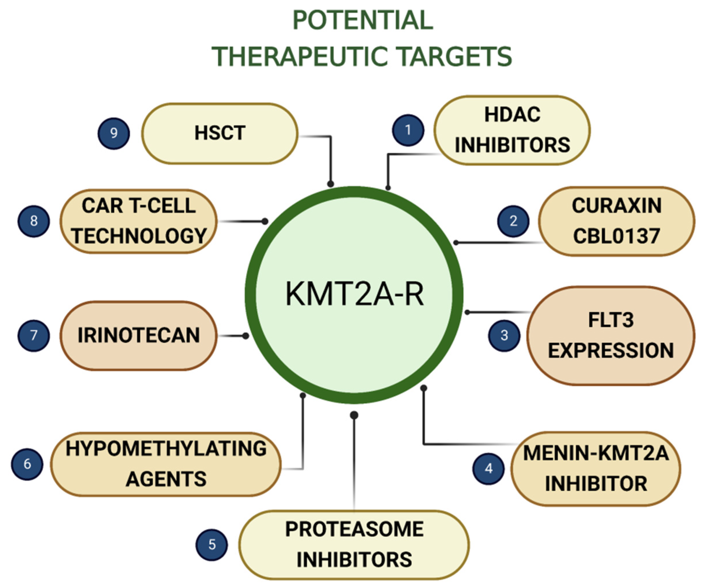
6. Potential Therapeutic Targets

Several potential therapeutic targets that will be discussed in this review are presented in Figure 3.

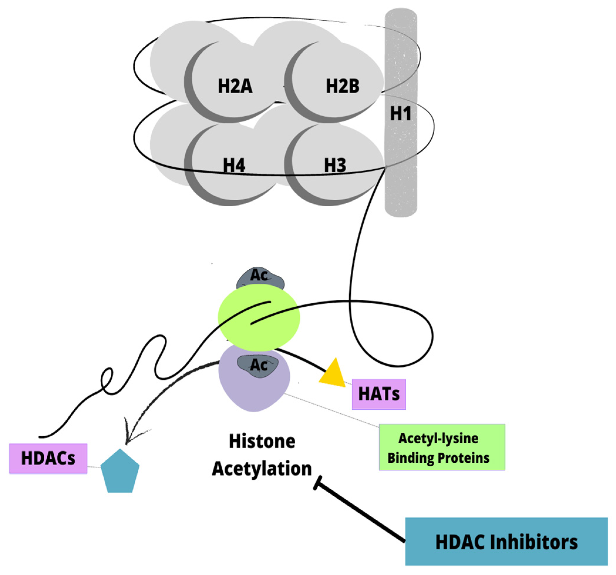
6.1. Histone Deacetylase Inhibitors

Histone deacetylase inhibitors (HDACi) are enzymes that remove acetyl groups from histones and other proteins, thereby regulating chromatin accessibility and target gene expression (Figure 4) [77,78]. Several studies have shown that HDACi are frequently overexpressed in leukemia cases, leading to the increased expression of tumor-driven genes and abnormal chromatin structure. A limited number of anti-cancer drugs have been approved by the US Food and Drug Administration (FDA). The most researched and prominent HDACi is SAHA [77].
The study by Yao J. and colleagues used an indole-3-butyric acid with phenyl groups in the linker as an HDAC inhibitor. They examined the effect of I1 on HDAC inhibitory activity by determining the level of acetylated histone protein H3 and H4 by Western blotting. Their findings show that the HDAC inhibitor I1, which is a chromatin remodeling factor, has a pronounced anti-proliferative effect on KMT2A-r ALL cells by inhibiting cell proliferation by inducing a G0/G1 cell cycle exit. Additionally, I1 inhibits HDAC more effectively than SAHA. I1 inhibited HDAC and also activated the signaling pathway of hematopoietic cell lines when they were treated with I1 at a concentration of 2 μM. Furthermore, I1 inhibited HDAC more effectively in THP-1 cells than in MOLM-13 cells, which is consistent with the IC50 values of I1 in MOLM-13 and THP-1 cells. Accordingly, I1 was able to overcome the cell differentiation block of KMT2A-r ALL cells, suggesting potential epigenetic drug potential including in vivo studies and anti-proliferation activity. Furthermore, it would be promising to induce cell differentiation to treat ALL [79].

6.2. Curaxin CBL0137

The curaxin family of compounds can activate p53 and inhibit NF-B at the same time. CBL0137 belongs to the second-generation curaxin and shows a very high potential for clinical applications due to the fact that it is soluble in water and has higher metabolic stability in mice compared with other members of the curaxin family [80]. In preclinical in vitro and in vivo models of KMT2A-r leukemia, curaxin CBL0137 has antileukemic effects and potentiates the effects of established chemotherapeutic treatments used in the treatment of pediatric high-risk ALL. In studies using CBL0137, nongenotoxic anticancer effects were demonstrated, such as activation of the p53 pathway and the induction of IFN by its chromatin-destabilizing properties [81]. By activating apoptosis and/or causing cell cycle arrest, all antileukemic mechanisms induced by CBL0137 can delay leukemia cell growth, which may have contributed to the KMT2A-r ALL growth delay [82].
Based on the evidence of CBL0137’s action in KMT2A-r leukemia in vivo, it would be worthwhile to apply this compound with other targeted approaches to KMT2A-r ALL that are currently being tested in clinical trials. As a result, the doses of chemotherapeutic drugs can be further reduced and safety may be enhanced. Considering CBL0137’s chromatin-destabilizing effect, one would expect an enhanced therapeutic effect when combined with histone deacetylase inhibitors (HDACi) such as vorinostat or panobinostat. There is evidence that HDACi exerts anti-leukemic effects in vitro, and in particular, LBH589 (panobinostat) shows a promising therapeutic index, since nanomolar concentrations specifically target primary KMT2A-r infant ALL cells [83]. Additionally, in a recent study in mouse models, it was observed that panobinostat alone induced the accumulation of DNA damage in the splenocytes of treated mice but did not significantly change the mean leukemia burden or survival. However, the combination of CBL0137 and panobinostat showed the greatest inhibition of leukemia progression and was well tolerated. Curaxin CBL0137 inhibits KMT2A-r leukemia cell growth by rapidly inducing apoptosis, and the addition of HDAC increased CBL0137-induced apoptosis. Similar effects were obtained after using CBL0137 in combination with another HDAC inhibitor, entinostat [84,85].

6.3. FLT3 Expression

The fms-like tyrosine kinase 3 (FLT3) is a proto-oncogene expressed on hematopoietic progenitor cells and plays an important role in hematopoiesis. There is a possibility that leukemia can be caused by mutations in the FLT3 receptor [86]. Patients with KMT2A-r ALL frequently exhibit constitutive FLT3 activation or increased FLT3 expression resulting from mutations in the tyrosine kinase domain [87]. The use of FLT3 inhibitors has been successful in treating AML with FLT3-activating mutations [88,89].
The Pediatric Oncology Group (COG) AALL0631 investigated whether adding lestaurtinib, a first-generation FLT3 inhibitor, to post-induction chemotherapy improves event-free survival. Following the induction of KMT2A-r chemotherapy, infants received chemotherapy alone or chemotherapy with lestaurtinib. The 3-year EFS was not different between chemotherapy plus lestaurtinib (n = 67, 36 + 6%) and chemotherapy alone (n = 54, 39 + 7%, p = 0.67) [90]. Despite the lack of the benefit of lestaurtinib, which may be partially due to pharmacological limitations, it illustrates the possibility of testing new targeted therapies in this high-risk group and lays the groundwork for international collaboration [50].
In contrast, FLT3 inhibitors, which potently inhibit FLT3 autophosphorylation, only partially impair the survival of KMT2A-r ALL cells. The FLT3 protein can also undergo post-translational modifications, such as glycosylation and ubiquitylation, although it has not been confirmed whether these modifications affect the FLT3 function. The predominant type of protein arginine methyltransferases (PRMTs) is PRMT1, which generates approximately 85% of the asymmetric dimethylarginine (ADMA) proteins. The study revealed that PRMT1 methylates FLT3 at arginine residues at its C-terminus and facilitates the recruitment of signaling adaptor proteins. By modulating FLT3 arginine methylation, PRMT1 contributes to KMT2A-r ALL cell survival and growth. Yinghui Zhu et al. consider an important mechanism for the PRMT1-mediated inhibition of FLT3 methylation as a potential treatment for KMT2A-r ALL and encourage the further evaluation of MS023 or other potent PRMT1 inhibitors [53].

6.4. Menin-KMT2A Inhibitor

Iterative structure-based drug design and X-ray co-crystallography were combined to develop VTP50469, a small molecule inhibitor of Menin–KMT2A interaction [91,92]. Despite the fact that the loss of Menin on the chromatin does not lead to a global loss of KMT2A chromatin binding, the KMT2A, DOT1L, and SLC43A2 bonds are broken. DOT1L chromatin occupancy was probably decreased as a result of the desalination of the DOT1L protein. H3K79me2 levels did not decline globally following VTP50469 treatment because the amount of DOT1L destabilized and displaced from chromatin was limited. However, researchers have proven that DOT1L occupancy and H3K79me2 are lost from a subset of KMT2A fusion target genes. Moreover, treatment with VTP50469 removed KMT2A only from a limited subset of KMT2A fusion target genes, which are the same as those losing DOT1L and H3K79me2 occupation [91]. Orally administered as a single agent for 28 days, VTP50469 significantly improved survival and completely eradicated aggressive KMT2A-r ALL in a high percentage of mice [91,93].
The inhibition of the Menin–KMT2A interaction in KMT2A-r AML and ALL causes similar transcriptional changes as the inhibition of DOT1L methyltransferase activity. Comparing Menin-KMT2A inhibition with other methods, the anti-proliferative and differential effects were significantly higher. Therefore, the inhibition of Menin-KMT2A leads to the overall decreased recruitment of ENL and other elongation factors (such as DOT1L), which then leads to the observed suppression of HOXA10, MEIS1, and MYB and the upregulation of CD11b [94].
In specific KMT2A-fusion target genes (e.g., MEF2C, MEIS1, JMJD1C), concordant gene expression decreases, while a different set (e.g., HOXA, MYB) is much less vulnerable to Menin-KMT2A perturbation [91,93]. S. Kłossowski et al. developed a highly potent Menin-KMT2A inhibitor MI-3454 which is the second generation of Menin-KMT2A inhibitors with higher potency and optimized drug-like properties. They also found that MEIS1 expression was particularly sensitive to MI-3454 treatment in KMT2A-r ALL, identifying MEIS1 expression as a potential pharmacodynamic biomarker for the clinical translation of Menin-KMT2A inhibitors [94]. Given the impressive efficacy observed in the PDX models, these data provide strong support for the development of this approach to clinical evaluation in humans [91].

6.5. Proteasome Inhibitors

Proteasomes are intracellular complexes that remove damaged proteins and degrade short-lived regulatory proteins. It is likely that cancer and autoimmune disease may lead to elevated levels of proteasomes in extracellular body fluids. At present, however, little is known about the biological origin and mechanisms of the extracellular transport of these complexes [95].
Glucocorticosteroids (GCs) are the primary component of standard chemotherapy in most acute lymphoblastic leukemia (ALL) regimens; however, KMT2A-r ALL is characterized by resistance to GCs. Mousavian Z. et al., in their study, analyzed differential co-expression (DC) networks and protein–protein interactions (PPIs) in KMT2A-r infant ALL patients to identify protein modules associated with GC resistance. The results of this work support that proteasome inhibitors and asparagine-depletion drugs can be used as components of the chemotherapy treatment of childhood ALL for patients showing resistance to glucocorticoids [96]. Cheung LC et al. characterized eight infants’ ALL cell lines for immunophenotypic and cytogenetic features. They found that higher doses of a selective proteasome inhibitor, carfilzomib, had a cytotoxic effect against infant cell lines with KMT2A-r [97]. In addition to this, Jenkins TW et al., in their study, proved that KMT2A::AF4 cells, the same as T-cell ALL lines, are sensitive to pharmacologically relevant concentrations of specific immunoproteasome inhibitor ONX-0914. Treatment with this inhibitor truly delayed the growth of orthotopic ALL xenograft tumors in mice [98].

6.6. Hypomethylating Agents

Hypomethylating agents (HMA) such as cytosine analogs azacitidine (AZA) or decitabine (DEC) inhibit DNA methyltransferases (DNMT) by their incorporation into DNA and by preventing cytosine methylation during cell division. This results in genome-wide demethylation. Both drugs are used in the treatment of acute myeloid leukemia (AML) [99].
Roolf C. et al., in a 2018 mouse study of the biological effects of HMA in BCP-ALL with KMT2A, analyzed the effectiveness of drugs in mono-application and in combination with conventional cytostatic drugs. It has been shown that HMA reduces the cell proliferation and viability of BCP-ALL. Low-dose drug concentrations were used in combination studies. Researchers have noticed that a combination of low doses of HMA with low doses of cytostatic drugs caused partly stronger anti-proliferative effects compared to the use of a single drug. In addition, it was found that DEC (decitabine) treatment did not eradicate ALL but delayed disease progression in xenograft models [99].
Zhang G. et al., in their 2021 study, stimulated human T-cell acute lymphoblastic leukemia molt4 cells with decitabine in vitro and analyzed cell proliferation, apoptosis, and the cell cycle. The results showed that decitabine (especially in low concentrations) reduced viability by inhibiting the PI3K/AKT/mTOR pathway via PTEN. When decitabine 50 µM was used instead of 10 µM, PTEN expression was downregulated by downregulating PI3K, AKT, mTOR, P70S6, and EIF 4E-binding protein-1, whereas 10 m upregulated PTEN expression by downregulating these enzymes. As a result of decitabine treatment, lipid droplets and autophagosomes were increased. In addition to inducing mitochondrial damage, decitabine inhibits cell proliferation and arrests the G2 phase of the cell cycle. The results of this study emphasize the importance of the appropriate dose of decitabine in treatment [100]. Additionally, a study conducted in 2020 by Schneider P. et al. on mice showed that decitabine moderately delays the progression of leukemia in KMT2A-r ALL. It has been reported that low-dose of decitabine with long-term pretreatment lightly sensitizes KMT2A-r ALL cells to conventional chemotherapeutics, epigenetic compound-based compounds, and anti-neoplastic agents [101].

6.7. Irinotecan

Many cancers have been treated with irinotecan, which is a topoisomerase I inhibitor. Topoisomerase I inhibitors act in a dose-dependent manner, increase enzyme inhibition, and increase cellular topoisomerase levels. Tumor cells are characterized by higher levels of topoisomerase, which makes them more sensitive to irinotecan. Topoisomerase complex with irinotecan prevents the release of topoisomerase and disables the relegation of the nicked strand, which ultimately leads to cell death [102].
Kerstjens M. et al. performed in vitro drug screening studies on various models of human ALL cell lines. All ALL cell lines, including those with KMT2A-r ALL, were tested with 3685 compounds, and the alkaloid Camptothecin (CPT), at very low concentrations, as well as 10-hydroxycamtothecin (10-HCPT) and 7-ethyl-10-hydroxycamtothecin (SN-38, an active metabolite of irinotecan), had the best clinical effect. After implantation, irinotecan completely blocked the expansion of leukemia in mouse xenografts of a pediatric KMT2A-r ALL cell line. Irinotecan monotherapy induced sustained remission in KMT2A-r ALL xenotransplanted mice with advanced leukemia [103].

6.8. Chimeric Antigen Receptor T-Cell Technology

Chimeric antigen receptor (CAR) T-cell technology is approved for the treatment of relapsed/refractory (r/r) acute lymphoblastic leukemia (B-ALL) and other hematological malignancies [104,105]. The first CAR-T therapy approved for this indication by the Food and Drug Administration (FDA) is tisagenlecleucel (Kymriah) [106]. CAR-T recognizes the B-lymphocyte antigen CD19 and can direct the patient’s T cells to kill CD19+ B-ALL [107,108]. This therapy consists of collecting T cells from the patient, then introducing the CAR construct, and, after lymphodepletion, infusing the modified T cells into the patient [108].
Patients with KMT2A-r ALL show CD19 antigen loss and immune escape after CAR-T therapy [109]. Therefore, the use of CAR-T therapy in these patients carries the risk of lineage change and relapse in the form of AML, as evidenced by individual cases of patients [105,109]. There is insufficient research on this topic [107].

6.9. Hematopoietic Stem Cell Transplantation

Studies show that Hematopoietic stem cell transplantation (HSCT) is more beneficial in infants with KMT2A-r ALL with one high-risk (HR) feature: younger age (<6 months old), high white blood cell count (WBC) (≥300,000/μL), or poor response to prednisone [110]. However, due to the late effects of HSCT, some researchers believe that the indication for HSCT should be limited or eliminated in the future [72].
In the Japanese Pediatric Leukemia/Lymphoma Study Group trial MLL-10, by introducing intensive chemotherapy, the indication of HSCT was restricted to patients with high-risk (HR) features only (KMT2A-r and either age <180 days or the presence of central nervous system leukemia). There were 56 HR patients, 49 of whom achieved complete remission. In the first remission setting, 43 patients received HSCT. Of those, 38 patients received protocol-specified HSCT with conditioning consisting of individualized doses of busulfan, etoposide, and cyclophosphamide. Overall survival was 80.2% (95% CI, 67.1% to 88.5%) and event-free survival was 56.8% (95% CI, 42.4% to 68.8%) after three years. According to Interfant-06, 14.4% of patients who had HSCT died from HSCT-related toxicity [72].
Researchers Cui Y. and Zhou M. et al. found that allo-HSCT should be recommended for patients with CR1 status. Prospective pEFS was higher in the allo-HSCT group than in the chemotherapy group [51]. The development of new HSCT techniques is necessary in order to avoid acute and late toxicities [111].

7. Future Prospects

Our research presents that the combination of curaxin CBL0137 with panobinostat (or another HDAC inhibitor) has a greater inhibition of leukemia progression than the usage of one of the above-mentioned drugs alone. Furthermore, this therapy was well tolerated. The usage of FLT3 inhibitors lays the groundwork for further international collaboration. However, the PRMT1-mediated inhibition of FLT3 methylation is a potential treatment for KMT2A-r ALL and it is worth further investigation. Proteasome and Menin-KMT2A inhibitors exhibit their efficacy on PDX mice, and still there is a need to continue the research to implement these methods of therapy in humans. The same thing concerns the evaluation of hypomethylating agents (HMA) in mouse studies—a combination of low doses of HMA with low doses of cytostatic drugs caused partly stronger anti-proliferative effects compared to the use of a single drug. Moreover, irinotecan monotherapy induced sustained remission in KMT2A-r ALL xenotransplanted mice with advanced leukemia, which gives us a reason to carry out further research on this therapy and, in the event of safety, implement this therapy in humans. (CAR) T-cell technology is an approved therapeutic strategy, but the fact that in some cases it causes lineage switch and relapse as AML needs to be examined more closely. HSCT therapy has a beneficial therapeutic effect, especially on infants with KMT2A-r ALL with some high-risk features, but it carries a high risk of negative late effects and HSCT-related toxicity. That is why it is crucial to work on other new HSCT techniques in order to avoid acute and late toxicities.

8. Conclusions

In conclusion, KMT2A-r ALL in children had moderate remission rates but was prone to relapse with low overall survival and poor outcomes for those treated with chemotherapy alone. For accurate risk stratification, it is important to screen for KMT2A partner genes and combine them with other prognostic factors. To conclude, in our review we tried to portray and prove how a thorough understanding of the details can lead us to a holistic view and an understanding of the whole that will finally lead to the achievement of the improvement of the patient’s condition. A careful look at the gene along with the protein it encodes and the processes of fusion with partners allows us to identify pathways that will be targeted for the use of new therapeutic strategies. Better investigation and understanding of genetics move novel therapeutic strategies forward. This review was supposed to be proof of how significantly research in this area can influence the selection of appropriate therapy, response to the treatment, improvement of the clinical condition, and, as a result, improvement of the quality of life and its extension, which is really what medicine in the 21st century is striving for.

Author Contributions

M.L. and J.Z. were responsible for the conception; M.L. was responsible for the design of the study; M.G., I.K., A.K. and J.B. were responsible for the acquisition of the literature for the manuscript; M.G., I.K., A.K. and J.B. wrote the original draft of the manuscript; M.L. and J.Z. reviewed and edited; M.L. and J.Z. supervised the paper. All authors have read and agreed to the published version of the manuscript.

Funding

This research was funded by Medical University of Lublin, grant numbers DS411 2022/2024.

Institutional Review Board Statement

Not applicable.

Informed Consent Statement

Not applicable.

Data Availability Statement

No new data were created or analyzed in this study. Data sharing is not applicable to this article.

Conflicts of Interest

The authors declare no conflict of interest.

References

  1. Dores, G.M.; Devesa, S.S.; Curtis, R.E.; Linet, M.S.; Morton, L.M. Acute leukemia incidence and patient survival among children and adults in the United States, 2001–2007. Blood 2012, 119, 34–43. [Google Scholar] [CrossRef] [PubMed]
  2. Pulte, D.; Jansen, L.; Gondos, A.; Katalinic, A.; Barnes, B.; Ressing, M.; Holleczek, B.; Eberle, A.; Brenner, H.; the GEKID Cancer Survival Working Group. Survival of adults with acute lymphoblastic leukemia in Germany and the United States. PLoS ONE 2014, 9, e85554. [Google Scholar] [CrossRef] [PubMed]
  3. Gökbuget, N.; Hoelzer, D. Treatment of adult acute lymphoblastic leukemia. Hematol. Am. Soc. Hematol. Educ. Program 2006, 1, 133–141. [Google Scholar] [CrossRef] [PubMed] [Green Version]
  4. Armstrong, S.A.; Staunton, J.E.; Silverman, L.B.; Pieters, R.; den Boer, M.L.; Minden, M.D.; Sallan, S.E.; Lander, E.S.; Golub, T.R.; Korsmeyer, S.J. MLL translocations specify a distinct gene expression profile that distinguishes a unique leukemia. Nat. Genet. 2002, 30, 41–47. [Google Scholar] [CrossRef]
  5. Entry—*159555—Lysine-Specific Methyltransferase 2A; KMT2A—OMIM. (n.d.). Available online: https://www.omim.org/entry/159555 (accessed on 22 January 2023).
  6. Harris, N.L.; Jaffe, E.S.; Diebold, J.; Flandrin, G.; Muller-Hermelink, H.K.; Vardiman, J.; Lister, T.A.; Bloomfield, C.D. The World Health Organization classification of neoplastic diseases of the hematopoietic and lymphoid tissues. Report of the Clinical Advisory Committee meeting, Airlie House, Virginia, November, 1997. Ann. Oncol. 1999, 10, 1419–1432. [Google Scholar] [CrossRef] [PubMed]
  7. Alaggio, R.; Amador, C.; Anagnostopoulos, I.; Attygalle, A.D.; Araujo, I.B.D.O.; Berti, E.; Bhagat, G.; Borges, A.M.; Boyer, D.; Calaminici, M.; et al. The 5th Edition of the World Health Organization Classification of Haematolymphoid Tumours: Lymphoid Neoplasms. Leukemia 2022, 36, 1720–1748. [Google Scholar] [CrossRef]
  8. Duffield, A.S.; Mullighan, C.G.; Borowitz, M.J. International Consensus Classification of acute lymphoblastic leukemia/lymphoma. Virchows Archiv. 2023, 482, 11–26. [Google Scholar] [CrossRef]
  9. Tkachuk, D.C.; Kohler, S.; Cleary, M.L. Involvement of a homolog of Drosophila trithorax by 11q23 chromosomal translocations in acute leukemias. Cell 1992, 71, 691–700. [Google Scholar] [CrossRef] [PubMed]
  10. Gu, Y.; Nakamura, T.; Alder, H.; Prasad, R.; Canaani, O.; Cimino, G.; Croce, C.M.; Canaani, E. The t(4;11) chromosome translocation of human acute leukemias fuses the ALL-1 gene, related to Drosophila trithorax, to the AF-4 gene. Cell 1992, 71, 701–708. [Google Scholar] [CrossRef]
  11. Ziemin-van der Poel, S.; McCabe, N.R.; Gill, H.J.; Espinosa, R.; Patel, Y.; Harden, A.; Rubinelli, P.; Smith, S.D.; LeBeau, M.M.; Rowley, J.D. Identification of a gene, MLL, that spans the breakpoint in 11q23 translocations associated with human leukemias. Proc. Natl. Acad. Sci. USA 1991, 88, 10735–10739. [Google Scholar] [CrossRef] [Green Version]
  12. Castiglioni, S.; Di Fede, E.; Bernardelli, C.; Lettieri, A.; Parodi, C.; Grazioli, P.; Colombo, E.A.; Ancona, S.; Milani, D.; Ottaviano, E.; et al. KMT2A: Umbrella Gene for Multiple Diseases. Genes 2022, 13, 514. [Google Scholar] [CrossRef] [PubMed]
  13. Fahrner, J.A.; Bjornsson, H.T. Mendelian Disorders of the Epigenetic Machinery: Tipping the Balance of Chromatin States. Annu. Rev. Genom. Hum. Genet. 2014, 15, 269–293. [Google Scholar] [CrossRef] [PubMed] [Green Version]
  14. Bjornsson, H.T. The Mendelian Disorders of the Epigenetic Machinery. Genome Res. 2015, 25, 1473–1481. [Google Scholar] [CrossRef] [PubMed] [Green Version]
  15. Fahrner, J.A.; Bjornsson, H.T. Mendelian Disorders of the Epigenetic Machinery: Postnatal Malleability and Therapeutic Prospects. Hum. Mol. Genet. 2019, 28, R254–R264. [Google Scholar] [CrossRef] [PubMed]
  16. WSS Foundation. Available online: http://www.wssfoundation.org/wiedemann-steiner-syndrome/ (accessed on 23 January 2023).
  17. Husmann, D.; Gozani, O. Histone Lysine Methyltransferases in Biology and Disease. Nat. Struct. Mol. Biol. 2019, 26, 880–889. [Google Scholar] [CrossRef] [PubMed]
  18. Faundes, V.; Newman, W.G.; Bernardini, L.; Canham, N.; Clayton-Smith, J.; Dallapiccola, B.; Davies, S.J.; Demos, M.K.; Goldman, A.; Gill, H.; et al. Histone Lysine Methylases and Demethylases in the Landscape of Human Developmental Disorders. Am. J. Hum. Genet. 2018, 102, 175–187. [Google Scholar] [CrossRef] [PubMed]
  19. Rodríguez-Paredes, M.; Esteller, M. Cancer Epigenetics Reaches Mainstream Oncology. Nat. Med. 2011, 17, 330–339. [Google Scholar] [CrossRef]
  20. Steinhilber, D.; Marschalek, R. How to effectively treat acute leukemia patients bearing MLL-rearrangements? Biochem. Pharmacol. 2018, 147, 183–190. [Google Scholar] [CrossRef]
  21. Marschalek, R. Systematic classification of mixed-lineage leukemia fusion partners predicts additional cancer pathways. Ann. Lab. Med. 2016, 36, 85–100. [Google Scholar] [CrossRef] [Green Version]
  22. Ono, R.; Kumagai, H.; Nakajima, H.; Hishiya, A.; Taki, T.; Horikawa, K.; Takatsu, K.; Satoh, T.; Hayashi, Y.; Kitamura, T.; et al. Mixed-lineage-leukemia (MLL) fusion protein collaborates with Ras to induce acute leukemia through aberrant Hox expression and Raf activation. Leukemia 2009, 23, 2197–2209. [Google Scholar] [CrossRef]
  23. Chen, C.W.; Armstrong, S.A. Targeting DOT1L and HOX gene expression in MLL- rearranged leukemia and beyond. Exp. Hematol. 2015, 43, 673–684. [Google Scholar] [CrossRef] [Green Version]
  24. Winters, A.C.; Bernt, K.M. MLL-Rearranged Leukemias-An Update on Science and Clinical Approaches. Front. Pediatr. 2017, 5, 4. [Google Scholar] [CrossRef] [PubMed] [Green Version]
  25. Muntean, A.G.; Hess, J.L. The pathogenesis of mixed-lineage leukemia. Annu. Rev. Pathol. 2012, 7, 283–301. [Google Scholar] [CrossRef] [PubMed] [Green Version]
  26. Roberts, K.G.; Gu, Z.; Payne-Turner, D.; McCastlain, K.; Harvey, R.C.; Chen, I.M.; Pei, D.; Iacobucci, I.; Valentine, M.; Pounds, S.B.; et al. High Frequency and Poor Outcome of Philadelphia Chromosome-Like Acute Lymphoblastic Leukemia in Adults. J. Clin. Oncol. Off. J. Am. Soc. Clin. Oncol. 2017, 35, 394–401. [Google Scholar] [CrossRef] [PubMed] [Green Version]
  27. Inaba, H.; Greaves, M.; Mullighan, C.G. Acute lymphoblastic leukaemia. Lancet 2013, 381, 1943–1955. [Google Scholar] [CrossRef] [Green Version]
  28. Matlawska-Wasowska, K.; Kang, H.; Devidas, M.; Wen, J.; Harvey, R.C.; Nickl, C.K.; Ness, S.A.; Rusch, M.; Li, Y.; Onozawa, M.; et al. MLL rearrangements impact outcome in HOXA-deregulated T-lineage acute lympho- blastic leukemia: A Children’s Oncology Group Study. Leukemia 2016, 30, 1909–1912. [Google Scholar] [CrossRef] [Green Version]
  29. Liu, Y.; Easton, J.; Shao, Y.; Maciaszek, J.; Wang, Z.; Wilkinson, M.R.; McCastlain, K.; Edmonson, M.; Pounds, S.B.; Shi, L.; et al. The genomic landscape of pediatric and young adult T-lineage acute lymphoblastic leukemia. Nat. Genet. 2017, 49, 1211–1218. [Google Scholar] [CrossRef] [Green Version]
  30. Belver, L.; Ferrando, A. The genetics and mechanisms of T cell acute lymphoblastic leukaemia. Nat. Rev. Cancer 2016, 16, 494–507. [Google Scholar] [CrossRef]
  31. Meyer, C.; Burmeister, T.; Gröger, D.; Tsaur, G.; Fechina, L.; Renneville, A.; Sutton, R.; Venn, N.C.; Emerenciano, M.; Pombo-de-Oliveira, M.S.; et al. The MLL recombinome of acute leukemias in 2017. Leukemia 2018, 32, 273–284. [Google Scholar] [CrossRef] [PubMed]
  32. t(11;19)(q23;p13.3) KMT2A/MLLT1. Available online: https://atlasgeneticsoncology.org/haematological/1071/t(11;19)(q23;p13-3) (accessed on 26 February 2023).
  33. Wen, J.; Zhou, M.; Shen, Y.; Long, Y.; Guo, Y.; Song, L.; Xiao, J. Poor treatment responses were related to poor outcomes in pediatric B cell acute lymphoblastic leukemia with KMT2A rearrangements. BMC Cancer 2022, 22, 859. [Google Scholar] [CrossRef]
  34. Ries, R.E.; Leonti, A.R.; Triche, T.J.; Gerbing, R.B.; Hirsch, B.A.; Raimondi, S.C.; Smith, J.L.; Cooper, T.M.; Farrar, J.E.; Deshpande, A.J.; et al. Structural Variants Involving MLLT10/AF10 Are Associated with Adverse Outcome in AML Regardless of the Partner Gene—A COG/Tpaml Study. Blood 2019, 134, 461. [Google Scholar] [CrossRef]
  35. t(1;11)(p32;q23) KMT2A/EPS15. Available online: https://atlasgeneticsoncology.org/haematological/1046/t(1;11)(p32;q23) (accessed on 26 February 2023).
  36. t(6;11)(q27;q23) KMT2A/AFDN. Available online: https://atlasgeneticsoncology.org/haematological/1015/t(6;11)(q27;q23) (accessed on 26 February 2023).
  37. t(9;11)(p21;q23) KMT2A/MLLT3. Available online: https://atlasgeneticsoncology.org/haematological/1001/t(9;11)(p21;q23) (accessed on 23 January 2023).
  38. Andersson, A.K.; Ma, J.; Wang, J.; Chen, X.; Gedman, A.L.; Dang, J.; Nakitandwe, J.; Holmfeldt, L.; Parker, M.; Easton, J.; et al. The landscape of somatic mutations in infant MLL-rearranged acute lymphoblastic leukemias. Nat. Genet. 2015, 47, 330–337. [Google Scholar] [CrossRef] [PubMed]
  39. Agraz-Doblas, A.; Bueno, C.; Bashford-Rogers, R.; Roy, A.; Schneider, P.; Bardini, M.; Ballerini, P.; Cazzaniga, G.; Moreno, T.; Revilla, C.; et al. Unraveling the cellular origin and clinical prognostic markers of infant B-cell acute lymphoblastic leukemia using genome-wide analysis. Haematologica 2019, 104, 1176–1188. [Google Scholar] [CrossRef]
  40. Liang, D.C.; Chen, S.H.; Liu, H.C.; Yang, C.P.; Yeh, T.C.; Jaing, T.H.; Hung, I.J.; Hou, J.Y.; Lin, T.H.; Lin, C.H.; et al. Mutational status of NRAS, KRAS, and PTPN11 genes is associated with genetic/cytogenetic features in children with B-precursor acute lymphoblastic leukemia. Pediatr. Blood Cancer 2018, 65, e26786. [Google Scholar] [CrossRef] [PubMed]
  41. Fedders, H.; Alsadeq, A.; Schmäh, J.; Vogiatzi, F.; Zimmermann, M.; Möricke, A.; Lenk, L.; Stadt, U.Z.; Horstmann, M.A.; Pieters, R.; et al. The role of constitutive activation of FMS-related tyrosine kinase-3 and NRas/KRas mutational status in infants with KMT2A-rearranged acute lymphoblastic leukemia. Haematologica 2017, 102, e438–e442. [Google Scholar] [CrossRef] [PubMed] [Green Version]
  42. Trentin, L.; Bresolin, S.; Giarin, E.; Bardini, M.; Serafin, V.; Accordi, B.; Fais, F.; Tenca, C.; De Lorenzo, P.; Valsecchi, M.G.; et al. Deciphering KRAS and NRAS mutated clone dynamics in MLL-AF4 paediatric leukaemia by ultra deep sequencing analysis. Sci. Rep. 2016, 6, 34449. [Google Scholar] [CrossRef] [PubMed] [Green Version]
  43. Emerenciano, M.; Barbosa Tda, C.; de Almeida Lopes, B.; Meyer, C.; Marschalek, R.; Pombo-de-Oliveira, M.S. Subclonality and prenatal origin of RAS mutations in KMT2A (MLL)-rearranged infant acute lymphoblastic leukaemia. Br. J. Haematol. 2015, 170, 268–271. [Google Scholar] [CrossRef]
  44. Driessen, E.M.; van Roon, E.H.; Spijkers-Hagelstein, J.A.; Schneider, P.; de Lorenzo, P.; Valsecchi, M.G.; Pieters, R.; Stam, R.W. Frequencies and prognostic impact of RAS mutations in MLL-rearranged acute lymphoblastic leukemia in infants. Haematologica 2013, 98, 937–944. [Google Scholar] [CrossRef]
  45. Prelle, C.; Bursen, A.; Dingermann, T.; Marschalek, R. Secondary mutations in t(4;11) leukemia patients. Leukemia 2013, 27, 1425–1427. [Google Scholar] [CrossRef] [PubMed]
  46. Khalidi, H.S.; Chang, K.L.; Medeiros, L.J.; Brynes, R.K.; Slovak, M.L.; Murata-Collins, J.L.; Arber, D.A. Acute lymphoblastic leukemia. Survey of immunophenotype, French-American-British classification, frequency of myeloid antigen expression, and karyotypic abnormalities in 210 pediatric and adult cases. Am. J. Clin. Pathol. 1999, 111, 467–476. [Google Scholar] [CrossRef]
  47. Tamai, H.; Miyake, K.; Takatori, M.; Miyake, N.; Yamaguchi, H.; Dan, K.; Shimada, T.; Inokuchi, K. Activated K-Ras protein accelerates human MLL/AF4-induced leukemo-lymphomogenicity in a transgenic mouse model. Leukemia 2011, 25, 888–891. [Google Scholar] [CrossRef] [PubMed] [Green Version]
  48. Lanza, C.; Gaidano, G.; Cimino, G.; Pastore, C.; Nomdedeu, J.; Volpe, G.; Vivenza, C.; Parvis, G.; Mazza, U.; Basso, G.; et al. Distribution of TP53 mutations among acute leukemias with MLL rearrangements. Genes Chromosom. Cancer 1996, 15, 48–53. [Google Scholar] [CrossRef]
  49. Stengel, A.; Schnittger, S.; Weissmann, S.; Kuznia, S.; Kern, W.; Kohlmann, A.; Haferlach, T.; Haferlach, C. TP53 mutations occur in 15.7% of ALL and are associated with MYC-rearrangement, low hypodiploidy, and a poor prognosis. Blood 2014, 124, 251–258. [Google Scholar] [CrossRef] [PubMed]
  50. Brown, P.; Pieters, R.; Biondi, A. How I treat infant leukemia. Blood 2019, 133, 205–214. [Google Scholar] [CrossRef] [PubMed]
  51. Cui, Y.; Zhou, M.; Zou, P.; Liao, X.; Xiao, J. Mature B cell acute lymphoblastic leukaemia with KMT2A-MLLT3 transcripts in children: Three case reports and literature reviews. Orphanet J. Rare Dis. 2021, 16, 331. [Google Scholar] [CrossRef] [PubMed]
  52. Gruber, T.A.; Pei, D.; Choi, J.; Cheng, C.; Coustan-Smith, E.; Campana, D.; Swanson, H.D.; Pauley, J.L.; Inaba, H.; Metzger, M.L.; et al. Clofarabine treatment of KMT2Ar infantile patients with acute lymphoblastic leukemia in St. Jude Total Therapy Study 16. Blood Adv. 2022, 6, 6131–6134. [Google Scholar] [CrossRef]
  53. Zhu, Y.; He, X.; Lin, Y.C.; Dong, H.; Zhang, L.; Chen, X.; Wang, Z.; Shen, Y.; Li, M.; Wang, H.; et al. Targeting PRMT1-mediated FLT3 methylation disrupts maintenance of MLL-rearranged acute lymphoblastic leukemia. Blood 2019, 134, 1257–1268. [Google Scholar] [CrossRef]
  54. Pieters, R.; De Lorenzo, P.; Ancliffe, P.; Aversa, L.A.; Brethon, B.; Biondi, A.; Campbell, M.; Escherich, G.; Ferster, A.; Gardner, R.A.; et al. Outcome of Infants Younger Than 1 Year With Acute Lymphoblastic Leukemia Treated With the Interfant-06 Protocol: Results From an International Phase III Randomized Study. J. Clin. Oncol. 2019, 37, 2246–2256. [Google Scholar] [CrossRef]
  55. Stutterheim, J.; de Lorenzo, P.; van der Sluis, I.M.; Alten, J.; Ancliffe, P.; Attarbaschi, A.; Aversa, L.; Boer, J.M.; Biondi, A.; Brethon, B.; et al. Minimal residual disease and outcome characteristics in infant KMT2A-germline acute lymphoblastic leukaemia treated on the Interfant-06 protocol. Eur. J. Cancer 2022, 160, 72–79. [Google Scholar] [CrossRef]
  56. Stutterheim, J.; van der Sluis, I.M.; de Lorenzo, P.; Alten, J.; Ancliffe, P.; Attarbaschi, A.; Brethon, B.; Biondi, A.; Campbell, M.; Cazzaniga, G.; et al. Clinical Implications of Minimal Residual Disease Detection in Infants With KMT2A-Rearranged Acute Lymphoblastic Leukemia Treated on the Interfant-06 Protocol. J. Clin. Oncol. 2021, 39, 652–662. [Google Scholar] [CrossRef]
  57. Pui, C.H.; Rubnitz, J.E.; Hancock, M.L.; Downing, J.R.; Raimondi, S.C.; Rivera, G.K.; Sandlund, J.T.; Ribeiro, R.C.; Head, D.R.; Relling, M.V.; et al. Reappraisal of the clinical and biologic significance of myeloid-associated antigen expression in childhood acute lymphoblastic leukemia. J. Clin. Oncol. 1998, 16, 3768–3773. [Google Scholar] [CrossRef]
  58. Sanjuan-Pla, A.; Bueno, C.; Prieto, C.; Acha, P.; Stam, R.W.; Marschalek, R.; Menéndez, P. Revisiting the biology of infant t(4;11)/MLL-AF4+ B-cell acute lymphoblastic leukemia. Blood 2015, 126, 2676–2686. [Google Scholar] [CrossRef] [PubMed] [Green Version]
  59. Motlló, C.; Ribera, J.M.; Morgades, M.; Granada, I.; Montesinos, P.; Brunet, S.; Bergua, J.; Tormo, M.; García-Boyero, R.; Sarrà, J.; et al. Frequency and prognostic significance of t(v;11q23)/KMT2A rearrangements in adult patients with acute lymphoblastic leukemia treated with risk-adapted protocols. Leuk Lymphoma. 2017, 58, 145–152. [Google Scholar] [CrossRef] [PubMed]
  60. Chiaretti, S.; Zini, G.; Bassan, R. Diagnosis and subclassification of acute lymphoblastic leukemia. Mediterr. J. Hematol. Infect. Dis. 2014, 6, e2014073. [Google Scholar] [CrossRef] [PubMed] [Green Version]
  61. Bueno, C.; Montes, R.; Martín, L.; Prat, I.; Hernandez, M.C.; Orfao, A.; Menendez, P. NG2 antigen is expressed in CD34+ HPCs and plasmacytoid dendritic cell precursors: Is NG2 expression in leukemia dependent on the target cell where leukemogenesis is triggered? Leukemia 2008, 22, 1475–1478. [Google Scholar] [CrossRef] [PubMed] [Green Version]
  62. Menendez, P.; Bueno, C. Expression of NG2 antigen in MLL-rearranged acute leukemias: How complex does it get? Leuk. Res. 2011, 35, 989–990. [Google Scholar] [CrossRef] [PubMed]
  63. Prieto, C.; López-Millán, B.; Roca-Ho, H.; Stam, R.W.; Romero-Moya, D.; Rodríguez-Baena, F.J.; Sanjuan-Pla, A.; Ayllón, V.; Ramírez, M.; Bardini, M.; et al. NG2 antigen is involved in leukemia invasiveness and central nervous system infiltration in MLL-rearranged infant B-ALL. Leukemia 2018, 32, 633–644. [Google Scholar] [CrossRef]
  64. Smith, F.O.; Rauch, C.; Williams, D.E.; March, C.J.; Arthur, D.; Hilden, J.; Lampkin, B.C.; Buckley, J.D.; Buckley, C.V.; Woods, W.G.; et al. The human homologue of rat NG2, a chondroitin sulfate proteoglycan, is not expressed on the cell surface of normal hematopoietic cells but is expressed by acute myeloid leukemia blasts from poor-prognosis patients with abnormalities of chromosome band 11q23. Blood 1996, 87, 1123–1133. [Google Scholar]
  65. Behm, F.G.; Smith, F.O.; Raimondi, S.C.; Pui, C.H.; Bernstein, I.D. Human homologue of the rat chondroitin sulfate proteoglycan, NG2, detected by monoclonal antibody 7.1, identifies childhood acute lymphoblastic leukemias with t(4;11)(q21;q23) or t(11;19)(q23;p13) and MLL gene rearrangements. Blood 1996, 1, 1134–1139. [Google Scholar] [CrossRef]
  66. Wuchter, C.; Harbott, J.; Schoch, C.; Schnittger, S.; Borkhardt, A.; Karawajew, L.; Ratei, R.; Ruppert, V.; Haferlach, T.; Creutzig, U.; et al. Detection of acute leukemia cells with mixed lineage leukemia (MLL) gene rearrangements by flow cytometry using monoclonal antibody 7.1. Leukemia 2000, 14, 1232–1238. [Google Scholar] [CrossRef] [Green Version]
  67. Lopez-Millan, B.; Sanchéz-Martínez, D.; Roca-Ho, H.; Gutiérrez-Agüera, F.; Molina, O.; Diaz de la Guardia, R.; Torres-Ruiz, R.; Fuster, J.L.; Ballerini, P.; Suessbier, U.; et al. NG2 antigen is a therapeutic target for MLL-rearranged B-cell acute lymphoblastic leukemia. Leukemia 2019, 33, 1557–1569. [Google Scholar] [CrossRef] [PubMed] [Green Version]
  68. Hilden, J.M.; Dinndorf, P.A.; Meerbaum, S.O.; Sather, H.; Villaluna, D.; Heerema, N.A.; McGlennen, R.; Smith, F.O.; Woods, W.G.; Salzer, W.L.; et al. Analysis of prognostic factors of acute lymphoblastic leukemia in infants: Report on CCG 1953 from the Children’s Oncology Group. Blood 2006, 108, 441–451. [Google Scholar] [CrossRef] [PubMed]
  69. Guest, E.M.; Stam, R.W. Updates in the biology and therapy for infant acute lymphoblastic leukemia. Curr. Opin. Pediatr. 2017, 29, 20–26. [Google Scholar] [CrossRef] [PubMed]
  70. Van der Linden, M.H.; Valsecchi, M.G.; De Lorenzo, P.; Möricke, A.; Janka, G.; Leblanc, T.M.; Felice, M.; Biondi, A.; Campbell, M.; Hann, I.; et al. Outcome of congenital acute lymphoblastic leukemia treated on the Interfant-99 protocol. Blood 2009, 114, 3764–3768. [Google Scholar] [CrossRef] [PubMed]
  71. Takachi, T.; Watanabe, T.; Miyamura, T.; Moriya Saito, A.; Deguchi, T.; Hori, T.; Yamada, T.; Ohmori, S.; Haba, M.; Aoki, Y.; et al. Hematopoietic stem cell transplantation for infants with high-risk KMT2A gene-rearranged acute lymphoblastic leukemia. Blood Adv. 2021, 5, 3891–3899. [Google Scholar] [CrossRef]
  72. Tomizawa, D.; Miyamura, T.; Imamura, T.; Watanabe, T.; Moriya Saito, A.; Ogawa, A.; Takahashi, Y.; Hirayama, M.; Taki, T.; Deguchi, T.; et al. A risk-stratified therapy for infants with acute lymphoblastic leukemia: A report from the JPLSG MLL-10 trial. Blood 2020, 136, 1813–1823. [Google Scholar] [CrossRef]
  73. Montaño, A.; Forero-Castro, M.; Marchena-Mendoza, D.; Benito, R.; Hernández-Rivas, J.M. New Challenges in Targeting Signaling Pathways in Acute Lymphoblastic Leukemia by NGS Approaches: An Update. Cancers 2018, 10, 110. [Google Scholar] [CrossRef] [Green Version]
  74. Montaño, A.; Hernández-Sánchez, J.; Forero-Castro, M.; Matorra-Miguel, M.; Lumbreras, E.; Miguel, C.; Santos, S.; Ramírez-Maldonado, V.; Fuster, J.L.; de Las Heras, N.; et al. Comprehensive Custom NGS Panel Validation for the Improvement of the Stratification of B-Acute Lymphoblastic Leukemia Patients. J. Pers. Med. 2020, 10, 137. [Google Scholar] [CrossRef]
  75. Qiu, K.Y.; Zhou, D.H.; Liao, X.Y.; Huang, K.; Li, Y.; Xu, H.G.; Weng, W.J.; Xu, L.H.; Fang, J. Prognostic value and outcome for acute lymphocytic leukemia in children with MLL rearrangement: A case-control study. BMC Cancer 2022, 22, 1257. [Google Scholar] [CrossRef]
  76. Popov, A.; Tsaur, G.; Permikin, Z.; Fominikh, V.; Verzhbitskaya, T.; Riger, T.; Demina, A.; Shorikov, E.; Kustanovich, A.; Movchan, L.; et al. Incidence and prognostic value of central nervous system involvement in infants with B-cell precursor acute lymphoblastic leukemia treated according to the MLL-Baby protocol. Pediatr. Blood Cancer 2022, 69, e29860. [Google Scholar] [CrossRef]
  77. Zhao, M.; Duan, Y.; Wang, J.; Liu, Y.; Zhao, Y.; Wang, H.; Zhang, L.; Chen, Z.S.; Hu, Z.; Wei, L. Histone Deacetylase Inhibitor I3 Induces the Differentiation of Acute Myeloid Leukemia Cells with t (8; 21) or MLL Gene Translocation and Leukemic Stem-Like Cells. J. Oncol. 2022, 2022, 3345536. [Google Scholar] [CrossRef]
  78. Cheng, Y.; He, C.; Wang, M.; Ma, X.; Mo, F.; Yang, S.; Han, J.; Wei, X. Targeting epigenetic regulators for cancer therapy: Mechanisms and advances in clinical trials. Signal Transduct. Target Ther. 2019, 4, 62. [Google Scholar] [CrossRef] [PubMed] [Green Version]
  79. Yao, J.; Li, G.; Cui, Z.; Chen, P.; Wang, J.; Hu, Z.; Zhang, L.; Wei, L. The Histone Deacetylase Inhibitor I1 Induces Differentiation of Acute Leukemia Cells With MLL Gene Rearrangements via Epigenetic Modification. Front. Pharmacol. 2022, 13, 876076. [Google Scholar] [CrossRef] [PubMed]
  80. Ding, H.; Jiang, M.; Lau, C.W.; Luo, J.; Chan, A.M.; Wang, L.; Huang, Y. Curaxin CBL0137 inhibits endothelial inflammation and atherogenesis via suppression of the Src-YAP signalling axis. Br. J. Pharmacol. 2022, 1–18. [Google Scholar] [CrossRef] [PubMed]
  81. Leonova, K.; Safina, A.; Nesher, E.; Sandlesh, P.; Pratt, R.; Burkhart, C.; Lipchick, B.; Gitlin, I.; Frangou, C.; Koman, I.; et al. TRAIN (Transcription of Repeats Activates INterferon) in response to chromatin destabilization induced by small molecules in mammalian cells. eLife 2018, 7, e30842. [Google Scholar] [CrossRef] [PubMed]
  82. Somers, K.; Kosciolek, A.; Bongers, A.; El-Ayoubi, A.; Karsa, M.; Mayoh, C.; Wadham, C.; Middlemiss, S.; Neznanov, N.; Kees, U.R.; et al. Potent antileukemic activity of curaxin CBL0137 against MLL-rearranged leukemia. Int. J. Cancer 2020, 1, 1902–1916. [Google Scholar] [CrossRef]
  83. Garrido Castro, P.; van Roon, E.H.J.; Pinhanços, S.S.; Trentin, L.; Schneider, P.; Kerstjens, M.; Te Kronnie, G.; Heidenreich, O.; Pieters, R.; Stam, R.W. The HDAC inhibitor panobinostat (LBH589) exerts in vivo anti-leukaemic activity against MLL-rearranged acute lymphoblastic leukaemia and involves the RNF20/RNF40/WAC-H2B ubiquitination axis. Leukemia 2018, 32, 323–331. [Google Scholar] [CrossRef] [PubMed]
  84. Xiao, L.; Karsa, M.; Ronca, E.; Bongers, A.; Kosciolek, A.; El-Ayoubi, A.; Revalde, J.L.; Seneviratne, J.A.; Cheung, B.B.; Cheung, L.C.; et al. The Combination of Curaxin CBL0137 and Histone Deacetylase Inhibitor Panobinostat Delays KMT2A-Rearranged Leukemia Progression. Front. Oncol. 2022, 12, 863329. [Google Scholar] [CrossRef]
  85. Gurova, K.V. Chromatin Stability as a Target for Cancer Treatment. Bioessays 2019, 41, e1800141. [Google Scholar] [CrossRef]
  86. Frikha, R.; Abdellaoui, N.; Kassar, O.; Rebai, T. Lack of FLT3-ITD in Tunisian childhood acute lymphoblastic leukemia. Afr. Health Sci. 2022, 22, 318–322. [Google Scholar] [CrossRef]
  87. El Chaer, F.; Keng, M.; Ballen, K.K. MLL-Rearranged Acute Lymphoblastic Leukemia. Curr. Hematol. Malig. Rep. 2020, 15, 83–89. [Google Scholar] [CrossRef]
  88. Cooper, T.M.; Cassar, J.; Eckroth, E.; Malvar, J.; Sposto, R.; Gaynon, P.; Chang, B.H.; Gore, L.; August, K.; Pollard, J.A.; et al. A Phase I Study of Quizartinib Combined with Chemotherapy in Relapsed Childhood Leukemia: A Therapeutic Advances in Childhood Leukemia & Lymphoma (TACL) Study. Clin. Cancer Res. 2016, 22, 4014–4022. [Google Scholar] [CrossRef] [PubMed] [Green Version]
  89. Uckun, F.M.; Qazi, S. Tyrosine kinases in KMT2A/MLL-rearranged acute leukemias as potential therapeutic targets to overcome cancer drug resistance. Cancer Drug Resist. 2022, 9, 902–916. [Google Scholar] [CrossRef] [PubMed]
  90. Guest, E.M.; Kairalla, J.A.; Hilden, J.M.; Dreyer, Z.E.; Carroll, A.J.; Heerema, N.A.; Wang, C.Y.; Devidas, M.; Gore, L.; Salzer, W.L.; et al. Outstanding outcomes in infants with KMT2A-germline acute lymphoblastic leukemia treated with chemotherapy alone: Results of the Children’s Oncology Group AALL0631 trial. Haematologica 2022, 1, 1205–1208. [Google Scholar] [CrossRef] [PubMed]
  91. Krivtsov, A.V.; Evans, K.; Gadrey, J.Y.; Eschle, B.K.; Hatton, C.; Uckelmann, H.J.; Ross, K.N.; Perner, F.; Olsen, S.N.; Pritchard, T.; et al. A Menin-MLL Inhibitor Induces Specific Chromatin Changes and Eradicates Disease in Models of MLL-Rearranged Leukemia. Cancer Cell 2019, 36, 660–673. [Google Scholar] [CrossRef] [PubMed]
  92. Heikamp, E.B.; Henrich, J.A.; Perner, F.; Wong, E.M.; Hatton, C.; Wen, Y.; Barwe, S.P.; Gopalakrishnapillai, A.; Xu, H.; Uckelmann, H.J.; et al. The menin-MLL1 interaction is a molecular dependency in NUP98-rearranged AML. Blood 2022, 139, 894–906. [Google Scholar] [CrossRef] [PubMed]
  93. Brzezinka, K.; Nevedomskaya, E.; Lesche, R.; Steckel, M.; Eheim, A.L.; Haegebarth, A.; Stresemann, C. Functional diversity of inhibitors tackling the differentiation blockage of MLL-rearranged leukemia. J. Hematol. Oncol. 2019, 12, 66. [Google Scholar] [CrossRef] [PubMed] [Green Version]
  94. Klossowski, S.; Miao, H.; Kempinska, K.; Wu, T.; Purohit, T.; Kim, E.; Linhares, B.M.; Chen, D.; Jih, G.; Perkey, E.; et al. Menin inhibitor MI-3454 induces remission in MLL1-rearranged and NPM1-mutated models of leukemia. J. Clin. Investig. 2020, 130, 981–997. [Google Scholar] [CrossRef] [PubMed] [Green Version]
  95. Ben-Nissan, G.; Katzir, N.; Füzesi-Levi, M.G.; Sharon, M. Biology of the Extracellular Proteasome. Biomolecules 2022, 12, 619. [Google Scholar] [CrossRef] [PubMed]
  96. Mousavian, Z.; Nowzari-Dalini, A.; Rahmatallah, Y.; Masoudi-Nejad, A. Differential network analysis and protein-protein interaction study reveals active protein modules in glucocorticoid resistance for infant acute lymphoblastic leukemia. Mol. Med. 2019, 25, 36. [Google Scholar] [CrossRef] [PubMed] [Green Version]
  97. Cheung, L.C.; de Kraa, R.; Oommen, J.; Chua, G.A.; Singh, S.; Hughes, A.M.; Ferrari, E.; Ford, J.; Chiu, S.K.; Stam, R.W.; et al. Preclinical Evaluation of Carfilzomib for Infant KMT2A-Rearranged Acute Lymphoblastic Leukemia. Front. Oncol. 2021, 11, 631594. [Google Scholar] [CrossRef]
  98. Jenkins, T.W.; Downey-Kopyscinski, S.L.; Fields, J.L.; Rahme, G.J.; Colley, W.C.; Israel, M.A.; Maksimenko, A.V.; Fiering, S.N.; Kisselev, A.F. Activity of immunoproteasome inhibitor ONX-0914 in acute lymphoblastic leukemia expressing MLL-AF4 fusion protein. Sci. Rep. 2021, 11, 10883. [Google Scholar] [CrossRef]
  99. Roolf, C.; Richter, A.; Konkolefski, C.; Knuebel, G.; Sekora, A.; Krohn, S.; Stenzel, J.; Krause, B.J.; Vollmar, B.; Murua Escobar, H.; et al. Decitabine demonstrates antileukemic activity in B cell precursor acute lymphoblastic leukemia with MLL rearrangements. J. Hematol. Oncol. 2018, 11, 62. [Google Scholar] [CrossRef] [PubMed] [Green Version]
  100. Zhang, G.; Gao, X.; Zhao, X.; Wu, H.; Yan, M.; Li, Y.; Zeng, H.; Ji, Z.; Guo, X. Decitabine inhibits the proliferation of human T-cell acute lymphoblastic leukemia molt4 cells and promotes apoptosis partly by regulating the PI3K/AKT/mTOR pathway. Oncol. Lett. 2021, 21, 340. [Google Scholar] [CrossRef]
  101. Schneider, P.; Castro, P.G.; Pinhanços, S.M.; Kerstjens, M.; van Roon, E.H.; Essing, A.H.W.; Dolman, M.E.M.; Molenaar, J.J.; Pieters, R.; Stam, R.W. Decitabine mildly attenuates MLL-rearranged acute lymphoblastic leukemia in vivo, and represents a poor chemo-sensitizer. EJHaem 2020, 1, 527–536. [Google Scholar] [CrossRef] [PubMed]
  102. Kciuk, M.; Marciniak, B.; Kontek, R. Irinotecan-Still an Important Player in Cancer Chemotherapy: A Comprehensive Overview. Int. J. Mol. Sci. 2020, 21, 4919. [Google Scholar] [CrossRef] [PubMed]
  103. Kerstjens, M.; Garrido Castro, P.; Pinhanços, S.S.; Schneider, P.; Wander, P.; Pieters, R.; Stam, R.W. Irinotecan Induces Disease Remission in Xenograft Mouse Models of Pediatric MLL-Rearranged Acute Lymphoblastic Leukemia. Biomedicines 2021, 9, 711. [Google Scholar] [CrossRef]
  104. Hayden, P.J.; Roddie, C.; Bader, P.; Basak, G.W.; Bonig, H.; Bonini, C.; Chabannon, C.; Ciceri, F.; Corbacioglu, S.; Ellard, R.; et al. Management of adults and children receiving CAR T-cell therapy: 2021 best practice recommendations of the European Society for Blood and Marrow Transplantation (EBMT) and the Joint Accreditation Committee of ISCT and EBMT (JACIE) and the European Haematology Association (EHA). Ann. Oncol. 2022, 33, 259–275. [Google Scholar] [CrossRef] [PubMed]
  105. Wölfl, M.; Rasche, M.; Eyrich, M.; Schmid, R.; Reinhardt, D.; Schlegel, P.G. Spontaneous reversion of a lineage switch following an initial blinatumomab-induced ALL-to-AML switch in MLL-rearranged infant ALL. Blood Adv. 2018, 2, 1382–1385. [Google Scholar] [CrossRef] [PubMed] [Green Version]
  106. O’Leary, M.C.; Lu, X.; Huang, Y.; Lin, X.; Mahmood, I.; Przepiorka, D.; Gavin, D.; Lee, S.; Liu, K.; George, B.; et al. FDA Approval Summary: Tisagenlecleucel for Treatment of Patients with Relapsed or Refractory B-cell Precursor Acute Lymphoblastic Leukemia. Clin. Cancer Res. 2019, 25, 1142–1146. [Google Scholar] [CrossRef] [Green Version]
  107. Liao, W.; Kohler, M.E.; Fry, T.; Ernst, P. Does lineage plasticity enable escape from CAR-T cell therapy? Lessons from MLL-r leukemia. Exp. Hematol. 2021, 100, 1–11. [Google Scholar] [CrossRef] [PubMed]
  108. Martino, M.; Alati, C.; Canale, F.A.; Musuraca, G.; Martinelli, G.; Cerchione, C. A Review of Clinical Outcomes of CAR T-Cell Therapies for B-Acute Lymphoblastic Leukemia. Int. J. Mol. Sci. 2021, 22, 2150. [Google Scholar] [CrossRef]
  109. Britten, O.; Ragusa, D.; Tosi, S.; Kamel, Y.M. MLL-Rearranged Acute Leukemia with t(4;11)(q21;q23)-Current Treatment Options. Is There a Role for CAR-T Cell Therapy? Cells 2019, 8, 1341. [Google Scholar] [CrossRef] [PubMed] [Green Version]
  110. Dreyer, Z.E.; Dinndorf, P.A.; Camitta, B.; Sather, H.; La, M.K.; Devidas, M.; Hilden, J.M.; Heerema, N.A.; Sanders, J.E.; McGlennen, R.; et al. Analysis of the role of hematopoietic stem-cell transplantation in infants with acute lymphoblastic leukemia in first remission and MLL gene rearrangements: A report from the Children’s Oncology Group. J. Clin. Oncol. 2011, 29, 214–222. [Google Scholar] [CrossRef] [PubMed] [Green Version]
  111. Balduzzi, A.; Buechner, J.; Ifversen, M.; Dalle, J.H.; Colita, A.M.; Bierings, M. Acute Lymphoblastic Leukaemia in the Youngest: Haematopoietic Stem Cell Transplantation and Beyond. Front. Pediatr. 2022, 10, 807992. [Google Scholar] [CrossRef] [PubMed]
Figure 1. Relationship between KMT2A fusion partner and median survival rate in infant KMT2A-r ALL.
Biomedicines 11 00821 g001
Figure 2. Relationship between KMT2A fusion partner and median survival rate in pediatric KMT2A-r ALL.
Biomedicines 11 00821 g002
Figure 3. KMT2A-r ALL—potential therapeutic targets discussed in the review. HDAC—histone deacetylase; FLT3—fms-like tyrosine kinase 3; CART T-cell—chimeric antigen receptor T-cell; HSCT—hematopoietic stem cell transplantation.
Biomedicines 11 00821 g003
Figure 4. Mechanism of action of HDAC inhibitors. HATs-histone acetylases, HDACs-histone deacetylases.
Biomedicines 11 00821 g004
Table 1. Examples of common KMT2A fusion partners in KMT2A-r ALL, with their frequency and prognosis. The frequency rate is based on the study group of 1005 KMT2A-r ALL infant and pediatric patients (out of 2345 study groups) [31].
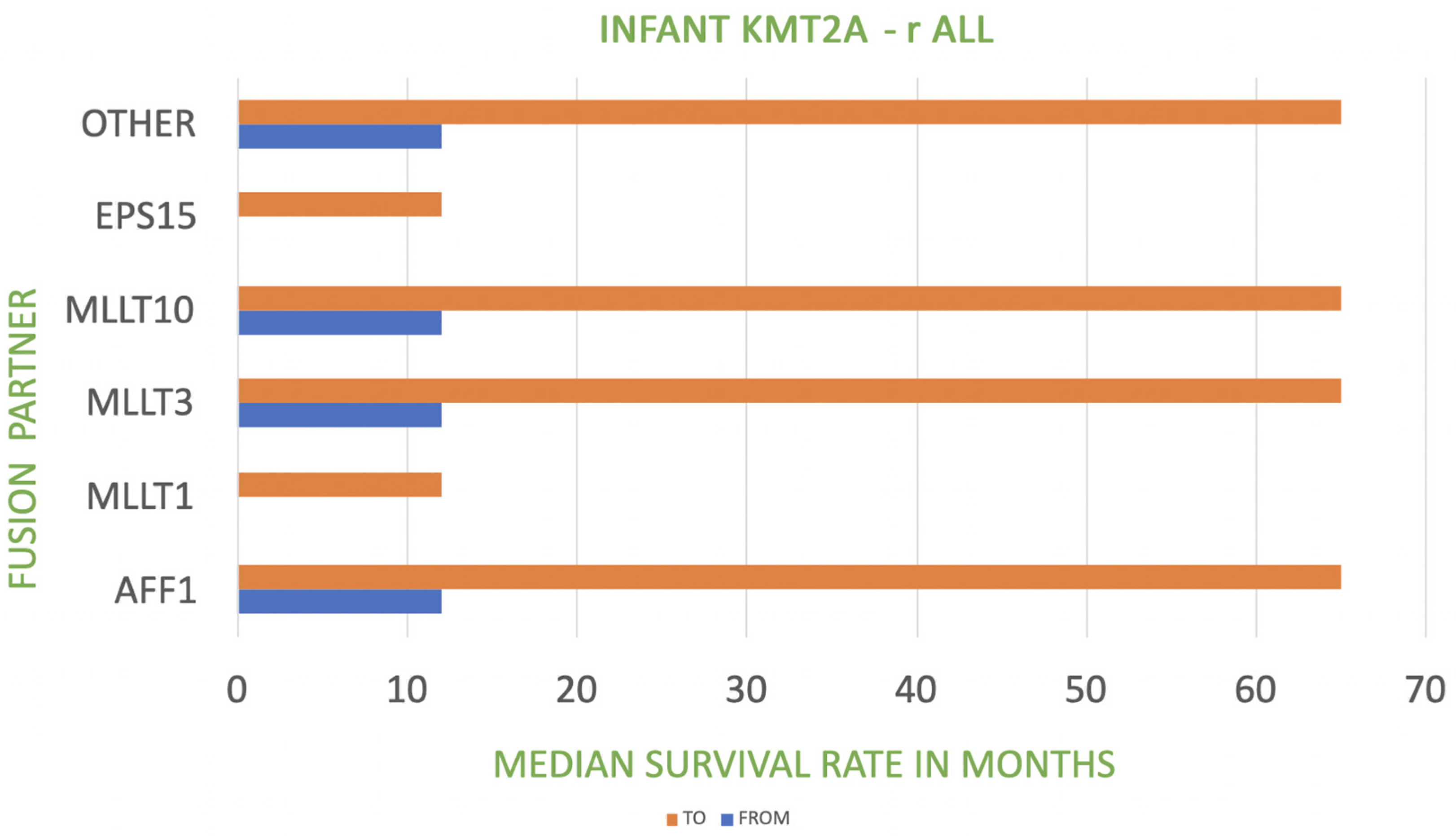
KMT2A Fusion Partner in Infant KMT2A-r ALLFrequencyPrognosis
AFF1-4q2149%poor
MLLT1-19p1322%very poor
MLLT3-9p2116%poor to intermediate
MLLT10-10p126%poor
EPS15-1p322%very poor
Other5%poor
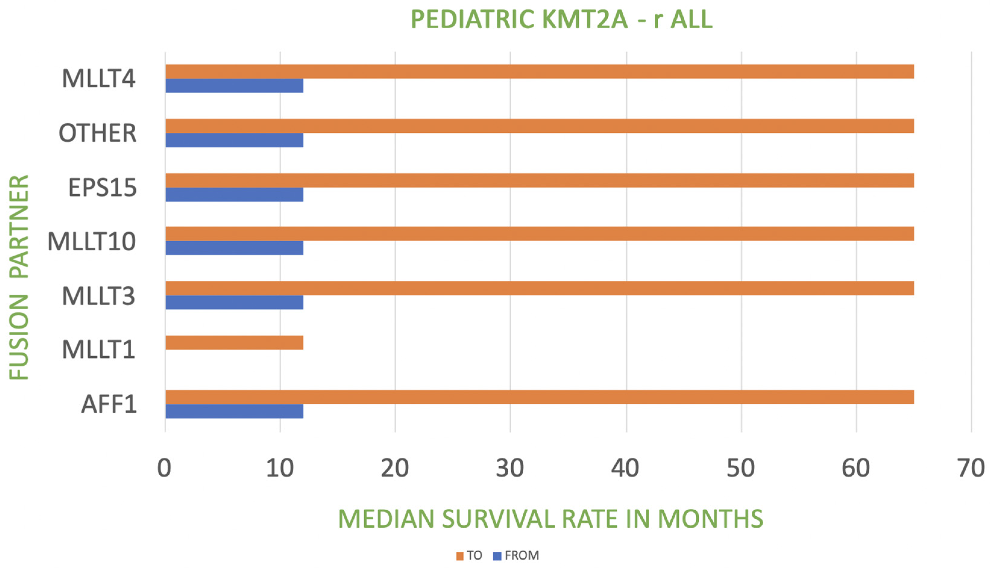
KMT2A Fusion Partner in Pediatric KMT2A-r ALLFrequencyPrognosis
AFF1-4q2144%poor
MLLT3-9p2118%poor to intermediate
MLLT1-19p1318%very poor
MLLT10-10p125%poor
MLLT4-6q275%poor
EPS15-1p322%poor
other8%poor
Table 2. Risk stratification in infants with KMT2A.
RiskInterfantCOGJPLSGApproximate EFS, %
highKMT2A-r and age <180 days and WBCs ≥ 300,000/μL KMT2A-r and age <90 daysKMT2A-r and (age <180 days or CNS leukemia or poor prednisone response)20
intermediateother KMT2A-r other KMT2A-r other KMT2A-r 50
lowKMT2A-gKMT2A-gKMT2A-g75
Disclaimer/Publisher’s Note: The statements, opinions and data contained in all publications are solely those of the individual author(s) and contributor(s) and not of MDPI and/or the editor(s). MDPI and/or the editor(s) disclaim responsibility for any injury to people or property resulting from any ideas, methods, instructions or products referred to in the content.

