Article

The von Willebrand Factor Antigen Reflects the Juvenile Dermatomyositis Disease Activity Score

1
Department of Biological Sciences, Wellesley College, Wellesley, MA 02481, USA
2
Department of Pediatrics, College of Medicine, Umm Al-Qura University, Makkah 21421, Saudi Arabia
3
Division of Pediatric Rheumatology, Ann & Robert H. Lurie Children’s Hospital of Chicago, Cure-JM Center of Excellence in Juvenile Myositis Research and Care, Chicago, IL 60611, USA
4
Computational Research, Children’s Hospital of Orange County Research Institute, Orange, CA 92868, USA
5
Feinberg School of Medicine, Northwestern University, Chicago, IL 60611, USA
*
Author to whom correspondence should be addressed.
These authors contributed equally to this work.
Biomedicines 2023, 11(2), 552; https://doi.org/10.3390/biomedicines11020552
Submission received: 29 December 2022 / Revised: 3 February 2023 / Accepted: 6 February 2023 / Published: 14 February 2023
(This article belongs to the Special Issue Mechanisms and Novel Therapeutic Approaches for Muscle Disease)

Abstract

:
Objective: This study determined if an accessible, serologic indicator of vascular disease activity, the von Willebrand factor antigen (vWF:Ag), was useful to assess disease activity in children with juvenile dermatomyositis (JDM), a rare disease, but the most common of the pediatric inflammatory myopathies. Methods: A total of 305 children, median age 10 years, 72.5% female, 76.5% white, with definite/probable JDM at diagnosis, were enrolled in the Ann & Robert H. Lurie Cure JM Juvenile Myositis Repository, a longitudinal database. Disease Activity Score (DAS) and vWF:Ag data were obtained at each visit. These data were analyzed using generalized estimating equation (GEE) models (both linear and logistic) to determine if vWF:Ag reflects disease severity in children with JDM. A secondary analysis was performed for untreated active JDM to exclude the effect of medications on vWF:Ag. Result: The vWF:Ag test was elevated in 25% of untreated JDM. We found that patients with elevated vWF:Ag had a 2.55-fold higher DAS total (CI95: 1.83–3.27, p < 0.001). Patients with difficulty swallowing had 2.57 higher odds of elevated vWF:Ag (CI95: 1.5–4.38, p < 0.001); those with more generalized skin involvement had 2.58-fold higher odds of elevated vWF:Ag (CI95: 1.27–5.23, p = 0.006); and those with eyelid peripheral blood vessel dilation had 1.32-fold higher odds of elevated vWF:Ag (CI95: 1.01–1.72, p = 0.036). Untreated JDM with elevated vWF:Ag had more muscle weakness and higher muscle enzymes, neopterin and erythrocyte sedimentation rate compared to JDM patients with a normal vWF:Ag. Conclusion: vWF:Ag elevation is a widely accessible concomitant of active disease in 25% of JDM.

1. Introduction

Juvenile dermatomyositis (JDM) is a rare pediatric autoimmune disease with an incidence of approximately 3–4 cases/million children/year [1]. JDM is defined as a systemic vasculopathy [2]. It often presents with truncal and symmetrical proximal muscle weakness as well as a distinctive heliotrope rash that includes Gottron’s papules, occurring most often on the upper extremity, where the skin bends [3,4,5]. The diagnosis of JDM subtypes is facilitated by the relatively recent identification of protein-specific antibodies: the most common antibody in the Western world is anti-Tif-1-γ (p155/140), while anti-MDA-5 is dominant in the East [6,7,8]. JDM complications include macrophage activation syndrome [9]. Gastrointestinal damage may be as severe as pneumatosis intestinalis [10]. Other less common symptoms in JDM include weakened eye muscles, also specific targets in children with orbital myositis [11].
A variety of methods are available to assess JDM skin and muscle symptoms, including the disease activity score (DAS), used in this study, [12] or the childhood myositis assessment scale (CMAS) and the DASI [13]. More recently, the European League Against Rheumatism and American College of Rheumatology developed new classification criteria for JDM [14,15]. The DAS has 20 points and is divided into skin (0–9 points) and muscle (0–11 points) symptoms [12]. The skin evaluation includes an estimation of the extent of involvement, distribution of skin symptoms, vasculitis, and Gottron’s papules. Muscle weakness is usually associated with loss of specific muscle functional status as well [12,16]. In addition to the clinical tools, there is an urgent need for a robust, accessible, and inexpensive serological biomarker that indicates the severity of disease activity in children with JDM, especially with respect to vascular injury. The association of von Willebrand factor antigen (vWF:Ag) and the diagnosis of JDM had been previously reported [17,18]. However, the use of vWF:Ag as a reliable indicator of “disease activity” is not well established.
The aim of the present study was to examine the relationship between vWF:Ag and DAS components in children with JDM. We utilized a longitudinal data set collected over the whole disease course from the Cure JM Juvenile Dermatomyositis Registry, which includes data on over 500 children diagnosed with JDM. The first part of the study included an analysis of both treated and untreated JDM children, while the second part focused specifically on untreated JDM children to determine if an elevated vWF:Ag is associated with more severe JDM clinical manifestations than JDM with a normal vWF:Ag level.

2. Methods

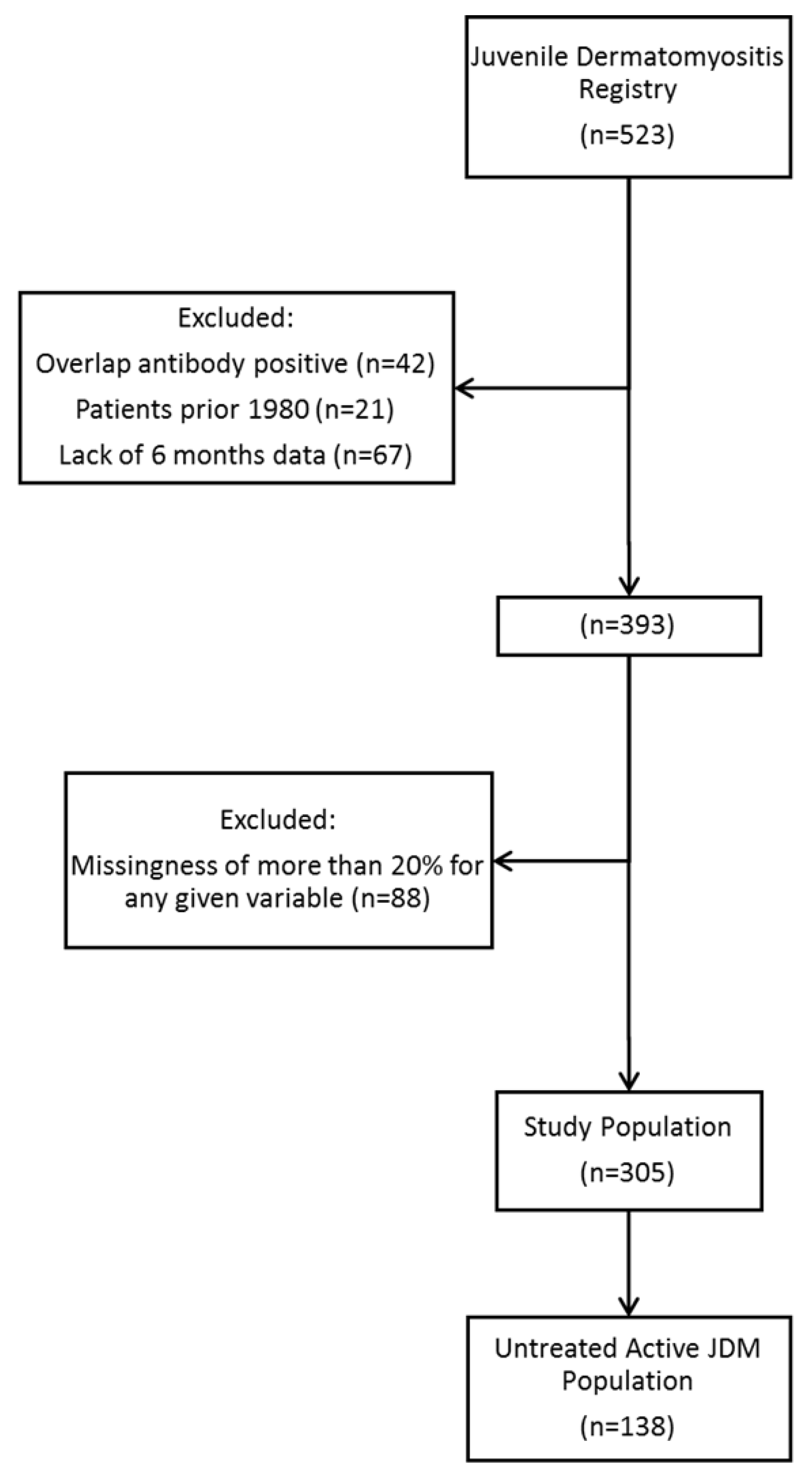
2.1. Study Design, Approval, and Population

We created and utilized the Juvenile Dermatomyositis Registry (1971 through 2020) at Ann & Robert H. Lurie Children’s Hospital of Chicago, hereafter referred to as Lurie Children’s. Each patient in the registry has been diagnosed with definite/probable JDM, based on the Bohan and Peter criteria [4,19]. The data included in the registry of JDM were first obtained at the inception of the study in 1971, when we first recognized the extensive variability of clinical symptoms, and potential etiologies of JDM. There are over 1400 variables per child over time. Written patient informed consent was obtained for enrollment into the registry (Lurie Children’s Hospital IRB 2010-14117, last yearly approval 2 June 2022). Upon enrollment, data obtained throughout the course of routine clinical care for JDM is entered into a REDCap database. For this retrospective study, these secondary clinical data were queried for all patients meeting inclusion criteria, beginning in 1980 (through 2020), having at least six months’ follow-up data. Inclusion criteria were all subjects who had vWF:Ag level and definite/probable JDM according to the internationally accepted Bohan and Peter criteria (study was approved by the IRB of Lurie Children’s Hospital IRB 2008-1345, last yearly approval 28 August 2022). The Bohan and Peter diagnostic criteria include typical skin rash (heliotrope rash and Gottron’s sign) in addition to 3 out 4 of the following criteria: symmetric proximal muscle weakness, elevated serum muscle enzymes, abnormal electromyography and muscle biopsy [4,14,19]. We excluded subjects with overlap syndrome, such as positive anti-PM-Scl, anti-U1 RNP, or anti-U2 RNP, from the study. Of note, one patient was found to have a positive anti-Jo-1, 25 patients did not have myositis-specific or -associated antibody (MSA/MAA) data available, as their visits were prior to MSA/MAA testing being regularly tested around the year 2000, and one patient post-2000 did not have MSA/MAA data available due to a laboratory issue. Continually, 12 patients had one instance of indeterminate or possible U1RNP, U2RNP, U4RNP, U5RNP, U6RNP antibodies, with seven of these patients negative for the above antibodies upon retest.

2.2. Study Variables

The variables used in this study were vWF:Ag level, vWF:Ag category (normal, elevated), age, sex, race, duration of untreated disease, blood type, neopterin, erythrocyte sedimentation rate (ESR), muscle enzymes, T and B cell flow cytometry, treatment status at the first visit, and DAS components. As vWF:Ag is blood type-specific, the normal ranges are: type A = 48–234%, type B = 57–241%, type AB = 64–238%, and type O = 36–157%. The T and B cell flow cytometry was performed in the clinical immunology lab at Lurie Children’s at the time of diagnosis. Various antibodies manufactured by BD Biosciences (Franklin Lakes, NJ, USA) were used to evaluate CD45, CD3, CD4, CD8, CD16, CD56, and CD19. The DAS, assessed by a physician, is divided into skin (0–9 points) and muscle (0–11 points) components [12]. Both clinical assessment and laboratory testing were typically performed at the same visit. Generally, the cause of missing data was due to a failure by the clinician to collect the necessary information during patient visits or failure to order necessary laboratory tests for the patient. Patients meeting a threshold of less than 20% of missingness for any given variable were included. A secondary analysis was done for untreated active JDM (n = 138) to exclude the effect of medications on the vWF:Ag data (Figure 1).

2.3. Statistical Analysis

Data were analyzed using R software Version 4.1 in R Studio Version 1.4.1717. After variables were selected, summary statistics were obtained by finding the median or (count when appropriate) of demographic variables along with treatment status, DAS and vWF:Ag. Data that were consistent throughout a patient’s life (i.e., sex at birth and race) were evaluated for each patient, while data that changed depending on visit (such as vWF:Ag and DAS) were evaluated by visit. To assess the association of DAS subcomponents and vWF:Ag, vWF:Ag was stratified into “normal” and “elevated” for specific reference range for the subjects’ blood group. Two sets of models were built: (1) assessing DAS total, DAS skin, and DAS muscle as outcome variables, and (2) assessing the individual components of the DAS skin and DAS muscle scores. In the second model, vWF:Ag was selected as the outcome variable to simplify the analyses and preclude the need to build as many models as there are components of the DAS skin and muscle scores.
In other words, the association between DAS total, DAS skin, and DAS muscle, and vWF:Ag was tested using a generalized estimating equation linear model [20,21] that accounts for repeated measures (that is, the longitudinal nature of the data on disease activity). We used an unstructured correlation matrix [20,21] in this study to avoid making assumptions about the direction of correlations between repeated measures of the DAS. Final model selection was carried out using backward elimination controlling for demographics. Bonferroni correction was applied to adjust for multiple testing by multiplying each p value by corresponding number of tests across all models built.
A secondary analysis of the first visit data of untreated JDM patients (n = 138) was also performed to evaluate the utility of vWF:Ag, without the confounding effect of medications. In this analysis, we divided the study subjects into two groups (normal vWF:Ag and elevated vWF:Ag) based on the reference range of the vWF:Ag for the JDM child’s blood group. The Mann–Whitney test compared the medians of DAS, muscle enzymes, ESR, neopterin and flow cytometry results between the two groups. GraphPad Prism version 9.4.1 was used to generate the figures.

3. Results

3.1. Patient Background and Demographic Analysis

There were 393 JDM patients that met inclusion criteria for this study, but when assessed for missingness of less than 20% for any given variable, 305 patients with 5818 visits were included in the analysis. The average number of visits per patient was 19.1 (±14.0 SD), and median age for all visits of 10.7 (IQR = 7.3–14.9) years. The median age of registry enrollment was 7.1 (IQR = 4.4–10.3) years; 72.5% were female (n = 221), and most patients were white (76.5%), followed by Hispanic (15.7%), black (4.3%), Asian (2%), and other (1.3%) (Table 1). For those patients enrolled in the registry after initiation of treatment for JDM, many were referred from other centers and had variably reported medications prior to their first visit at Lurie Children’s; however, their mean duration of months from first therapy to registry enrollment was 9.0 (IQR = 1.3–29.9) (Table 1).
Since vWF:Ag levels differ depending on blood type, these data were collected and controlled for in later statistical tests. Most patients were blood type O (45%), followed by type A (36.7%), type B (13.1%), and AB (4.3%), frequencies that are relatively consistent with the general population [22]. The median total DAS for all visits was 4 out of 20 (IQR = 5.5), with the skin DAS median reported as 3 (IQR = 5) out of 9 and muscle DAS as 0 out of 11 (IQR = 2), and most patients had vWF:Ag levels available (89.9% in the overall database and 81% for the untreated JDM patients). It should be noted that age, DAS, and vWF:Ag medians were derived at the time of the encounter.

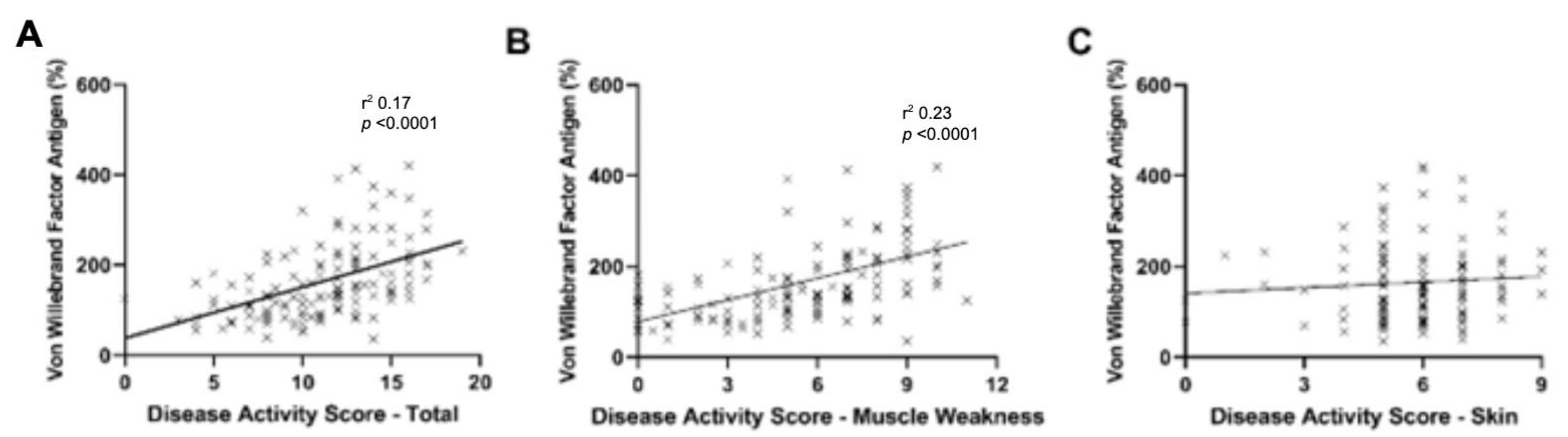
3.2. vWF:Ag Effect on DAS

We investigated the association between DAS total, DAS skin components, and DAS muscle components scores with demographic variables, duration of untreated disease, treatment status at first visit, and vWF:Ag to identify statistical associations. We found that age is associated with DAS total such that disease activity is highest at younger ages (effect size = −0.14, p < 0.001); having been treated elsewhere prior to their first visit at Lurie Children’s JM Clinic was associated with higher DAS total score with (effect size = 0.94, p = 0.014); and most notably, elevated vWF:Ag was associated with higher DAS total score (effect size = 2.55, p < 0.001) (Table 2). Differences in total DAS scores were not associated with sex (p = 0.624), duration of untreated disease (p = 1.000), or racial/ethnic group (p = 1.000).
For the DAS skin component scores, we found that higher scores were associated with African American race (effect size = −1.12, p = 0.009) than any other racial group (white as reference group, Hispanic p = 1.000, Asian p = 1.000, other race p = 0.677) and higher vWF:Ag levels (effect size = 0.96, p < 0.001) (Table 1). Neither duration of untreated disease (p = 0.726) nor sex (p = 0.485) was associated with DAS skin.
Finally, for muscle DAS components, we found that age is associated with DAS muscles such that it is highest at younger ages (effect size = −0.13, p < 0.001). Having been treated at the first visit was associated with higher DAS skin scores (effect size = 0.52, p < 0.005), and elevated vWF:Ag was also associated with higher DAS muscle scores (effect size = 1.59, p < 0.001) (Table 2). Differences in DAS muscle scores were not associated with sex (p = 1.000), race (white as reference group: black/African American, p = 0.084; Hispanic, p = 1.000; Asian, p = 1.000; other race, p = 1.000), and duration of untreated disease (p = 1.000).
For all components of DAS, no significant relationship with blood type was found (type O as reference group: DAS total and muscle, all blood types, p = 1.000; DAS skin, type A, p = 0.979; type B, p = 1.000; type AB, p = 1.000). When comparing the effect size of muscle and skin components, we find that an elevated vWF:Ag has a higher association with muscle DAS components (effect size = 1.59, p < 0.001) than with skin symptoms (effect size = 0.96, p < 0.001) as shown in Table 2.

3.3. Subcomponents Associated with vWF:Ag

We subsequently assessed subcomponents of the DAS to determine their association with the increased vWF:Ag levels. We found that skin involvement at specific focal points (i.e., a joint-related skin rash) had a 46% increased odds of higher vWF:Ag levels (p = 0.013), generalized skin involvement had 158% increased odds (p = 0.006), and eyelid peripheral blood vessel dilation had 32% increased odds (p = 0.036) (Table 3).
Data concerning muscle weaknesses, such as the inability to clear scapula (56% increased odds, p < 0.001), lower proximal muscle weakness (40% increased odds, p = 0.009), Gower’s sign (58% increased odds, p = 0.004), and difficulty swallowing (157% increased odds, p < 0.001), were all associated with a higher vWF:Ag (Table 3). Muscle functional status in children with JDM, especially with respect to the impact on their lives, was also significantly associated with vWF:Ag data at all levels. JDM with minimal limitations had 81% increased odds of higher vWF:Ag (p < 0.001), those with mild limitations had 159% increased odds of higher vWF:ag levels (p < 0.001), while JDM with moderate limitations had 265% increased odds (p < 0.001) and JDM with severe limitations had a 819% increased odds (p < 0.001). Therefore, there is a strong correlation between increasing levels of the severity of the loss of muscle function and increased odds of a higher value for vWF:Ag in a given individual.
Of note, age did not significantly increase odds of higher vWF:Ag (p = 1.000), nor male sex (p = 1.000) or race/ethnic group (white as reference group; black/African American, p = 0.156; Hispanic, p = 1.000; Asian, p = 1.000; other race, p = 0.170). Additionally, while vWF:Ag reference range is blood type-specific, no blood type had significantly increased odds of higher vWF:Ag (type O as reference group: type A, p = 0.053; type B, p = 0.149; type AB, p = 0.361).

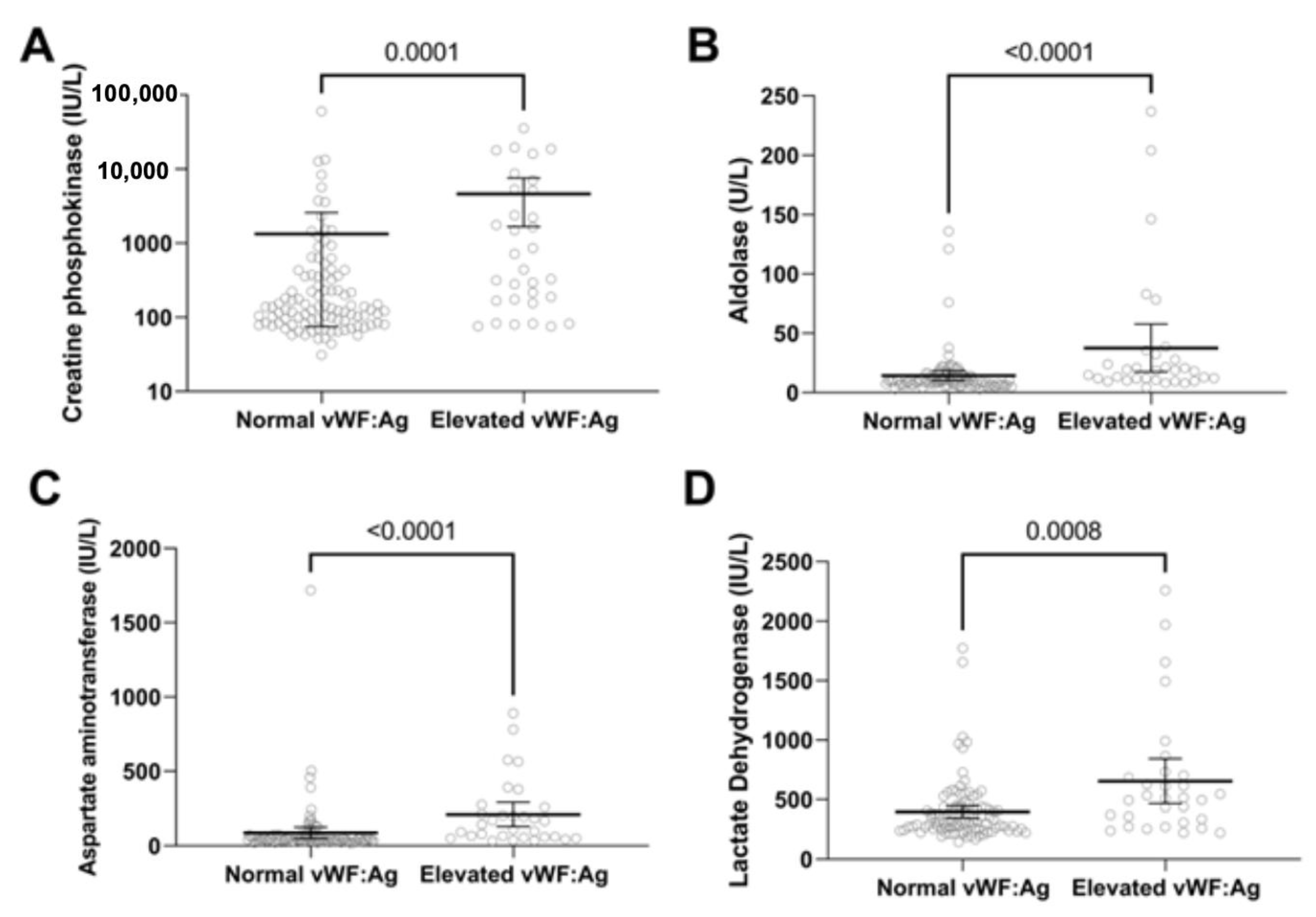
3.4. Untreated JDM with Active Disease

Many patients arrive at Lurie Children’s before receiving any medical treatment. In this study, 45.2% (n = 138) were untreated at registry enrollment, their median duration of untreated disease was 4.3 months (IQR = 2.0–10.3), median age at diagnosis was 6.1 (IQR = 4.0–9.4) years and 76.1% (n = 105) were female (Table 1). For untreated active JDM, 25% (35 of 138) had elevated vWF:Ag levels. Patients with increased vWF:Ag levels were older and had shorter duration of untreated disease than those with normal vWF:Ag levels (Table 4). As expected from the previous analysis, the groups with elevated vWF:Ag than normal vWF:Ag had higher median total DAS (13 vs. 11, p < 0.0001) and muscle DAS (8 vs. 5, p < 0.0001) (Figure 2). In addition, muscle enzymes such as creatine phosphokinase, aspartate aminotransferase, lactate dehydrogenase and aldolase (Figure 3) were higher in the elevated vWF:Ag group (Table 5). The inflammatory markers neopterin and ESR were higher in the elevated vWF:Ag group as well (Figure 4A,B,C and Table 5). Lymphocyte subset analyses by flow cytometry showed that an elevated vWF:Ag was associated with a decreased NK (natural killer) cell count than normal vWF:Ag (73 vs. 140 cells/mm3, p = 0.0005) (Figure 4C).

4. Discussion

In this study, we document a significant correlation between vWF:Ag level and disease activity score in JDM. Interestingly, muscle DAS components had a greater association with elevated vWF:Ag levels than skin DAS, especially in the untreated subjects. These data have also shown that untreated children with elevated vWF:Ag had more muscle weakness and higher muscle enzymes than JDM patients with normal vWF:Ag, documenting the utility of this biomarker in revealing muscle involvement. Furthermore, both neopterin and erythrocyte sedimentation rate were more elevated in the high vWF:Ag group, which confirms the association of vWF:Ag with other indicators of inflammation in children with JDM. This study’s results align with findings from a previous study that demonstrated that vWF:Ag levels are higher in JDM patients with increased serum neopterin, a marker of macrophage activation [23]. vWF:Ag has also been associated with downregulated miRNA-10a, a proinflammatory mediator [24]. RNA transcriptomes have shown that JDM can be associated with evidence of increased inflammation, even when the child, on examination, appears to be clinically inactive [25]. The appearance of clinical quiescence usually triggers reduction in immunosuppressive medication, resulting in cycles of disease flares and prolonging disease chronicity. Therefore, utilizing vWF:Ag level and other inflammatory markers in clinical practice may help in recognizing disease activity in the apparently clinically quiescent patients.
In this study, JDM patients with eyelid marginal blood vessel dilation were 32% more likely to have elevated vWF:Ag (Table 3), supporting the use of vWF:Ag as a biomarker for vasculitis in JDM. This finding is consistent with previous research linking elevated vWF:Ag to active CNS vasculitis and ANCA vasculitis [26,27,28], which is not unexpected as vWF:Ag is primarily synthesized in the endothelial cells and megakaryocytes [27,28,29]. Additionally, elevated vWF:Ag activity has been linked to a greater likelihood of severe COVID-19 requiring ICU admissions [30] and it also predicts portal hypertension and mortality in patients with liver cirrhosis [31,32]. We did not find any significant difference in the mean vWF:Ag level for JDM patients diagnosed before and after 1 January 2020, which corresponds to the date of the initial cases of COVID-19 in Chicago (manuscript under review).
Although vWF:Ag is a relatively inexpensive indicator of endothelial cell damage [33,34], it is not always reliable for the diagnosis of JDM, as a majority of untreated JDM patients still have normal vWF:Ag levels. Nailfold capillary loss, a common feature of JDM, is a more reliable indicator of small vessel injury [3,35]. Notably, baseline vWF:Ag varies depending on blood group, with group O having the lowest baseline and group AB having the highest [36]. Other current biomarkers for JDM, such as muscle enzymes and inflammatory markers, also have limitations [37]. For example, although elevated muscle enzymes are an excellent tool early in the disease course, they tend to normalize in patients with untreated disease for ≥4.7 months [38]. Traditional inflammatory markers such as ESR and C-reactive protein are not useful for most children with JDM, as most patients have a normal ESR [23]. On the other hand, neopterin, a small molecule produced by activated macrophages, is a good marker of disease activity, but has limited availability in commercial labs [23]. In summary, to accurately assess JDM disease activity, a thorough physical examination and the use of multiple biomarkers should be utilized to address the limitations of any single marker.
Study limitations include the following. Only 25% of untreated JDM had an elevated vWF:Ag. However, those patients who did have an elevated vWF:Ag had more severe symptoms, such as muscle weakness and difficulty swallowing, as well as higher levels of inflammatory markers. A significant portion of the primary cohort (55%, n = 305) had been treated at other centers, and the details of their treatment and initial clinical presentation were unclear. Therefore, we conducted a secondary analysis on the untreated group (n = 138) to avoid confounding effects of medication use.

5. Conclusions

These results establish vWF:Ag as an accessible but not very sensitive biomarker for JDM diagnosis. Potentially, this assay can be employed to monitor disease activity in order to avoid premature termination of medical therapy. Finally, vWF:Ag is an affordable and readily available test, making it a possible biomarker for clinicians to consider, even in geographic areas with limited availability of resources.

Author Contributions

Conceptualization, L.M.P.; methodology, E.G., L.E. and L.M.P.; formal analysis, E.G., A.K. and L.E.; investigation, G.M. and L.M.P.; tesources, L.E. and L.M.P.; data curation, G.M.; writing—original draft preparation, E.G., A.K. and L.M.P.; writing—review and editing, E.G., A.K., G.M., L.E. and L.M.P.; visualization, E.G., A.K. and L.E.; supervision, L.E. and L.M.P.; project administration, L.M.P.; funding acquisition, L.E. and L.M.P. All authors have read and agreed to the published version of the manuscript.

Funding

Supported in part by the Vivian Allison and Daniel J. Pachman Fund, the DenUyl Family Fund, the Cure JM Foundation, Children’s Health of Orange County (CHOC) Chief Scientific Officer (CSO) Award, and other much-appreciated donors. We also acknowledge the contribution of Wellesley College Honors Program. The REDCap database is supported by NUCATS and funded in part by a Clinical and Translational Science Award (CTSA) grant from the National Institutes of Health (NIH), [UL1TR001422]. Amer Khojah would like to thank the Deanship of Scientific Research at Umm Al-Qura University for supporting this work (grant 22UQU4300201DSR07).

Institutional Review Board Statement

The study was conducted in accordance with the Declaration of Helsinki, and approved by the Institutional Review Board of Lurie Children’s Hospital (IRB 2008-134).

Informed Consent Statement

Informed consent was obtained from all subjects involved in the study.

Data Availability Statement

Data for study is not publicly available. Please reach out to corresponding author for further inquiries.

Conflicts of Interest

The authors declare no conflict of interest.

References

  1. Mendez, E.P.; Lipton, R.; Ramsey-Goldman, R.; Roettcher, P.; Bowyer, S.; Dyer, A.; Pachman, L.M.; NIAMS Juvenile DM Registry Physician Referral Group. US incidence of juvenile dermatomyositis, 1995-1998: Results from the National Institute of Arthritis and Musculoskeletal and Skin Diseases Registry. Arthritis Rheum 2003, 49, 300–305. [Google Scholar] [CrossRef]
  2. Tezak, Z.; Hoffman, E.P.; Lutz, J.L.; Fedczyna, T.O.; Stephan, D.; Bremer, E.G.; Krasnoselska-Riz, I.; Kumar, A.; Pachman, L.M. Gene expression profiling in DQA1*0501+ children with untreated dermatomyositis: A novel model of pathogenesis. J. Immunol. 2002, 168, 4154–4163. [Google Scholar] [CrossRef] [PubMed]
  3. Pachman, L.M.; Khojah, A.M. Advances in Juvenile Dermatomyositis: Myositis Specific Antibodies Aid in Understanding Disease Heterogeneity. J. Pediatr. 2018, 195, 16–27. [Google Scholar] [CrossRef] [PubMed]
  4. Bohan, A.; Peter, J.B. Polymyositis and dermatomyositis (first of two parts). N. Engl. J. Med. 1975, 292, 344–347. [Google Scholar] [CrossRef] [PubMed]
  5. Cao, H.; Xia, Q.; Pan, M.; Zhao, X.; Li, X.; Shi, R.; Zhou, M.; Ding, X.; Kuwana, M.; Zheng, J. Gottron Papules and Gottron Sign with Ulceration: A Distinctive Cutaneous Feature in a Subset of Patients with Classic Dermatomyositis and Clinically Amyopathic Dermatomyositis. J. Rheumatol. 2016, 43, 1735–1742. [Google Scholar] [CrossRef]
  6. Tansley, S.L.; Simou, S.; Shaddick, G.; Betteridge, Z.E.; Almeida, B.; Gunawardena, H.; Thomson, W.; Beresford, M.W.; Midgley, A.; Muntoni, F.; et al. Autoantibodies in juvenile-onset myositis: Their diagnostic value and associated clinical phenotype in a large UK cohort. J. Autoimmun 2017, 84, 55–64. [Google Scholar] [CrossRef]
  7. Li, D.; Tansley, S.L. Juvenile Dermatomyositis-Clinical Phenotypes. Curr. Rheumatol. Rep. 2019, 21, 74. [Google Scholar] [CrossRef]
  8. Boyarchuk, O.; Kuka, A.; Yuryk, I. Clinical and autoantibody phenotypes of juvenile dermatomyositis. Reumatologia 2022, 60, 281–291. [Google Scholar] [CrossRef]
  9. Poddighe, D.; Dauyey, K. Macrophage activation syndrome in juvenile dermatomyositis: A systematic review. Rheumatol. Int. 2020, 40, 695–702. [Google Scholar] [CrossRef]
  10. Miyamae, T.; Ishiguro, N.; Yonezawa, M.; Tokushige, K.; Yamanaka, H. Pneumatosis Intestinalis Associated with Juvenile Dermatomyositis. Case Rep. Rheumatol. 2016, 2016, 6497357. [Google Scholar] [CrossRef] [Green Version]
  11. Briones, M.R.; Morgan, G.A.; Amoruso, M.C.; Rahmani, B.; Ryan, M.E.; Pachman, L.M. Decreased CD3-CD16+CD56+ natural killer cell counts in children with orbital myositis: A clue to disease activity. RMD Open 2017, 3, e000385. [Google Scholar] [CrossRef]
  12. Bode, R.K.; Klein-Gitelman, M.S.; Miller, M.L.; Lechman, T.S.; Pachman, L.M. Disease activity score for children with juvenile dermatomyositis: Reliability and validity evidence. Arthritis Rheum 2003, 49, 7–15. [Google Scholar] [CrossRef]
  13. Rider, L.G.; Werth, V.P.; Huber, A.M.; Alexanderson, H.; Rao, A.P.; Ruperto, N.; Herbelin, L.; Barohn, R.; Isenberg, D.; Miller, F.W. Measures of adult and juvenile dermatomyositis, polymyositis, and inclusion body myositis: Physician and Patient/Parent Global Activity, Manual Muscle Testing (MMT), Health Assessment Questionnaire (HAQ)/Childhood Health Assessment Questionnaire (C-HAQ), Childhood Myositis Assessment Scale (CMAS), Myositis Disease Activity Assessment Tool (MDAAT), Disease Activity Score (DAS), Short Form 36 (SF-36), Child Health Questionnaire (CHQ), physician global damage, Myositis Damage Index (MDI), Quantitative Muscle Testing (QMT), Myositis Functional Index-2 (FI-2), Myositis Activities Profile (MAP), Inclusion Body Myositis Functional Rating Scale (IBMFRS), Cutaneous Dermatomyositis Disease Area and Severity Index (CDASI), Cutaneous Assessment Tool (CAT), Dermatomyositis Skin Severity Index (DSSI), Skindex, and Dermatology Life Quality Index (DLQI). Arthritis Care Res. 2011, 63 (Suppl. S11), S118–S157. [Google Scholar] [CrossRef]
  14. Leclair, V.; Lundberg, I.E. New Myositis Classification Criteria-What We Have Learned Since Bohan and Peter. Curr. Rheumatol. Rep. 2018, 20, 18. [Google Scholar] [CrossRef]
  15. Lundberg, I.E.; Tjarnlund, A.; Bottai, M.; Werth, V.P.; Pilkington, C.; Visser, M.; Alfredsson, L.; Amato, A.A.; Barohn, R.J.; Liang, M.H.; et al. 2017 European League Against Rheumatism/American College of Rheumatology classification criteria for adult and juvenile idiopathic inflammatory myopathies and their major subgroups. Ann. Rheum. Dis. 2018, 77, e91. [Google Scholar] [CrossRef]
  16. Huber, A.M.; Feldman, B.M.; Rennebohm, R.M.; Hicks, J.E.; Lindsley, C.B.; Perez, M.D.; Zemel, L.S.; Wallace, C.A.; Ballinger, S.H.; Passo, M.H.; et al. Validation and clinical significance of the Childhood Myositis Assessment Scale for assessment of muscle function in the juvenile idiopathic inflammatory myopathies. Arthritis Rheum 2004, 50, 1595–1603. [Google Scholar] [CrossRef] [PubMed]
  17. Kishi, T.; Chipman, J.; Evereklian, M.; Nghiem, K.; Stetler-Stevenson, M.; Rick, M.E.; Centola, M.; Miller, F.W.; Rider, L.G. Endothelial Activation Markers as Disease Activity and Damage Measures in Juvenile Dermatomyositis. J. Rheumatol. 2020, 47, 1011–1018. [Google Scholar] [CrossRef]
  18. Guzman, J.; Petty, R.E.; Malleson, P.N. Monitoring disease activity in juvenile dermatomyositis: The role of von Willebrand factor and muscle enzymes. J. Rheumatol. 1994, 21, 739–743. [Google Scholar]
  19. Bohan, A.; Peter, J.B. Polymyositis and dermatomyositis (second of two parts). N. Engl. J. Med. 1975, 292, 403–407. [Google Scholar] [CrossRef]
  20. Ziegler, A.; Vens, M. Generalized estimating equations. Notes on the choice of the working correlation matrix. Methods Inf. Med. 2010, 49, 421–425, discussion 426–432. [Google Scholar] [CrossRef]
  21. Hardin, J.W.; Joseph, M. Generalized Estimating Equations; Chapman and Hall/CRC: Boca Raton, FL, USA, 2002. [Google Scholar]
  22. Garratty, G.; Glynn, S.A.; McEntire, R.; Retrovirus Epidemiology Donor, S. ABO and Rh(D) phenotype frequencies of different racial/ethnic groups in the United States. Transfusion 2004, 44, 703–706. [Google Scholar] [CrossRef]
  23. Khojah, A.; Morgan, G.; Pachman, L.M. Clues to Disease Activity in Juvenile Dermatomyositis: Neopterin and Other Biomarkers. Diagnostics 2021, 12, 8. [Google Scholar] [CrossRef]
  24. Xu, D.; Huang, C.C.; Kachaochana, A.; Morgan, G.A.; Bonaldo, M.F.; Soares, M.B.; Costa, F.; Sarwark, J.; Sredni, S.T.; Pachman, L.M. MicroRNA-10a Regulation of Proinflammatory Mediators: An Important Component of Untreated Juvenile Dermatomyositis. J. Rheumatol. 2016, 43, 161–168. [Google Scholar] [CrossRef]
  25. Roberson, E.D.O.; Mesa, R.A.; Morgan, G.A.; Cao, L.; Marin, W.; Pachman, L.M. Transcriptomes of peripheral blood mononuclear cells from juvenile dermatomyositis patients show elevated inflammation even when clinically inactive. Sci. Rep. 2022, 12, 275. [Google Scholar] [CrossRef] [PubMed]
  26. Salmela, A.; Ekstrand, A.; Joutsi-Korhonen, L.; Raisanen-Sokolowski, A.; Lassila, R. Activation of endothelium, coagulation and fibrinolysis is enhanced and associates with renal anti-neutrophil cytoplasmic antibody-associated vasculitis. Nephrol. Dial. Transplant. 2015, 30 (Suppl. S1), i53–i59. [Google Scholar] [CrossRef]
  27. Cellucci, T.; Tyrrell, P.N.; Pullenayegum, E.; Benseler, S.M. von Willebrand factor antigen--a possible biomarker of disease activity in childhood central nervous system vasculitis? Rheumatology 2012, 51, 1838–1845. [Google Scholar] [CrossRef]
  28. Brogan, P.; Eleftheriou, D. Vasculitis update: Pathogenesis and biomarkers. Pediatr. Nephrol. 2018, 33, 187–198. [Google Scholar] [CrossRef] [PubMed]
  29. Chen, J.; Chung, D.W. Inflammation, von Willebrand factor, and ADAMTS13. Blood 2018, 132, 141–147. [Google Scholar] [CrossRef]
  30. Rostami, M.; Mansouritorghabeh, H.; Parsa-Kondelaji, M. High levels of Von Willebrand factor markers in COVID-19: A systematic review and meta-analysis. Clin. Exp. Med. 2022, 22, 347–357. [Google Scholar] [CrossRef]
  31. Ferlitsch, M.; Reiberger, T.; Hoke, M.; Salzl, P.; Schwengerer, B.; Ulbrich, G.; Payer, B.A.; Trauner, M.; Peck-Radosavljevic, M.; Ferlitsch, A. von Willebrand factor as new noninvasive predictor of portal hypertension, decompensation and mortality in patients with liver cirrhosis. Hepatology 2012, 56, 1439–1447. [Google Scholar] [CrossRef]
  32. Takaya, H.; Kawaratani, H.; Tsuji, Y.; Nakanishi, K.; Saikawa, S.; Sato, S.; Sawada, Y.; Kaji, K.; Okura, Y.; Shimozato, N.; et al. von Willebrand factor is a useful biomarker for liver fibrosis and prediction of hepatocellular carcinoma development in patients with hepatitis B and C. United Eur. Gastroenterol. J. 2018, 6, 1401–1409. [Google Scholar] [CrossRef] [Green Version]
  33. von Vischer, U.M. Willebrand factor, endothelial dysfunction, and cardiovascular disease. J. Thromb. Haemost. 2006, 4, 1186–1193. [Google Scholar] [CrossRef] [PubMed]
  34. Gragnano, F.; Sperlongano, S.; Golia, E.; Natale, F.; Bianchi, R.; Crisci, M.; Fimiani, F.; Pariggiano, I.; Diana, V.; Carbone, A.; et al. The Role of von Willebrand Factor in Vascular Inflammation: From Pathogenesis to Targeted Therapy. Mediators Inflamm. 2017, 2017, 5620314. [Google Scholar] [CrossRef]
  35. Khojah, A.; Liu, V.; Savani, S.I.; Morgan, G.; Shore, R.; Bellm, J.; Pachman, L.M. Association of p155/140 Autoantibody With Loss of Nailfold Capillaries but not Generalized Lipodystrophy: A Study of Ninety-Six Children With Juvenile Dermatomyositis. Arthritis Care Res. 2022, 74, 1065–1069. [Google Scholar] [CrossRef]
  36. Gill, J.C.; Endres-Brooks, J.; Bauer, P.J.; Marks, W.J., Jr.; Montgomery, R.R. The effect of ABO blood group on the diagnosis of von Willebrand disease. Blood 1987, 69, 1691–1695. [Google Scholar] [CrossRef] [PubMed]
  37. Pachman, L.M.; Nolan, B.E.; DeRanieri, D.; Khojah, A.M. Juvenile Dermatomyositis: New Clues to Diagnosis and Therapy. Curr. Treatm. Opt. Rheumatol. 2021, 7, 39–62. [Google Scholar] [CrossRef]
  38. Pachman, L.M.; Abbott, K.; Sinacore, J.M.; Amoruso, L.; Dyer, A.; Lipton, R.; Ilowite, N.; Hom, C.; Cawkwell, G.; White, A.; et al. Duration of illness is an important variable for untreated children with juvenile dermatomyositis. J. Pediatr. 2006, 148, 247–253. [Google Scholar] [CrossRef]
Figure 1. Inclusion criteria flow diagram. Patients selected from the Juvenile Dermatomyositis Registry at Lurie Children’s meeting inclusion criteria for the study.
Biomedicines 11 00552 g001
Figure 2. Untreated JDM vWF:Ag levels and DAS. Untreated JDM patients with higher vWF:Ag had higher (A) DAS total and (B) DAS muscle weakness, but not (C) DAS skin.
Biomedicines 11 00552 g002
Figure 3. vWF:Ag groups and muscle enzyme levels in untreated JDM. JDM disease activity indicators in untreated JDM (n = 138) with normal vWF:Ag and elevated vWF:Ag. Untreated JDM with elevated vWF:Ag showed (A) higher creatine phosphokinase levels, (B) higher aldolase levels, (C) higher aspartate aminotransferase levels, and (D) higher lactate dehydrogenase levels.
Biomedicines 11 00552 g003
Figure 4. vWF:Ag groups and inflammatory markers in untreated JDM. JDM disease activity indicators in untreated JDM (n = 138) with normal vWF:Ag and elevated vWF:Ag. Untreated JDM with elevated vWF:Ag showed (A) higher ESR levels, (B) higher neopterin levels, and (C) lower NK cell absolute counts.
Biomedicines 11 00552 g004
Table 1. Demographics and Clinical Characteristics of 305 Patients with JDM.
Total JDM Population
n = 305
Untreated JDM
n = 138
Demographics
Age at enrollment (yr), median (IQR)7.1 (4.4–10.3)6.1 (4.0–9.4)
Age at onset of symptoms (yr), median (IQR)5.6 (3.1–8.4)5.5 (3.4–8.4)
Sex
      Male, n (%)84 (27.5)33 (23.9)
      Female, n (%)221 (72.5)105 (76.1)
Race/Ethnicity
      White, n (%)234 (76.7)101 (73.2)
      Hispanic, n (%)48 (15.7)25 (18.1)
      Black/African American, n (%)13 (4.3)6 (4.3)
      Asian, n (%)6 (2.0)5 (3.6)
      Other, n (%)4 (1.3)1 (0.7)
Clinical Factorsmedian (IQR)median (IQR)
Duration of untreated disease (mo)4.3 (2.0–10.3) n = 3026.0 (3.1–12.8)
Duration from 1st treatment to enrollment a (mo)9.0 (1.3–29.9)n/a
DAS
      DAS Total (0–20)11.0 (7.5–13.0) n = 27312.0 (8.9–14.0) n = 133
      DAS Skin (0–9)6.0 (5.0–7.0) n = 2736.0 (5.0–7.0) n = 133
      DAS Muscle (0–11)5.0 (2.0–8.0) n = 2766.0 (3.0–8.0) n = 133
Laboratory Disease Activity Indicators
      Neopterin (nmol/liter)12.9 (7.0–19.4) n = 25717.2 (11.0–23.9) n = 124
      ESR (mm/hr)12.0 (7.0–20.0) n = 21815.0 (9.0–23.0) n = 104
      vWF:Ag (%)140.0 (99.8–203.5) n = 260140.0 (99.0–206.3)
      Muscle Enzymes
           CK (IU/L)114.0 (71.0–341.5) n = 267154.5 (85.5–631.8) n = 132
           LDH (IU/L)298.5 (223.0–433.3) n = 264350.5 (264.0–527.3) n = 131
           AST(SGOT) (IU/L)40.5 (28.0–67.3) n = 25049.0 (36.0–107.0) n = 125
           Aldolase (U/L)8.8 (6.3–14.0) n = 24910.3 (7.6–18.0) n = 123
a Patients enrolled after starting treatment for JDM.
Table 2. Association of DAS components (total, skin, and muscle) with JDM demographic and disease factors a.
DAS TotalDAS SkinDAS Muscle
Effect Size (95% CI)p ValueEffect Size (95% CI)p ValueEffect Size (95% CI)p Value
Age (yr)−0.14
(−0.21, −0.06)
<0.001−0.01
(−0.06, 0.04)
1.000−0.13
(−0.16, −0.09)
<0.001
Treatment Status
at 1st Visit
      UntreatedReference Reference Reference
      Treated0.94
(0.13, 1.76)
0.0140.42
(−0.18, 1.02)
0.3850.52
(0.11, 0.94)
0.005
vWF:Ag
      Elevated2.55
(1.83, 3.27)
<0.0010.96
(0.59, 1.34)
<0.0011.59
(1.13, 2.04)
<0.001
a All reported statistics adjusted for multiple comparison using Bonferroni correction.
Table 3. Association of specific DAS components with vWF:Ag levels using odds ratios.
Odds Ratio (95% CI)p Value
Duration of untreated disease (mo)0.95 (0.91, 0.995)0.025
Treatment status at 1st visit
      UntreatedReference
      Treated1.03 (0.66, 1.61)1.000
DAS Skin Elements
Skin Involvement Distribution
      NoneReference
      Focal (including areas of joint related skin)1.46 (1.07, 2.00)0.013
      Diffuse (including extensor surfaces of limbs/shawl)1.38 (0.88, 2.17)0.211
      Generalized (including trunk involvement)2.58 (1.27, 5.23)0.006
      Eyelid blood vessel dilation, present1.32 (1.01, 1.72)0.036
DAS Muscle Elements
Functional Status
      Within Normal LimitsReference
      Minimal Limitations1.81 (1.27, 2.58)<0.001
      Mild Limitations2.59 (1.81, 3.70)<0.001
      Moderate Limitations3.65 (2.14, 6.23)<0.001
      Severe Limitations9.19 (3.33, 25.38)<0.001
Neck flexor weakness1.32 (0.997, 1.75)0.053
Unable to clear scapula 1.56 (1.22, 2.01)<0.001
Lower proximal muscle weakness1.40 (1.07, 1.83)0.009
Gower’s Sign1.58 (1.13, 2.19)0.004
Difficulty Swallowing2.57 (1.50, 4.38)<0.001
Table 4. vWF:Ag in 138 untreated children with JDM: demographic characteristics.
Normal vWF:AgElevated vWF:Agp Value
Number of subjects, n (%)103 (74.6%)35 (25.4%)
Age at onset of symptoms in years, median (IQR)5.7 (3.9–8.7)8.4 (5.4–9.9)0.017
Duration of untreated disease in months, median (IQR)6.4 (3.6–13.5)3.8 (1.9–7.2)0.004
Sex, n (%)
      Female76 (74%)29 (83%)0.277
      Male27 (26%)6 (17%)
Race/ethnicity, n (%)
      White79 (77%)22 (63%)0.184
      Hispanic17 (16%)3 (7%)
      African American3 (3%)8 (23%)
      Asian4 (4%)1 (3%)
      Others0 (0%)1(3%)
Table 5. vWF:Ag levels in 138 untreated children with JDM: disease activity markers.
Normal vWF:AgElevated vWF:Agp Value
Median (IQR)Median (IQR)
DAS
      DAS Total11 (8–13)13 (12–16)<0.0001
      DAS Skin6 (5–7)5.5 (5–7)0.704
      DAS Muscle5 (2.5–7)8 (6–9)<0.0001
Laboratory Disease Activity Indicators
      Neopterin (nmol/L)15.3 (9.5–22.4)21.6 (14.8–28)0.0098
      ESR (mm/h)13 (8–18)23<0.0001
      Muscle Enzymes
           CK (IU/L)130 (83–356)579 (169–5283)0.0001
           LDH (IU/L)315 (253–435)512 (307–703)0.0008
           AST(SGOT) (IU/L)44 (33–64)143 (58–258)<0.0001
           Aldolase (U/L)9.6 (6.9–14.2)18.4 (11–31)<0.0001
      Flow Cytometry (% lymphocytes)
           Total T cells (CD3+) 62.9 (57–68)69 (63–74)0.0008
           T helper cells (CD3+ CD4+)42 (37–48)48 (43–54)0.0006
           T cytotoxic cells (CD3+ CD8+) 19 (16–22)20 (17.5–23)0.323
           B cells (CD19+) 29.5 (24–35)25 (19.8–30.8)0.049
           NK cells (CD16+/CD56+) 6 (4–9)4 (3–7)0.0097
Disclaimer/Publisher’s Note: The statements, opinions and data contained in all publications are solely those of the individual author(s) and contributor(s) and not of MDPI and/or the editor(s). MDPI and/or the editor(s) disclaim responsibility for any injury to people or property resulting from any ideas, methods, instructions or products referred to in the content.

