
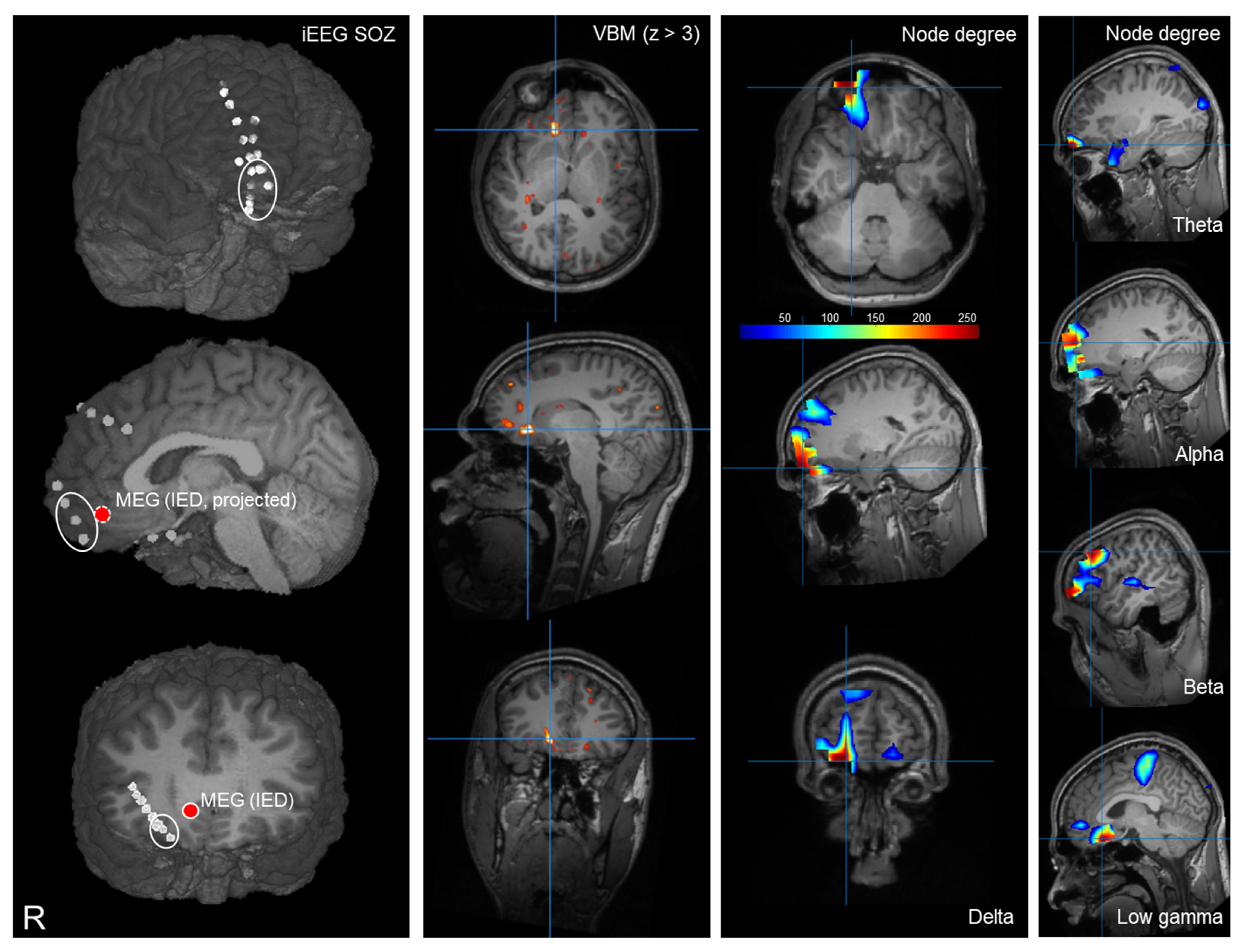
MEG Node Degree for Focus Localization: Comparison with Invasive EEG
 ,
,
Abstract
:1. Introduction
2. Materials and Methods
2.1. Participants
2.2. MEG Acquisition
2.3. MRI Acquisition
2.4. Presurgical Evaluation
2.5. MEG Analysis
2.6. Preprocessing
2.7. Source Analysis
2.8. Connectivity and Graph Analysis
2.9. Statistical Evaluation
3. Results
3.1. Patients
3.2. Presurgical Evaluation
3.3. Node Degree
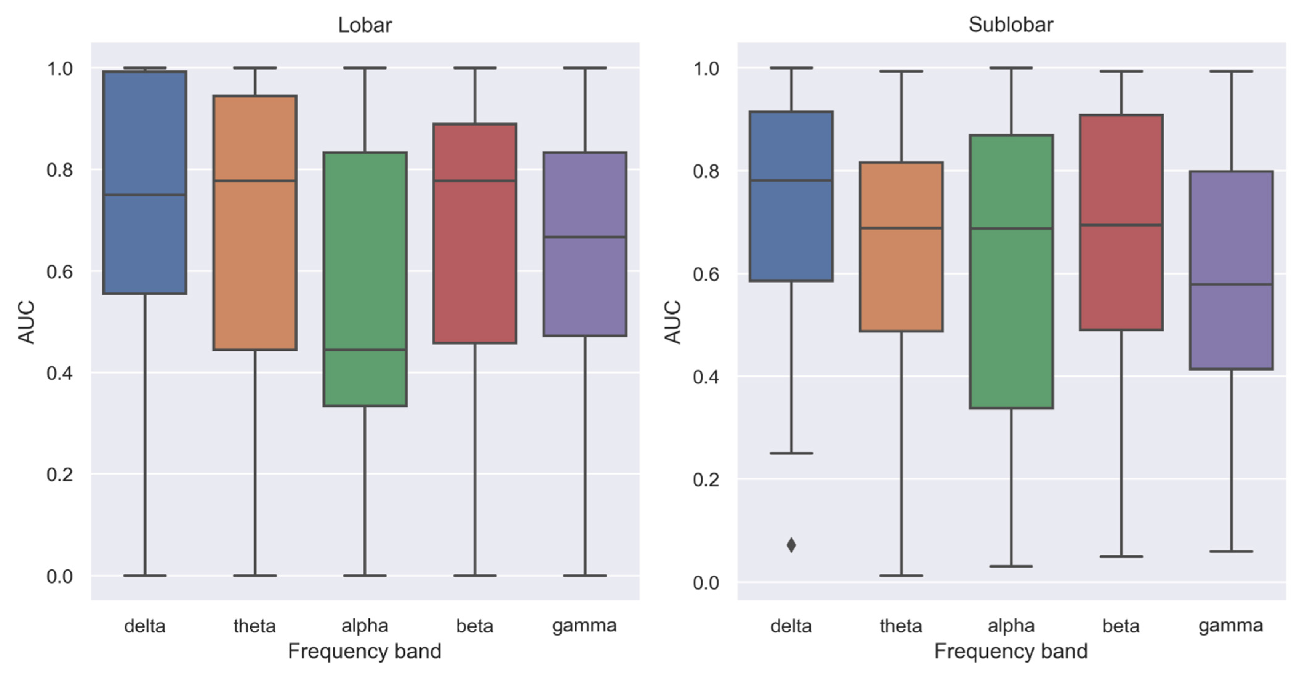
3.4. Lobar Level
3.5. Sublobar Level
4. Discussion
4.1. Connectivity Alterations in Epilepsy
4.2. Differences between Frequency Bands
4.3. Clinical Aspects
4.4. Limitations
5. Conclusions
Supplementary Materials
Author Contributions
Funding
Institutional Review Board Statement
Informed Consent Statement
Data Availability Statement
Conflicts of Interest
References
- Behr, C.; Goltzene, M.A.; Kosmalski, G.; Hirsch, E.; Ryvlin, P. Epidemiology of epilepsy. Rev. Neurol. 2016, 172, 27–36. [Google Scholar] [CrossRef] [PubMed]
- Blumcke, I.; Spreafico, R.; Haaker, G.; Coras, R.; Kobow, K.; Bien, C.G.; Pfafflin, M.; Elger, C.; Widman, G.; Schramm, J.; et al. Histopathological Findings in Brain Tissue Obtained during Epilepsy Surgery. N. Engl. J. Med. 2017, 377, 1648–1656. [Google Scholar] [CrossRef] [PubMed]
- Rosenow, F.; Luders, H. Presurgical evaluation of epilepsy. Brain 2001, 124, 1683–1700. [Google Scholar] [CrossRef]
- Rampp, S.; Stefan, H.; Wu, X.; Kaltenhauser, M.; Maess, B.; Schmitt, F.C.; Wolters, C.H.; Hamer, H.; Kasper, B.S.; Schwab, S.; et al. Magnetoencephalography for epileptic focus localization in a series of 1000 cases. Brain 2019, 142, 3059–3071. [Google Scholar] [CrossRef] [PubMed]
- van Mierlo, P.; Vorderwulbecke, B.J.; Staljanssens, W.; Seeck, M.; Vulliemoz, S. Ictal EEG source localization in focal epilepsy: Review and future perspectives. Clin. Neurophysiol. 2020, 131, 2600–2616. [Google Scholar] [CrossRef]
- Sharma, P.; Seeck, M.; Beniczky, S. Accuracy of Interictal and Ictal Electric and Magnetic Source Imaging: A Systematic Review and Meta-Analysis. Front. Neurol. 2019, 10, 1250. [Google Scholar] [CrossRef]
- Brodbeck, V.; Spinelli, L.; Lascano, A.M.; Wissmeier, M.; Vargas, M.I.; Vulliemoz, S.; Pollo, C.; Schaller, K.; Michel, C.M.; Seeck, M. Electroencephalographic source imaging: A prospective study of 152 operated epileptic patients. Brain 2011, 134, 2887–2897. [Google Scholar] [CrossRef]
- Mouthaan, B.E.; Rados, M.; Boon, P.; Carrette, E.; Diehl, B.; Jung, J.; Kimiskidis, V.; Kobulashvili, T.; Kuchukhidze, G.; Larsson, P.G.; et al. Diagnostic accuracy of interictal source imaging in presurgical epilepsy evaluation: A systematic review from the E-PILEPSY consortium. Clin. Neurophysiol. 2019, 130, 845–855. [Google Scholar] [CrossRef]
- Abdallah, C.; Maillard, L.G.; Rikir, E.; Jonas, J.; Thiriaux, A.; Gavaret, M.; Bartolomei, F.; Colnat-Coulbois, S.; Vignal, J.P.; Koessler, L. Localizing value of electrical source imaging: Frontal lobe, malformations of cortical development and negative MRI related epilepsies are the best candidates. Neuroimage Clin. 2017, 16, 319–329. [Google Scholar] [CrossRef]
- Schneider, F.; Irene Wang, Z.; Alexopoulos, A.V.; Almubarak, S.; Kakisaka, Y.; Jin, K.; Nair, D.; Mosher, J.C.; Najm, I.M.; Burgess, R.C. Magnetic source imaging and ictal SPECT in MRI-negative neocortical epilepsies: Additional value and comparison with intracranial EEG. Epilepsia 2013, 54, 359–369. [Google Scholar] [CrossRef]
- Rampp, S.; Rossler, K.; Hamer, H.; Illek, M.; Buchfelder, M.; Doerfler, A.; Pieper, T.; Hartlieb, T.; Kudernatsch, M.; Koelble, K.; et al. Dysmorphic neurons as cellular source for phase-amplitude coupling in Focal Cortical Dysplasia Type II. Clin. Neurophysiol. 2021, 132, 782–792. [Google Scholar] [CrossRef]
- Heers, M.; Hirschmann, J.; Jacobs, J.; Dumpelmann, M.; Butz, M.; von Lehe, M.; Elger, C.E.; Schnitzler, A.; Wellmer, J. Frequency domain beamforming of magnetoencephalographic beta band activity in epilepsy patients with focal cortical dysplasia. Epilepsy Res. 2014, 108, 1195–1203. [Google Scholar] [CrossRef]
- Frauscher, B.; Bartolomei, F.; Kobayashi, K.; Cimbalnik, J.; van’t Klooster, M.A.; Rampp, S.; Otsubo, H.; Holler, Y.; Wu, J.Y.; Asano, E.; et al. High-frequency oscillations: The state of clinical research. Epilepsia 2017, 58, 1316–1329. [Google Scholar] [CrossRef]
- Schonherr, M.; Stefan, H.; Hamer, H.M.; Rossler, K.; Buchfelder, M.; Rampp, S. The delta between postoperative seizure freedom and persistence: Automatically detected focal slow waves after epilepsy surgery. Neuroimage Clin. 2017, 13, 256–263. [Google Scholar] [CrossRef]
- Vogel, S.; Kaltenhauser, M.; Kim, C.; Muller-Voggel, N.; Rossler, K.; Dorfler, A.; Schwab, S.; Hamer, H.; Buchfelder, M.; Rampp, S. MEG Node Degree Differences in Patients with Focal Epilepsy vs. Controls-Influence of Experimental Conditions. Brain Sci. 2021, 11, 1590. [Google Scholar] [CrossRef]
- Tzourio-Mazoyer, N.; Landeau, B.; Papathanassiou, D.; Crivello, F.; Etard, O.; Delcroix, N.; Mazoyer, B.; Joliot, M. Automated anatomical labeling of activations in SPM using a macroscopic anatomical parcellation of the MNI MRI single-subject brain. Neuroimage 2002, 15, 273–289. [Google Scholar] [CrossRef]
- Oostenveld, R.; Fries, P.; Maris, E.; Schoffelen, J.M. FieldTrip: Open source software for advanced analysis of MEG, EEG, and invasive electrophysiological data. Comput. Intell. Neurosci. 2011, 2011, 156869. [Google Scholar] [CrossRef]
- Nolte, G. The magnetic lead field theorem in the quasi-static approximation and its use for magnetoencephalography forward calculation in realistic volume conductors. Phys. Med. Biol. 2003, 48, 3637–3652. [Google Scholar] [CrossRef]
- Jung, T.-P.; Makeig, S.; Bell, A.J.; Sejnowski, T.J. Independent Component Analysis of Electroencephalographic and Event-Related Potential Data. In Central Auditory Processing and Neural Modeling; Springer: Singapore, 1998; pp. 189–197. [Google Scholar]
- Nolte, G.; Bai, O.; Wheaton, L.; Mari, Z.; Vorbach, S.; Hallett, M. Identifying true brain interaction from EEG data using the imaginary part of coherency. Clin. Neurophysiol. 2004, 115, 2292–2307. [Google Scholar] [CrossRef]
- Yekutieli, D.; Benjamini, Y. Resampling-based false discovery rate controlling multiple test procedures for correlated test statistics. J. Stat. Plan. Inference 1999, 82, 171–196. [Google Scholar] [CrossRef]
- Xu, N.; Shan, W.; Qi, J.; Wu, J.; Wang, Q. Presurgical Evaluation of Epilepsy Using Resting-State MEG Functional Connectivity. Front. Hum. Neurosci. 2021, 15, 649074. [Google Scholar] [CrossRef] [PubMed]
- Li Hegner, Y.; Marquetand, J.; Elshahabi, A.; Klamer, S.; Lerche, H.; Braun, C.; Focke, N.K. Increased Functional MEG Connectivity as a Hallmark of MRI-Negative Focal and Generalized Epilepsy. Brain Topogr. 2018, 31, 863–874. [Google Scholar] [CrossRef] [PubMed]
- Niso, G.; Carrasco, S.; Gudin, M.; Maestu, F.; Del-Pozo, F.; Pereda, E. What graph theory actually tells us about resting state interictal MEG epileptic activity. Neuroimage Clin. 2015, 8, 503–515. [Google Scholar] [CrossRef] [PubMed]
- Pourmotabbed, H.; Wheless, J.W.; Babajani-Feremi, A. Lateralization of epilepsy using intra-hemispheric brain networks based on resting-state MEG data. Hum. Brain Mapp. 2020, 41, 2964–2979. [Google Scholar] [CrossRef]
- Routley, B.; Shaw, A.; Muthukumaraswamy, S.D.; Singh, K.D.; Hamandi, K. Juvenile myoclonic epilepsy shows increased posterior theta, and reduced sensorimotor beta resting connectivity. Epilepsy Res. 2020, 163, 106324. [Google Scholar] [CrossRef]
- Krishnan, B.; Vlachos, I.; Wang, Z.I.; Mosher, J.; Najm, I.; Burgess, R.; Iasemidis, L.; Alexopoulos, A.V. Epileptic focus localization based on resting state interictal MEG recordings is feasible irrespective of the presence or absence of spikes. Clin. Neurophysiol. 2015, 126, 667–674. [Google Scholar] [CrossRef]
- Aydin, Ü.; Pellegrino, G.; Ali, O.B.K.B.; Abdallah, C.; Dubeau, F.; Lina, J.-M.; Kobayashi, E.; Grova, C. Magnetoencephalography resting state connectivity patterns as indicatives of surgical outcome in epilepsy patients. J. Neural Eng. 2020, 17, 035007. [Google Scholar] [CrossRef]
- Ramaraju, S.; Wang, Y.; Sinha, N.; McEvoy, A.W.; Miserocchi, A.; de Tisi, J.; Duncan, J.S.; Rugg-Gunn, F.; Taylor, P.N. Removal of Interictal MEG-Derived Network Hubs Is Associated With Postoperative Seizure Freedom. Front. Neurol. 2020, 11, 563847. [Google Scholar] [CrossRef]
- Englot, D.J.; Hinkley, L.B.; Kort, N.S.; Imber, B.S.; Mizuiri, D.; Honma, S.M.; Findlay, A.M.; Garrett, C.; Cheung, P.L.; Mantle, M.; et al. Global and regional functional connectivity maps of neural oscillations in focal epilepsy. Brain 2015, 138, 2249–2262. [Google Scholar] [CrossRef]
- Fujiwara, H.; Kadis, D.S.; Greiner, H.M.; Holland, K.D.; Arya, R.; Aungaroon, G.; Fong, S.L.; Arthur, T.M.; Kremer, K.M.; Lin, N.; et al. Clinical validation of magnetoencephalography network analysis for presurgical epilepsy evaluation. Clin. Neurophysiol. 2022, 142, 199–208. [Google Scholar] [CrossRef]
- Nissen, I.A.; van Klink, N.E.; Zijlmans, M.; Stam, C.J.; Hillebrand, A. Brain areas with epileptic high frequency oscillations are functionally isolated in MEG virtual electrode networks. Clin. Neurophysiol. 2016, 127, 2581–2591. [Google Scholar] [CrossRef]
- Vespa, S.; Baroumand, A.G.; Ferrao Santos, S.; Vrielynck, P.; De Tourtchaninoff, M.; Feys, O.; Strobbe, G.; Raftopoulos, C.; Van Mierlo, P.; El Tahry, R. Ictal EEG source imaging and connectivity to localize the seizure onset zone in extratemporal lobe epilepsy. Seizure 2020, 78, 18–30. [Google Scholar] [CrossRef] [PubMed]
- He, B.J. Scale-free brain activity: Past, present, and future. Trends Cogn. Sci. 2014, 18, 480–487. [Google Scholar] [CrossRef] [PubMed]
- Buzsáki, G.; Wang, X.-J. Mechanisms of Gamma Oscillations. Annu. Rev. Neurosci. 2012, 35, 203–225. [Google Scholar] [CrossRef] [PubMed]
- Sherman, M.A.; Lee, S.; Law, R.; Haegens, S.; Thorn, C.A.; Hämäläinen, M.S.; Moore, C.I.; Jones, S.R. Neural mechanisms of transient neocortical beta rhythms: Converging evidence from humans, computational modeling, monkeys, and mice. Proc. Natl. Acad. Sci. USA 2016, 113, E4885–E4894. [Google Scholar] [CrossRef]
- Van Diepen, R.M.; Foxe, J.J.; Mazaheri, A. The functional role of alpha-band activity in attentional processing: The current zeitgeist and future outlook. Curr. Opin. Psychol. 2019, 29, 229–238. [Google Scholar] [CrossRef]
- Isaacson, J.S.; Scanziani, M. How inhibition shapes cortical activity. Neuron 2011, 72, 231–243. [Google Scholar] [CrossRef]
- Kaltenhauser, M.; Scheler, G.; Rampp, S.; Paulini, A.; Stefan, H. Spatial intralobar correlation of spike and slow wave activity localisations in focal epilepsies: A MEG analysis. Neuroimage 2007, 34, 1466–1472. [Google Scholar] [CrossRef]
- De Stefano, P.; Carboni, M.; Marquis, R.; Spinelli, L.; Seeck, M.; Vulliemoz, S. Increased delta power as a scalp marker of epileptic activity: A simultaneous scalp and intracranial electroencephalography study. Eur. J. Neurol. 2022, 29, 26–35. [Google Scholar] [CrossRef]
- Duez, L.; Tankisi, H.; Hansen, P.O.; Sidenius, P.; Sabers, A.; Pinborg, L.H.; Fabricius, M.; Rasonyi, G.; Rubboli, G.; Pedersen, B.; et al. Electromagnetic source imaging in presurgical workup of patients with epilepsy: A prospective study. Neurology 2019, 92, e576–e586. [Google Scholar] [CrossRef] [Green Version]
- Baroumand, A.G.; van Mierlo, P.; Strobbe, G.; Pinborg, L.H.; Fabricius, M.; Rubboli, G.; Leffers, A.M.; Uldall, P.; Jespersen, B.; Brennum, J.; et al. Automated EEG source imaging: A retrospective, blinded clinical validation study. Clin. Neurophysiol. 2018, 129, 2403–2410. [Google Scholar] [CrossRef]


| Number | Sex | Age at MEG | iEEG SOZ | Etiology | Epilepsy Surgery Outcome | Follow-Up | |
|---|---|---|---|---|---|---|---|
| Side | Lobe | (Engel) | (Months) | ||||
| 1 | F | 21 | L | parietal | FCD | 1A | 48 |
| 2 | M | 31 | R | occipital | Unclear lesion | 1A | 72 |
| 3 | F | 20 | R | frontal | Unclear | ||
| 4 | M | 18 | L | temporal | FCD | ||
| 5 | F | 28 | L | frontal | FCD | 3A | 24 |
| 6 | F | 29 | L | frontal | FCD | ||
| 7 | F | 29 | L | frontal | FCD | 1A | 29 |
| 8 | F | 26 | R | parietal | Unclear | 3A | 24 |
| 9 | M | 43 | R | frontal | FCD | 1A | 43 |
| 10 | F | 52 | L | frontal | FCD | ||
| 11 | F | 44 | L | temporal | HS | ||
| 12 | F | 50 | L | temporal | Unclear | ||
| 13 | M | 53 | R | temporal | Unclear | 1A | 6 |
| 14 | F | 24 | L | temporal | HS | 1A | 36 |
| 15 | M | 18 | R | frontal | FCD | 3A | 33 |
| 16 | F | 42 | R | frontal | FCD | 1A | 36 |
| 17 | M | 24 | R | frontal | FCD | ||
| 18 | M | 26 | R | parietal | Unclear | ||
| 19 | M | 36 | L | temporal | Unclear | 3A | 36 |
| 20 | F | 23 | R | frontal | FCD | 3A | 12 |
| 21 | M | 21 | L | insel | Unclear | ||
| 22 | F | 34 | R | frontal | Unclear | ||
| 23 | M | 28 | L | frontal | Unclear | 2D | 24 |
| 24 | M | 20 | R | frontal | Unclear | ||
| 25 | F | 29 | L | parietal | Unclear | ||
| 26 | F | 30 | L | temporal | HS | 1A | 24 |
| 27 | M | 27 | L | frontal | MCD | 1A | 24 |
| 28 | M | 28 | L | frontal | FCD | 1B | 6 |
| 29 | F | 42 | Unclear | 1A | 24 | ||
| 30 | M | 33 | R | temporal | Unclear | 1A | 12 |
| 31 | M | 20 | R | parietal | Unclear | 1A | 6 |
| 32 | F | 48 | R | frontal | FCD | 3A | 6 |
| 33 | F | 36 | L | frontal | FCD | 1A | 34 |
| 34 | M | 26 | L | frontal | FCD | 3A | 30 |
| 35 | M | 47 | R | frontal | Periv. heterotopia. | ||
| 36 | M | 42 | L | insula | FCD | ||
| 37 | F | 19 | L | parietal | Postischemic | ||
| 38 | M | 22 | R | parietal | Unclear | ||
| 39 | M | 22 | R | frontal | FCD | 1A | 6 |
| 40 | F | 19 | R | frontal | Unclear | 1A | 12 |
| 41 | M | 32 | L | frontal | PMG | 3A | 12 |
| 42 | F | 26 | R | frontal | Unclear | ||
| 43 | F | 48 | L | temporal | Unclear | 1A | 18 |
| 44 | F | 24 | L | occipital | Unclear | 3A | 3 |
| 45 | F | 33 | L | parietal | FCD | 1A | 3 |
| 46 | F | 33 | R | temporal | HS | ||
| 47 | M | 32 | R | frontal | FCD | ||
| 48 | F | 26 | R | temporal | Unclear | ||
| 49 | F | 33 | L | temporal | Unclear | ||
| R | parietal | ||||||
| 50 | M | 33 | L | insula | Unclear | ||
| Frequency Band | Inside:Outside | 1st–3rd Quartile | p (Uncorrected) | p (FDR-Adjusted) | Adjusted Significance Level |
|---|---|---|---|---|---|
| Lobar | |||||
| Delta | 1.19 | 0.98–1.55 | <0.0001 | <0.0001 | *** |
| Theta | 1.14 | 0.97–1.48 | <0.001 | <0.001 | *** |
| Alpha | 1.02 | 0.85–1.28 | 0.28 | 0.28 | |
| Beta | 1.20 | 0.96–1.45 | <0.001 | <0.001 | *** |
| Low gamma | 1.11 | 0.93–1.26 | 0.016 | 0.02 | * |
| Sublobar (AAL) | |||||
| Delta | 1.38 | 1.07–1.64 | <0.0001 | <0.0001 | *** |
| Theta | 1.22 | 0.99–1.44 | <0.0005 | <0.001 | *** |
| Alpha | 1.36 | 0.44–2.59 | 0.009 | 0.011 | * |
| Beta | 1.13 | 0.92–1.63 | 0.001 | 0.002 | ** |
| Low gamma | 1.02 | 0.84–1.30 | 0.16 | 0.16 |
| Frequency Band | Median AUC | 1st–3rd Quartile | p (Uncorrected) | p (FDR-Adjusted) | Adjusted Significance Level |
|---|---|---|---|---|---|
| Lobar | |||||
| Delta | 0.75 | 0.56–1.0 | <0.0001 | <0.0001 | *** |
| Theta | 0.78 | 0.44–0.94 | <0.0005 | <0.001 | *** |
| Alpha | 0.44 | 0.33–0.83 | 0.23 | 0.23 | |
| Beta | 0.78 | 0.44–0.89 | <0.0001 | <0.0001 | *** |
| Low gamma | 0.67 | 0.44–0.83 | <0.001 | <0.001 | *** |
| Sublobar (AAL) | |||||
| Delta | 0.78 | 0.58–0.92 | <0.0001 | <0.0001 | *** |
| Theta | 0.69 | 0.47–0.82 | <0.0001 | <0.0001 | *** |
| Alpha | 0.69 | 0.33–0.87 | 0.009 | 0.011 | * |
| Beta | 0.69 | 0.49–0.91 | <0.0002 | <0.0002 | *** |
| Low gamma | 0.58 | 0.41–0.80 | 0.037 | 0.037 | * |
| Comparison | p (Uncorrected) | p (FDR-Adjusted) | Adjusted Significance Level |
|---|---|---|---|
| Lobar | |||
| Delta—Theta | 0.25 | 0.36 | |
| Delta—Alpha | 0.003 | 0.015 | * |
| Delta—Beta | 0.48 | 0.59 | |
| Delta—Low gamma | 0.088 | 0.18 | |
| Theta—Alpha | 0.009 | 0.03 | * |
| Theta—Beta | 0.56 | 0.62 | |
| Theta—Low gamma | 0.63 | 0.63 | |
| Alpha—Beta | <0.001 | 0.006 | *** |
| Alpha—Low Gamma | 0.051 | 0.13 | |
| Beta—Low Gamma | 0.25 | 0.36 | |
| Sublobar (AAL) | |||
| Delta—Theta | 0.18 | 0.35 | |
| Delta—Alpha | 0.07 | 0.25 | |
| Delta—Beta | 0.22 | 0.37 | |
| Delta—Low gamma | 0.01 | 0.12 | |
| Theta—Alpha | 0.41 | 0.46 | |
| Theta—Beta | 0.92 | 0.92 | |
| Theta—Low gamma | 0.10 | 0.26 | |
| Alpha—Beta | 0.36 | 0.45 | |
| Alpha—Low Gamma | 0.35 | 0.45 | |
| Beta—Low Gamma | 0.04 | 0.22 |
Disclaimer/Publisher’s Note: The statements, opinions and data contained in all publications are solely those of the individual author(s) and contributor(s) and not of MDPI and/or the editor(s). MDPI and/or the editor(s) disclaim responsibility for any injury to people or property resulting from any ideas, methods, instructions or products referred to in the content. |
© 2023 by the authors. Licensee MDPI, Basel, Switzerland. This article is an open access article distributed under the terms and conditions of the Creative Commons Attribution (CC BY) license (https://creativecommons.org/licenses/by/4.0/).
Share and Cite
Rampp, S.; Kaltenhäuser, M.; Müller-Voggel, N.; Doerfler, A.; Kasper, B.S.; Hamer, H.M.; Brandner, S.; Buchfelder, M. MEG Node Degree for Focus Localization: Comparison with Invasive EEG. Biomedicines 2023, 11, 438. https://doi.org/10.3390/biomedicines11020438
Rampp S, Kaltenhäuser M, Müller-Voggel N, Doerfler A, Kasper BS, Hamer HM, Brandner S, Buchfelder M. MEG Node Degree for Focus Localization: Comparison with Invasive EEG. Biomedicines. 2023; 11(2):438. https://doi.org/10.3390/biomedicines11020438
Chicago/Turabian StyleRampp, Stefan, Martin Kaltenhäuser, Nadia Müller-Voggel, Arnd Doerfler, Burkhard S. Kasper, Hajo M. Hamer, Sebastian Brandner, and Michael Buchfelder. 2023. "MEG Node Degree for Focus Localization: Comparison with Invasive EEG" Biomedicines 11, no. 2: 438. https://doi.org/10.3390/biomedicines11020438