
Design and Manufacture of Bone Cements Based on Calcium Sulfate Hemihydrate and Mg, Sr-Doped Bioactive Glass
Abstract
:1. Introduction
2. Materials and Methods
2.1. Initial Powders Preparation
2.2. Cement Pastes Preparation
2.3. Setting Time, Porosity and Compressive Strength
2.4. Phase Composition
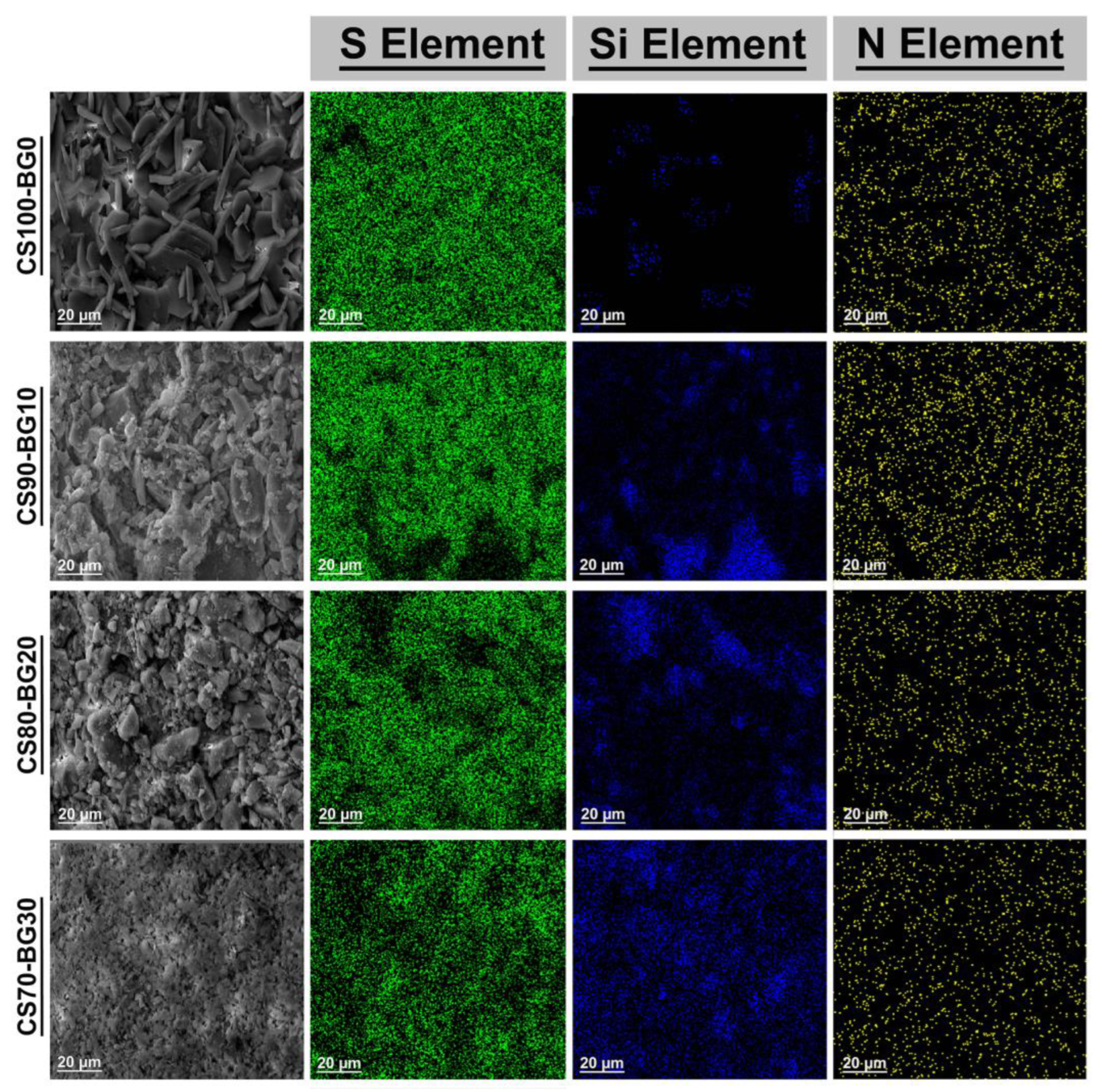
2.5. SEM Observation
2.6. Injectability
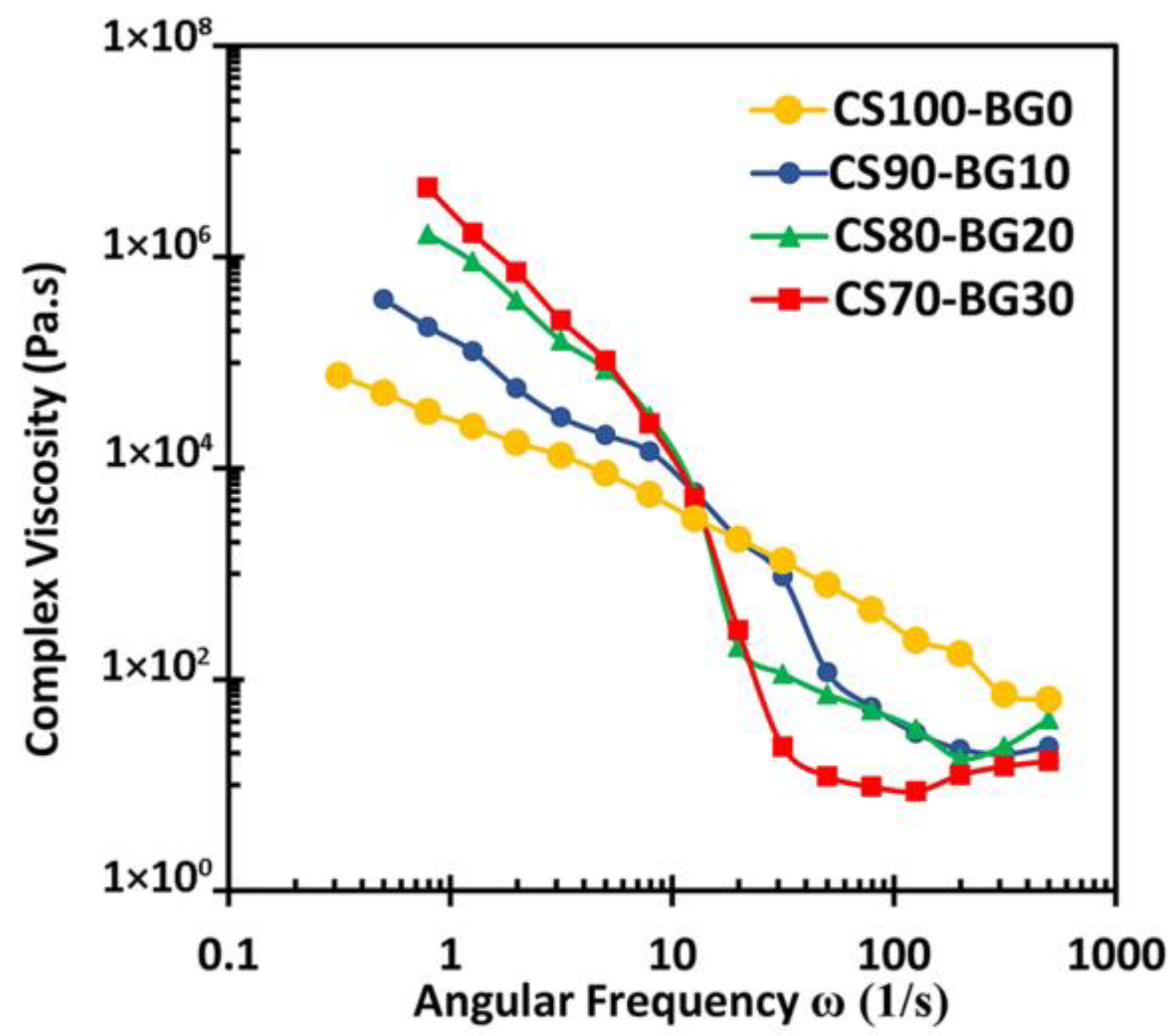
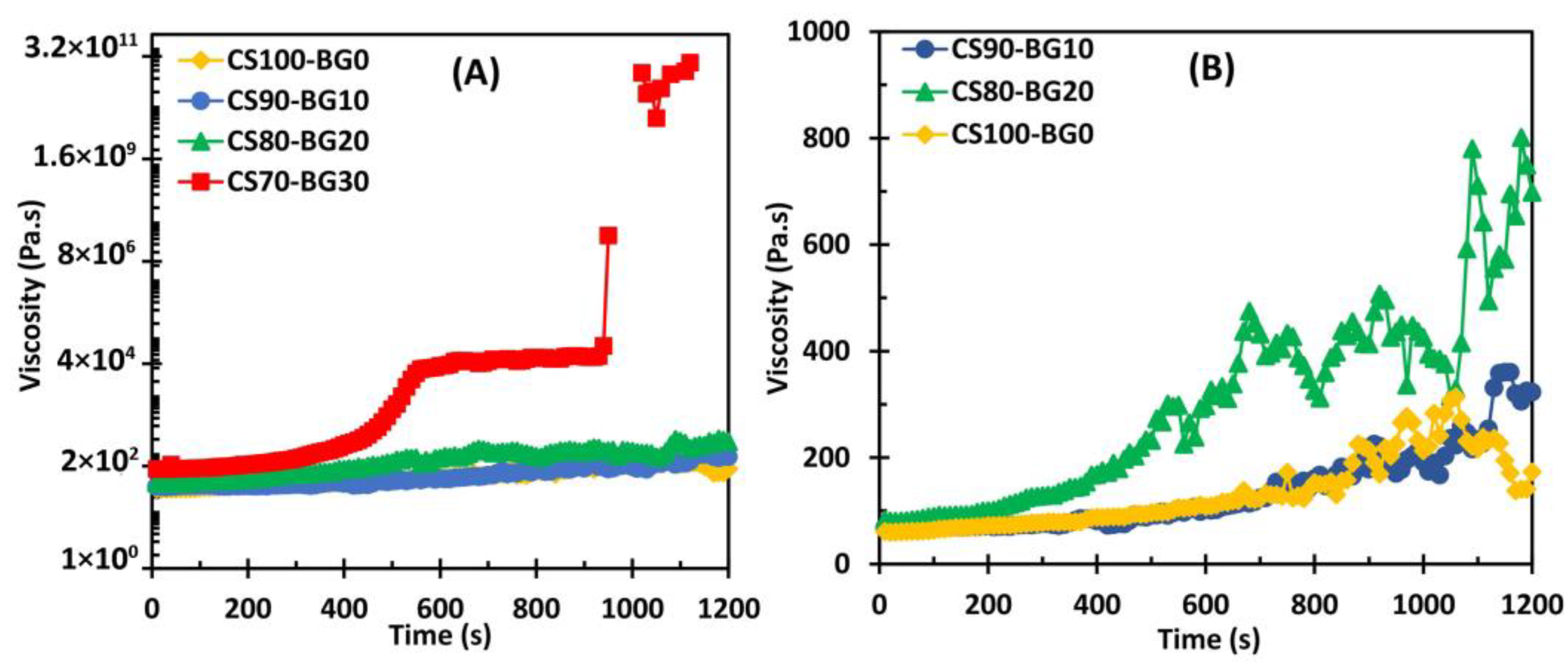
2.7. The Viscosity Measurements
2.8. The Cell Studies
2.8.1. Cell Survival, Viability, and Growth
2.8.2. DAPI Staining
2.8.3. Calcein Staining
2.9. Statistical Analysis
3. Results
3.1. Phase Composition
3.2. Setting Time
3.3. SEM Observation
3.4. Injectability
3.5. Viscosity
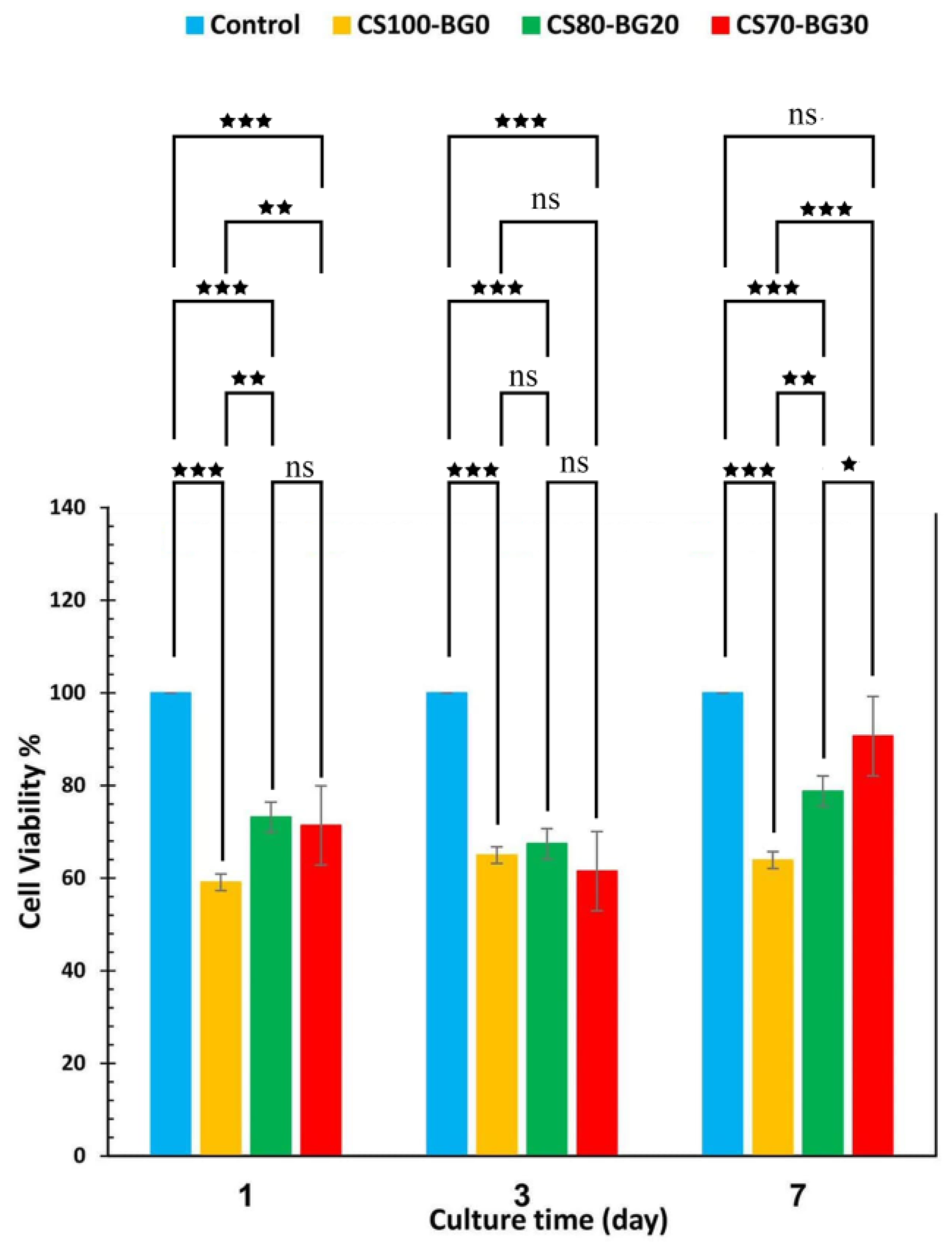
3.6. Cell Studies
3.6.1. Cell Viability and DAPI Staining
3.6.2. Calcein Staining
4. Discussion
5. Conclusions
Author Contributions
Funding
Data Availability Statement
Acknowledgments
Conflicts of Interest
References
- Zhang, F.; Zhou, M.; Gu, W.; Shen, Z.; Ma, X.; Lu, F.; Yang, X.; Zheng, Y.; Gou, Z. Zinc-/copper-substituted dicalcium silicate cement: Advanced biomaterials with enhanced osteogenesis and long-term antibacterial properties. J. Mater. Chem. B 2020, 8, 1060–1070. [Google Scholar] [CrossRef]
- Chiu, Y.H.; Chen, I.C.; Su, C.Y.; Tsai, H.H.; Young, T.H.; Fang, H.W. Development of injectable calcium sulfate and self-setting calcium phosphate composite bone graft materials for minimally invasive surgery. Int. J. Mol. Sci. 2022, 23, 7590. [Google Scholar] [CrossRef] [PubMed]
- Rezaei, H.; Shahrezaee, M.; Jalali Monfared, M.; Ghorbani, F.; Zamanian, A.; Sahebalzamani, M. Mussel-inspired polydopamine induced the osteoinductivity to ice-templating PLGA–gelatin matrix for bone tissue engineering application. Biotechnol. Appl. Biochem. 2021, 68, 185–196. [Google Scholar] [CrossRef]
- Haghighizadeh, E.; Shahrezaee, M.; Sharifzadeh, S.R.; Momeni, M. Transforming growth factor-β3 relation with osteoporosis and osteoporotic fractures. J. Res. Med. Sci. 2019, 24, 46. [Google Scholar]
- Thomas, M.V.; Puleo, D.A. Calcium sulfate: Properties and clinical applications. J. Biomed. Mater. Res. Part B Appl. Biomater. 2009, 88, 597–610. [Google Scholar] [CrossRef] [PubMed]
- Rezaei, H.; Shahrezaee, M.; Jalali Monfared, M.; Fathi Karkan, S.; Ghafelehbashi, R. Simvastatin-loaded graphene oxide embedded in polycaprolactone-polyurethane nanofibers for bone tissue engineering applications. J. Polym. Eng. 2021, 41, 375–386. [Google Scholar] [CrossRef]
- Jensen, T.; Jakobsen, T.; Baas, J.; Nygaard, J.V.; Dolatshahi-Pirouz, A.; Hovgaard, M.B.; Foss, M.; Bünger, C.; Besenbacher, F.; Søballe, K. Hydroxyapatite nanoparticles in poly-D, L-lactic acid coatings on porous titanium implants conducts bone formation. J. Biomed. Mater. Res. Part A 2010, 95, 665–672. [Google Scholar] [CrossRef]
- Jensen, T.; Dolatshahi-Pirouz, A.; Foss, M.; Baas, J.; Lovmand, J.; Duch, M.; Pedersen, F.S.; Kassem, M.; Bünger, C.; Søballe, K.; et al. Interaction of human mesenchymal stem cells with osteopontin coated hydroxyapatite surfaces. Colloids Surf. B Biointerfaces 2010, 75, 186–193. [Google Scholar] [CrossRef]
- Morley, R.; Lopez, F.; Webb, F. Calcium sulphate as a drug delivery system in a deep diabetic foot infection. Foot 2016, 27, 36–40. [Google Scholar] [CrossRef]
- Stubbs, D.; Deakin, M.; Chapman-Sheath, P.; Bruce, W.; Debes, J.; Gillies, R.M.; Walsh, W.R. In vivo evaluation of resorbable bone graft substitutes in a rabbit tibial defect model. Biomaterials 2004, 25, 5037–5044. [Google Scholar] [CrossRef]
- Thomas, M.V.; Puleo, D.A.; Al-Sabbagh, M. Calcium sulfate: A review. J. Long-Term Eff. Med. Implant. 2005, 15, 599–607. [Google Scholar] [CrossRef] [PubMed]
- Beenken, K.E.; Smith, J.K.; Skinner, R.A.; Mclaren, S.G.; Bellamy, W.; Gruenwald, M.J.; Spencer, H.J.; Jennings, J.A.; Haggard, W.O.; Smeltzer, M.S. Chitosan coating to enhance the therapeutic efficacy of calcium sulfate-based antibiotic therapy in the treatment of chronic osteomyelitis. J. Biomater. Appl. 2014, 29, 514–523. [Google Scholar] [CrossRef] [PubMed]
- Chen, Z.; Liu, H.; Liu, X.; Cui, F.Z. Injectable calcium sulfate/mineralized collagen-based bone repair materials with regulable self-setting properties. J. Biomed. Mater. Res. Part A 2011, 99, 554–563. [Google Scholar] [CrossRef]
- Shuai, C.; Zhou, J.; Wu, P.; Gao, C.; Feng, P.; Xiao, T.; Deng, Y.; Peng, S. Enhanced stability of calcium sulfate scaffolds with 45S5 bioglass for bone repair. Materials 2015, 8, 7498–7510. [Google Scholar] [CrossRef] [PubMed]
- Hsu, P.Y.; Kuo, H.C.; Tuan, W.H.; Shih, S.J.; Naito, M.; Lai, P.L. Manipulation of the degradation behavior of calcium sulfate by the addition of bioglass. Prog. Biomater. 2019, 8, 115–125. [Google Scholar] [CrossRef]
- Shams, M.; Nezafati, N.; Poormoghadam, D.; Zavareh, S.; Zamanian, A.; Salimi, A. Synthesis and characterization of electrospun bioactive glass nanofibers-reinforced calcium sulfate bone cement and its cell biological response. Ceram. Int. 2020, 46, 10029–10039. [Google Scholar] [CrossRef]
- Chamansara, A.; Behnamghader, A.; Zamanian, A. Preparation and characterization of injectable gelatin/alginate/chondroitin sulfate/α-calcium sulfate hemihydrate composite paste for bone repair application. J. Biomater. Appl. 2022, 36, 1758–1774. [Google Scholar] [CrossRef]
- Czechowska, J.; Zima, A.; Siek, D.; Ślósarczyk, A. The importance of chitosan and nano-TiHA in cement-type composites on the basis of calcium sulfate. Ceram. Int. 2016, 42, 15559–15567. [Google Scholar] [CrossRef]
- Zeimaran, E.; Pourshahrestani, S.; Fathi, A.; bin Abd Razak, N.A.; Kadri, N.A.; Sheikhi, A.; Baino, F. Advances in bioactive glass-containing injectable hydrogel biomaterials for tissue regeneration. Acta Biomater. 2021, 136, 1–36. [Google Scholar] [CrossRef]
- Jones, J.R. Review of bioactive glass: From Hench to hybrids. Acta Biomater. 2013, 9, 4457–4486. [Google Scholar] [CrossRef]
- Ruan, Z.; Yao, D.; Xu, Q.; Liu, L.; Tian, Z.; Zhu, Y. Effects of mesoporous bioglass on physicochemical and biological properties of calcium sulfate bone cements. Appl. Mater. Today 2017, 9, 612–621. [Google Scholar] [CrossRef]
- Jennings, J.A.; Bumgardner, J.D. (Eds.) Chitosan Based Biomaterials Volume 1: Fundamentals; Woodhead Publishing: Cambridge, UK, 2016. [Google Scholar]
- Patel, S.K.; Kim, J.H.; Kalia, V.C.; Lee, J.K. Antimicrobial activity of amino-derivatized cationic polysaccharides. Indian J. Microbiol. 2019, 59, 96–99. [Google Scholar] [CrossRef] [PubMed]
- Demir-Oğuz, Ö.; Boccaccini, A.R.; Loca, D. Injectable bone cements: What benefits the combination of calcium phosphates and bioactive glasses could bring? Bioact. Mater. 2023, 19, 217–236. [Google Scholar] [CrossRef] [PubMed]
- Zima, A.; Czechowska, J.; Siek, D.; Ślósarczyk, A. Influence of magnesium and silver ions on rheological properties of hydroxyapatite/chitosan/calcium sulphate based bone cements. Ceram. Int. 2017, 43, 16196–16203. [Google Scholar] [CrossRef]
- Sharifianjazi, F.; Moradi, M.; Abouchenari, A.; Pakseresht, A.H.; Esmaeilkhanian, A.; Shokouhimehr, M.; Asl, M.S. Effects of Sr and Mg dopants on biological and mechanical properties of SiO2–CaO–P2O5 bioactive glass. Ceram. Int. 2020, 46, 22674–22682. [Google Scholar] [CrossRef]
- ASTM C266-08; Standard Test Method for Time of Setting of Hydraulic Cement Paste by Gilmore Needles. ASTM International: West Conshohocken, PA, USA, 2009.
- Zou, Y.; Malzbender, J. Development and optimization of porosity measurement techniques. Ceram. Int. 2016, 42, 2861–2870. [Google Scholar] [CrossRef]
- Borhan, S.; Hesaraki, S.; Behnamghader, A.A.; Ghasemi, E. Rheological evaluations and in vitro studies of injectable bioactive glass–polycaprolactone–sodium alginate composites. J. Mater. Sci. Mater. Med. 2016, 27, 137. [Google Scholar] [CrossRef]
- Kaffashi, B.; Rezapour-Sarabi, M. Polymers Rheology; Boroon Sepehr Publishing: Washington, DC, USA, 2022; p. 141. [Google Scholar]
- Fatimi, A.; Tassin, J.F.; Bosco, J.; Deterre, R.; Axelos, M.A.; Weiss, P. Injection of calcium phosphate pastes: Prediction of injection force and comparison with experiments. J. Mater. Sci. Mater. Med. 2012, 23, 1593–1603. [Google Scholar] [CrossRef]
- Medrano-David, D.; Lopera, A.M.; Londoño, M.E.; Araque-Marín, P. Formulation and characterization of a new injectable bone substitute composed PVA/borax/CaCO3 and Demineralized Bone Matrix. J. Funct. Biomater. 2021, 12, 46. [Google Scholar] [CrossRef]
- Sarda, S.; Fernández, E.; Llorens, J.; Martinez, S.; Nilsson, M.; Planell, J.A. Rheological properties of an apatitic bone cement during initial setting. J. Mater. Sci. Mater. Med. 2001, 12, 905–909. [Google Scholar] [CrossRef]
- Le Gouellec, Y.A.; Elimelech, M. Calcium sulfate (gypsum) scaling in nanofiltration of agricultural drainage water. J. Membr. Sci. 2002, 205, 279–291. [Google Scholar] [CrossRef]
- Shamsi, M.; Karimi, M.; Ghollasi, M.; Nezafati, N.; Shahrousvand, M.; Kamali, M.; Salimi, A. In vitro proliferation and differentiation of human bone marrow mesenchymal stem cells into osteoblasts on nanocomposite scaffolds based on bioactive glass (64SiO2-31CaO-5P2O5)-poly-l-lactic acid nanofibers fabricated by electrospinning method. Mater. Sci. Eng. C 2017, 78, 114–123. [Google Scholar] [CrossRef]
- Barreto, M.E.; Medeiros, R.P.; Shearer, A.; Fook, M.V.; Montazerian, M.; Mauro, J.C. Gelatin and Bioactive Glass Composites for Tissue Engineering: A Review. J. Funct. Biomater. 2022, 14, 23. [Google Scholar] [CrossRef] [PubMed]
- Hesaraki, S.; Moztarzadeh, F.; Nezafati, N. Evaluation of a bioceramic-based nanocomposite material for controlled delivery of a non-steroidal anti-inflammatory drug. Med. Eng. Phys. 2009, 31, 1205–1213. [Google Scholar] [CrossRef]
- Sony, S.; Suresh Babu, S.; Nishad, K.V.; Varma, H.; Komath, M. Development of an injectable bioactive bone filler cement with hydrogen orthophosphate incorporated calcium sulfate. J. Mater. Sci. Mater. Med. 2015, 26, 31. [Google Scholar] [CrossRef] [PubMed]
- Hesaraki, S.; Zamanian, A.; Moztarzadeh, F. The influence of the acidic component of the gas-foaming porogen used in preparing an injectable porous calcium phosphate cement on its properties: Acetic acid versus citric acid. J. Biomed. Mater. Res. Part B Appl. Biomater. 2008, 86, 208–216. [Google Scholar] [CrossRef]
- Yokoyama, A.; Yamamoto, S.; Kawasaki, T.; Kohgo, T.; Nakasu, M. Development of calcium phosphate cement using chitosan and citric acid for bone substitute materials. Biomaterials 2002, 23, 1091–1101. [Google Scholar] [CrossRef]
- Zhao, Y.; Jia, L.; Liu, K.; Gao, P.; Ge, H.; Fu, L. Inhibition of calcium sulfate scale by poly (citric acid). Desalination 2016, 392, 1–7. [Google Scholar] [CrossRef]
- Muzzarelli, R.A. Natural chelating polymers. Alginic acid, chitin and chitosan. In International Series of Monographs in Analytical Chemistry; Pergamon Press: Oxford, UK, 1973. [Google Scholar]
- Krajewska, B. Diffusion of metal ions through gel chitosan membranes. React. Funct. Polym. 2001, 47, 37–47. [Google Scholar] [CrossRef]
- Hesaraki, S.; Moztarzadeh, F.; Nemati, R.; Nezafati, N. Preparation and characterization of calcium sulfate–biomimetic apatite nanocomposites for controlled release of antibiotics. J. Biomed. Mater. Res. Part B Appl. Biomater. 2009, 91, 651–661. [Google Scholar] [CrossRef]
- Zhang, X.; Jia, W.; Gu, Y.; Xiao, W.; Liu, X.; Wang, D.; Zhang, C.; Huang, W.; Rahaman, M.N.; Day, D.E.; et al. Teicoplanin-loaded borate bioactive glass implants for treating chronic bone infection in a rabbit tibia osteomyelitis model. Biomaterials 2010, 31, 5865–5874. [Google Scholar] [CrossRef] [PubMed]
- Rauschmann, M.A.; Wichelhaus, T.A.; Stirnal, V.; Dingeldein, E.; Zichner, L.; Schnettler, R.; Alt, V. Nanocrystalline hydroxyapatite and calcium sulphate as biodegradable composite carrier material for local delivery of antibiotics in bone infections. Biomaterials 2005, 26, 2677–2684. [Google Scholar] [CrossRef] [PubMed]
- Bernardo, M.P.; da Silva, B.C.; Hamouda, A.E.; de Toledo, M.A.; Schalla, C.; Rütten, S.; Goetzke, R.; Mattoso, L.H.; Zenke, M.; Sechi, A. PLA/Hydroxyapatite scaffolds exhibit in vitro immunological inertness and promote robust osteogenic differentiation of human mesenchymal stem cells without osteogenic stimuli. Sci. Rep. 2022, 12, 2333. [Google Scholar] [CrossRef]
- Liang, H.; Xu, X.; Feng, X.; Ma, L.; Deng, X.; Wu, S.; Liu, X.; Yang, C. Gold nanoparticles-loaded hydroxyapatite composites guide osteogenic differentiation of human mesenchymal stem cells through Wnt/β-catenin signaling pathway. Int. J. Nanomed. 2019, 14, 6151–6163. [Google Scholar] [CrossRef] [PubMed]


| Code | Powder Phase | Liquid Phase | |
|---|---|---|---|
| CSH | Solution of 19.2% citric acid and 2% chitosan (w/v) | ||
| CS100-BG0 | 100 | 0 | |
| CS90-BG10 | 90 | 10 | |
| CS80-BG20 | 80 | 20 | |
| CS70-BG30 | 70 | 30 | |
| Sample | Initial Setting Time (min) | Final Setting Time (min) | Compressive Strength (MPa) | Porosity (%) |
|---|---|---|---|---|
| Cement made of pure CS and distilled water | 25 ± 3 | 35 ± 3 | - | - |
| Cement made of pure CS and citric acid | 180 ± 15 | 240 ± 20 | - | - |
| CS100-BG0 | >300 | >600 | 0.29 ± 0.02 | 50 ± 2 |
| CS90-BG10 | 60 ± 5 | 150 ± 20 | 0.20 ± 0.07 | 61 ± 3 |
| CS80-BG20 | 32 ± 5 | 120 ± 10 | 1.25 ± 0.11 | 38 ± 1 |
| CS70-BG30 | 6 ± 1 | 20 ± 4 | 4.25 ± 0.85 | 36 ± 2 |
Disclaimer/Publisher’s Note: The statements, opinions and data contained in all publications are solely those of the individual author(s) and contributor(s) and not of MDPI and/or the editor(s). MDPI and/or the editor(s) disclaim responsibility for any injury to people or property resulting from any ideas, methods, instructions or products referred to in the content. |
© 2023 by the authors. Licensee MDPI, Basel, Switzerland. This article is an open access article distributed under the terms and conditions of the Creative Commons Attribution (CC BY) license (https://creativecommons.org/licenses/by/4.0/).
Share and Cite
Moazeni, N.; Hesaraki, S.; Behnamghader, A.; Esmaeilzadeh, J.; Orive, G.; Dolatshahi-Pirouz, A.; Borhan, S. Design and Manufacture of Bone Cements Based on Calcium Sulfate Hemihydrate and Mg, Sr-Doped Bioactive Glass. Biomedicines 2023, 11, 2833. https://doi.org/10.3390/biomedicines11102833
Moazeni N, Hesaraki S, Behnamghader A, Esmaeilzadeh J, Orive G, Dolatshahi-Pirouz A, Borhan S. Design and Manufacture of Bone Cements Based on Calcium Sulfate Hemihydrate and Mg, Sr-Doped Bioactive Glass. Biomedicines. 2023; 11(10):2833. https://doi.org/10.3390/biomedicines11102833
Chicago/Turabian StyleMoazeni, Nazanin, Saeed Hesaraki, Aliasghar Behnamghader, Javad Esmaeilzadeh, Gorka Orive, Alireza Dolatshahi-Pirouz, and Shokoufeh Borhan. 2023. "Design and Manufacture of Bone Cements Based on Calcium Sulfate Hemihydrate and Mg, Sr-Doped Bioactive Glass" Biomedicines 11, no. 10: 2833. https://doi.org/10.3390/biomedicines11102833