Review

Association between Immunosuppressive Therapy Utilized in the Treatment of Autoimmune Disease or Transplant and Cancer Progression

Department of Medical Oncology & Therapeutics Research, City of Hope National Medical Center, Duarte, CA 91010, USA
*
Author to whom correspondence should be addressed.
Biomedicines 2023, 11(1), 99; https://doi.org/10.3390/biomedicines11010099
Submission received: 3 December 2022 / Revised: 28 December 2022 / Accepted: 28 December 2022 / Published: 30 December 2022
(This article belongs to the Special Issue Immune Checkpoints and Autoimmunity)

Abstract

:
Autoimmunity and cancer rates have both been on the rise in Western civilization prompting many to investigate the link between the two entities. This review will investigate the complex interactions between the activation and deactivation of the immune system and the development of malignancy. Additional focus will be placed on the main classes of immune inhibitor therapy utilized in transplant patients and in autoimmune disease including TNF-alpha, Calcineurin, mTOR, purine synthesis antagonists and IMPDH inhibitors.

1. Introduction

The concept of an immune system whereby innate cells identify and destroy foreign or malfunctioning cells has intrigued scientists for decades. As knowledge expanded so did the complexity of investigations. In the 1950s–1970s, researchers Burnet and Thomas formulated the concept of cancer immunosurveillance whereby a functioning immune system, at the time thought to be cells derived from the Thymus, can recognize ‘transformed’ cells or tumor cells [1,2]. However, there was limited proof of this concept in animal experimental models several decades after the initial theory was published [3,4,5]. More recently this concept of the protective immunosurveillance has been redefined into a broad topic of cancer immunoediting with 3 distinct phases termed elimination, equilibrium and escape [6] The phases covered variety of functions including both the innate and adaptive immunity as well as tumor recognition and tumor modification [6]. Additional support of this concept is demonstrated by the discovery of mice without properly functioning lymphocytes developed malignancy at a significantly higher rate compared to the wild type [7]. Thus, properly functioning immune systems are beneficial in the targeting and destruction of tumor cells.
Further investigation into the effects of upregulation of immune systems as seen in autoimmunity and downregulation seen in immunodeficiency/immunosuppression is needed. Autoimmunity and cancer rates have both been on the rise in Western civilization leading many to question if there is a link between the two entities. Likewise, the association between immunodeficiency or induced immunodeficiency and malignancy has been at the forefront of medical research with particular interest in the transplanted patients, HIV patients and patients with autoimmune disease requiring chronic immunosuppression. As the complexity and intricacy of immunosuppressive agents expands there is intensified interest in investigating down-stream effects of these agents
This paper aims to focus on the intersection of immune function and cancer with a review of the literature surrounding these two subjects. This is especially relevant in the age of exponentially increased use of immune checkpoint inhibitors in the treatment of cancers.

2. Autoimmunity and Cancer Correlation

Several studies have highlighted the connection between chronic inflammation and the development of malignancy [8]. While the exact mechanism of this relationship is not known, some speculate chronic inflammation leading to antigen specific cell damage, activation of Type 2 immune response mediated by IL-4 and IL-13, and relative CTLA-4 deficiency are potential triggers [8]. Other studies stipulate inflammatory cells themselves are a potential mechanism such as tumor-associated macrophages which can stimulate tumor growth and angiogenesis [9]. Observational representation of this concept can be seen in the increase in rates of autoimmune gastritis and its associated increase in rates of gastric cancer in younger women who have the highest incidence of autoimmune disease in comparison with a previously high prevalence of gastric cancer in men [10].
The association between autoimmunity induced inflammation and malignancy does not have a negative outcome always. A review by Zityogel et al. discussed the concept of ‘beneficial autoimmunity’ and identified examples of therapy-induced (such as immune checkpoint inhibitors) as well as idiopathic or spontaneous autoimmune disease conferring favorable outcomes against oncologic disease [11]. A higher level of T cells that recognize tumor-specific antigens available for anticancer immunosurveillance is thought to be another mechanism of beneficial autoimmunity [11]. In a large SEER [Surveillance, Epidemiology, and End Results program] database study, 13.5% of lung cancer patients were diagnosed with autoimmune disease during or after their cancer diagnosis [12]. However, a retrospective cohort study comparing lung cancer patients with and without autoimmune disease did not find any significant difference in progression-free survival per stage among the two groups, even though the autoimmune group was less likely to receive the standard of care inferring some protective benefit from the autoimmune disease [13]. In paraneoplastic encephalomyelitis, most commonly seen in patients with small cell lung cancer [SCLC], there was a favorable response to chemotherapy observed in patients who tested positive for anti-hu antibodies, autoantibodies against neuronal RNA binding proteins [14,15].
Given the evidence of beneficial autoimmunity, it would appear immunosuppression is a larger contributor to malignancy risk compared to autoimmune disease itself. Further evidence demonstrated in a 2020 review on celiac’s disease [treated with dietary modification] which found that overall malignancy risk was low and even negligible one year after diagnosis [16]. Likewise, a European study of autoimmune thyroiditis treated with hormone therapy did not find an elevated risk of thyroid cancer after a 10-year follow up period [17,18]. On the other hand, a Taiwanese study did find an increased risk of both thyroid cancer and colorectal cancer among Hashimoto’s thyroiditis patient population [19]. From this mixed data autoimmune disease must have a role in the development of malignancy.
Interestingly, malignancy has also been shown to stimulate autoimmunity, most notably through well-known paraneoplastic syndromes [20]. Paraneoplastic rheumatologic conditions such paraneoplastic polyarthritis and hypertrophic osteoarthropathy are a few notable examples and are thought be related to upregulation of VEGF and fibroblast growth factor 23 (FGF23) in tumors [20]. SCLC is another typical example of malignancy and associated with a high frequency of paraneoplastic syndromes that can proceed the diagnosis or be the first sign of relapse [21].

3. Immunodeficiency and Cancer Correlation

While overactive immune systems are a risk for malignancy, studies have also demonstrated that immunodeficiency may also be an independent risk factor [22]. This concept was first examined in mice models. In a research study, mice without an innate immune system were more vulnerable to both spontaneous and externally induced cancers [23]. A study involving the US Immune Deficiency Network Database examine primary immunodeficiency patients which included patients from 39 academic medical centers and >300 single gene mutations of the immune system in comparison to the age-adjusted SEER population. They identified a 1.42-fold increase in relative risk of malignancy in the primary immunodeficient population [22]. Interestingly, in subgroup analysis, men with primary immunodeficiency had the highest increase in relative risk, 1.91, compared to the age-adjusted population, while women were diagnosed with malignancy at similar rates [22].
Additional data provided by a large meta-analysis of HIV/AIDS patients [7 studies] and transplanted patients [5 studies] showed immunodeficiency and immunosuppression conferred a higher risk for malignancy in 20 out of the 28 types of cancer [24]. The authors proposed several mechanisms for the increased risk of malignancy including increased infections, immunodeficiency, increased cancer screening, and lifestyle factors [24]. However, the latter two theories were thought to be less likely as rates of screened cancers, breast and colon, were not elevated in both study populations and lifestyle factors were not standard across the study populations. Infection was thought to be an independent factor as many of the cancers in the analysis had an infectious cause [i.e., pylori, hepatitis, EBV] and several AIDS-defining malignancies were included [24]. However, notable exceptions were non-melanoma skin cancer and lip cancer that have no known infectious etiology and were found to have higher rates of malignancy in transplant recipients. Interestingly, the common epithelial-derived cancers, colon, breast, rectum, and ovarian, did not have higher rates among the immunosuppressed population [24].

4. Immunosuppression in Autoimmune Disease and Cancer Correlation

Autoimmune disease can be organ specific or systemic, but treatment and presentation vary greatly within each category. The mainstay of treatment in autoimmune disease can be separated into two categories, replacement therapy vs. immunosuppressive [25]. A NEJM review proposed that immunosuppressive treatment can be further characterized into 4 subtypes: alteration of thresholds of immune activation, modulation of antigen specific cells, reconstitution of the immune system and sparing of target organs [26]. Reconstitution of the immune system involves bone marrow ablation with or without the addition of stem cells, reserved for severe refractory disease as is an aggressive treatment [27]. More commonly used treatments target inflammatory pathways [26]. One such example are inhibitors of the TNF receptor as binding of its substrate triggers downstream signaling leading to upregulation of inflammation and apoptosis (Figure 1) [28].

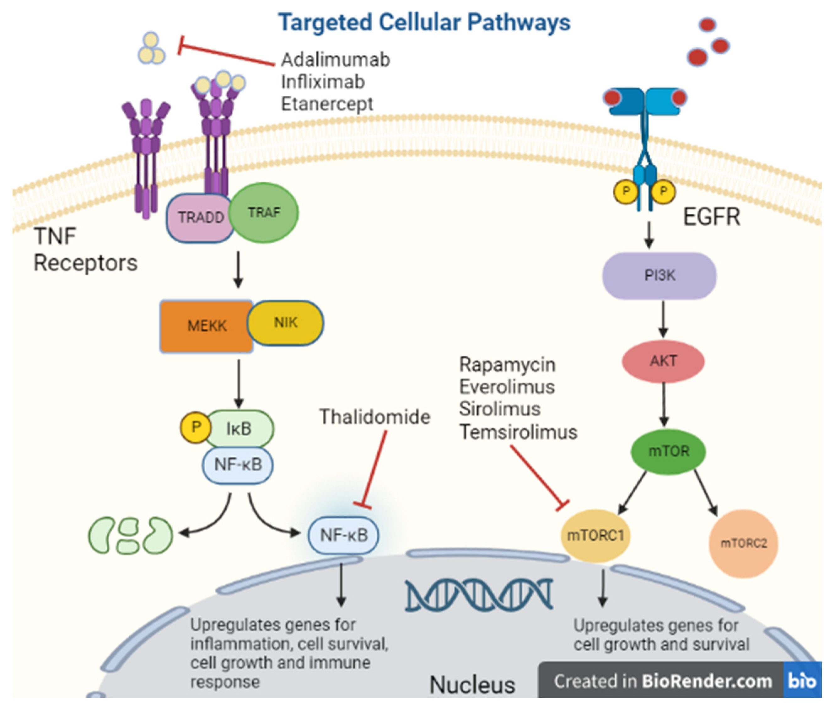
Tumor Necrosis Factor-Alpha Inhibitors

Tumor Necrosis Factor-Alpha [TNF-alpha] has been synonymous with pro-inflammation given its association with pro-inflammatory cytokines IL-1, IL-6, IL-8 and VEGF; therefore, its inhibitors are used for immunosuppression (Figure 2) [29]. However, there is some concern regarding the diverse functionality of TNF-alpha as low levels promoted angiogenesis while higher levels were anti-angiogenetic [30]. Many molecular pathways of TNF-alpha are associated with the upregulation of matrix metallopeptidase 9 [MMP9] which directly degrades the extracellular matrix allowing tumor migration and indirectly promotes cytokines that support cell tumor growth [31]. Despite this mixed data on TNF-alpha, researchers believed blocking its action could be a potential target for malignancy.
Thalidomide, an inhibitor of TNF-alpha protein synthesis (Figure 1), has been proven effective against certain cancer types including multiple myeloma, renal, breast, colon, and prostate among others, due to its inhibition of various growth factors, including VEGF, basic FGF, and Hepatocyte Growth Factor, as well as inhibiting tumor DNA synthesis [32,33]. In a clinical trial of 84 patients with refractory myeloma, 76 refractories to high-dose chemotherapy, were treated with escalating doses of Thalidomide to 800 mg over two months, the amount of myeloma protein determined response in serum or Bence Jones protein in urine [34]. Overall, there was a 32% response rate with greater than 90% reduction in 10 of the patients and the majority responded in two months. At 12 month follow up, overall survival was 58%, and event-free survival was 22% [34]. While the data on thalidomide is substantial, other medications in its class had significant adverse effects prompting further investigation. As a class, TNF-alpha inhibitors can promote lymphoproliferative disorders by an unclear mechanism [35]. One study by Askling et al. found an increased risk of leukemia and lymphoma in rheumatoid arthritis patients who were treated with TNF-alpha inhibitors [36]. Another found an increased risk of lymphoma in rheumatoid arthritis patients treated with etanercept or infliximab [37]. There is a dose-dependent relationship between TNF-alpha inhibitor use and the risk of malignancy [38]. Additionally, in a meta-analysis of 36 global clinical trials with 19,041 patients treated with adalimumab [Humira] for rheumatoid arthritis, ankylosing spondylitis, juvenile idiopathic arthritis, psoriatic arthritis, psoriasis, or Crohn’s disease had increased standardized incident ratio of non-melanoma skin cancer and lymphoma collectively [39]. With a few notable exceptions, caution should be used when treating patients with medications that modulate TNF-alpha.
Given this concern for malignancy risk, additional agents were developed with new targets of the inflammatory cascade; namely a monoclonal antibody targeting IL-12 and IL23 [40] and a4b7 integrin antagonist [41]. Contrastingly, both drugs [Ustekinumab and Vedolizumab] were not found to have an increased risk of new or recurrent malignancy in patients with IBD and history of previous malignancy [42,43].

5. Transplant Immunosuppression and Cancer Correlation

As mentioned above, immunodeficiency may be an independent risk factor for malignancy, affecting the transplanted population given the extensive immunosuppression to prevent rejection. In a cohort study using the US Scientific Registry of Transplant Recipients [SRTR], Non-Hodgkin’s lymphoma had the highest incidence apart from non-melanoma skin cancer among the most transplanted organs [kidney, liver, heart, lung] [Table 1] [44]. A study demonstrated that 1351 lung transplanted patients in Spain had higher rates of non-melanoma skin cancer [32%], lymphoproliferative diseases [18%] and lung cancer [16.5%] (Table 1) [45]. Further, another study of 2150 bone marrow transplant patients showed an elevated SIR of 11.6 of all cancer combined with the highest risk in b-cell lymphoproliferative disease [BLPD] [46]. In a more recent 2022 study on 2814 patients [only 23 met criteria for analysis] who received a liver transplant from 2008–2020 at a high-volume center found once again the highest risk of de novo development of non-melanoma skin cancer [21.7%] followed by gynecological cancers [17.3%] [47]. In addition, Non-Hodgkin’s lymphoma and Post-transplant lymphoproliferative disease were grouped separately unlike other studies in which they were grouped as the same entity [47].
Several studies have found that the higher rates of immunosuppression seen in heart transplanted patients conferred the highest rates of malignancy with skin cancer being the most common [48]. Additional data showed that post-kidney transplanted patients on immunosuppression had higher rates of lung cancer compared to rates prior to transplant while on dialysis [49]. Immunosuppression regimens can differ but commonly include calcineurin inhibitors, mTOR inhibitors, corticosteroids and anti-proliferative/antimetabolite [azathioprine]. According to the SRTR, the most common regimen used in 60–70% of kidney, heart, lung, liver, kidney-pancreas transplanted patients consist of tacrolimus, prednisone and mycophenolate mofetil [Table 1] [50]. While the risk of de novo malignancy after transplant has been noted in several studies, donor-derived malignancy is another entity.
In a highly publicized case from 2008, 4 patients who received organs from a 53-year-old donor with no known history of cancer developed molecularly similar breast cancer, confirmed by DNA microsatellites, within 16 months to 6 years of trans-plant [51]. Three of the four patients died from widely metastatic disease. Still the fourth patient, despite also having metastatic disease, was ultimately cured with removal of the transplanted organ and cessation of immunosuppression and chemotherapy [51]. While this was considered an infrequent event from estimates with a risk is 0.01–0.05%, it elicited further investigation into donor-derived malignancy [52]. A review from 2020 of 72 case reports, 50 case series, and six registries comprising of 234 patients found the most frequent donor-derived malignancy was lymphoma [20.5%], followed by renal cancer [17.9%], melanoma [17.1%], and non-small cell lung cancer [5.6%] [53]. Interestingly, in the analysis of the donor demographics, the donor had an ongoing history of malignancy in 17% of the cases while in 18.5% of the cases, the donors were not screened with imaging [53]. Whether the latent/dormant malignancies in donors evolved into aggressive disease as a consequence of immunosuppression alone or if there are other factors involved remains to be seen. Each Immunosuppressive agent requires further examination to evaluate malignancy risk.

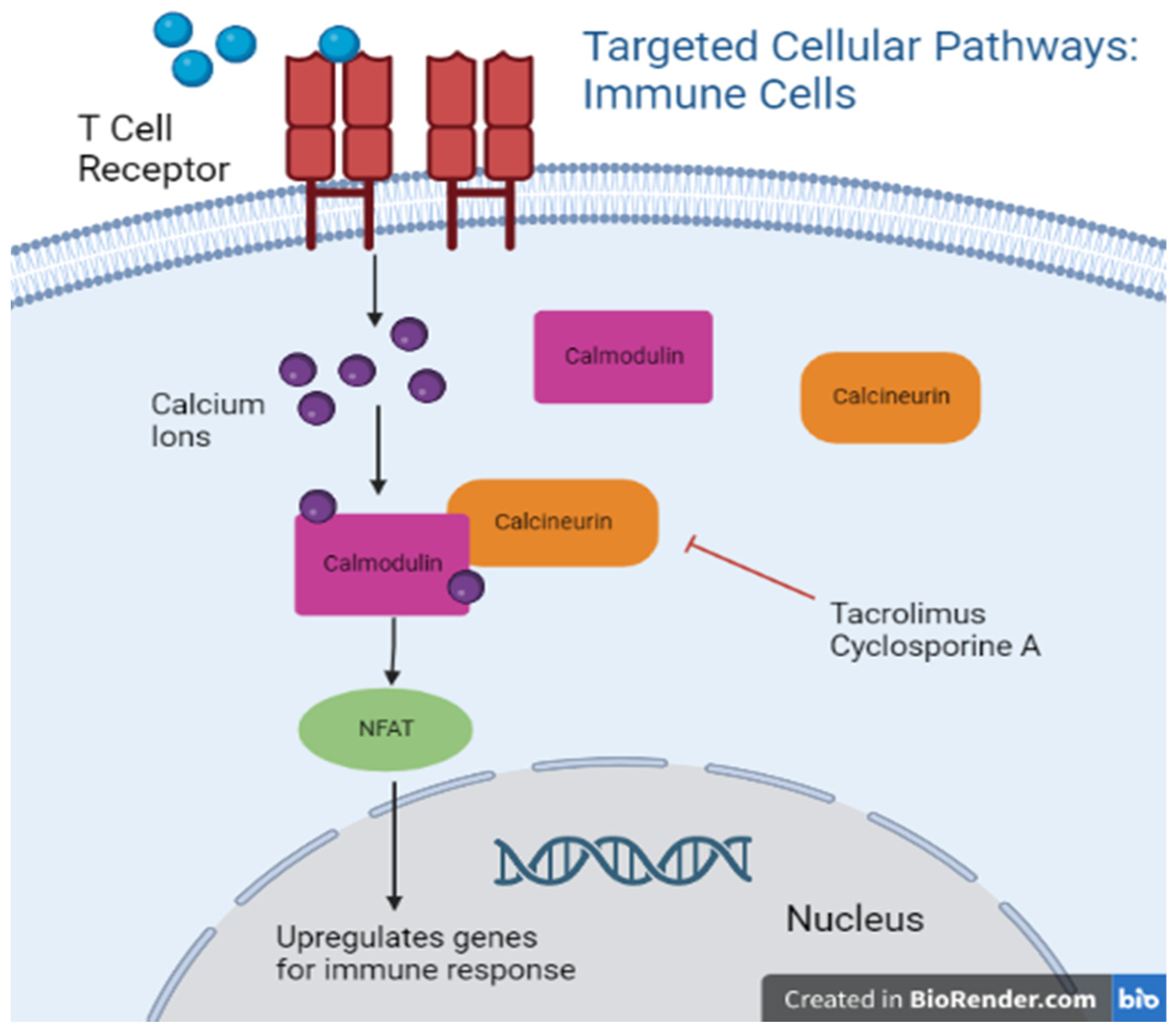
5.1. Calcineurin Inhibitors

Calcineurin inhibitors, namely cyclosporine, have been employed in the transplant population for decades after the discovery of its selective inhibitory effect on T cells [54] (Figure 2). Later discovered to be the direct action of Cyclosporine A and Tacrolimus on calcineurin which uninhibited facilitates NF-AT translocation to the nucleus and activation of genes involved in the regulation of T cell proliferation (Figure 3) [55]. Popularity of this class of immunosuppressants increased after the observation of limited bone marrow suppression in animal models even at high doses [56].
Much research has been done to investigate the association between calcineurin inhibitors and their mechanism of action, prompting an investigation into potential malignancy concerns. A large meta-analysis on topical calcineurin inhibitors by Lam et al. did not find a significant increase in the risk of melanoma and keratinocyte carcinoma but identified a significant association with lymphoma. However, this was thought to be overall a minimal risk [57]. Systemic calcineurin inhibitors utilized in transplants, Cyclosporin and Tacrolimus, have been shown to promote tumorigenesis by increasing VEGF, TGF-B, and IL-6 in mice models [58]. In another large case–control study by Stewart et al., Cyclosporin A combined with other immunosuppressant medications, steroids and azathioprine, was associated with a decreased incidence of de novo breast cancer in female patients after kidney or heart transplants [59]. However, there was an increased incidence in all other types of malignancy reviewed compared to the ‘expected’ incidence obtained from an international data collection study in 1990s [59]. An additional investigation into a patient population of both sexes by the same team found a decreased incidence of rectal cancer among immunosuppressed patients but similar studies with prolonged follow-up intervals found an increased incidence [60]. From our current data, it remains difficult to form any unifying conclusions regarding calcineurin inhibitors and malignancy. Still, topical calcineurin inhibitors have a higher safety profile when compared to their systemic counterparts.

5.2. mTOR Inhibitors

MTOR inhibitors were first utilized in transplant in 1999 (Figure 2) [61] after the discovery of their activity blocking the proliferation of IL-2 stimulated T cells by halting the cell cycle progression from G1 to S [62]. Specifically, studies have demonstrated the binding and complex formation of Rapamycin with FKBP12 inhibits mTOR, most notably the mTORC1 portion (Figure 1) [63]. In addition, mTOR inhibitors have been shown to have a significant role in promoting T-cell anergy both in vitro [64] and in vivo studies [65]. Due to these functions, mTOR inhibitors remain an essential part of immunosuppression. Based on the literature, MTOR inhibitors play a more beneficial role regarding malignancy compared to other classes of immunosuppressants. Several in vitro studies have shown that rapamycin displays tumor targeting effects by triggering cell apoptosis (Figure 1) [66]. When rapamycin was used as a sole agent, it had the lowest incidence of skin cancers among the immunosuppressed population [67]. Additionally, rapamycin has been associated with decreased cancer risk when given without Cyclosporin A or when Cyclosporin A was withdrawn shortly after treatment initiation in renal transplant patients followed two years after the transplant [68]. However, the authors highlighted that short follow-up was a limitation of the study and additional studies with prolonged follow-up are needed to further quantify malignancy risk.
Chemical variations of the initial compounds were created to improve bioavailability and started being utilized to target and treat certain cancer types. The first, temsirolimus, an mTOR signaling inhibitor (Figure 2), was FDA approved in 2007 for advanced renal cell carcinoma after a phase III multicenter randomized open label trial of 626 previously untreated renal cell carcinoma patients with poor prognosis compared temsirolimus vs. temsirolimus plus IFN-alpha vs. IFN-alpha alone and found OS benefit of 10.9 months with temsirolimus monotherapy compared to 7.3 months with IFN-alpha alone [69]. Additional success was seen in mantle cell lymphoma but not in solid tumors [70]. One explanation is the cytostatic rather than cytotoxic effect of the drug which then induces cancer cell resistance rather than destruction [71]. To overcome this limitation, researchers combined the drugs with other proven effective cytotoxic agents, such as taxanes and carboplatin in ovarian cancer and metastatic melanoma [72,73]. A phase II of temsirolimus with carboplatin and paclitaxel followed by temsirolimus consolidation therapy in first-line stage III–IV clear cell carcinoma did not find statistically significantly improvement in progression-free survival. However, treatment was well tolerated and did find benefit in patients who were sufficiently debulked [72]. Less success was seen in the Alliance trial, a randomized phase 2 trial investigating carboplatin, paclitaxel, bevacizumab with and without everolimus in the treatment of stage IV metastatic melanoma, which found that the addition of everolimus was too toxic without additional survival benefit in comparison with the control arm, with progression free survival of 5 months in the 149 patients studied [73].
Everolimus was more successful when combined with endocrine therapy as it was shown to improve median progression-free survival with hormone therapy to 10.6 months compared to 4.1 months with hormone therapy alone in HR-positive breast cancer as second line in patients previously treated with aromatase inhibitors, according to the BOLERO-2, a phase 3 randomized trial of 724 patients [74]. Selective mTOR inhibitors were subsequentially developed to have a more targeted approach and in the hopes of achieving less toxicity and higher potency [75,76]. Several are currently in clinical trials. One such example is vistusertib (AZD2014), which is being evaluated in the treatment of recurrent grade II-III meningiomas (NCT03071874). Limitations to this drug class remain; a phase I clinical trial investigating BEZ235 in the treatment of advanced renal cell carcinoma (NCT00620594) was concluded early due to lack of efficacy and extensive toxicity associated with the drug with 50% of patients experiencing grade 3–4 toxicity [77]. Further, an additional randomized phase II trial of apitolisib, a combination mTOR/PI3K inhibitor in comparison with everolimus had shorter progression-free survival and a high rate of grade 3–4 toxicities [78]. Everolimus and temsirolimus have proven benefit in the treatment of select malignancies, combination mTOR and PI3K inhibitors should be monitored closely as toxicity is high.

6. Immunosuppression in Both Autoimmune Disease/Transplant and Cancer Correlation

Purine Synthesis Antagonists

IMPDH is an essential enzyme in the synthesis of purines, catalyzing the rate-limiting conversion of IMP to XMP, with the greatest impact in rapidly growing cells [79]. Mycophenolate mofetil (MMF), inhibitor of IMPDH, was first utilized as an immunosuppressant in rheumatologic disease (Figure 2) [80]. However, it is more commonly used in solid organ transplant in combination with calcineurin inhibitors [81]. The development of MMF stems from significant toxicity from Azathioprine and discovery of the selectivity of MMF for IMPDH in B and T cells [82,83]. Since this discovery, the utilization and interest in IMPDH inhibitors considerably increased.
In the late 1960s, researchers investigated the relationship of IMPDH inhibitors and anti-tumor properties; initial data was promising with anti-tumor activity against leukemia and sarcoma [84,85]. IMPDH inhibitors have been shown to decrease cell proliferation in several cancer types including leukemia, lymphoma, pancreatic, and non-small cell lung cancer [NSCLC] [86]. Unfortunately, this concept never made it past phase II clinical trials as effectiveness was limited at that time [87]. In a phase I dose escalation trial of mycophenolate mofetil in 11 relapsed and refractory multiple myeloma patients, disease progression was seen in 6/11 patients with stable disease in 4/11 and partial response in 1 patient [88]. In addition, severe GI side effects from direct enterocyte toxicity remained a limiting factor [89], as well as limited bioavailability due to degradation from glucuronidation [87].
As advancements in medical technology were made, methods were developed to overcome these shortcomings, such as the development of MPA nanofiber that could deliver a concentrated amount of the medication to a specific target for a sustained period in glioblastoma cells [90]. Additional investigation into the tumor targeting effects of IMPDH inhibition has identified benefits in tuberous sclerosis complex [91]. In addition to the benefits of IMPDH inhibitor’s tumor targeting effects, several studies have shown that transplant patients who received immunosuppression with IMPDH inhibitors, namely mycophenolate mofetil, had a decreased risk of malignancy including post-transplant lymphoproliferative disorders [92,93]. One proposed mechanism for the above finding is the IMPDH inhibitors’ antiviral properties exhibited against HIV and hepatitis, especially as an uncontrolled infection is associated with certain cancer tumorigenesis [94]. Additional research is currently underway to further explore molecular pathways of IMPDH and its downstream effects. Until such data exists to support both the efficacy and safety of IMPDH inhibitors in the treatment of malignancy, caution should be used prior to initiating treatment.
Antimetabolite Azathioprine, an additional antagonist of purine synthesis, has been utilized in the transplanted population for immunosuppression since 1960 (Figure 2) [95]. Its success as an immunosuppressant prompted further studies in the 1970s into its use in autoimmune disease, notably autoimmune hepatitis [96] where it has been proven beneficial in long-term remission after steroids have been withdrawn [97]. With its widespread and long-term use, questions of safety surfaced. A cohort study of patients with inflammatory bowel disease treated with azathioprine in Denmark from 1997–2008 found an increased overall cancer risk of 1.41 [95% CI 1.15–1.74] in particular lymphoid [2.42] and urinary tract cancer [2.84] [98]. Another investigation into the use of azathioprine and inflammatory bowel disease identified a higher risk of non-melanoma skin cancer with an odds ratio of 5.0 [95% CI of 1.1–22.8] [99]. Contrastingly, a meta-analysis of patients with myasthenia gravis on long-term azathioprine therapy did not find a significant increase in the risk of malignancy [100].
6-Mercaptopurine [6-MP], another antagonist of purine synthesis, was first used in the treatment of leukemias and related diseases in children in 1950s (Figure 2) [101]. More recently it was used as part of the consolidation protocol ALL2008 for pediatric acute lymphoblastic leukemia [NCT 00816049]. Like azathioprine, 6-MP was also utilized in the treatment of inflammatory bowel disease, but a review of 591 patients treated with 6-MP from 1969–1997 for an average of 5 years did not support higher malignancy risk [102]. A meta-analysis on both Azathioprine and 6-MP used in inflammatory bowel disease found an association with increased risk of lymphoma [non-Hodgkin’s], however, on further review, only one study of the six studies reviewed pertained to 6-MP, the others exclusively azathioprine [102,103]. Therefore, the data on 6-MP remains limited with the current studies and reviews.

7. Immune Checkpoint Inhibitors in Transplant and Autoimmune Disease

As the utility and wide distribution of immune checkpoint inhibitors [ICI] exponentially increased over the last decade, the question of their use in autoimmune disease and transplanted patients became more prominent. Given the concern for adverse effects, patients with autoimmune disease and transplanted patients have been excluded from the initial clinical trials limiting data [104]. This is further compounded by the fact there are no known methods or biomarkers to predict which patients will have severe immune related adverse events [105]. In an analysis of 49 publications and 129 patients with autoimmune disease, 75% had a flare of their pre-existing autoimmune disease while on ICI [106]. Interestingly, there was no difference in flares in active vs. inactive pre-existing disease [106]. A retrospective case series with 4438 patients, 283 who had autoimmune disease, found a significant increase in hospitalizations for immune related adverse events compared to patients without autoimmune disease [107]. However, a recent review found similar efficacy of ICI therapy in patients with pre-existing autoimmune disease compared to patients without [108]. There is some data that some patients with autoimmune disease benefit immensely from ICI treatment; in one case report, a patient with active Crohn’s disease requiring immunosuppression had a complete response of his metastatic melanoma with Pembrolizumab [109]. Less promising data was seen with ICI in solid organ transplants as the rate of allograft rejection was 41% in one review and 37% in another [110,111] but cause of death was more often from progression of cancer rather than transplanted organ failure [110]. Therefore, utilization of ICI in patients with autoimmune disease and solid organ transplants may be considered on a case-by-case basis with careful evaluation of risks and benefits.

8. Discussion

Based on the literature evidence, immunosuppressive treatment rather than autoimmune disease or transplant status confers a high risk for malignancy. Therefore, limiting the use may prove to be beneficial. Several of the investigations into the use of immunosuppressants in the transplanted population as well as the autoimmune group had long-term follow-up as many patients have been on immunosuppression for years. However, there was not distinction between continuous and intermittent use. If intermittent dosing is sufficient to prevent flairs and maintain disease stability, then patients may be spared at least a portion of malignancy risk. For patients on immunosuppression for decades, this potential modification may have exponential risk reduction. Additionally, for patients with both autoimmune disease and malignancy who might benefit from ICI therapy, alternative dosing of immunosuppressants may be optimal to facilitate this.
In compiling the many reviews and studies on transplanted patients, the highest risks overall were non-melanoma skin cancers [basal cell, squamous cell most common] and post-transplant lymphoproliferative disease [non-Hodgkin’s most common]. In addition, there is non-negligible risk of direct donor transmission of malignancy which may further complicate the matter and highlights the importance of strict donor screening protocols. Based on the US Scientific Registry of Transplants, tacrolimus with steroids and mycophenolate mofetil have been most commonly used in all transplanted solid organs by far for the last several years. This apparent standardization of immunosuppression may explain the similarity in cancer risk among the most transplanted organs. Not surprisingly, an elevated risk of cancer of the organ transplanted was noted except for heart transplants, likely related to the very low frequency of primary heart cancers. If another regimen of immunosuppression becomes utilized more frequently in the future, it would be interesting to evaluate the new regimen’s risk of malignancy and if any changes are seen. Current rates of graft rejection with ICI therapy in solid organ transplant are high but novel immunosuppressants may be able to mitigate this risk. Likewise, in the treatment of autoimmune disease, TNF-alpha inhibitors are at the center, but given the progression of targeted drug development these drugs may be replaced by alternative agents with their own set of risks.
Overall, various studies have demonstrated a direct association between malignancy and the immune system. It has also been determined that the relationship between malignancy and the immune system varies depending on whether the system is activated or deactivated. There are no absolutes regarding the downstream effects of overactivation or underactivation of the immune system as many factors are involved. The heterogeneity of malignancy itself complicates the matter further and makes drawing unifying conclusions from reviews and large studies difficult. Years of research have shown that certain immunosuppression agents carry a higher risk of malignancy while others have cancer targeting effects. This data may be helpful when selecting an immunosuppressive agent in transplant patients and patients with autoimmune disease. More research into the molecular intricacies of both malignancy and immunosuppression may help identify additional actionable connections and eventually potential therapeutic targets. Additionally, further investigation into this topic may reveal a potential biomarker or methodology to determine relative safety of ICI therapy in patients with autoimmune disease.

Author Contributions

Conceptualization, A.R., A.M., R.P., E.M.; original draft preparation and writing, A.R.; review and editing, all authors. All authors have read and agreed to the published version of the manuscript.

Funding

This research received no external funding.

Conflicts of Interest

Grant/Research Support from Merck, & Co., Inc. Consultant for Bristol Myers Squibb, Daiichi Sankyo, Eli Lilly and Company, Genentech/Roche, Janssen Scientific Affairs, LLC, and Sanofi. On the Speakers Bureau for AstraZeneca, Eli Lilly and Company, Merck & Co., Inc., and Takeda Pharmaceuticals.

References

  1. Burnet, F.M. The Concept of Immunological Surveillance. Prog. Tumor Res. 1970, 13, 1–27. [Google Scholar] [CrossRef]
  2. Thomas, L.; Lawrence, H. Cellular and Humoral Aspects of the Hypersensitive States; Hoeber-Harper: New York, NY, USA, 1959; pp. 529–532.
  3. Thomas, L. On immunosurveillance in human cancer. Yale J. Biol. Med. 1982, 55, 329. [Google Scholar]
  4. Nozawa, M.; Weil, R.; Mcintosh, R.; Reemtsma, K. Is immunological surveillance not a cell-mediated immune function? Transplantation 1974, 17, 135–139. [Google Scholar] [CrossRef]
  5. Stutman, O. Chemical carcinogenesis in nude mice: Comparison between nude mice from homozygous matings and heterozygous matings and effect of age and carcinogen dose. J. Natl. Cancer Inst. 1979, 62, 353–358. [Google Scholar] [PubMed]
  6. Dunn, G.P.; Old, L.J.; Schreiber, R.D. The Immunobiology of Cancer Immunosurveillance and Immunoediting. Immunity 2004, 21, 137–148. [Google Scholar] [CrossRef] [PubMed] [Green Version]
  7. Shankaran, V.; Ikeda, H.; Bruce, A.T.; White, J.M.; Swanson, P.E.; Old, L.J.; Schreiber, R.D. IFNγ and lymphocytes prevent primary tumour development and shape tumour immunogenicity. Nature 2001, 410, 1107–1111. [Google Scholar] [CrossRef]
  8. Li, C.; Chen, Z. Autoimmunity as an Etiological Factor of Cancer: The Transformative Potential of Chronic Type 2 Inflammation. Front. Cell Dev. Biol. 2021, 9, 664305. [Google Scholar] [CrossRef]
  9. Balkwill, F.; Mantovani, A. Inflammation and cancer: Back to Virchow? Lancet 2001, 357, 539–545. [Google Scholar] [CrossRef]
  10. Anderson, W.F.; Rabkin, C.S.; Turner, N.; Fraumeni, J.F., Jr.; Rosenberg, P.S.; Camargo, M.C. The Changing Face of Noncardia Gastric Cancer Incidence Among US Non-Hispanic Whites. J. Natl. Cancer Inst. 2018, 110, 608–615. [Google Scholar] [CrossRef] [Green Version]
  11. Zityogel, L.; Perreault, C.; Finn, O.J. Beneficial autoimmunity improves cancer prognosis. Nat. Rev. Clin. Oncol. 2021, 18, 591–602. [Google Scholar] [CrossRef]
  12. Khan, S.; Pruitt, S.; Xuan, L. Prevalence of autoimmune disease among patients with lung cancer: Implications for immunotherapy treatment options. JAMA Oncol. 2016, 2, 1507–1508. [Google Scholar] [CrossRef]
  13. Jacob, S.; Rahbari, K.; Tegtmeyer, K. Lung Cancer Survival in Patients with Autoimmune Disease. JAMA Netw. Open 2020, 3, e2029917. [Google Scholar] [CrossRef] [PubMed]
  14. Douglas, C.; Ellershaw, J. Anti-Hu antibodies may indicate a positive response to chemotherapy in paraneoplastic syndrome secondary to small cell lung cancer. Palliat. Med. 2003, 17, 638–639. [Google Scholar] [CrossRef] [PubMed]
  15. Manley, G.T.; Smitt, P.S.; Dalmau, J.; Posner, J.B. Hu antigens: Reactivity with hu antibodies, tumor expression, and major immunogenic sites. Ann. Neurol. 1995, 38, 102–110. [Google Scholar] [CrossRef] [PubMed]
  16. Marafini, I.; Monteleone, G.; Stolfi, C. Association Between Celiac Disease and Cancer. Int. J. Mol. Sci. 2020, 21, 4155. [Google Scholar] [CrossRef] [PubMed]
  17. Singer, P.A.; Cooper, D.S.; Levy, E.G.; Ladenson, P.W.; Braverman, L.E.; Daniels, G.; Greenspan, F.S.; McDougall, I.R.; Nikolai, T.F. Treatment guidelines for patients with hyperthyroidism and hypothyroidism. JAMA 1995, 273, 808–812. [Google Scholar] [CrossRef] [PubMed]
  18. Rotondi, M.; Groppelli, G.; Croce, L.; Latrofa, F.; Ancona, G.; Coperchini, F.; Pasquali, D.; Cappelli, C.; Fugazza, A.; Guazzoni, V.; et al. Patients with chronic autoimmune thyroiditis are not at higher risk for developing clinically overt thyroid cancer: A 10-year follow-up study. Eur. J. Endocrinol. 2020, 183, 317–323. [Google Scholar] [CrossRef]
  19. Chen, Y.K.; Lin, C.L.; Cheng, F.T.F.; Sung, F.C.; Kao, C.H. Cancer risk in patients with Hashimoto’s thyroiditis: A nationwide cohort study. Br. J. Cancer 2013, 109, 2496–2501. [Google Scholar] [CrossRef] [PubMed] [Green Version]
  20. Manger, B.; Schett, G. Paraneoplastic syndromes in rheumatology. Nat. Rev. Rheumatol. 2014, 10, 662–670. [Google Scholar] [CrossRef] [PubMed]
  21. Soomro, Z.; Youssef, M.; Yust-Katz, S.; Jalali, A. Paraneoplastic Syndromes in Small Cell Lung Cancer. J. Thorac. Dis. 2020, 12, 6253. Available online: https://jtd.amegroups.com/article/view/38423 (accessed on 11 October 2022). [CrossRef]
  22. Mayor, P.C.; Eng, K.H.; Singel, K.L.; Abrams, S.I.; Odunsi, K.; Moysich, K.B.; Fuleihan, R.; Garabedian, E.; Lugar, P.; Ochs, H.D.; et al. Cancer in primary immunodeficiency diseases: Cancer incidence in the United States Immune Deficiency Network Registry. J. Allergy Clin. Immunol. 2018, 141, 1028–1035. [Google Scholar] [CrossRef]
  23. Schreiber, R.D.; Old, L.J.; Smyth, M.J. Cancer immunoediting: Integrating immunity’s roles in cancer suppression and promotion. Science 2011, 331, 1565–1570. [Google Scholar] [CrossRef] [PubMed] [Green Version]
  24. Grulich, A.E.; Van Leeuwen, M.T.; Falster, M.O.; Vajdic, C.M. Incidence of cancers in people with HIV/AIDS compared with immunosuppressed transplant recipients: A meta-analysis. Lancet 2007, 370, 59–67. [Google Scholar] [CrossRef] [PubMed]
  25. Chandrashekara, S. The treatment strategies of autoimmune disease may need a different approach from conventional protocol: A review. Indian J. Pharm. 2012, 44, 665–671. [Google Scholar] [CrossRef] [PubMed]
  26. Davidson, A.; Diamond, B. Autoimmune Diseases. N. Engl. J. Med. 2001, 345, 340–350. [Google Scholar] [CrossRef]
  27. Marmont, A.M. New horizons in the treatment of autoimmune diseases: Immunoablation and stem cell transplantation. Annu. Rev. Med. 2000, 51, 115–134. [Google Scholar] [CrossRef] [PubMed]
  28. Wallach, D.; Varfolomeev, E.E.; Malinin, N.L.; Goltsev, Y.V.; Kovalenko, A.V.; Boldin, M.P. Tumor necrosis factor receptor and Fas signaling mechanisms. Annu. Rev. Immunol. 1999, 17, 331–367. [Google Scholar] [CrossRef]
  29. Mocellin, S.; Riccardo Rossi, C.; Pilati, P.; Nitti, D. Tumor necrosis factor, cancer and anticancer therapy. Cytokine Growth Factor Rev. 2004, 16, 35–53. [Google Scholar] [CrossRef] [PubMed]
  30. Fajardo, L.; Kwan, H.; Kowalski, J.; Prionas, S. Dual role of tumor necrosis factor-alpha in angiogenesis. Am. J. Pathol. 1992, 140, 539–544. [Google Scholar]
  31. Overall, C.; Kleifeld, O. Tumour microenvironment-opinion: Validating matrix metalloproteinases as drug targets and antitargets for cancer therapy. Nat. Rev. Cancer 2006, 6, 227–239. [Google Scholar] [CrossRef]
  32. Kumar, S.; Witzig, T.E.; Rajkumar, S.V. Thalidomide as an anticancer agent. J. Cell. Mol. Med. 2022, 6, 160–174. [Google Scholar] [CrossRef] [PubMed]
  33. Teo, S.K. Properties of Thalidomide and its analogues: Implications for anticancer therapy. AAPS J. 2005, 7, E14–E15. [Google Scholar] [CrossRef] [PubMed]
  34. Singhal, S.; Mehta, J.; Desikan, R.; Ayers, D.; Roberson, P.; Eddlemon, P.; Munshi, N.; Anaissie, E.; Wilson, C.; Dhodapkar, M.; et al. Antitumor Activity of Thalidomide in Refractory Multiple Myeloma. N. Engl. J. Med. 1999, 341, 1565–1571. [Google Scholar] [CrossRef] [PubMed] [Green Version]
  35. Keystone, E. Advances in targeted therapy: Safety of biological agents. Ann. Rheum. Dis. 2003, 62 (Suppl. 2), ii34–ii36. [Google Scholar] [CrossRef] [Green Version]
  36. Askling, J.; Fored, C.M.; Baecklund, E.; Brandt, L.; Backlin, C.; Ekbom, A.; Sundström, C.; Bertilsson, L.; Cöster, L.; Geborek, P.; et al. Haematopoietic malignancies in rheumatoid arthritis: Lymphoma risk and characteristics after exposure to tumour necrosis factor antagonists. Ann. Rheum. Dis. 2005, 64, 1414. [Google Scholar] [CrossRef] [Green Version]
  37. Geborek, P.; Bladström, A.; Turesson, C.; Gulfe, A.; Petersson, I.F.; Saxne, T.; Olsson, H.; Jacobsson, L.T. Tumour necrosis factor blockers do not increase overall tumour risk in patients with rheumatoid arthritis, but may be associated with an increased risk of lymphomas. Ann. Rheum. Dis. 2005, 64, 699. [Google Scholar] [CrossRef] [Green Version]
  38. Solomon, D. The comparative safety and effectiveness of TNFalpha antagonists. J. Manag. Care Pharm. 2007, 13 (Suppl. 1), S7–S18. [Google Scholar] [CrossRef]
  39. Burmester, G.R.; Mease, P.; Dijkmans, B.A.; Gordon, K.; Lovell, D.; Panaccione, R.; Perez, J.; Pangan, A.L. Adalimumab safety and mortality rates from global clinical trials of six immune-mediated inflammatory diseases. Ann. Rheum. Dis. 2009, 68, 1863–1869. [Google Scholar] [CrossRef]
  40. Benson, J.M.; Peritt, D.; Scallon, B.J.; Heavner, G.A.; Shealy, D.J.; Giles-Komar, J.M.; Mascelli, M.A. Discovery and mechanism of ustekinumab: A human monoclonal antibody targeting interleukin-12 and interleukin-23 for treatment of immune-mediated disorders. mAbs 2011, 3, 535–545. [Google Scholar] [CrossRef] [Green Version]
  41. Wyant, T.; Fedyk, E.; Abhyankar, B. An Overview of the Mechanism of Action of the Monoclonal Antibody Vedolizumab. J. Crohn’s Colitis 2016, 10, 1437–1444. [Google Scholar] [CrossRef] [Green Version]
  42. Hasan, B.; Tandon, K.S.; Miret, R.; Khan, S.; Riaz, A.; Gonzalez, A.; Rahman, A.U.; Charles, R.; Narula, N.; Castro, F.J. Ustekinumab does not increase risk of new or recurrent cancer in inflammatory bowel disease patients with prior malignancy. J. Gastroenterol. Hepatol. 2022, 37, 1016–1021. [Google Scholar] [CrossRef] [PubMed]
  43. Hong, S.J.; Zenger, C.; Pecoriello, J.; Pang, A.; Vallely, M.; Hudesman, D.P.; Chang, S.; E Axelrad, J. Ustekinumab and Vedolizumab Are Not Associated With Subsequent Cancer in IBD Patients with Prior Malignancy. Inflamm. Bowel Dis. 2022, 28, 1826–1832. [Google Scholar] [CrossRef]
  44. Engels, E.A.; Pfeiffer, R.M.; Fraumeni, J.F.; Kasiske, B.L.; Israni, A.K.; Snyder, J.J.; Wolfe, R.A.; Goodrich, N.P.; Bayakly, A.R.; Clarke, C.A.; et al. Spectrum of Cancer Risk Among US Solid Organ Transplant Recipients. JAMA 2011, 306, 1891–1901. [Google Scholar] [CrossRef] [PubMed]
  45. Berastegui, C.; LaPorta, R.; López-Meseguer, M.; Romero, L.; Gómez-Ollés, S.; Riera, J.; Monforte, V.; Sáez, B.; Bravo, C.; Roman, A.; et al. Epidemiology and Risk Factors for Cancer After Lung Transplantation. Transplant. Proc. 2017, 49, 2285–2291. [Google Scholar] [CrossRef] [PubMed]
  46. Bhatia, S.; Ramsay, N.K.; Steinbuch, M.; Dusenbery, K.E.; Shapiro, R.S.; Weisdorf, D.J.; Robison, L.L.; Miller, J.S.; Neglia, J.P. Malignant Neoplasms Following Bone Marrow Transplantation. Blood 1996, 87, 3633–3639. [Google Scholar] [CrossRef] [Green Version]
  47. Saglam, K.; Sahin, T.T.; Ogut, Z.; Ince, V.; Usta, S.; Yilmaz, S. De Novo Malignancies After Liver Transplantation: Experience of a High-Volume Center. J. Gastrointest. Cancer 2021, 53, 1020–1027. [Google Scholar] [CrossRef]
  48. Caforio, A.; Fortina, A.; Piaserico, S.; Alaibac, M. Skin cancer in heart transplant recipients: Risk factor analysis and relevance of immunosuppressive therapy. Circulation 2000, 102, III222–III227. [Google Scholar] [CrossRef]
  49. Vajdic, C.; McDonald, S.; McCredie, M. Cancer incidence before and after kidney transplantation. JAMA 2006, 296, 2823–2831. [Google Scholar] [CrossRef] [PubMed] [Green Version]
  50. Enderby, C.; Keller, C.A. An overview of immunosuppression in solid organ transplantation. Am. J. Manag. Care 2015, 21 (Suppl. 1), S12–S23. [Google Scholar] [PubMed]
  51. Matser, Y.A.; Terpstra, M.L.; Nadalin, S.; Nossent, G.D.; de Boer, J.; van Bemmel, B.C.; van Eeden, S.; Budde, K.; Brakemeier, S.; Bemelman, F.J. Transmission of breast cancer by a single multiorgan donor to 4 transplant recipients. Am. J. Transplant. 2018, 18, 1810–1814. [Google Scholar] [CrossRef] [Green Version]
  52. Desai, R.; Collett, D.; Watson, C.J.; Johnson, P.; Evans, T.; Neuberger, J. Cancer Transmission From Organ Donors—Unavoidable But Low Risk. Transplantation 2012, 94, 1200–1207. Available online: https://journals.lww.com/transplantjournal/Fulltext/2012/12270/Cancer_Transmission_From_Organ_Donors_Unavoidable.5.aspx (accessed on 20 September 2022). [CrossRef] [PubMed]
  53. Eccher, A.; Girolami, I.; Motter, J.D.; Marletta, S.; Gambaro, G.; Momo, R.E.; Nacchia, F.; Donato, P.; Boschiero, L.; Boggi, U.; et al. Donor-transmitted cancer in kidney transplant recipients: A systematic review. J. Nephrol. 2020, 33, 1321–1332. [Google Scholar] [CrossRef]
  54. Borel, J.F.; Feurer, C.; Magnée, C.; Stähelin, H. Effects of the new anti-lymphocytic peptide cyclosporin A in animals. Immunology 1977, 32, 1017–1025. [Google Scholar]
  55. Ho, S.; Clipstone, N.; Timmermann, L.; Northrop, J.; Graef, I.; Fiorentino, D.; Nourse, J.; Crabtree, G.R. The mechanism of action of cyclosporin A and FK506. Clin. Immunol. Immunopathol. 1996, 80 Pt 2, S40–S45. [Google Scholar] [CrossRef] [PubMed]
  56. Tutschka, P.J.; Beschorner, W.E.; Allison, A.C.; Burns, W.H.; Santos, G.W. Use of cyclosporin A in allogeneic bone marrow transplantation in the rat. Nature 1979, 280, 148–151. [Google Scholar] [CrossRef] [PubMed]
  57. Lam, M.; Zhu, J.W.; Tadrous, M.; Drucker, A. Association Between Topical Calcineurin Inhibitor Use and Risk of Cancer, Including Lymphoma, Keratinocyte Carcinoma, and Melanoma: A Systematic Review and Meta-analysis. JAMA Dermatol. 2021, 157, 549–558. [Google Scholar] [CrossRef] [PubMed]
  58. Dantal, J.; Soulillou, J.P. Immunosuppressive Drugs and the Risk of Cancer after Organ Transplantation. N. Engl. J. Med. 2005, 352, 1371–1373. [Google Scholar] [CrossRef] [PubMed]
  59. Stewart, T.; Tsai, S.C.; Grayson, H.; Henderson, R.; Opelz, G. Incidence of de-novo breast cancer in women chronically immunosuppressed after organ transplantation. Lancet 1995, 346, 796–798. [Google Scholar] [CrossRef]
  60. Stewart, T.; Henderson, R. Reduced incidence of rectal cancer, compared to gastric and colonic cancer, in a population of 73,076 men and women chronically immunosuppressed. Clin. Cancer Res. 1997, 3, 51–55. [Google Scholar] [PubMed]
  61. Miller, J.L. Sirolimus approved with renal transplant indication. Am. J. Health-Syst. Pharm. 1999, 56, 2177–2178. [Google Scholar] [CrossRef]
  62. Morice, W.; Brunn, G.J.; Wiederrecht, G.; Siekierka, J.; Abraham, R. Rapamycin-induced inhibition of p34cdc2 kinase activation is associated with G1/S-phase growth arrest in T lymphocytes. J. Biol. Chem. 1993, 268, 3734–3737. [Google Scholar] [CrossRef] [PubMed]
  63. Sabers, C.J.; Martin, M.M.; Brunn, G.J.; Williams, J.M.; Dumont, F.J.; Wiederrecht, G.; Abraham, R.T. Isolation of a Protein Target of the FKBP12-Rapamycin Complex in Mammalian Cells. J. Biol. Chem. 1995, 270, 815–8220. [Google Scholar] [CrossRef] [PubMed] [Green Version]
  64. Powell, J.D.; Lerner, C.G.; Schwartz, R.H. Inhibition of cell cycle progression by rapamycin induces T cell clonal anergy even in the presence of costimulation. J. Immunol. 1999, 162, 2775–2784. [Google Scholar] [PubMed]
  65. Vanasek, T.L.; Khoruts, A.; Zell, T.; Mueller, D.L. Antagonistic roles for CTLA-4 and the mammalian target of rapamycin in the regulation of clonal anergy: Enhanced cell cycle progression promotes recall antigen responsiveness. J. Immunol. 2001, 167, 5636–5644. [Google Scholar] [CrossRef] [PubMed] [Green Version]
  66. Luan, F.L.; Hojo, M.; Maluccio, M.; Yamaji, K.; Suthanthiran, M. Rapamycin blocks tumor progression: Unlinking immunosuppression from antitumor efficacy. Transplantation 2002, 73, 1565–1572. [Google Scholar] [CrossRef] [Green Version]
  67. Euvrard, S.; Ulrich, C.; Lefrancois, N. Immunosuppressants and Skin Cancer in Transplant Patients: Focus on Rapamycin. Dermatol. Surg. 2004, 30, 628–633. [Google Scholar] [CrossRef]
  68. Mathew, T.; Kreis, H.; Friend, P. Two-year incidence of malignancy in sirolimus-treated renal transplant recipients: Results from five multicenter studies. Clin. Transpl. 2004, 18, 446–449. [Google Scholar] [CrossRef]
  69. Kwitkowski, V.E.; Prowell, T.M.; Ibrahim, A.; Farrell, A.T.; Justice, R.; Mitchell, S.S.; Sridhara, R.; Pazdur, R. FDA Approval Summary: Temsirolimus as Treatment for Advanced Renal Cell Carcinoma. Oncologist 2010, 15, 428–435. [Google Scholar] [CrossRef] [Green Version]
  70. Carew, J.S.; Kelly, K.R.; Nawrocki, S.T. Mechanisms of mTOR inhibitor resistance in cancer therapy. Target. Oncol. 2011, 6, 17–27. [Google Scholar] [CrossRef]
  71. Huang, S.; Bjornsti, M.A.; Houghton, P.J. Rapamycins: Mechanisms of Action and Cellular Resistance. Cancer Biol. Ther. 2003, 2, 222–232. [Google Scholar] [CrossRef] [Green Version]
  72. Farley, J.H.; Brady, W.E.; Fujiwara, K.; Nomura, H.; Yunokawa, M.; Tokunaga, H.; Saitou, M.; Gershenson, D.M. A phase II evaluation of temsirolimus in combination with carboplatin and paclitaxel followed by temsirolimus consolidation as first-line therapy in the treatment of stage III-IV clear cell carcinoma of the ovary. JCO 2016, 34, 5531. [Google Scholar] [CrossRef]
  73. McWilliams, R.R.; Allred, J.B.; Slostad, J.A.; Katipamula, R.; Dronca, R.S.; Rumilla, K.M.; Erickson, L.A.; Bryce, A.H.; Joseph, R.W.; Kottschade, L.A.; et al. NCCTG N0879 (Alliance): A randomized phase 2 cooperative group trial of carboplatin, paclitaxel, and bevacizumab ± everolimus for metastatic melanoma. Cancer 2018, 124, 537–545. [Google Scholar] [CrossRef]
  74. Baselga, J.; Campone, M.; Piccart, M.; Burris, I.I.I.H.A.; Rugo, H.S.; Sahmoud, T.; Noguchi, S.; Gnant, M.; Pritchard, K.I.; Lebrun, F.; et al. Everolimus in Postmenopausal Hormone-Receptor–Positive Advanced Breast Cancer. N. Engl. J. Med. 2012, 366, 520–529. [Google Scholar] [CrossRef] [PubMed]
  75. Pallet, N.; Legendre, C. Adverse events associated with mTOR inhibitors. Expert Opin. Drug Saf. 2013, 12, 177–186. [Google Scholar] [CrossRef]
  76. Pike, K.G.; Malagu, K.; Hummersone, M.G.; Menear, K.A.; Duggan, H.M.; Gomez, S.; Martin, N.M.; Ruston, L.; Pass, S.L.; Pass, M. Optimization of potent and selective dual mTORC1 and mTORC2 inhibitors: The discovery of AZD8055 and AZD2014. Bioorg. Med. Chem. Lett. 2013, 23, 1212–1216. [Google Scholar] [CrossRef] [PubMed]
  77. Carlo, M.I.; Molina, A.M.; Lakhman, Y.; Patil, S.; Woo, K.; DeLuca, J.; Lee, C.H.; Hsieh, J.J.; Feldman, D.R.; Motzer, R.J.; et al. A phase Ib study of BEZ235, a dual inhibitor of phosphatidylinositol 3-kinase (PI3K) and mammalian target of rapamycin (mTOR), in patients with advanced renal cell carcinoma. Oncologist 2016, 21, 787–788. [Google Scholar] [CrossRef] [Green Version]
  78. Powles, T.; Lackner, M.R.; Oudard, S.; Escudier, B.; Ralph, C.; Brown, J.E.; Hawkins, R.E.; Castellano, D.; Rini, B.I.; Staehler, M.D.; et al. Randomized open-label phase II trial of apitolisib (GDC-0980), a novel inhibitor of the PI3K/mammalian target of rapamycin pathway, versus everolimus in patients with metastatic renal cell carcinoma. J. Clin. Oncol. 2016, 34, 1660. [Google Scholar] [CrossRef]
  79. Jackson, R.C.; Weber, G.; Morris, H.P. IMP dehydrogenase, an enzyme linked with proliferation and malignancy. Nature 1975, 256, 331–333. [Google Scholar] [CrossRef]
  80. Allison, A.; Eugui, E. Immunosuppressive and other anti-rheumatic activities of mycophenolate mofetil. Agents Actions Suppl. 1993, 44, 165–188. [Google Scholar]
  81. Staatz, C.E.; Tett, S.E. Pharmacology and toxicology of mycophenolate in organ transplant recipients: An update. Arch. Toxicol. 2014, 88, 1351–1389. [Google Scholar] [CrossRef]
  82. Allison, A.C.; Eugui, E.M. The design and development of an immunosuppressive drug, mycophenolate mofetil. Springer Semin. Immunopathol. 1993, 14, 353–380. [Google Scholar] [CrossRef]
  83. Allison, A.C.; Eugui, E.M. Mycophenolate mofetil and its mechanisms of action. Immunopharmacology 2000, 47, 85–118. [Google Scholar] [CrossRef] [PubMed]
  84. Suzuki, S.; Kimura, T.; Ando, K.; Sawada, M.; Tamura, G. Antitumor activity of mycophenolic acid. J. Antibiot. 1969, 22, 297–302. [Google Scholar] [CrossRef] [PubMed] [Green Version]
  85. Williams, R.H.; Lively, D.H.; DeLong, D.C.; Cline, J.C.; Sweeny, M.J. Mycophenolic acid: Antiviral and antitumor properties. J. Antibiot. 1968, 21, 463–464. [Google Scholar] [CrossRef]
  86. Tressler, R.J.; Garvin, L.J.; Slate, D.L. Anti-tumor activity of mycophenolate mofetil against human and mouse tumors in vivo. Int. J. Cancer 1994, 57, 568–573. [Google Scholar] [CrossRef] [PubMed]
  87. Naffouje, R.; Grover, P.; Yu, H.; Sendilnathan, A.; Wolfe, K.; Majd, N.; Smith, E.P.; Takeuchi, K.; Senda, T.; Kofuji, S.; et al. Anti-Tumor Potential of IMP Dehydrogenase Inhibitors: A Century-Long Story. Cancers 2019, 11, 1346. [Google Scholar] [CrossRef] [Green Version]
  88. Takebe, N.; Cheng, X.; Wu, S.; Bauer, K.; Goloubeva, O.G.; Fenton, R.G.; Heyman, M.; Rapoport, A.P.; Badros, A.; Shaughnessy, J.; et al. Phase I Clinical Trial of the Inosine Monophosphate Dehydrogenase Inhibitor Mycophenolate Mofetil (Cellcept) in Advanced Multiple Myeloma Patients. Clin. Cancer Res. 2004, 10, 8301–8308. [Google Scholar] [CrossRef] [Green Version]
  89. Behrend, M. Adverse Gastrointestinal Effects of Mycophenolate Mofetil. Drug Saf. 2001, 24, 645–663. [Google Scholar] [CrossRef]
  90. Han, D.; Sasaki, M.; Yoshino, H.; Kofuji, S.; Sasaki, A.T.; Steckl, A.J. In-vitro evaluation of MPA-loaded electrospun coaxial fiber membranes for local treatment of glioblastoma tumor cells. J. Drug Deliv. Sci. Technol. 2017, 40, 45–50. [Google Scholar] [CrossRef]
  91. Valvezan, A.J.; McNamara, M.C.; Miller, S.K.; Torrence, M.E.; Asara, J.M.; Henske, E.P.; Manning, B.D. IMPDH inhibitors for antitumor therapy in tuberous sclerosis complex. JCI Insight 2020, 5, 135071. [Google Scholar] [CrossRef] [Green Version]
  92. Robson, R.; Cecka, J.M.; Opelz, G.; Budde, M.; Sacks, S. Prospective Registry-Based Observational Cohort Study of the Long-Term Risk of Malignancies in Renal Transplant Patients Treated with Mycophenolate Mofetil. Am. J. Transplant. 2005, 5, 2954–2960. [Google Scholar] [CrossRef] [PubMed]
  93. Cherikh, W.S.; Kauffman, H.M.; McBride, M.A.; Maghirang, J.; Swinnen, L.J.; Hanto, D.W. Association of the type of induction immunosuppression with posttransplant lymphoproliferative disorder, graft survival, and patient survival after primary kidney transplantation. Transplantation 2003, 76, 1289–1293. Available online: https://journals.lww.com/transplantjournal/Fulltext/2003/11150/Association_of_the_type_of_induction.4.aspx (accessed on 16 October 2022). [CrossRef] [PubMed]
  94. Allison, A. Mechanisms of action of mycophenolare mofetil. Lupus 2005, 14 (Suppl. 3), 2–8. [Google Scholar] [CrossRef]
  95. Murray, J.E.; Merrill, J.P.; Dammin, G.J.; Dealy, J.B., Jr.; Alexandre, G.W.; Harrison, J.H. Kidney transplantation in modified recipients. Ann. Surg. 1962, 156, 337. [Google Scholar] [CrossRef] [PubMed]
  96. Murray-Lyon, I.; Stern, R.; Williams, R. Controlled trial of prednisone and azathioprine in active chronic hepatitis. Lancet 1973, 301, 735–737. [Google Scholar] [CrossRef]
  97. Johnson, P.J.; McFarlane, I.G.; Williams, R. Azathioprine for Long-Term Maintenance of Remission in Autoimmune Hepatitis. N. Engl. J. Med. 1995, 333, 958–963. [Google Scholar] [CrossRef]
  98. Pasternak, B.; Svanström, H.; Schmiegelow, K.; Jess, T.; Hviid, A. Use of Azathioprine and the Risk of Cancer in Inflammatory Bowel Disease. Am. J. Epidemiol. 2013, 177, 1296–1305. [Google Scholar] [CrossRef]
  99. Setshedi, M.; Epstein, D.; Winter, T.A.; Myer, L.; Watermeyer, G.; Hift, R. Use of thiopurines in the treatment of inflammatory bowel disease is associated with an increased risk of non-melanoma skin cancer in an at-risk population: A cohort study. J. Gastroenterol. Hepatol. 2012, 27, 385–389. [Google Scholar] [CrossRef]
  100. Zhang, Z.; Wang, M.; Jiang, B.; Jin, T.; Shi, T.; Xu, B. Cancer occurrence following azathioprine treatment in myasthenia gravis patients: A systematic review and meta-analysis. J. Clin. Neurosci. 2021, 88, 70–74. [Google Scholar] [CrossRef]
  101. Burchenal, J.H.; Murphy, M.L.; Ellison, R.R.; Sykes, M.P.; Tan, T.C.; Leone, L.A.; Karnof-Sky, D.A.; Craver, L.F.; Dargeon, H.W.; Rhoads, C.P. Clinical Evaluation of a New Antimetabolite, 6-Mercaptopurine, in the Treatment of Leukemia and Allied Diseases. Blood 1953, 8, 965–999. [Google Scholar] [CrossRef] [Green Version]
  102. Korelitz, B.I.; Mirsky, F.J.; Fleisher, M.R.; Warman, J.I.; Wisch, N.; Gleim, G.W. Malignant neoplasms subsequent to treatment of inflammatory bowel disease with 6-mercaptopurine. Am. J. Gastroenterol. 1999, 94, 3248–3253. [Google Scholar] [CrossRef] [PubMed]
  103. Kandiel, A.; Fraser, A.G.; Korelitz, B.I.; Brensinger, C.; Lewis, J.D. Increased risk of lymphoma among inflammatory bowel disease patients treated with azathioprine and 6-mercaptopurine. Gut 2005, 54, 1121–1125. [Google Scholar] [CrossRef] [PubMed] [Green Version]
  104. Kennedy, L.C.; Bhatia, S.; Thompson, J.A.; Grivas, P. Preexisting autoimmune disease: Implications for immune checkpoint inhibitor therapy in solid tumors. J. Natl. Compr. Cancer Netw. 2019, 17, 750–757. [Google Scholar] [CrossRef] [PubMed] [Green Version]
  105. Dietz, H.; Weinmann, S.C.; Salama, A.K. Checkpoint Inhibitors in Melanoma Patients with Underlying Autoimmune Disease. Cancer Manag. Res. 2021, 13, 8199–8208. [Google Scholar] [CrossRef]
  106. Abdel-Wahab, N.; Shah, M.; Lopez-Olivo, M.A.; Suarez-Almazor, M.E. Use of Immune Checkpoint Inhibitors in the Treatment of Patients With Cancer and Preexisting Autoimmune Disease. Ann. Intern. Med. 2018, 168, 121–130. [Google Scholar] [CrossRef]
  107. Kehl, K.L.; Yang, S.; Awad, M.M.; Palmer, N.; Kohane, I.S.; Schrag, D. Pre-existing autoimmune disease and the risk of immune-related adverse events among patients receiving checkpoint inhibitors for cancer. Cancer Immunol. Immunother. 2019, 68, 917–926. [Google Scholar] [CrossRef]
  108. Tang, H.; Zhou, J.; Bai, C. The Efficacy and Safety of Immune Checkpoint Inhibitors in Patients With Cancer and Preexisting Autoimmune Disease. Front. Oncol. 2021, 11, 625872. [Google Scholar] [CrossRef]
  109. Frohne, C.C.; Llano, E.M.; Perkovic, A.; Cohen, R.D.; Luke, J.J. Complete response of metastatic melanoma in a patient with Crohn’s disease simultaneously receiving anti-α4β7 and anti-PD1 antibodies. J. ImmunoTherapy Cancer 2019, 7, 1. [Google Scholar] [CrossRef] [Green Version]
  110. Fisher, J.; Zeitouni, N.; Fan, W.; Samie, F.H. Immune checkpoint inhibitor therapy in solid organ transplant recipients: A patient-centered systematic review. J. Am. Acad. Derm. 2020, 82, 1490–1500. [Google Scholar] [CrossRef]
  111. Kumar, V.; Shinagare, A.B.; Rennke, H.G.; Ghai, S.; Lorch, J.H.; Ott, P.A.; Rahma, O.E. The Safety and Efficacy of Checkpoint Inhibitors in Transplant Recipients: A Case Series and Systematic Review of Literature. Oncologist 2020, 25, 505–514. [Google Scholar] [CrossRef]
Figure 1. Targeted cellular pathways.
Biomedicines 11 00099 g001
Figure 2. Immunosuppressants: a timeline of drug development.
Biomedicines 11 00099 g002
Figure 3. Targeted cellular pathways: immune cells.
Biomedicines 11 00099 g003
Table 1. Organ transplants associated with highest cancer SIR.
Organ TransplantedHighest Cancer SIRs 1Most Common Suppression 4
KidneyNMSC 2, PTLD 3, KidneyTacro + MMF + steroids
LiverNMSC 2, PTLD 3, LiverTacro + MMF + steroids
LungNMSC 2, PTLD 3, LungTacro + MMF + steroids
HeartNMSC 2, PTLD 3, LungTacro + MMF + steroids
PancreasNMSC 2, PTLD 3, PancreasTacro + MMF + steroids
1 Standardized incident ratio 2 Non-melanoma Skin cancer 3 Post Transplant Lymphoproliferative disorder 4 Per the US Scientific Registry of Transplant Recipients 2020 data.
Disclaimer/Publisher’s Note: The statements, opinions and data contained in all publications are solely those of the individual author(s) and contributor(s) and not of MDPI and/or the editor(s). MDPI and/or the editor(s) disclaim responsibility for any injury to people or property resulting from any ideas, methods, instructions or products referred to in the content.

