Article

The Impact of Antipsychotic Treatment on Neurological Soft Signs in Patients with Predominantly Negative Symptoms of Schizophrenia

by
Cristian Petrescu
1,2,†,
Ioana R. Papacocea
3,
Crisanda Vilciu
4,5,
Oana A. Mihalache
1,5,
Diana M. Vlad
4,5,†,
Gabriela Marian
6,7,
Brindusa E. Focseneanu
2,7,
Cristian T. Sima
2,
Constantin A. Ciobanu
8,*,
Sorin Riga
9,10 and
Adela M. Ciobanu
1,2,*
1
Neuroscience Department, Discipline of Psychiatry, Faculty of Medicine, ‘Carol Davila’ University of Medicine and Pharmacy, 020021 Bucharest, Romania
2
Department of Psychiatry, ‘Prof. Dr. Alexandru Obregia’ Clinical Hospital of Psychiatry, 041914 Bucharest, Romania
3
Discipline of Physiology, Faculty of Medicine, ‘Carol Davila’ University of Medicine and Pharmacy, 020021 Bucharest, Romania
4
Department of Neurology, ‘Carol Davila’ University of Medicine and Pharmacy, 020021 Bucharest, Romania
5
Neurology Clinic ‘Fundeni‘ Clinical Institute, 022328 Bucharest, Romania
6
Academy of Romanian Scientists‘, 927180 Bucharest, Romania
7
Department of Psychiatry and Psychology, ‘Titu Maiorescu‘ University of Medicine, 040051 Bucharest, Romania
8
Faculty of Medicine, ‘Titu Maiorescu‘ University of Medicine, 040051 Bucharest, Romania
9
Department of Stress Research and Prophylaxis, ‘Prof. Dr. Alexandru Obregia’ Clinical Hospital of Psychiatry, 041914 Bucharest, Romania
10
Romanian Academy of Medical Sciences, 927180 Bucharest, Romania
*
Authors to whom correspondence should be addressed.
These authors contributed equally to this work.
Biomedicines 2022, 10(11), 2939; https://doi.org/10.3390/biomedicines10112939
Submission received: 13 October 2022 / Revised: 7 November 2022 / Accepted: 11 November 2022 / Published: 15 November 2022
(This article belongs to the Special Issue Antipsychotics: 70 Years)

Abstract

:
Schizophrenia is a complex and incompletely elucidated pathology that affects sensorimotor function and also produces numerous therapeutic challenges. The aims of this cross-sectional study were to identify the profile of neurological soft signs (NSS) in patients with predominantly negative symptoms of schizophrenia (PNS) compared with patients with schizophrenia who do not present a predominance of negative symptoms (NPNS) and also to objectify the impact of treatment on the neurological function of these patients. Ninety-nine (n = 99; 56 females and 43 males) patients diagnosed with schizophrenia according to DSM-V were included; these patients were undergoing antipsychotic (4 typical antipsychotics, 86 atypical antipsychotics, and 9 combinations of two atypical antipsychotics) or anticholinergic treatment (24 out of 99) at the time of evaluation, and the PANSS was used to identify the patients with predominantly negative symptoms (n = 39), the Neurological Evaluation Scale (NES) was used for the evaluation of neurological soft signs (NSS), and the SAS was used for the objectification of the extrapyramidal side effects induced by the neuroleptic treatment, which was converted to chlorpromazine equivalents (CPZE). The study’s main finding was that, although the daily dose of CPZE did not represent a statistically significant variable, in terms of neurological soft signs, patients with PNS had higher rates of NSS.

1. Introduction

Schizophrenia is a neuropsychiatric disorder with a complex pathophysiology that involves major impairments of thinking, perception, emotions, and behaviour, and it is associated with substantial morbidity, disability, and decreased quality of life [1,2,3,4].
Patients affected by this disorder might exhibit the presence of some neurological abnormalities that are not specific to the disease, but the incidence of these in such patients is greater than in people with other mental illnesses and the normal population. Furthermore, these neurological signs, defined as “soft”, are nonlocalized abnormalities without an exact known relationship to a specific brain lesion and without a clear neurologic diagnostic specificity.
To date, numerous studies [5,6,7,8,9,10,11,12] have concluded that neurological soft signs (NSS) are found in variable proportions in patients with schizophrenia compared with healthy subjects or first-degree relatives and are associated with an early age of onset [6,11], a chronic course of illness [6,11,13,14], negative symptoms [15,16,17], lower IQ, lower education achievements, and a higher score on the Positive and Negative Syndrome Scale (PANSS). The presence of NSS translates into defects in sensory integration (SI), motor coordination (MC), integrative sensory functioning, and complex motor sequencing [18].
Moreover, some authors have advocated the use of NSS scales for the staging of schizophrenia [19,20].
The sensorimotor domain in schizophrenia involves numerous neurological abnormalities that are not limited to neurological disorders caused by adverse reactions to antipsychotic treatments [21,22], as demonstrated by studies on treatment-naïve patients with first-episode schizophrenia who featured more NSS than healthy control subjects [23,24,25,26,27].
Since the majority of authors have agreed on the presence of NSS in patients with schizophrenia, the current desire is to find the substrate of the cerebral damage leading to their presence and to establish treatment guidelines for patients with schizophrenia and NSS [28], especially for cases of treatment resistance, as they tend to have more prominent negative symptoms and a severe course of illness [1].
Regarding the correlation between the presence of NSS in patients with schizophrenia and sociodemographic characteristics, the literature is inconclusive. Bombin et al. [12] determined that although NSS were present in patients with schizophrenia, no correlation could be established between the severity of NSS and the patients’ sex, age, or level of education, in contrast with studies that supported the correlation of NSS with a low education level [27,29,30].
Regarding the substrate of NSS, through progress in neuroimaging, a conclusion that negative symptoms of schizophrenia have common neural substrates in the cerebellothalamic–prefrontal network with NSS has been reached in patients with schizophrenia or first-episode psychosis [31,32,33]. Moreover, imaging studies of brain structure have proposed a prognostic value for a poorer outcome and the presence of predominantly negative symptoms in patients with both brain structural abnormalities and the presence of NSS [34,35].
Key features of schizophrenia include the negative symptoms, which are responsible for a significant proportion of patients’ long-term morbidity and poor functional results [36]. Although described as the most frequent initial symptom of schizophrenia, negative symptoms can occur at any time over the course of the illness. When it comes to the underlying pathophysiology of schizophrenia, the negative symptoms might be the main symptomatology [37]. The European Medicines Agency (EMA) acknowledges negative symptoms as the characteristics of schizophrenia that are currently not properly addressed by existing antipsychotic medications, and these are carefully considered when new drugs are being developed [38,39,40,41]. According to Akinsulore et al. [42], the presence of negative symptoms might predict greater disability in patients with schizophrenia. Multiple studies have found stronger correlations of NSS with cognitive impairment [12], disorganised thinking, working memory deficits [43], or negative symptoms rather than positive symptoms [30,31,44,45,46].
The centrepiece in the treatment of schizophrenia is antipsychotic medication [47]. Positive symptoms are effectively managed by dopamine D2 receptor antagonists or partial agonists [37]; nonetheless, negative symptoms generally do not respond to these antipsychotics, and their therapy may necessitate alternate techniques for their management [41]. There is currently no definitive agreement about the impact of these drugs on the severity of NSS. The vast majority of authors support the fact that medication of any type has little to no influence on the existence or evolution of NSS [7,23,24,25,26]. A study [30] that compared patients under treatment with clozapine versus patients treated with conventional neuroleptics concluded that the patients’ NSS scores did not substantially differ between the groups. Another study that used MRI to correlate structural modifications of the brain with NSS in patients with schizophrenia concluded that there was no relationship between cerebellar volumetric measurements and PANSS, neuroleptic dosage, or treatment period, although the total Neurological Evaluation Scale (NES) scores were correlated with marked atrophy in the central white substance of the cerebellum [48]. On the other hand, according to one study, haloperidol therapy tends to cause a drop in NSS [49]. In a 10-week comparative longitudinal study, Buchanan et al. [24] found no differences in NSS between the haloperidol and clozapine treatment groups, except for the scores for the motor coordination items, which decreased in the clozapine group and increased in the haloperidol group. The authors hypothesised that the greater extrapyramidal symptoms (EPS) in the group receiving haloperidol may have contributed to the higher scores for the motor coordination measures. A later study came to the same conclusion that EPS associated with medication might influence the expression of NSS. For the haloperidol (n = 37) and risperidone groups (n = 19), there were statistically significant associations between the EPS and overall NES score compared with a group of patients treated with clozapine (n = 34) [50].
In a study by de Bartolomeis et al. [51], it was determined that higher illness severity, higher antipsychotic doses, and high scores on the NES subscales of sensory integration and other signs were major predictors of treatment-resistant schizophrenia.
NSS are considered more likely to be an intrinsic component of schizophrenia rather than a side effect caused by neuroleptic therapy, as they are present in patients who are receiving neuroleptic medication and in untreated or first-episode schizophrenic patients [50,52,53,54,55,56,57]. Even so, medication-related EPS may have an impact on the expression of NSS [51], as typical or conventional neuroleptic agents may cause Parkinsonian symptoms or akathisia. To clarify this hypothesis, a study by Schröder et al. [58] demonstrated that dopamine receptor D2 (D2R) occupancy in the nigrostriatal dopamine system (the presumed cause of extrapyramidal side effects [59]) was not correlated with NSS. Instead, the single-photon emission computed tomography (SPECT) results showed that the upregulation of the striatal dopamine D2 receptor was significantly correlated with the scores for Parkinsonian side effects but not with NSS. Moreover, a 2010 meta-analysis [60] that included 12 studies regarding Parkinsonism and dyskinesia in antipsychotic-naive schizophrenia patients concluded that dyskinesia and Parkinsonism were found to be substantially correlated with schizophrenia, and the results showed that these movement disturbances were connected to schizophrenia itself and could not be explained on the basis of the use of antipsychotic medication.
To sum up, studies have indicated that NSS are present in a significant proportion of patients with schizophrenia and are not necessarily related to the patients’ age or stage of schizophrenia, information which may be useful in clinical assessments of and research into this pathology [9].
The development of chlorpromazine (CPZ) in the 1950s completely changed how schizophrenia was treated [61]. Later, in the 1960s, clozapine, the first of the atypical antipsychotics, was developed [62]. For many years, it has been the major therapy for treatment-resistant schizophrenia [1].
A 2014 meta-analysis [27] of 17 studies, which also included neuroleptic treatments, showed that the majority of reviewed studies indicated a decline in NSS in individuals receiving any type of neuroleptic medication, but the exact moment when the treatment was initialised was not completely defined in all the studies included in the meta-analysis. In a more recent review [63], numerous studies were taken into consideration by the authors, who showed links between motor coordination and negative symptoms, as well as positive associations over time between overall NSS scores and negative symptoms. Furthermore, the authors implied no strong correlation between the daily dose of antipsychotics and NSS scores.
The present study was aimed to describe the profile of NSS in treated schizophrenia patients and whether NSS have correlations with sociodemographic characteristics, the severity of symptoms, or the daily dosage of the treatment. We also aimed to estimate the frequency of extrapyramidal symptoms and neurological soft signs in a sample of schizophrenic patients and to analyse whether neurological soft signs are more pronounced in patients with predominantly negative symptoms. Another purpose of this study was to examine groups of schizophrenic patients treated with antipsychotics to determine if the presence and/or severity of EPS influenced the expression of NSS.

2. Materials and Methods

2.1. Setting and Subjects

This cross-sectional study included 99 psychiatric inpatients (56 females and 43 males) recruited from the Prof. Dr. Alexandru Obregia Psychiatry Hospital in Bucharest, with ages ranging from 18 to 64 years and who met the DSM V criteria [64] for schizophrenia. The patients had been on antipsychotic medication for more than three weeks, with a mean daily dose of antipsychotics of 424 mg of chlorpromazine equivalent (CPZE) [65,66,67,68]. The research received approval from the Prof. Dr. Alexandru Obregia Psychiatry Hospital Ethics Committee (approval number 89, 7 June 2022).
All the participants provided written informed consent after the procedures of the study had been fully explained, in accordance with the Declaration of Helsinki and according to the country’s law. The exclusion criteria of the study were as follows: patients who refused to participate in the study or did not provide informed consent; those with mental retardation, an organic brain disorder, a history of substance dependence/abuse as defined by DSM-V [64], a history of severe head trauma, a history of neurological disorders or other severe medical diseases, nonschizophrenia psychotic disorders (including brief psychotic disorder, schizophreniform disorder, schizoaffective disorder, delusional disorder, schizotypal personality disorder, and affective psychosis), and/or a history of other nonpsychiatric drugs with neurological side effects; and those aged outside the 18–65 year range.
As it is considered that negative symptoms have a higher burden of illness and are a valid target for drug development [38,39,40], the included patients were divided into a subgroup of schizophrenia patients with predominantly negative symptoms (PNS) and a subgroup of patients with non-predominantly negative symptoms (NPNS). Regarding the medication, 4 patients were receiving conventional neuroleptic treatment, 86 were atypical neuroleptics, and 9 patients had a combination of 2 atypical neuroleptics. The mean daily dose of biperiden equivalent received for the 24 patients undergoing anticholinergic treatment was 1.54 mg (SD = 0.58). The investigators did not interfere in the choice of neuroleptics or in the given daily dosage.

2.2. Measurements

Sociodemographic and medical data were collected from the participants and their families through verbal responses to several questions and from the patients’ medical written or electronic files. They included the patients’ medical history, years of education, marital status, socioeconomic level, psychiatric and medical history, duration of illness, age of onset, age of first hospitalization, number of hospitalizations, and administered treatment.

2.2.1. Assessment of Clinical Symptoms

The clinical symptoms of schizophrenia were assessed with the Positive and Negative Syndrome Scale (PANSS) [69]. Clinical assessments of patients were performed on the same day as their neurological assessments.
As the EMA (European Medicines Agency) [40] guidelines require predominantly negative symptoms in medical trials to study the effect of drugs on negative symptoms in schizophrenia, the following definitions of predominantly negative symptoms were applied:
(1) A baseline score of ≥4 (moderate) on at least 3 or ≥5 (moderately severe) on at least 2 of the 7 negative subscale items and a PANSS positive scale score of less than 19 [70];
(2) A PANSS negative subscale score of ≥ 6 points over the PANSS positive subscale score [71];
(3) A PANSS negative subscale score of at least 21 and at least 1 point greater than the PANSS positive subscale [72].
Furthermore, Rabinowitz et al. 2013 [73] validated this definition in their study.
To correlate the PANSS score with the Clinical Global Impression (CGI) [74] score, the method validated by Leucht et al. [75] was used (Table 1).

2.2.2. Assessment of Neurological Signs

Neurological Evaluation Scale (NES)
The neurological soft signs in each group were assessed with the Neurological Evaluation Scale (NES) [6]. The NES is a structured scale that provides scores in four subscales: motor coordination (MC), sequencing of complex motor acts (SCMA), sensory integration (SI), and a subscale comprising cerebral dominance, short-term memory, unusual eye movements, and primitive reflexes. It encompasses a wide spectrum of neurological symptoms in 26 items. According to its standardised guidelines, each item is assessed on a scale of 0 to 2 (0 being typical, 1 being a little disruptive, and 2 being significantly disruptive). The total score and the scores for each of the four subscales were used to assess the degree of neurological impairment.
Simpson–Angus Scale (SAS)
In the patient group, the extrapyramidal side effects of neuroleptics were rated using the Simpson–Angus Scale (SAS) [76]. The Simpson–Angus Scale was devised in 1970 as a tool for evaluating drug-induced Parkinsonism (DIP) and its associated extrapyramidal side effects. Ten items make up the scale: one measures gait (hypokinesia), six quantify rigidity, and another three measure glabellar tap sign, tremor, and salivation. The scoring system for each item is a five-point scale (0–4). The sum of the individual items is divided by 10 to determine the final score. Extrapyramidal symptoms are indicated by a total score over 0.3 [77].

2.3. Statistical Analysis and Data Evaluation

For the statistical analysis, R software was used with the following packages: getsummary, Table 1, and leaps (Table 2).

3. Results

The clinical and demographic parameters used in our study served as the independent variables in a simple/multiple univariate linear regression, with the dependent variable being the total NES score (Table 3).
A paired two-sample t-test was performed for the means, and the results are presented in Table 4.
A simple/multiple univariate linear regression was used, with the dependent variable being the SAS score and the independent variables being the demographic, clinical, and paraclinical variables followed in our study, to determine whether there are any predictors for the severity of hard neurological syndromes (quantified by the SAS scores).
The results of the simple univariate linear regression are presented in Table 5.
Multiple univariate linear regression was carried out with the forward selection of the final model. All predictors in the model with statistical significance are listed in Table 6.
The Pearson’s correlation matrix for the NES, daily dose of CPZE, and SAS is given in Table 7.
All the NES subscales and the total NES score had a positive correlation with the SAS score, but almost no correlation was found between the total NES score and the daily dose of CPZE.
For the whole sample, we found weak to moderate positive correlations between all the subscales and the total NES score and a moderately positive correlation between the daily dose of CPZE and the SAS score. Almost no statistical correlation using Pearson’s correlation coefficient (r) was noted between the daily dose of CPZE and the total NES score. A negligible positive correlation (r = 0.15) was obtained between the CPZE and the NES-MC and NES-SCMA subscales (Appendix A). Out of the total 99 participants, 91 (91.92%) exhibited extrapyramidal symptoms, and 7 (7.69%) of those participants had extrapyramidal symptoms regarded as “clinically significant degree of movement disorder” according to the Simpson–Angus Scale (SAS). Regarding the presence of NSS, 71 patients presented abnormalities included in the sensory integration (SI) subscale of the NES, 83 patients presented abnormalities in the motor coordination (MC) subscale, 94 patients presented abnormalities in the sequencing of complex motor acts (SCMA) subscale, and 88 presented abnormalities in the “Other” subscale. Out of the total patients, 98 presented with at least one neurological abnormality scored using the NES.
A comparison of predominantly negative PANSS patients with the remainder is shown in Table 8.
The Pearson correlation matrix for PNS is given in Table 9.
After using Pearson’s correlation for the subgroup of PNS (Table 9), we found the following correlations for the SAS: a weak positive correlation with the patients’ age, duration of illness, number of hospitalizations, and total period of hospitalisation. Regarding correlations with the NES, a weak correlation was found for the sensory integration and the sequencing of complex motor acts subscales and the total value of NES. A moderate correlation (r = 0.69) was found with the daily dose of CPZE.
Regarding the NES, the total NES score had a weak to moderate correlation with the DOI (of all the subscales, “other” had the highest positive correlation, namely, r = 0.53), number of hospitalizations, and total period of hospitalisation. In terms of the daily dose of CPZE, the highest correlation was with the sequence of complex motor acts NES subscale (r = 0.24).
Scores for negative PANSS symptoms were negatively correlated with age of onset and years of education. A positive correlation was observed with the sensory integration and motor coordination subscales of the NES and with the NES total score.

4. Discussion

Clinicians should be cautious in diagnosing dyskinesia in all patients with neuroleptic treatment considering that certain neurological signs are an intrinsic part of a disease and not necessarily an adverse reaction to treatment. This research highlights the significance of evaluating all movement abnormalities before beginning antipsychotic medication.
The total NES score was correlated with males (it was higher in males, with an average increase of 3.3 points in the total NES), thus strengthening other previous reports [78], and age (an additional year was found to be associated with a 0.19 point increase in the total NES score), as shown in Table 3. We found no evidence of a correlation between the total NES score and years of education but a strong correlation between the total NES score and retired patients. This was probably due to the disability caused by the disease that required the retirement of these patients (Appendix A).
The results showed a moderate correlation with the age of onset of schizophrenia (1 year more being associated with an increase of 0.23 points in the NES) and a high correlation with the duration of illness (1 year more being associated with an increase of 0.25 points in the NES), which are comparable with the results of other previous studies [6,14,43].
The results also showed a low correlation with age at first treatment and age of first hospitalisation (an increase in age of 1 year being associated with an increase of 0.54 points in the NES) but a strong correlation with the total number of hospitalisations (another hospitalisation being associated with an increase of 0.54 points in the NES) and the total time spent in the hospital (another month of hospitalisation was associated with an increase of 0.80 points in the NES).
We found no correlation with the daily dose of CPZE, which was in line with a study by Herold CJ et al. [79], in which the authors also found no correlation between the total NES score and the daily dose of CPZE of 80 chronic and sub-chronic patients with schizophrenia. Moreover, there was no correlation associated with the lack of the administration of anticholinergic treatment. A strong correlation with left-handed patients was observed.

4.1. Correlations with Extrapyramidal Side Effects Documented with the SAS

We found no correlation of the SAS with the age of onset, duration of illness, age at first treatment, or age at first hospitalisation, but we found a strong correlation with total hospitalisations and the amount of time spent in hospital (for the number of hospitalisations, an increase by one hospitalisation was associated with an increase of 0.15 in the SAS; for the cumulative duration of hospitalisations, an increase of 1 month was associated with an increase of 0.21 in the SAS score), as presented in Table 5. These results are similar to those of another study [80] and could be explained by the fact that a long period of time spent in the hospital is caused by a more severe symptomatology, which probably requires higher doses of treatment that produce extrapyramidal adverse reactions. Regarding the correlations between extrapyramidal side effects and treatment, there was a strong correlation, where an increase of 100 mg in the daily dose of CPZE was associated with an increase of 1 point in the SAS; moreover, a moderate correlation was found for the association of the SAS with anticholinergic medication (its absence being related to a decrease of 1.2 points in the SAS), which is similar to the results of another study [81] and a 2009 meta-analysis [82]. Moreover, anticholinergic medications have also been linked in other studies to cognitive impairments in people with schizophrenia [83,84].
Regarding the type of treatment, when we compared treatment with typical antipsychotics with atypical or two atypical antipsychotics, we observed that the administration of atypical antipsychotics was associated with a decrease of 3.9 points in the SAS and the administration of two atypical antipsychotics was associated with a decrease of 3.6 points in the SAS.

4.2. Patients with Predominantly Negative Symptoms (PNS) (n = 39) vs. Patients with Non-Predominantly Negative Symptoms NPNS (n = 60)

There was no statistical correlation between the two subgroups regarding the years of education, age of onset, duration of illness, age at first treatment, number of hospitalizations, or total period spent in hospital. Overall, as shown by Table 8, the patients with predominantly negative symptoms (n = 39) had almost the same years of education as the NPNS patients (mean (SD) = 12.38 (1.90) vs. 12.42 (1.99), respectively). For the daily dose of CPZE, there was no statistically significant difference between the two subgroups (p = 0.38; for CPZE (mg), mean (SD) = 446.79 (179.84) for PNS patients vs. mean (SD) = 409.58 (241.99) for NPNS patients).
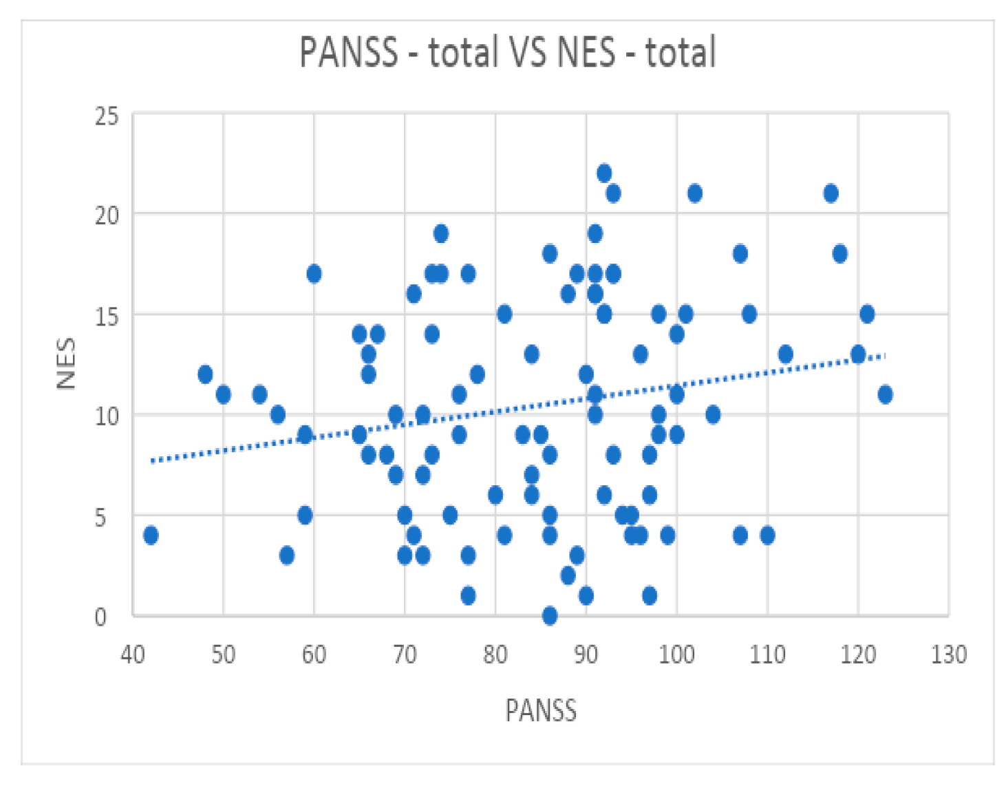
We found no correlation between the total NES score and the total PANSS score of PNS patients or between the total PANSS score of NPNS patients with the “Other” subscale of the NES. Moderate evidence of a correlation between the total PANSS score and the sensory integration, motor coordination, and sequencing of complex motor acts NES subscales in patients with PNS compared with those with NPNS was found. Additionally, there was a strong statistical correlation between the total PANSS score and the total NES score in the PNS vs. NPNS subgroups. These results are in line with previous reports [12,51,85,86] but are in contrast to others [9], in which the authors found weak correlations between the NES and PANSS scores, especially with the negative symptoms, and concluded that the NES score is a variable independent of the PANSS score. A potential explanation for the relationship between NSS and negative symptoms in schizophrenia can be sought at the level of brain architecture. Studies using imaging techniques have shown a relationship between specific brain changes, such as a decreased grey matter volume of the frontotemporal cortex and orbitofrontal cortical thinning, and the presence of negative symptoms in schizophrenia, particularly avolition and apathy, suggesting that these symptoms originate in the prefrontal network [87,88,89,90,91]. On the other hand, other studies that used brain imaging methods, which aimed to identify brain structural changes that correlate with the presence of NSS in patients with schizophrenia or a first psychotic episode, identified structural changes in similar regions [92,93,94] to those correlated with negative symptoms. Future research is thus needed to confirm whether there are certain areas or common structural changes in the brain that correlate with both the onset of negative symptoms and the pathogenesis mechanisms of NSS.
The present study had several limitations. First, patients in this study were not first-episode patients, all were undergoing neuroleptic treatment, and some were also on benzodiazepine medication at the time of evaluation. Nevertheless, as we stated previously, according to the specialised literature, neuroleptic treatment does not seem to significantly influence NSS. For studies regarding only the presence of NSS in patients on the schizophrenia spectrum, it is our opinion that it would be ideal to include drug-naïve patients. Secondly, the fact that every individual in the present study had a history of hospitalisation may indicate that the majority of them have had a more severe course of disease, making it unlikely that the present findings may be applied to other generalised contexts.
Another possible limitation of the present study was the use of the PANSS to quantify negative symptoms. The PANSS negative symptoms subscale contains certain items that are no longer considered relevant in the classification of negative symptoms. With the emergence of new scales for the analysis of negative symptoms, such as The Clinical Assessment Interview for Negative Symptoms (CAINS) and The Brief Negative Symptom Scale (BNSS) [95,96], we consider it appropriate to use them in future studies that exclusively target negative symptoms in schizophrenia. However, the present study aimed to analyse not only negative symptoms but also the entire psychopathological symptomatology. In addition, we used criteria to identify those patients with predominantly negative symptoms according to studies that validated this definition by using the PANSS. Moreover, some authors suggest only using the N1, N2, N3, N4, and N6 items when considering analyses of negative symptom by using the PANSS [97]. Additionally, future research of this kind may apply scales, such as the Calgary Depression Scale for Schizophrenia CDSS [98], to better differentiate depressive symptoms because they might affect the ratings for negative symptoms. One more significant constraint was the total number of patients included in the study. Finally, another important limitation was the cross-sectional design of the study, in contrast to a longitudinal one, that allowed us to better observe the impact of the treatment on the psychiatric and neurological symptoms, thus acting as a call for a large, multicentre longitudinal study.

5. Conclusions

This study was conducted to demonstrate the presence of neurological soft signs among patients with schizophrenia and to show the correlations between neurological soft signs and the presence of predominantly negative symptoms, treatment, and extrapyramidal side effects. By comparing patients on the basis of how negative their symptoms were in relation to their overall disease status and the presence of NSS, the current analysis was aimed to address the problem of the specificity of the effects of therapy on negative symptoms. Our research demonstrated that after schizophrenia patients were divided into PNS and NPNS subgroups, the most significant correlations were lost in the NPNS patients but preserved and even increased in the PNS patients who, for instance, had higher total NES and PANSS scores, required higher daily doses of antipsychotic drugs, and had longer cumulative hospitalised periods. In conclusion, we found that neurological soft signs were more prevalent in schizophrenic patients with predominantly negative symptoms compared with NPNS patients. This result raises the possibility that PNS patients might have a significant mediating role in the relationships between NSS and the variables examined in the present investigation.
The overall quality of life of persons suffering from schizophrenia is influenced by both the disorder and how it is treated, which results in a variety of adverse effects of antipsychotic drugs. Thus, the goals of future studies should be to improve the quality of life for patients with schizophrenia and to decrease the frequency of negative symptoms by extending the arsenal of psychiatrists with next-generation medication.
The most significant findings in relation to extrapyramidal symptoms were that they were strongly correlated with the daily dose of CPZE, the absence of anticholinergic treatment, and the type of treatment, particularly when two antipsychotics were combined. It is important to note that our current findings do not support the routine use of antipsychotic polypharmacy. Furthermore, at this point, it is impossible to say with certainty that a technique of this kind would never have an acceptable risk–benefit ratio, but more information on the possible effects of combination therapy will undoubtedly arise from future studies.
Regarding patients with predominantly negative symptoms, the present study could not objectify statistically significant differences between these patients and NPNS patients in terms of treatment; instead, an important finding was the fact that patients with PNS presented significantly more NSS than NPNS, thus strengthening the argument that negative symptoms in schizophrenia are distinguished by a unique set of neuropsychiatric features. In addition, compared with the rest of the schizophrenic patients in the present study, these patients displayed higher total scores for the PANSS.
Once again, we want to emphasise the importance of identifying the optimal antipsychotic treatment of the negative symptoms associated with schizophrenia and to reinforce the hypothesis that NSS comprise a trait that rather correlated with negative symptoms. There is a potential issue of subjectivity in the challenge of accurately judging negative symptoms in the presence of rather severe positive symptoms. Thus, it is important to distinguish between the decision to rank or treat a symptom. The issue is not one of operational criteria alone but of whether we view negative symptoms as a physiologically independent characteristic of schizophrenia, with a separate road to functional handicap and a separate opportunity for a particular treatment.

Author Contributions

Conceptualization, C.P. and A.M.C.; methodology, C.P., A.M.C., G.M., C.V. and B.E.F.; validation, A.M.C., G.M., I.R.P., O.A.M., C.V. and B.E.F.; formal analysis, C.P. and C.T.S.; investigation, C.P., C.T.S. and B.E.F.; resources, C.P., A.M.C., S.R., G.M., C.V., B.E.F. and D.M.V.; data curation, C.P., A.M.C., B.E.F. and C.T.S.; writing—original draft preparation, C.P., B.E.F., C.A.C., C.T.S. and D.M.V.; writing—review and editing, A.M.C., C.V. and G.M.; visualization, C.P. and C.T.S.; supervision, A.M.C., G.M., I.R.P., C.V. and B.E.F. All authors have read and agreed to the published version of the manuscript.

Funding

This research received no specific grant from any funding agency or commercial or not-for-profit sectors.

Institutional Review Board Statement

The study was conducted in accordance with the Declaration of Helsinki and approved by the Institutional Review Board of Prof. Dr. Alexandru Obregia Psychiatry Hospital Bucharest (approval number 89, 7 June 2022) and the hospital manager (approval number 23094 under the Operational Procedure regarding clinical studies PO-MED−83).

Informed Consent Statement

Informed consent was obtained from all the subjects involved in the study.

Data Availability Statement

All the data reported within the article are available in anonymised form upon request from the qualified investigators. The data presented in this study are available on request from the corresponding author.

Acknowledgments

This scientific material is part of a larger study of a PhD thesis currently under development by the main author, Petrescu Cristian, from the Carol Davila University of Medicine and Pharmacy, Bucharest, Faculty of Medicine, with Adela Magdalena Ciobanu as the thesis coordinator. We would like to thank all individuals who participated in this study.

Conflicts of Interest

All the authors have read and approved the final manuscript, and they confirm that there are no conflicts of interest.

Appendix A

Figure A1. Normal Q-Q Plot for NES Model Residuals.
Biomedicines 10 02939 g0a1
Figure A2. Density Plot For NES Model Residuals.
Biomedicines 10 02939 g0a2
Figure A3. Residual vs. Fitted For MSAS Model.
Biomedicines 10 02939 g0a3
Figure A4. Normal Q-Q Plot for MSAS Model Residuals.
Biomedicines 10 02939 g0a4
Figure A5. Density Plot For MSAS Model Residuals.
Biomedicines 10 02939 g0a5
Figure A6. Residual vs. Fitted For NES Model.
Biomedicines 10 02939 g0a6
Figure A7. NES vs. AGE.
Biomedicines 10 02939 g0a7
Figure A8. PANSS—total vs. NES—total.
Biomedicines 10 02939 g0a8
Figure A9. PANSS—total vs. NES—total.
Biomedicines 10 02939 g0a9
Figure A10. CPZE vs. NES.
Biomedicines 10 02939 g0a10

References

  1. Tsapakis, E.M.; Dimopoulou, T.; Tarazi, F.I. Clinical management of negative symptoms of schizophrenia: An update. Pharmacol. Ther. 2015, 153, 135–147. [Google Scholar] [CrossRef] [PubMed]
  2. Kandel, E.R.; Schwartz, J.H.; Jessel, T.M.; Siegelbaum, S. Principles of Neural Sciences, 4th ed.; McGraw—Hill Medical: New York, NY, USA, 2000. [Google Scholar]
  3. Nasrallah, H.A.; Weinberger, D.R. The Neurology of Schizophrenia; BV: Amsterdam, The Netherlands, 1986. [Google Scholar]
  4. Baldessarini, R.J. Schizophrenia. N. Engl. J. Med. 1977, 297, 988–995. [Google Scholar] [CrossRef] [PubMed]
  5. Bachmann, S.; Beck, M.; Tsai, D.-H.; Haupt, F. Neurological Soft Signs (NSS) in Census-Based, Decade-Adjusted Healthy Adults, 20 to >70 Years of Age. Front. Psychiatry 2021, 12, 670539. [Google Scholar] [CrossRef]
  6. Buchanan, R.W.; Heinrichs, D.W. The neurological evaluation scale (NES): A structured instrument for the assessment of neurological signs in schizophrenia. Psychiatry Res. 1989, 27, 335–350. [Google Scholar] [CrossRef]
  7. Chen, E.Y.-H.; Hui, C.L.-M.; Chan, R.C.-K.; Dunn, E.L.-W.; Miao, M.Y.-K.; Yeung, W.-S.; Wong, C.-K.; Chan, W.-F.; Tang, W.-N. A 3-year prospective study of neurological soft signs in first-episode schizophrenia. Schizophr. Res. 2005, 75, 45–54. [Google Scholar] [CrossRef]
  8. Dazzan, P.; Morgan, K.; Orr, K.G.; Hutchinson, G.; Chitnis, X.; Suckling, J.; Fearon, P.; Salvo, J.; McGuire, P.; Mallett, R.M.; et al. The structural brain correlates of neurological soft signs in AeSOP first-episode psychoses study. Brain 2004, 127, 143–153. [Google Scholar] [CrossRef] [Green Version]
  9. Fountoulakis, K.N.; Panagiotidis, P.; Gonda, X.; Kimiskidis, V.; Nimatoudis, I. Neurological soft signs significantly differentiate schizophrenia patients from healthy controls. Acta Neuropsychiatr. 2017, 30, 97–105. [Google Scholar] [CrossRef] [PubMed]
  10. Fountoulakis, K.N.; Panagiotidis, P.; Tegos, T.; Kimiskidis, V.; Nimatoudis, I. Paternal age and specific neurological soft signs as reliable and valid neurobiological markers for the diagnosis of patients with schizophrenia. Eur. Arch. Psychiatry Clin. Neurosci. 2022, 272, 1087–1096. [Google Scholar] [CrossRef] [PubMed]
  11. Schröder, J.; Niethammer, R.; Geider, F.-J.; Reitz, C.; Binkert, M.; Jauss, M.; Sauer, H. Neurological soft signs in schizophrenia. Schizophr. Res. 1991, 6, 25–30. [Google Scholar] [CrossRef]
  12. Bombin, I.; Arango, C.; Buchanan, R.W. Significance and Meaning of Neurological Signs in Schizophrenia: Two Decades Later. Schizophr. Bull. 2005, 31, 962–977. [Google Scholar] [CrossRef]
  13. Rossi, A.; De Cataldo, S.; Di Michele, V.; Manna, V.; Ceccoli, S.; Stratta, P.; Casacchia, M. Neurological Soft Signs in Schizophrenia. Br. J. Psychiatry 1990, 157, 735–739. [Google Scholar] [CrossRef] [PubMed]
  14. Chen, E.Y.; Lam, L.C.; Chen, R.Y.; Nguyen, D.G. Neurological Signs, Age, and Illness Duration in Schizophrenia. J. Nerv. Ment. Dis. 1996, 184, 339–348. [Google Scholar] [CrossRef] [PubMed]
  15. Merriam, A.E.; Kay, S.R.; Opler, L.A.; Kushner, S.F.; van Praag, H.M. Neurological signs and the positive-negative dimension in schizophrenia. Biol. Psychiatry 1990, 28, 181–192. [Google Scholar] [CrossRef]
  16. A Flashman, L.; Flaum, M.; Gupta, S.; Andreasen, N.C. Soft signs and neuropsychological performance in schizophrenia. Am. J. Psychiatry 1996, 153, 526–532. [Google Scholar] [CrossRef]
  17. Wong, A.H.; Voruganti, L.N.; Heslegrave, R.J.; Awad, A.G. Neurocognitive deficits and neurological signs in schizophrenia. Schizophr. Res. 1997, 23, 139–146. [Google Scholar] [CrossRef]
  18. Chan, R.C.; Xu, T.; Heinrichs, R.W.; Yu, Y.; Gong, Q.-Y. Neurological soft signs in non-psychotic first-degree relatives of patients with schizophrenia: A systematic review and meta-analysis. Neurosci. Biobehav. Rev. 2010, 34, 889–896. [Google Scholar] [CrossRef]
  19. Prikryl, R.; Ceskova, E.; Tronerova, S.; Kasparek, T.; Kucerova, H.P.; Ustohal, L.; Venclikova, S.; Vrzalova, M. Dynamics of neurological soft signs and its relationship to clinical course in patients with first-episode schizophrenia. Psychiatry Res. 2012, 200, 67–72. [Google Scholar] [CrossRef]
  20. White, C.; Stirling, J.; Hopkins, R.; Morris, J.; Montague, L.; Tantam, D.; Lewis, S. Predictors of 10-year outcome of first-episode psychosis. Psychol. Med. 2009, 39, 1447–1456. [Google Scholar] [CrossRef]
  21. Hirjak, D.; Van Harten, P.; Wolf, R. Commentary on Motor system dysfunction in the schizophrenia diathesis: Neural systems to neurotransmitters. Eur. Psychiatry 2017, 48, 49–50. [Google Scholar] [CrossRef]
  22. Hirjak, D.; Wolf, R.C.; Stieltjes, B.; Hauser, T.; Seidl, U.; Schröder, J.; Thomann, P.A. Cortical Signature of Neurological Soft Signs in Recent Onset Schizophrenia. Brain Topogr. 2013, 27, 296–306. [Google Scholar] [CrossRef]
  23. Boks, M.P.M.; Liddle, P.F.; Burgerhof, J.G.M.; Knegtering, R.; Bosch, R.-J.V.D. Neurological soft signs discriminating mood disorders from first episode schizophrenia. Acta Psychiatr. Scand. 2004, 110, 29–35. [Google Scholar] [CrossRef] [PubMed]
  24. Buchanan, R.W.; Koeppl, P.; Breier, A. Stability of neurological signs with clozapine treatment. Biol. Psychiatry 1994, 36, 198–200. [Google Scholar] [CrossRef]
  25. Scheffer, R.E. Abnormal neurological signs at the onset of psychosis. Schizophr. Res. 2004, 70, 19–26. [Google Scholar] [CrossRef]
  26. Dazzan, P.; Lloyd, T.; Morgan, K.D.; Zanelli, J.; Morgan, C.; Orr, K.; Hutchinson, G.; Fearon, P.; Allin, M.; Rifkin, L.; et al. Neurological abnormalities and cognitive ability in first-episode psychosis. Br. J. Psychiatry 2008, 193, 197–202. [Google Scholar] [CrossRef] [PubMed] [Green Version]
  27. Bachmann, S.; Degen, C.; Geider, F.J.; Schröder, J. Neurological soft signs in the clinical course of schizophrenia: Results of a meta-analysis. Front. Psychiatry 2014, 5, 185. [Google Scholar] [CrossRef] [Green Version]
  28. Lieberman, J.A.; Murray, R.M. Comprehensive Care of Schizophrenia: A Textbook of Clinical Management; London Malden MA: London, UK, 2001. [Google Scholar]
  29. Wimberley, T.; Støvring, H.; Sørensen, H.J.; Horsdal, H.T.; MacCabe, J.H.; Gasse, C. Predictors of treatment resistance in patients with schizophrenia: A population-based cohort study. Lancet Psychiatry 2016, 3, 358–366. [Google Scholar] [CrossRef] [Green Version]
  30. Jahn, T.; Hubmann, W.; Karr, M.; Mohr, F.; Schlenker, R.; Heidenreich, T.; Cohen, R.; Schröder, J. Motoric neurological soft signs and psychopathological symptoms in schizophrenic psychoses. Psychiatry Res. 2006, 142, 191–199. [Google Scholar] [CrossRef]
  31. Galderisi, S.; Maj, M.; Mucci, A.; Cassano, G.B.; Invernizzi, G.; Rossi, A.; Vita, A.; Dell’Osso, L.; Daneluzzo, E.; Pini, S. Historical, Psychopathological, Neurological, and Neuropsychological Aspects of Deficit Schizophrenia: A Multicenter Study. Am. J. Psychiatry 2002, 159, 983–990. [Google Scholar] [CrossRef] [Green Version]
  32. Mittal, V.A.; Dean, D.J.; Bernard, J.A.; Orr, J.; Pelletier-Baldelli, A.; Carol, E.E.; Gupta, T.; Turner, J.; Leopold, D.R.; Robustelli, B.L.; et al. Neurological Soft Signs Predict Abnormal Cerebellar-Thalamic Tract Development and Negative Symptoms in Adolescents at High Risk for Psychosis: A Longitudinal Perspective. Schizophr. Bull. 2013, 40, 1204–1215. [Google Scholar] [CrossRef] [Green Version]
  33. Mouchet-Mages, S.; Rodrigo, S.; Cachia, A.; Mouaffak, F.; Olie, J.P.; Meder, J.F.; Oppenheim, C.; Krebs, M.O. Correlations of cerebello-thalamo-prefrontal structure and neurological soft signs in patients with first-episode psychosis. Acta Psychiatr. Scand. 2011, 123, 451–458. [Google Scholar] [CrossRef]
  34. Rubin, P.; Vorstrup, S.; Hemmingsen, R.; Andersen, H.S.; Bendsen, B.B.; Strømsø, N.; Larsen, J.K.; Bolwig, T.G. Neurological abnormalities in patients with schizophrenia or schizophreniform disorder at first admission to hospital: Correlations with computerized tomography and regional cerebral blood flow findings. Acta Psychiatr. Scand. 1994, 90, 385–390. [Google Scholar] [CrossRef] [PubMed]
  35. Ismail, B.T.; Cantor-Graae, E.; Cardenal, S.; McNeil, T.F. Neurological abnormalities in schizophrenia: Clinical, etiological and demographic correlates. Schizophr. Res. 1998, 30, 229–238. [Google Scholar] [CrossRef]
  36. Giordano, G.M.; Caporusso, E.; Pezzella, P.; Galderisi, S. Updated perspectives on the clinical significance of negative symptoms in patients with schizophrenia. Expert Rev. Neurother 2022, 22, 541–555. [Google Scholar] [CrossRef] [PubMed]
  37. Correll, C.U.; Schooler, N.R. Negative Symptoms in Schizophrenia: A Review and Clinical Guide for Recognition, Assessment, and Treatment. Neuropsychiatr. Dis. Treat. 2020, 16, 519–534. [Google Scholar] [CrossRef] [PubMed] [Green Version]
  38. Laughren, T.; Levin, R. Food and Drug Administration Perspective on Negative Symptoms in Schizophrenia as a Target for a Drug Treatment Claim. Schizophr. Bull. 2005, 32, 220–222. [Google Scholar] [CrossRef] [PubMed] [Green Version]
  39. Laughren, T.; Levin, R. Food and Drug Administration Commentary on Methodological Issues in Negative Symptom Trials. Schizophr. Bull. 2011, 37, 255–256. [Google Scholar] [CrossRef] [PubMed] [Green Version]
  40. European Medicines Agency. Guideline on Clinical Investigation of Medicinal Products, Including Depot Preparations in the Treatment of Schizophrenia; European Medicines Agency: London, UK, 2012. [Google Scholar]
  41. Németh, G.; Laszlovszky, I.; Czobor, P.; Szalai, E.; Szatmári, B.; Harsányi, J.; Barabássy, Á.; Debelle, M.; Durgam, S.; Bitter, I.; et al. Cariprazine versus risperidone monotherapy for treatment of predominant negative symptoms in patients with schizophrenia: A randomised, double-blind, controlled trial. Lancet 2017, 389, 1103–1113. [Google Scholar] [CrossRef]
  42. Akinsulore, A.; Mapayi, B.M.; Aloba, O.O.; Oloniniyi, I.; Fatoye, F.O.; Makanjuola, R.O.A. Disability assessment as an outcome measure: A comparative study of Nigerian outpatients with schizophrenia and healthy control. Ann. Gen. Psychiatry 2015, 14, 40. [Google Scholar] [CrossRef] [Green Version]
  43. Schröder, J.; Tittel, A.; Stockert, A.; Karr, M. Memory deficits in subsyndromes of chronic schizophrenia. Schizophr. Res. 1996, 21, 19–26. [Google Scholar] [CrossRef]
  44. Whitty, P.; Clarke, M.; McTigue, O.; Browne, S.; Gervin, M.; Kamali, M.; Lane, A.; Kinsella, A.; Waddington, J.; Larkin, C.; et al. Diagnostic specificity and predictors of neurological soft signs in schizophrenia, bipolar disorder and other psychoses over the first 4 years of illness. Schizophr. Res. 2006, 86, 110–117. [Google Scholar] [CrossRef]
  45. Yazıcı, A.H.; Demir, B.; Yazıcı, K.M.; Gou, A.; Göğüş, A. Neurological soft signs in schizophrenic patients and their nonpsychotic siblings. Schizophr. Res. 2002, 58, 241–2466. [Google Scholar] [CrossRef]
  46. Chan, R.C.K.; Geng, F.-L.; Lui, S.S.Y.; Wang, Y.; Ho, K.K.Y.; Hung, K.S.Y.; Gur, R.E.; Gur, R.C.; Cheung, E.F.C. Course of neurological soft signs in first-episode schizophrenia: Relationship with negative symptoms and cognitive performances. Sci. Rep. 2015, 5, 11053. [Google Scholar] [CrossRef] [PubMed] [Green Version]
  47. Ciobanu, A.; Dionisie, V.; Neagu, C.; Bolog, O.; Riga, S.; Popa-Velea, O. Psychopharmacological Treatment, Intraocular Pressure and the Risk of Glaucoma: A Review of Literature. J. Clin. Med. 2021, 10, 2947. [Google Scholar] [CrossRef]
  48. Thomann, P.A.; Roebel, M.; Dos Santos, V.; Bachmann, S.; Essig, M.; Schröder, J. Cerebellar substructures and neurological soft signs in first-episode schizophrenia. Psychiatry Res. Neuroimaging 2009, 173, 83–87. [Google Scholar] [CrossRef]
  49. Mittal, V.A.; Hasenkamp, W.; Sanfilipo, M.; Wieland, S.; Angrist, B.; Rotrosen, J.; Duncan, E.J. Relation of neurological soft signs to psychiatric symptoms in schizophrenia. Schizophr. Res. 2007, 94, 37–44. [Google Scholar] [CrossRef] [PubMed]
  50. Bersani, G.; Gherardelli, S.; Clemente, R.; Di Giannantonio, M.; Grilli, A.; Conti, C.M.V.; Exton, M.S.; Conti, P.; Doyle, R.; Pancheri, P. Neurologic Soft Signs in Schizophrenic Patients Treated With Conventional and Atypical Antipsychotics. J. Clin. Psychopharmacol. 2005, 25, 372–375. [Google Scholar] [CrossRef] [PubMed]
  51. de Bartolomeis, A.; Prinzivalli, E.; Callovini, G.; D’Ambrosio, L.; Altavilla, B.; Avagliano, C.; Iasevoli, F. Treatment resistant schizophrenia and neurological soft signs may converge on the same pathology: Evidence from explanatory analysis on clinical, psychopathological, and cognitive variables. Prog. Neuro-Psychopharmacol. Biol. Psychiatry 2018, 81, 356–366. [Google Scholar] [CrossRef] [PubMed]
  52. Gupta, S.; Rajaprabhakaran, R.; Arndt, S.; Flaum, M.; Andreasen, N.C. Premorbid adjustment as a predictor of phenomenological and neurobiological indices in schizophrenia. Schizophr. Res. 1995, 16, 189–197. [Google Scholar] [CrossRef]
  53. Smith, R.C.; Hussain, M.I.; Chowdhury, S.A.; Stearns, A. Stability of Neurological Soft Signs in Chronically Hospitalized Schizophrenic Patients. J. Neuropsychiatry Clin. Neurosci. 1999, 11, 91–96. [Google Scholar] [CrossRef]
  54. Browne, S.; Clarke, M.; Gervin, M.; Lane, A.; Waddington, J.L.; Larkin, C.; O’Callaghan, E. Determinants of neurological dysfunction in first episode schizophrenia. Psychol. Med. 2000, 30, 1433–1441. [Google Scholar] [CrossRef]
  55. Venkatasubramanian, G.; Latha, V.; Gangadhar, B.N.; Janakiramaiah, N.; Subbakrishna, D.K.; Jayakumar, P.N.; Keshavan, M.S. Neurological soft signs in never-treated schizophrenia. Acta Psychiatr. Scand. 2003, 108, 144–146. [Google Scholar] [CrossRef] [PubMed]
  56. Gupta, S.; Andreasen, N.C.; Arndt, S.; Flaum, M.; Schultz, S.K.; Hubbard, W.C.; Smith, M. Neurological soft signs in neuroleptic-naive and neuroleptic-treated schizophrenic patients and in normal comparison subjects. Am. J. Psychiatry 1995, 152, 191–196. [Google Scholar] [CrossRef] [PubMed] [Green Version]
  57. Caligiuri, M.P.; Lohr, J.B. A disturbance in the control of muscle force in neuroleptic-naive schizophrenic patients. Biol. Psychiatry 1994, 35, 104–111. [Google Scholar] [CrossRef]
  58. Schröder, J.; Silvestri, S.; Bubeck, B.; Karr, M.; Demisch, S.; Scherrer, S.; Geider, F.J.; Sauer, H. D2 Dopamine Receptor Up-Regulation, Treatment Response, Neurological Soft Signs, and Extrapyramidal Side Effects in Schizophrenia: A Follow-Up Study with 123I-Iodobenzamide Single Photon Emission Computed Tomography in the Drug-Naive State and after Neuroleptic Treatment. Biol. Psychiatry 1998, 43, 660–665. [Google Scholar] [CrossRef] [PubMed]
  59. Sykes, D.; Moore, H.; Stott, L.; Holliday, N.; Javitch, J.; Lane, J.R.; Charlton, S.J. Extrapyramidal side effects of antipsychotics are linked to their association kinetics at dopamine D2 receptors. Nat. Commun. 2017, 8, 763. [Google Scholar] [CrossRef] [Green Version]
  60. Koning, J.P.; Tenback, D.E.; Van Os, J.; Aleman, A.; Kahn, R.S.; Van Harten, P.N. Dyskinesia and Parkinsonism in Antipsychotic-Naive Patients with Schizophrenia, First-Degree Relatives and Healthy Controls: A Meta-analysis. Schizophr. Bull. 2010, 36, 723–731. [Google Scholar] [CrossRef] [Green Version]
  61. López-Muñoz, F.; Alamo, C.; Cuenca, E.; Shen, W.; Clervoy, P.; Rubio, G. History of the Discovery and Clinical Introduction of Chlorpromazine. Ann. Clin. Psychiatry 2005, 17, 113–135. [Google Scholar] [CrossRef]
  62. Crilly, J. The history of clozapine and its emergence in the US market A Review and analysis. Hist. Psychiatry 2007, 18, 39–60. [Google Scholar] [CrossRef]
  63. Bachmann, S.; Schröder, J. Neurological Soft Signs in Schizophrenia: An Update on the State- versus Trait-Perspective. Front. Psychiatry 2017, 8, 272. [Google Scholar] [CrossRef]
  64. American Psychiatric Association. Diagnostic and Statistical Manual of Mental Disorders, 5th ed.; American Psychiatric Association: Washington, DC, USA, 2013. [Google Scholar]
  65. Davis, J.M. Dose equivalence of the anti-psychotic drugs. J. Psychiatr. Res. 1974, 11, 65–69. [Google Scholar] [CrossRef]
  66. Venkatasubramanian, G.; Danivas, V. Current perspectives on chlorpromazine equivalents: Comparing apples and oranges! Indian J. Psychiatry 2013, 55, 207–208. [Google Scholar] [CrossRef] [PubMed]
  67. Leucht, S.; Samara, M.; Heres, S.; Davis, J.M. Dose Equivalents for Antipsychotic Drugs: The DDD Method: Table 1. Schizophr. Bull. 2016, 42, S90–S94. [Google Scholar] [CrossRef] [PubMed] [Green Version]
  68. Woods, S.W. Chlorpromazine Equivalent Doses for the Newer Atypical Antipsychotics. J. Clin. Psychiatry 2003, 64, 663–667. [Google Scholar] [CrossRef] [PubMed]
  69. Kay, S.R.; Fiszbein, A.; Opler, L.A. The Positive and Negative Syndrome Scale (PANSS) for Schizophrenia. Schizophr. Bull. 1987, 13, 261–276. [Google Scholar] [CrossRef]
  70. Stauffer, V.L.; Song, G.; Kinon, B.J.; Ascher-Svanum, H.; Chen, L.; Feldman, P.D.; Conley, R.R. Responses to antipsychotic therapy among patients with schizophrenia or schizoaffective disorder and either predominant or prominent negative symptoms. Schizophr. Res. 2012, 134, 195–201. [Google Scholar] [CrossRef] [PubMed]
  71. Olié, J.-P.; Spina, E.; Murray, S.; Yang, R. Ziprasidone and amisulpride effectively treat negative symptoms of schizophrenia: Results of a 12-week, double-blind study. Int. Clin. Psychopharmacol. 2006, 21, 143–151. [Google Scholar] [CrossRef] [PubMed] [Green Version]
  72. Riedel, M.; Müller, N.; Strassnig, M.; Spellmann, I.; Engel, R.R.; Musil, R.; Dehning, S.; Douhet, A.; Schwarz, M.J.; Möller, H. Quetiapine has equivalent efficacy and superior tolerability to risperidone in the treatment of schizophrenia with predominantly negative symptoms. Eur. Arch. Psychiatry Clin. Neurosci. 2005, 255, 432–437. [Google Scholar] [CrossRef]
  73. Rabinowitz, J.; Werbeloff, N.; Caers, I.; Mandel, F.S.; Stauffer, V.; Menard, F.; Kinon, B.J.; Kapur, S. Negative symptoms in schizophrenia—The remarkable impact of inclusion definitions in clinical trials and their consequences. Schizophr. Res. 2013, 150, 334–338. [Google Scholar] [CrossRef]
  74. Guy, W. ECDEU Assessment Manual for Psychopharmacology; Revised; US Department of Health, Education, and Welfare Publication (ADM), National Institute of Mental Health: Rockville, MD, USA, 1976; pp. 76–338. [Google Scholar]
  75. Leucht, S.; Barabássy, Á.; Laszlovszky, I.; Szatmári, B.; Acsai, K.; Szalai, E.; Harsányi, J.; Earley, W.; Németh, G. Linking PANSS negative symptom scores with the Clinical Global Impressions Scale: Understanding negative symptom scores in schizophrenia. Neuropsychopharmacology 2019, 44, 1589–1596. [Google Scholar] [CrossRef] [Green Version]
  76. Simpson, G.M.; Angus, J.W.S. Rating Scale for Extrapyramidal Side Effects. Acta Psychiatr. Scand. 1970, 45, 11–19. [Google Scholar] [CrossRef]
  77. Hawley, C.; Fineberg, N.; Roberts, A.; Baldwin, D.; Sahadevan, A.; Sharman, V. The use of the Simpson Angus Scale for the assessment of movement disorder: A training guide. Int. J. Psychiatry Clin. Pr. 2003, 7, 349–2257. [Google Scholar] [CrossRef] [PubMed]
  78. Merhej, G.; Hallit, S.; Haddad, C.; Hachem, D.; Haddad, G. Neurological soft signs in schizophrenia: Gender differences and promising suggestions. J. Psychopathol. 2017, 23, 74–78. [Google Scholar]
  79. Herold, C.J.; Duval, C.Z.; Lässer, M.M.; Schröder, J. Neurological soft signs (NSS) and cognitive impairment in chronic schizophrenia. Schizophr. Res. Cogn. 2019, 16, 17–24. [Google Scholar] [CrossRef] [PubMed]
  80. Miller, D.D.; McEvoy, J.P.; Davis, S.M.; Caroff, S.N.; Saltz, B.L.; Chakos, M.H.; Swartz, M.S.; Keefe, R.S.; Rosenheck, R.A.; Stroup, T.S.; et al. Clinical correlates of tardive dyskinesia in schizophrenia: Baseline data from the CATIE schizophrenia trial. Schizophr. Res. 2005, 80, 33–43. [Google Scholar] [CrossRef]
  81. Misdrahi, D.; Tessier, A.; Daubigney, A.; Meissner, W.G.; Schurhoff, F.; Boyer, L.; Godin, O.; Bulzacka, E.; Aouizerate, B.; Andrianarisoa, M.; et al. Prevalence of and Risk Factors for Extrapyramidal Side Effects of Antipsychotics. J. Clin. Psychiatry 2019, 80, 7055. [Google Scholar] [CrossRef]
  82. Leucht, S.; Corves, C.; Arbter, D.; Engel, R.R.; Li, C.; Davis, J.M. Second-generation versus first-generation antipsychotic drugs for schizophrenia: A meta-analysis. Lancet 2009, 373, 31–41. [Google Scholar] [CrossRef]
  83. Minzenberg, M.J.; Poole, J.H.; Benton, C.; Vinogradov, S. Association of Anticholinergic Load With Impairment of Complex Attention and Memory in Schizophrenia. Am. J. Psychiatry 2004, 161, 116–124. [Google Scholar] [CrossRef] [Green Version]
  84. Ogino, S.; Miyamoto, S.; Miyake, N.; Yamaguchi, N. Benefits and limits of anticholinergic use in schizophrenia: Focusing on its effect on cognitive function. Psychiatry Clin. Neurosci. 2014, 68, 37–49. [Google Scholar] [CrossRef]
  85. Prikryl, R.; Ceskova, E.; Kasparek, T.; Kucerova, H. Neurological soft signs, clinical symptoms and treatment reactivity in patients suffering from first episode schizophrenia. J. Psychiatr. Res. 2006, 40, 141–146. [Google Scholar] [CrossRef]
  86. Cvetić, T.; Vuković, O.; Britvić, D.; Ivković, M.; Dukić-Dejanović, S.; Lecić-Tosevski, D. Comparative analysis of soft neurological signs in positive and negative subtype of schizophrenia. Psychiatr. Danub. 2009, 21, 174–178. [Google Scholar]
  87. Bègue, I.; Kaiser, S.; Kirschner, M. Pathophysiology of negative symptom dimensions of schizophrenia—Current developments and implications for treatment. Neurosci. Biobehav. Rev. 2020, 116, 74–88. [Google Scholar] [CrossRef] [PubMed]
  88. Roth, R.M.; Garlinghouse, M.A.; Flashman, L.A.; Koven, N.S.; Pendergrass, J.C.; Ford, J.C.; McAllister, T.W.; Saykin, A.J. Apathy Is Associated with Ventral Striatum Volume in Schizophrenia Spectrum Disorder. J. Neuropsychiatry Clin. Neurosci. 2016, 28, 191–194. [Google Scholar] [CrossRef] [PubMed]
  89. Caravaggio, F.; Fervaha, G.; Iwata, Y.; Plitman, E.; Chung, J.K.; Nakajima, S.; Mar, W.; Gerretsen, P.; Kim, J.; Chakravarty, M.M.; et al. Amotivation is associated with smaller ventral striatum volumes in older patients with schizophrenia. Int. J. Geriatr. Psychiatry 2017, 33, 523–530. [Google Scholar] [CrossRef] [PubMed]
  90. Giordano, G.M.; Stanziano, M.; Papa, M.; Mucci, A.; Prinster, A.; Soricelli, A.; Galderisi, S. Functional connectivity of the ventral tegmental area and avolition in subjects with schizophrenia: A resting state functional MRI study. Eur. Neuropsychopharmacol. 2018, 28, 589–602. [Google Scholar] [CrossRef] [PubMed]
  91. Walton, E.; Hibar, D.P.; Van Erp, T.G.M.; Potkin, S.G.; Roiz-Santiañez, R.; Crespo-Facorro, B.; Suarez-Pinilla, P.; Van Haren, N.E.M.; De Zwarte, S.M.C.; Kahn, R.S.; et al. Prefrontal cortical thinning links to negative symptoms in schizophrenia via the ENIGMA consortium. Psychol. Med. 2017, 48, 82–94. [Google Scholar] [CrossRef]
  92. Kong, L.; Bachmann, S.; Thomann, P.A.; Essig, M.; Schröder, J. Neurological soft signs and gray matter changes: A longitudinal analysis in first-episode schizophrenia. Schizophr. Res. 2012, 134, 27–32. [Google Scholar] [CrossRef]
  93. Kong, L.; Herold, C.J.; Lässer, M.M.; Schmid, L.A.; Hirjak, D.; Thomann, P.A.; Essig, M.; Schröder, J. Association of Cortical Thickness and Neurological Soft Signs in Patients with Chronic Schizophrenia and Healthy Controls. Neuropsychobiology 2015, 71, 225–233. [Google Scholar] [CrossRef]
  94. Hirjak, D.; Kubera, K.M.; Wolf, R.C.; Thomann, A.K.; Hell, S.K.; Seidl, U.; Thomann, P.A. Local brain gyrification as a marker of neurological soft signs in schizophrenia. Behav. Brain Res. 2015, 292, 19–25. [Google Scholar] [CrossRef]
  95. Kring, A.M.; Gur, R.E.; Blanchard, J.J.; Horan, W.P.; Reise, S.P. The Clinical Assessment Interview for Negative Symptoms (CAINS): Final Development and Validation. Am. J. Psychiatry 2013, 170, 165–172. [Google Scholar] [CrossRef]
  96. Kirkpatrick, B.; Strauss, G.P.; Nguyen, L.; Fischer, B.A.; Daniel, D.G.; Cienfuegos, A.; Marder, S.R. The Brief Negative Symptom Scale: Psychometric Properties. Schizophr. Bull. 2010, 37, 300–305. [Google Scholar] [CrossRef] [Green Version]
  97. Galderisi, S.; Mucci, A.; Dollfus, S.; Nordentoft, M.; Falkai, P.; Kaiser, S.; Giordano, G.M.; Vandevelde, A.; Nielsen, M.; Glenthøj, L.B.; et al. EPA guidance on assessment of negative symptoms in schizophrenia. Eur. Psychiatry 2021, 64, e23. [Google Scholar] [CrossRef] [PubMed]
  98. Addington, D.; Addington, J.; Schissel, B. A depression rating scale for schizophrenics. Schizophr. Res. 1990, 3, 247–251. [Google Scholar] [CrossRef]
Table 1. Patient’s symptomatology corelated with CGI scores.
CGI Score n
1 = Normal, not at all ill 0
2 = Borderline mentally ill2
3 = Mildly ill 13
4 = Moderately ill24
5 = Markedly ill45
6 = Severely ill15
7 = Among the most extremely ill patients0
Total99
CGI, Clinical Global Impression; n, number of patients.
Table 2. Descriptive statistical analysis of the studied variables.
VariableGlobal
(n = 99)
Sex
F56 (56.6%)
M43 (43.4%)
Age
Mean (SD)30.6 (10.4)
Median (Min, Max)26.0 (18.0, 65.0)
Environment
R12 (12.1%)
U87 (87.9%)
Years of education
Mean (SD)12.4 (1.94)
Median (Min, Max)12.0 (8.00, 18.0)
Economic status
Employed 11 (11.1%)
Retired 44 (44.4%)
Unemployed 37 (37.4%)
Student 7 (7.1%)
Age of onset
Mean (SD)22.5 (4.67)
Median (Min, Max)21.0 (17.0, 40.0)
Duration of illness
Mean (SD)8.15 (7.78)
Median (Min, Max)5.00 (1.00, 35.0)
Age at first treatment
Mean (SD)22.8 (4.72)
Median (Min, Max)21.0 (18.0, 40.0)
Missing3 (3.0%)
Age at first hospitalisation
Mean (SD)23.3 (5.24)
Median (Min, Max)22.0 (18.0, 40.0)
N/A2 (2.0%)
Number of hospitalisations
Mean (SD)5.27 (4.21)
Median [Min, Max]4.00 (1.00, 25.0)
Cumulative hospitalised period
Mean (SD)3.98 (3.05)
Median (Min, Max)3.50 (0.500, 15.0)
PANSS, CGI correlation
Mean (SD)4.59 (0.969)
Median (Min, Max)5.00 (2.00, 6.00)
PANSS P
Mean (SD)21.6 (6.06)
Median (Min, Max)22.0 (8.00, 35.0)
PANSS N
Mean (SD)21.4 (6.32)
Median (Min, Max)21.0 (8.00, 39.0)
PANSS, general
Mean (SD)41.8 (8.66)
Median (Min, Max)41.0 (20.0, 65.0)
PANSS, total
Mean (SD)84.8 (16.8)
Median (Min, Max)86.0 (42.0, 123)
PANSS, predominantly negative
Yes39 (39.4%)
No60 (60.6%)
Type of treatment
TA4 (4.0%)
AA86 (86.9%)
2 AA9 (9.1%)
Daily dose of CPZE
Mean (SD)424 (219)
Median (Min, Max)400 (75.0, 1500)
Anticholinergic treatment
Yes24 (24.2%)
No75 (75.8%)
NES, sensory integration
Mean (SD)1.67 (1.52)
Median (Min, Max)2.00 (0, 7.00)
NES, motor coordination
Mean (SD)2.08 (1.60)
Median (Min, Max)2.00 (0, 8.00)
NES, sequencing of complex motor acts
Mean (SD)3.12 (1.98)
Median (Min, Max)3.00 (0, 8.00)
NES, other
Mean (SD)3.57 (2.62)
Median (Min, Max)3.00 (0, 10.0)
NES, total
Mean (SD)10.5 (5.50)
Median (Min, Max)10.0 (0, 22.0)
SAS
Mean (SD)3.04 (2.01)
Median (Min, Max)3.00 (0, 9.00)
F: females; M: males; R: rural; U: urban; PANSS: Positive and Negative Syndrome Scale; CGI: Clinical Global Impression; PANSS P: PANSS positive symptoms; PANSS N: PANSS negative symptoms; NES: Neurological Evaluation Scale; SAS: Simpson–Angus Scale; CPZE: chlorpromazine equivalent; TA: typical antipsychotic; AA: atypical antipsychotic; 2 AA: combination of two atypical antipsychotics.
Table 3. Simple univariate linear regression.
PredictorsnBeta (95% CI) p-Value
Sex99
F
M 3.3 (1.3 to 5.4)0.002
Age990.19 (0.09 to 0.29)<0.001
Environment99
R
U −2.5 (−5.8 to 0.78)0.138
Marital status99
With a partner
No partner 0.01 (−2.4 to 2.5)0.991
Years of education99−0.41 (−1.0 to 0.15)0.157
Economic status99
Employed
Retired 6.8 (3.4 to 10)<0.001
Unemployed 3.3 (−0.11 to 6.7)0.061
Student 1.9 (−2.9 to 6.7)0.436
Age of onset 990.23 (0.01 to 0.46)0.048
Duration of illness (years)990.25 (0.12 to 0.38)<0.001
Age at first treatment960.20 (−0.03 to 0.43)0.098
Age at first hospitalisation 970.20 (−0.01 to 0.40)0.069
Number of hospitalizations990.54 (0.30 to 0.77)<0.001
Cumulative hospitalised period (months)990.80 (0.47 to 1.1)<0.001
Daily dose of CPZE990.001 (−0.003 to 0.007)0.438
Anticholinergic99
Yes
No −0.45 (−3.0 to 2.1)0.732
Dominance99
L
R −5.2 (−8.5 to −1.9)0.003
F: females; M: males; R: rural; U: urban; CPZE: chlorpromazine equivalent; L: left; R: right. Average values, including standard deviations from the total score and the respective subscores of the NES at the time of hospitalisation, are presented.
Table 4. Paired two-sample t-test.
Paired Two-Sample t-Test for MeansPaired Two-Sample t-Test for Means
PANSS negativeTotal NES Total PANSSTotal NES
Mean21.4210.45Mean84.8410.45
Variance39.9630.27Variance281.41830.27
r0.33 r0.19
PANSS: Positive and Negative Syndrome Scale; NES: Neurological Evaluation Scale; r: Pearson’s correlation coefficient.
Table 5. Simple univariate linear regression.
PredictorsnBeta (95% CI) 1p-Value
Sex99
F
M 0.75 (−0.04 to 1.5)0.066
Age990.02 (−0.02 to 0.06)0.385
Environment99
R
U −0.71 (−1.9 to 0.50)0.253
Marital status99
With a partner
No partner 0.21 (−0.69 to 1.1)0.649
Years of education99−0.19 (−0.39 to 0.01)0.072
Economic status99
Employed
Retired 1.0 (−0.37 to 2.3)0.16
Unemployed 0.24 (−1.1 to 1.6)0.729
Student −0.26 (−2.2 to 1.6)0.789
Age at onset 99−0.03 (−0.11 to 0.06)0.544
Duration of illness (years)990.04 (−0.01 to 0.09)0.127
Age at first treatment96−0.02 (−0.10 to 0.06)0.645
Age at first hospitalisation970.00 (−0.08 to 0.08)0.983
Number of hospitalizations990.15 (0.06 to 0.24)0.001
Cumulative hospitalised period (months)990.21 (0.09 to 0.34)0.001
Type of treatment99
TA
AA −3.9 (−5.8 to −2.0)<0.001
2 AA −3.6 (−5.9 to −1.4)0.002
Daily dose of CPZE990.01 (0.00 to 0.01)<0.001
Anticholinergic99
Yes
No −1.2 (−2.1 to −0.31)0.010
Dominance99
L
R 0.05 (−1.2 to 1.3)0.944
F: females; M: males; R: rural; U: urban; CPZE: chlorpromazine equivalent; TA: typical antipsychotic; AA: atypical antipsychotic; 2 AA: combination of two atypical antipsychotics. 1 CI = confidence interval.
Table 6. Multiple univariate linear regression forward selection of the final model.
PredictorsBeta (95% CI) 1p-Value
Daily dose of CPZE0.004 (0.003 to 0.006)<0.001
Number of hospitalizations0.16 (0.09 to 0.23)<0.001
Type of treatment
TA
AA−2.1 (−3.8 to −0.51)0.011
2 AA−2.7 (−4.6 to −0.89)0.004
CPZE: chlorpromazine equivalent; TA: typical antipsychotic; AA: atypical antipsychotic; 2 AA: combination of two atypical antipsychotics. 1 CI = confidence interval.
Table 7. Pearson’s correlation matrix for the NES, daily dose of CPZE, and SAS.
NES-SINES-MCNES-SCMANES-OthersNES-TotalSASCLPZE MG
NES-SI1
NES-MC0.341
NES-SCMA0.390.341
NES-Other0.400.250.281
NES-Total0.710.630.70.761
SAS0.330.170.270.260.371
CPZE 0.030.150.15−0.070.070.571
NES: Neurological Evaluation Scale; NES-SI: NES sensory integration; NES-MC: NES motor coordination; NES-SCMA: NES sequencing of complex motor acts; SAS: Simpson–Angus Scale; CPZE: chlorpromazine equivalent (mg).
Table 8. Patients with predominantly negative symptoms (PNS) vs. non-predominantly negative symptoms (NPNS).
VariablesPNS
n = 39
NPNS
n = 60
p-Value 1
Ex, n (%) <0.001
F13 (33)43 (72)
M26 (67)17 (28)
Age, mean (SD)29.31 (8.72)31.47 (11.30)0.29
Environment, n (%) >0.99
R5 (13)7 (12)
U34 (87)53 (88)
Marital status, n (%) 0.093
With a partner (actual or historical)7 (18)20 (33)
No partner32 (82)40 (67)
Years of schooling (number of years of education), mean (SD)12.38 (1.90)12.42 (1.99)0.94
Economic status, n (%) 0.27
Employed2 (5.1)9 (15)
Retired16 (41)28 (47)
Unemployed17 (44)20 (33)
Student4 (10)3 (5.0)
Age at onset, mean (SD)21.85 (3.62)22.87 (5.23)0.25
Duration of illness (years), mean (SD)7.46 (7.22)8.60 (8.15)0.47
Age at first treatment, mean (SD)22.08 (3.73)23.29 (5.25)0.19
N/A12
Age at first hospitalisation, mean (SD)22.67 (4.37)23.72 (5.75)0.31
N/A02
Number of pre-evaluation (pre-diagnosis) hospitalizations, mean (SD)5.87 (4.73)4.88 (3.83)0.28
Cumulative hospitalised period, mean (SD)4.63 (3.73)3.56 (2.45)0.12
PANSS–CGI correlation, mean (SD)4.92 (0.70)4.37 (1.07)0.002
General PANSS, mean (SD)43.79 (8.08)40.48 (8.84)0.058
PANSS Negative, Mean (SD)26.15 (4.25)18.35 (5.51)<0.001
PANSS Positive, Mean (SD)20.44 (4.91)22.42 (6.63)0.091
Total PANSS, mean (SD)90.38 (13.90)81.25 (17.60)0.005
Daily dose of CPZE, mean (SD)446.79 (179.84)409.58 (241.99)0.38
SAS mean (SD)2.98 (1.97)3.13 (2.10)0.73
Anticholinergic, n (%) 0.83
Yes9 (23)15 (25)
No30 (77)45 (75)
Dominance, n (%) 0.11
L7 (18)4 (6.7)
R32 (82)56 (93)
NES—motor coordination, mean (SD)2.59 (1.71)1.75 (1.43)0.013
NES—sensory integration, mean (SD)2.13 (1.82)1.37 (1.21)0.024
NES—sequencing of complex motor acts, mean (SD)3.67 (2.07)2.77 (1.84)0.03
Other NES, mean (SD)4.05 (2.66)3.25 (2.57)0.14
Total NES, mean (SD)12.49 (5.35)9.13 (5.23)0.003
F: females; M: males; R: rural; U: urban; PANSS: Positive and Negative Syndrome Scale; PANSS P: positive PANSS symptoms; PANSS N: negative PANSS symptoms; NES: Neurological Evaluation Scale; CPZE: chlorpromazine equivalent; CGI: Clinical Global Impression. 1 Pearson’s chi-squared test, Welch’s two-sample t-test, and Fisher’s exact test.
Table 9. Pearson correlation matrix for PNS.
AgeYOEAAODOIAFTAFHNOHCHPPANSS PPANSS NPANSS, generalPANSS, totalCPZENES-SINES-MCNES-SCMANES, otherNES, totalSAS
Age1
YOE−0.021
AAO0.590.161
DOI (years)0.91−0.10.21
AFT (years)0.570.140.970.181
AFH (years)0.650.130.890.330.981
NOH0.5400.110.60.050.271
CHP0.44−0.10.030.51−0.010.170.921
PANSS P−0.32−0.22−0.21−0.28−0.25−0.24−0.090.011
PANSS N−0.37−0.35−0.19−0.36−0.19−0.14−0.090.090.691
PANSS, general0.2−0.270.090.20.030.040.10.120.460.291
PANSS, total−0.1−0.35−0.07−0.09−0.12−0.10.020.110.830.710.831
DD CPZE0.1−0.03−0.080.17−0.060.040.280.3−0.130.13−0.11−0.071
NES-SI0.17−0.220.180.110.170.240.340.330.130.3−0.040.110.091
NES-MC0.020.040.11−0.020.030.120.120.120.090.20.10.150.080.311
NES-SCMA0.2−0.10.110.190.140.070.270.380.010.090.310.210.240.220.161
NES, other0.54−0.210.240.530.270.260.250.230.060.010.270.180.020.280.070.221
NES, total0.43−0.180.270.380.270.290.410.420.10.20.280.260.170.670.520.620.71
SAS0.38−0.060.060.430.010.20.410.4−0.12−0.010−0.050.690.40.130.310.280.441
YOE: years of education; AAO: age at onset; DOI: duration of illness; AFT: age at first treatment; AFH: age at first hospitalisation; NOH: number of pre-evaluation (pre-diagnosis) hospitalizations; CHP: cumulative hospitalised period; PANSS: Positive and Negative Syndrome Scale; PANSS P: positive PANSS symptoms; PANSS N: negative PANSS symptoms; NES: Neurological Evaluation Scale; NES-SI: NES sensory integration; NES-MC: NES motor coordination; NES-SCMA:NES sequencing of complex motor acts; SAS: Simpson–Angus Scale.
Publisher’s Note: MDPI stays neutral with regard to jurisdictional claims in published maps and institutional affiliations.

