Article

Bacterial Pulmonary Co-Infections on ICU Admission: Comparison in Patients with SARS-CoV-2 and Influenza Acute Respiratory Failure: A Multicentre Cohort Study

1
Service de Pneumologie, CHU Gabriel Montpied, 63000 Clermont-Ferrand, France
2
Unité Mixte de Recherche (UMR) 1137, IAME, Université Paris Cité, 75018 Paris, France
3
Infection Control Program and WHO Collaborating Centre on Patient Safety, Faculty of Medicine, University of Geneva Hospitals, 1205 Geneva, Switzerland
4
Polyvalent Intensive Care Unit, Hôpital Foch, 92150 Suresnes, France
5
General Intensive Care Unit, Sud Essonne Hospital, 91150 Etampes, France
6
Intensive Care Unit, University Hospital Avicenne, AP-HP, 93000 Bobigny, France
7
Polyvalent Intensive Care Unit, André Mignot Hospital, 78150 Le Chesnay, France
8
Medical Intensive Care Unit, University Hospital of Reims, 51100 Reims, France
9
Medical Intensive Care Unit, University Hospital of Nantes, 44000 Nantes, France
10
Medical and Surgical Intensive Care, Montfermeil Hospital, 93370 Montfermeil, France
11
Medical Intensive Care Unit, University Hospital Grenoble-Alpes, 38000 Grenoble, France
12
Medical and Infectious Diseases Intensive Care Unit, Bichat Hospital, AP-HP, 75018 Paris, France
13
Medical Intensive Care Unit, University Hospital Gabriel Montpied, 63000 Clermont-Ferrand, France
14
Unité de Nutrition Humaine, INRAe, CRNH Auvergne, Université Clermont Auvergne, 63000 Clermont-Ferrand, France
*
Author to whom correspondence should be addressed.
Biomedicines 2022, 10(10), 2646; https://doi.org/10.3390/biomedicines10102646
Submission received: 27 September 2022 / Revised: 16 October 2022 / Accepted: 17 October 2022 / Published: 20 October 2022
(This article belongs to the Special Issue Issues and Challenges in Ventilator-Associated Pneumonia in COVID-19)

Abstract

:
Background: Few data are available on the impact of bacterial pulmonary co-infection (RespCoBact) during COVID-19 (CovRespCoBact). The aim of this study was to compare the prognosis of patients admitted to an ICU for influenza pneumonia and for SARS-CoV-2 pneumonia with and without RespCoBact. Methods: This was a multicentre (n = 11) observational study using the Outcomerea© database. Since 2008, all patients admitted with influenza pneumonia or SARS-CoV-2 pneumonia and discharged before 30 June 2021 were included. Risk factors for day-60 death and for ventilator-associated-pneumonia (VAP) in patients with influenza pneumonia or SARS-CoV-2 pneumonia with or without RespCoBact were determined. Results: Of the 1349 patients included, 157 were admitted for influenza and 1192 for SARS-CoV-2. Compared with the influenza patients, those with SARS-CoV-2 had lower severity scores, were more often under high-flow nasal cannula, were less often under invasive mechanical ventilation, and had less RespCoBact (8.2% for SARS-CoV-2 versus 24.8% for influenza). Day-60 death was significantly higher in patients with SARS-CoV-2 pneumonia with no increased risk of mortality with RespCoBact. Patients with influenza pneumonia and those with SARS-CoV-2 pneumonia had no increased risk of VAP with RespCoBact. Conclusions: SARS-CoV-2 pneumonia was associated with an increased risk of mortality compared with Influenza pneumonia. Bacterial pulmonary co-infections on admission were not associated with patient survival rates nor with an increased risk of VAP.

1. Introduction

Due to the scale of its spread, severe acute respiratory syndrome coronavirus 2 (SARS-CoV-2) was rapidly compared with the influenza virus, in particular with regard to pulmonary bacterial co-infection on admission (RespCoBact).
Respiratory co-infection on admission in patients with influenza pneumonia is known to be frequent—from 19% to 47% depending on the study [1,2,3]. Such co-infections are also reported to be associated with an added risk of mortality, which varies according to the type of documented pathogen and the presence of co-morbidities [2] (Table S1).
For patients with SARS-CoV-2 pneumonia admitted to an intensive care unit (ICU), data on these co-infections are scarce. A review of 10 studies published in 2021 [4] found that 4% of patients hospitalised for SARS-CoV-2 pneumonia had a documented co-infection on admission. The main pathogens were Staphyloccocus aureus, Streptococcus pneumoniae, and Haemophilus influenzae. The main risk factors for co-infection were age, chronic renal failure, heart failure, and diabetes. Co-infection was associated with an increased length of hospital stay and increased in-hospital mortality, but these findings were not reported in all studies and were heterogeneous between studies.
Few studies directly compared the impact of these co-infections in patients with influenza pneumonia and in patients with SARS-CoV-2 pneumonia. The most important study was published in 2021 [5] and included 1050 patients. The prevalence of bacterial co-infections was significantly lower in patients with SARS-CoV-2 pneumonia than in those with influenza pneumonia (9.7% vs. 33.6%). Bacterial co-infection tended to be associated with an increased risk of mortality at D28 in SARS-CoV-2 pneumonia but not in influenza pneumonia.
The results from one study to another thus seem heterogeneous and for several studies were based on a small cohort. Furthermore, risk factors of co-infections are scarcely reported. Due to the heterogeneity of the results and the paucity of the data, those results should be confirmed.
In that context, the purpose of this study was to analyze the epidemiology and prognostic impact of RespCoBact in ICUs in patients with SARS-CoV-2 pneumonia and influenza pneumonia.

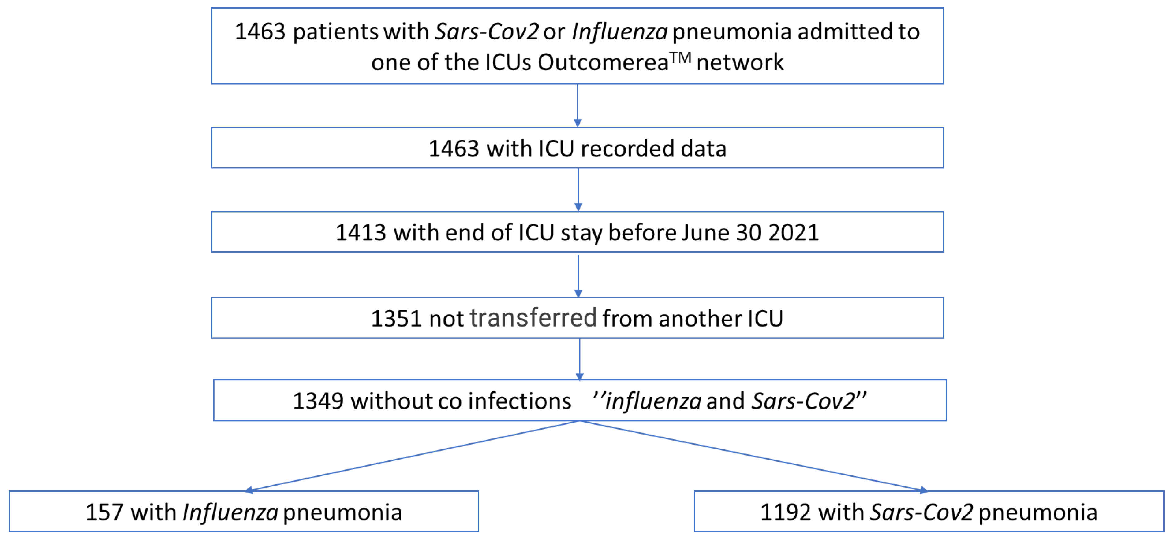
2. Materials and Methods

This was a French multicentre (n = 11) prospective observational study using data from the Outcomerea© database.

2.1. Database

In compliance with French law, the Outcomerea© database was approved by the Comité Consultatif sur le Traitement de l’Information en matière de Recherche dans le domaine de la Santé (CCTIRS) and by the Commission Nationale Information et Libertés (CNIL, No. 8999262). The database protocol was submitted to the research ethics committee (IRB) of the University of Clermont-Ferrand, which agreed that there was no requirement for informed consent. Information was given to the patient or their family.

2.2. Study Population

All patients over 18 years of age admitted to one of the ICUs in the Outcomerea© group could be included in the Outcomerea© database. For our study, we included patients with SARS-CoV-2 or influenza pneumonia admitted to the ICU after 1 January 2008 and ending their ICU stay before 30 June 2021. They had to have severe SARS-CoV-2 or influenza pneumonia with a positive SARS-CoV-2 or influenza RT-PCR.
Patients without a complete follow-up and patients infected with both influenza and SARS-CoV-2 were excluded.

2.3. Data Collection

Data that were collected prospectively from admission to the ICU included demographic data; chronic diseases including respiratory, cardiac, renal, and hepatic comorbidities according to the Knaus classification, SAPS II (Simplified Acute Physiology Score), and SOFA (Sequential Organ Failure Assessment) severity indices; and treatments received on admission including different antibiotic therapies and corticosteroid and immunomodulatory treatments. Other variables included with the clinical and biological variables were ventilation modalities; other organ support; lung diseases on admission and nosocomial infections arising in the ICU, including ventilator-associated pneumonia (VAP); and length of stay and patient outcome at 60 days.

2.4. Definitions

RespCoBact in the ICU was defined as the presence of a community-acquired or hospital-acquired bacterial pneumonia associated with influenza pneumonia or SARS-CoV-2 pneumonia during the ICU admission.
The presence of RespCoBact was defined as the presence of radiological and/or scanographic condensation, bacteriological documentation (a positive quantitative culture of lower respiratory tract samples collected as recommended (bronchoalveolar lavage, >104 CFU/mL, plugged telescoping catheter, >103 CFU/mL, endotracheal aspirate, >106 CFU/mL)), and/or presence of positive antigenuria, as defined by the European Centre for Disease Control and Prevention [6,7]. RespCoBact was deemed community-acquired if diagnosed within the first 48 h of hospital admission and hospital-acquired if diagnosed after 48 h. If bacterial pneumonia occurred at least 2 days after intubation, it was classified as VAP [8]. The risk period for VAP begins at 48 h after intubation and lasts until removal of the tracheal tube and weaning from the invasive ventilation, so it ends with extubation.
Causal agents of RespCoBact were defined as multidrug-resistant (MDR) on the following criteria: methicillin-resistant Staphylococcus aureus (MRSA), extended-spectrum beta-lactamase (ESBL)-producing Enterobacteriaceae, AmpC-producing Enterobacteriaceae, and Pseudomonas aeruginosa resistant to ticarcillin and/or imipenem and/or ceftazidime.
Invasive mechanical ventilation was defined as continuous mechanical ventilation using either an endotracheal tube or a tracheostomy tube. The other oxygenation modalities included face mask ventilation modes with noninvasive positive-pressure ventilation (NIPPV), continuous positive airway pressure (CPAP), and high-flow nasal cannula (HFNC). These techniques could be continuous or deferred.

2.5. Statistical Analysis

Patient characteristics were expressed as number and percentage for categorical variables and median and interquartile range for continuous variables. Comparisons were made using Fisher’s test for categorical variables and Wilcoxon’s test for continuous variables.
Univariate and multivariate analyses with a Cox survival model were performed to investigate the independent risk factors for death at 60 days. Variables reaching a p-value < 0.2 in the univariate analysis were tested in the multivariate analysis. Variables were then entered using a backward procedure and only those with a p-value < 0.05 were retained. In a similar way, factors associated with the occurrence of VAP among the patients at risk of VAP were sought. The Fine–Gray subdistribution hazard model was used while taking into account death and discharge alive from the ICU as competitive risks.
Finally, risk factors for respiratory co-infections were sought using a univariable logistic regression analysis.

3. Results

3.1. Comparison of Patients with Influenza Pneumonia versus SARS-CoV-2 Pneumonia

During the study period, 1463 patients were admitted to one of the Outcomerea© group ICUs with a diagnosis of SARS-CoV-2 or influenza-related pneumonia. Data from 1349 of these patients, including 157 patients with influenza and 1192 patients with SARS-CoV-2, were analysed (Figure 1). The main characteristics are reported in Table 1. Most of the patients in the influenza group were admitted before the COVID pandemic and after 2012 (n = 135 (84.7%)); 41.8% of the patients with SARS-CoV-2 were admitted to the ICU during the first wave of the pandemic (February–May 2020).
These two populations (SARS-CoV-2 pneumonia versus influenza pneumonia) differed in several ways. The patients in the SARS-CoV-2 pneumonia group were older and more often male. More were obese and more had cardiovascular comorbidities. In contrast, patients in the influenza pneumonia group had more comorbidities, especially chronic respiratory failure and immunosuppression.
The time from hospital admission to ICU admission was shorter for patients with influenza pneumonia than for patients with SARS-CoV-2 pneumonia (p < 0.01). On admission, the SAPS II and SOFA severity scores were significantly higher in the influenza pneumonia group, with more use of mechanical ventilation. Patients in the SARS-CoV-2 pneumonia group had more severe hypoxaemia and were predominantly managed using HFNC.
Regarding the treatments received on admission, patients with SARS-CoV-2 pneumonia more often received corticosteroids. Approximately 60% of the patients were receiving antibiotic therapy on admission to the ICU, with most using third-generation cephalosporins and macrolides in the SARS-CoV-2 pneumonia group and betalactam antibiotics with inhibitors, fluoroquinolones, or antistaphylococcal treatment in the influenza pneumonia group.
RespCoBact was more frequently observed in the influenza pneumonia group (24.8%) than in the SARS-CoV-2 pneumonia group (8.2%); p < 0.01. The proportion of community-acquired infections was higher in the patients with influenza pneumonia (84.6%) than in those with SARS-CoV-2 pneumonia (58.2%); p < 0.01.
During their ICU stay, the patients with influenza were more often intubated (p < 0.01). However, the duration of mechanical ventilation did not differ from that for patients with SARS-CoV-2 pneumonia. The patients with SARS-CoV-2 pneumonia were more often placed in a prone position (p = 0.07). There was no significant difference in the use of ECMO between the two groups (p = 0.19). Patients with influenza and those with SARS-CoV-2 pneumonia had nearly equivalent VAP rates (14.6% versus 17.5%, p = 0.37).

3.2. Comparison between Influenza Pneumonia and SARS-CoV-2 Pneumonia with and without Respiratory Bacterial Co-Infection at ICU Admission (Table 2)

3.2.1. Analysis of the Subgroup of Patients with Influenza Pneumonia

A subgroup analysis (Table 2) showed that subjects co-infected on admission were more severely ill and had higher SOFA scores. The use of vasopressors, renal replacement therapy, oseltamivir, and antibiotic therapy was more frequent in patients with co-infections. During their stays in the ICU, the use of ECMO and vasopressors was greater. There was no difference in the length of stay or mortality.
Table 2. Comparison of patients with influenza or SARS-CoV-2 pneumonia with and without respiratory bacterial co-infection on admission.
Variables (n (%)/Median [IQR])1. No FluRespCoBact2.
FluRespCoBact
P
1|2
3. No CovRespCoBact4.
CovRespCoBact
p
3|4
p
All
p
1|3
p
2|4
Number of patients11839 109498
Time from hospital admission to ICU (days)1 [1; 3]1 [1; 2]0.142 [1; 4]2 [1; 6]0.33<0.01<0.01<0.01
Age (years)59.8 [52.1; 71.4]61.1 [44.6; 72]0.5764.4 [54.3; 73.0]64.1 [55.2; 70.2]0.480.090.070.21
Sex (male)71 (60.2)22 (56.4)0.68790 (72.2)74 (75.5)0.48<0.01<0.010.03
Body mass index, kg/m²27.3 [24.0; 31.7]25.6 [21.2; 27.6]<0.0128.41 [25.1; 32.1]27.54 [23.9; 34.4]0.59<0.010.11<0.01
Comorbidities
Charlson score2 [1; 4]1 [0; 2]<0.011 [0; 3]2 [0; 4]0.02<0.01<0.010.05
Chronic cardiovascular disease20 (17.0)2 (5.1)0.07275 (25.1)31 (31.6)0.16<0.010.05<0.01
Chronic lung disease37 (31.4)12 (30.8)0.95123 (11.2)10 (10.2)0.75<0.01<0.01<0.01
Chronic kidney disease16 (13.6)2 (5.1)0.1598 (8.96)10 (10.2)0.680.310.100.34
Immunodepression *51 (43.2)8 (20.5)0.01132 (12.1)14 (14.3)0.52<0.01<0.010.37
Diabetes21 (17.8)5 (12.8)0.47165 (15.1)19 (19.4)0.260.580.440.36
Characteristics on admission
SAPS II score39.5 [27; 51]42 [32; 64]0.1033 [24; 43]33.5 [26; 48]0.16<0.01<0.010.02
SOFA score5 [3; 7]7 [5; 10]<0.015 [4; 7]6 [4; 8]<0.01<0.010.730.08
PaO2/FiO2 (missing data = 60)150 [103; 238]133 [82; 199]0.10107 [74; 180]110 [75; 154]0.66<0.01<0.010.20
Organ support at admisison
Invasive mechanical ventilation52 (44.1)27 (69.2)<0.01304 (27.8)41 (41.8)<0.01<0.01<0.01<0.01
ECMO02 (5.1)0.0117 (1.6)4 (4.1)0.070.040.170.79
Vasopressors10 (8.5)8 (20.5)0.04204 (18.7)27 (27.6)0.03<0.01<0.010.39
Renal replacement therapy4 (3.4)5 (12.8)0.0335 (3.2)10 (10.2)<0.01<0.010.910.66
Corticoids30 (25.4)9 (23.1)0.77634 (57.9)69 (70.4)0.02<0.01<0.01<0.01
Anti-Il-6 or anti-Il-100.83 (7.6)3 (3.1)0.02<0.01<0.01.
Ozeltamivir36 (30.5)15 (38.5)0.3629 (2.7)00.10<0.01<0.01<0.01
Other anti-infectious treatments on admission
Antibiotics55 (46.6)26 (66.7)0.03631 (57.7)73 (74.5)<0.01<0.010.020.36
Amoxicillin/clavulanic acid16 (13.5)11 (28.2)0.1366 (6.1)13 (13.2)<0.01<0.01<0.010.16
Ureido-carboxypenicillins21 (17.8)6 (15.4)0.7355 (5.0)8 (8.16)0.18<0.01<0.010.21
3rd-generation cephalosporin28 (23.7)17 (43.6)0.02453 (41.5)50 (51.0)0.07<0.01<0.010.43
4th-generation cephalosporin3 (2.5)1 (2.6)0.9946 (4.2)8 (8.2)0.070.190.380.23
Macrolides27 (22.9)16 (41.0)0.03312 (28.6)25 (25.5)0.520.160.190.07
Aminosides10 (8.5)3 (7.7)0.8840 (3.7)14 (14.3)<0.01<0.010.010.29
Fluoroquinolones8 (6.8)5 (12.8)0.2443 (3.9)11 (11.2)<0.01<0.010.140.79
Anti-MSSA and anti-MRSA §5 (4.2)5 (12.8)0.1622 (2.0)9 (9.2)<0.01<0.010.050.55
Bacteraemia on admission2 (1.7)4 (10.3)0.0231 (2.8)11 (11.2)<0.01<0.010.470.87
Organ support during stay in ICU
Invasive mechanical ventilation61 (51.7)28 (71.8)0.03484 (44.2)62 (63.3)<0.01<0.010.120.34
ECMO1 (0.9)4 (10.3)<0.0150 (4.6)10 (10.2)0.01<0.010.060.99
Vasopressors12 (10.2)9 (23.1)0.04347 (31.7)43 (43.9)0.01<0.01<0.010.02
Renal replacement therapy18 (15.3)10 (25.6)0.14161 (14.7)24 (24.5)0.010.020.880.89
VAP14 (11.9)9 (23.1)0.09187 (17.1)22 (22.5)0.180.160.150.94
Outcome
Duration of invasive mechanical ventilation (days)11 [4; 18]16 [7; 23.5]0.6812 [6; 21]12 [5; 18]0.680.450.770.77
Duration of oxygenation (days)8 [3; 17]12 [6; 25]0.098 [4; 15]8 [4; 16.5]0.090.130.350.35
Duration of ECMO (days)1 [1; 1]3.5 [2; 11.5]0.6510.5 [3; 16]15 [6; 24]0.650.170.080.08
Duration of RRT (days)9 [5; 14]3 [1; 14]0.208 [3; 17]5.5 [2; 14.5]0.200.610.740.74
Duration of ICU stay (days)6 [3; 14]13 [7; 28]<0.018 [4; 16]9.5 [5; 18]0.07<0.01<0.010.09
Duration of hospital stay (days)15 [8; 35]27.5 [16; 50.5]<0.0115 [10; 27]15.5 [9; 30]0.95<0.010.69<0.01
Mortality at D6023 (19.5)5 (12.8)0.35321 (29.3)35 (35.7)0.19<0.010.02<0.01
Flu: influenza, Cov: SARS-CoV-2, RespCoBact: bacterial respiratory co-infection on admission. * Organ transplants, AIDS, non-AIDS HIV, corticoids > 1 month or >2 mg/kg/j, chemotherapy, aplasia, or other immunodepression. § linezolid, daptomycin, vancomycin, cefazolin, or penicillin. SOFA: sequential organ failure assessment; SAPS: simplified acute physiology score; ECMO: extracorporeal membrane oxygenation; RRT: renal replacement therapy; MSSA: methicillin-susceptible Staphylococcus aureus; MRSA: methicillin-resistant Staphylococcus aureus; VAP: ventilator-associated pneumonia.

3.2.2. Analysis of the Subgroup of Patients with SARS-CoV-2 Pneumonia

Patients co-infected on admission were more likely to have comorbidity, higher Charlson scores, and a higher SOFA. The use of invasive mechanical ventilation, ECMO, vasopressors, and antibiotics was more frequent in patients with CovRespCoBact (Table 2). There was no difference in the length of stay or mortality in patients with SARS-CoV-2 pneumonia with and without CovRespCoBact.

3.2.3. Analysis of Patients in SARS-CoV-2 and Influenza Groups without RespCoBact

The differences between the two groups were those of the cohort (Table 1). Patients with influenza had longer hospital stays and lower mortality rates (Table 2).

3.2.4. Analysis of Patients in SARS-CoV-2 and Influenza Groups with RespCoBact

Patients with influenza had a lower body mass index and higher SAPS II severity scores and were more often intubated on admission, but this difference disappeared during their stay. Patients with influenza had a greater length of stay than patients with SARS-CoV-2 with no difference in mortality.

3.3. Microbiological Description of Lung Co-Infections on Admission

Of the 1349 patients included, 137 had a co-infection and 135 co-infections were documented. The microbiological description of RespCoBact is given in Table 3.
In the patients with influenza, most of the pathogens identified in RespCoBact were Gram-positive cocci (GPCs), with a predominance of S. pneumoniae over S. aureus. Among the Gram-negative bacilli (GNBs), Haemophilus spp. was the most frequent causal agent.
In patients with SARS-CoV-2, the most common organisms identified in RespCoBact were GNBs. These were mainly Enterobacter spp., E. coli, and Klebsiella spp., followed by Haemophilus spp. Among the GPCs, S. aureus was proportionally found more often found S. pneumoniae.
Very few cases of drug resistance were reported in the cohort, with ESBL-secreting Enterobacteriaceae being the most common in patients with SARS-CoV-2 pneumonia only.
The pathogens identified in RespCoBact with regard to community-acquired infection differed in patients with SARS-CoV-2 pneumonia, this time showing more than 40% GPCs with Staphylococcus being predominant.

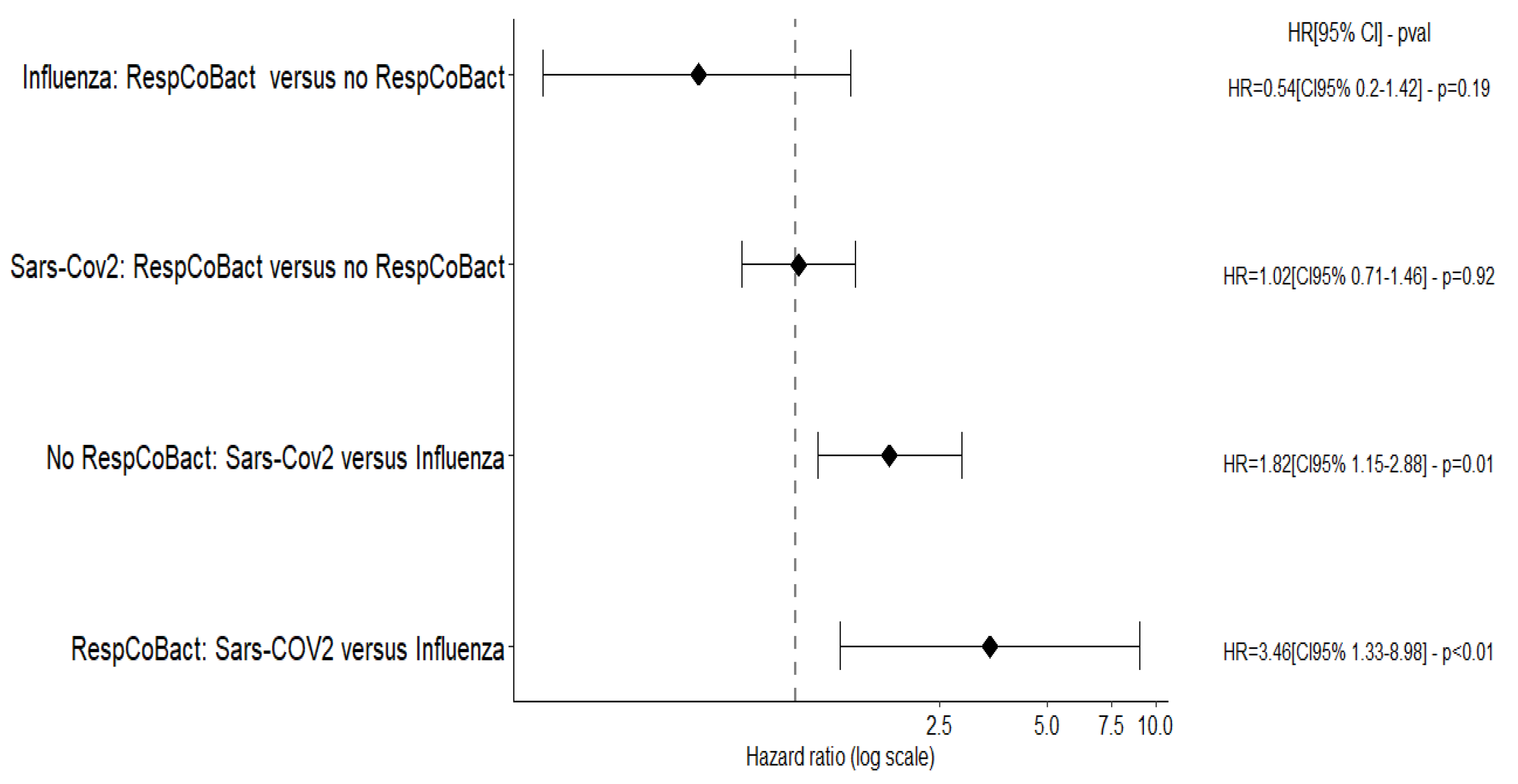
3.4. Risk Factors for Death at D60 (Figure 2; Tables S2 and S3)

After adjustment, having a bacterial co-infection on admission was not associated with an increased risk of death either in the whole cohort (aHR = 0.83 [CI95% 0.59; 1.15]; p = 0.26) or in the subgroups of patients with influenza pneumonia (aHR = 0.67 [CI95% 0.25; 1.81]; p = 0.43) or with SARS-CoV-2 pneumonia (aHR = 0.97 [CI95% 0.68; 1.39]; p = 0.88).
Figure 2. Association between SARS-CoV-2 or influenza and presence or absence of respiratory bacterial co-infection on admission and mortality at D60 (Cox multivariate survival model.) HR: hazard ratio at D60; RespCoBact: respiratory bacterial co-infection on admission in the Cox multivariate survival model adjusted for age, chronic cardiovascular diseases, immunodepression, PaO2/FiO2 < 150, vasopressors, extrarenal purification, and beta-lactamase inhibitors.
Biomedicines 10 02646 g002
There was no difference in impact between GPC and GNB pneumonia. Similar results were obtained in the subgroups of immunocompromised patients and patients receiving corticosteroids on admission.
Finally, whether there was co-infection or not, patients with SARS-CoV-2 pneumonia still had a higher risk of mortality than patients with influenza pneumonia (Figure 2).

3.5. Risk Factors for VAP (Tables S4 and S5)

Co-infections were not associated with an increased risk of VAP in either SARS-CoV-2 or Influenza pneumonia. Viral lung diseases were not associated with the occurrence of VAP.

3.6. Risk Factors for Co-Infections (Table S6)

Patients with SARS-CoV-2 pneumonia with cirrhosis (OR = 3.50 [CI95% 1.37; 8.94], p < 0.01) had more co-infections on admission. Immunocompromised or obese influenza patients had fewer co-infections on admission to the ICU (OR = 0.34 [CI95% 0.14; 0.8], p = 0.01; OR = 0.29 [0.1; 0.70], p = 0.02).

4. Discussion

This study found a lower prevalence of pulmonary bacterial co-infections on admission to an ICU in patients with SARS-CoV-2 pneumonia (8.2%) than in patients with influenza pneumonia (24.8%). In the literature, the rates of early bacterial co-infections at diagnosis of SARS-CoV-2 pneumonia at ICU admission ranged from 3% [8] to 20% [9], with a higher prevalence reported in cohorts that included a vast majority of patients on mechanical ventilation (Table S1) [4,8,9,10,11,12,13,14,15,16]. In 254 patients admitted to seven ICUs in England during the first wave, the rate of documented bacterial coinfections was 5.5% [10]. In this study’s population, 59.5% of patients received mechanical ventilation within 24 h of admission. In a French monocentric retrospective study performed in 92 ICU patients admitted for severe COVID-19 with 83 (90%) on mechanical ventilation on admission, the rate of co-infections was 19.2 [9]. The rate of 8.2% reported in our work was very similar to the rates reported by Rouzé et al. (9.7%) [5] and Pandey et al. (8.7%) [17]. These authors also notes a higher frequency of pulmonary co-infections in patients admitted for influenza pneumonia: 33.6% [5] and 25% [17]. In the study by Sarton et al. [18], the difference between the two populations was less important (16% for SARS-CoV-2 pneumonia and 33% for influenza pneumonia) but all patients included in this study were on mechanical ventilation for at least 48 h.
The higher prevalence of pulmonary co-infections during influenza pneumonia may be explained in several ways. Firstly, early intubation in influenza pneumonia: in our study, the proportion of patients intubated on admission was 50.3% for influenza pneumonia and 29% for SARS-CoV-2 pneumonia. Tracheal intubation facilitated the collection of distal respiratory secretions and bacteriological documentation and so made the diagnosis of a pulmonary bacterial co-infection easier. Secondly, in influenza, severe bacterial co-infection may be the prime reason for early intubation rather than the influenza infection itself. The mechanism of hypoxia leading to admission to the ICU differs between influenza and SARS-CoV-2 pneumonia. In SARS-CoV-2 pneumonia, chest imaging more frequently reveals extensive ground-glass lesions associated with initial diffuse involvement of the interstitium [19,20] and microthrombi in the microcirculation [21,22]. These mechanisms probably explain a slower development of lung injury in SARS-CoV-2 pneumonia, making major hypoxaemia more progressive and better tolerated. In contrast, chest imaging during severe influenza reveals influenza-specific lesions that are often less severe and less extensive and do not by themselves justify intensive care [20]. Additional alveolar condensations secondary to bacterial infection, the onset of which is more rapid and is sometimes associated with haemodynamic instability, could explain respiratory failure and earlier intubation. It is also important to highlight progress in noninvasive oxygen therapy techniques due to the considerable rise in recent years of HFNC in the management of acute hypoxaemic respiratory failure [23]. In our study, this therapeutic evolution hindered meaningful comparison of the outcomes observed between the cohort of influenza patients, who were mainly admitted in 2012–2020, and the cohort of patients with SARS-CoV-2 pneumonia admitted in 2020–2021. Finally, early antibiotic therapy could also be a confounding factor, but the proportion of patients receiving antibiotic therapy on admission was not different between patients with influenza and those with SARS-CoV-2 pneumonia.
Regarding the bacteriological documentation of bacterial co-infections, we identified a predominance of GPCs in influenza pneumonia, most often S. pneumoniae followed by S. aureus. In comparison, patients with SARS-CoV-2 pneumonia had more GNB co-infections; the causal agents isolated were, in descending order of frequency, Enterobacteriaceae and Haemophilus spp. Such results were in agreement with previous findings that in influenza pneumonia, the two bacteria most frequently identified were first S. Pneumoniae and then S. aureus with average rates of 40% and 20%, respectively [1,2,24]. In SARS-CoV-2 pneumonia, S aureus was more frequently prevalent than S. pneumoniae, accounting for 30% vs. 20% of documented co-infections [4,9,10,16]; other major etiologic agents of co-infections were Haemophilus in more than 10% of the cases [4,9,13,15] and Enterobacter spp. in more than 25% of the cases [9,14]. In the study by Rouzé et al. [5], GPCs were identified in 58% and 72% of co-infections and GNBs in 41.8% and 27.8% in SARS-CoV-2 pneumonia and Influenza pneumonia, respectively. In the study by Pandey et al. [17], most co-infections were due to S. aureus both in SARS-CoV-2 and in influenza pneumonia.
The microbiological differences observed between studies and between SARS-CoV-2 and influenza pneumonia could reflect the different study designs (for example, Rouzé et al. [5] included only mechanically ventilated patients) or the criteria for defining co-infections (whether only those diagnosed on admission to the ICU, or including the period of prior hospitalisation, or within the first 48 h in the ICU). Another explanation could be that patients with SARS-CoV-2 pneumonia spend more time hospitalized before ICU admission than patients with influenza pneumonia.
We found no excess mortality associated with the presence of a bacterial respiratory co-infection in patients with influenza or SARS-CoV-2 pneumonia. In SARS-CoV-2 pneumonia, while some studies did not report an increased risk of mortality due to co-infections [8], two systematic reviews found a longer hospital length of stay [4] and an increased risk of death [4,11]. However, the results of these reviews should be cautiously interpreted due to the great heterogeneity of the studies. In contrast, an increased risk of mortality was observed for respiratory bacterial co-infection in patients admitted to an ICU with severe influenza pneumonia [2]. For instance, Rice et al. reported that co-infections due to S. aureus were associated with an increased risk of death [24].
In our study, we noted that compared with influenza patients, the increased risk of mortality observed in patients with SARS-CoV-2 with or without bacterial respiratory co-infection persisted after adjustment. This result could be partly explained by the systemic inflammation and microthrombi involved in the pathophysiology of COVID-19 [25,26]. It is also likely that in patients with influenza, extensive prescription of oseltamivir in the initial phase of infection attenuated the intensity of the lesions by accelerating viral clearance. Finally, some of the patients included in the SARS-CoV-2 group were managed at the beginning of the pandemic and were not receiving corticosteroids or reinforced preventive and/or curative anticoagulation, the only treatments yet shown to be of benefit in the management of SARS-CoV-2 patients [27]. However, a causal link was recently shown between the use of steroids and ICU-acquired infections [28].
We also found that the patients most at risk of co-infection on admission were those that were most severely ill and that in patients with influenza, comorbidities were also associated with a higher risk of bacterial pulmonary co-infection on admission.
Contrary to other studies [29], we found no increased rate of VAP in patients with SARS-CoV-2 pneumonia compared with those with influenza pneumonia even if among the patients at risk of VAP, the VAP prevalence was higher among patients with SARS-CoV-2 pneumonia (40%) versus those with influenza pneumonia (27.4%). These findings could be explained by the unbalanced between patients with SARS-CoV-2 and influenza pneumonia in our cohort. The occurrence of RespCoBact was also not associated with an increased risk of VAP. Contrary to our results, bacterial respiratory co-infections were already reported to be associated with an increased risk of VAP [30,31]. Our results could be explained by the severity of the underlying viral diseases and the associated prolonged duration of invasive ventilation and increased risk of death, which minimized the impact of RespCoBact upon occurrence of VAP.
The main strengths of this study were the large size of its patient population and its multicentre nature, as well as its prospective data collection using a good-quality database, which allowed an accurate study of patients with influenza and with COVID-19.
One limitation was the imbalance in the number of patients between those with influenza and those with COVID-19. In addition, the difference in recruitment periods between patients with influenza and those with COVID-19 and progress in noninvasive oxygen therapy strategies over time prevented any direct comparison of the impact of co-infections between the two groups.
Finally, most of the patients with COVID-19 were recruited during the first COVID-19 pandemic wave in France, in particular before the introduction of corticosteroids, which was to greatly change the evolution of COVID-19 in hypoxaemic patients

5. Conclusions

This multicentre observational study confirmed that bacterial pulmonary co-infections on admission to an ICU were less frequent in COVID-19 patients than in influenza patients.
The increased risk of mortality observed in patients with COVID-19 compared with influenza patients was not specifically related to the presence of a bacterial co-infection but rather to the specific mechanisms of the SARS-CoV-2 respiratory infection.
It would be of interest to repeat this study in the current period of antiviral therapies, delayed intubation strategies [32], and widespread COVID-19 vaccination.

Supplementary Materials

The following supporting information can be downloaded at: https://www.mdpi.com/article/10.3390/biomedicines10102646/s1, Table S1: Results of studies comparing co-infections in patients with influenza or SARS-CoV2 pneumonia or comparing the two diseases; Table S2: Comparison of living and deceased patients in influenza and/or SARS-CoV2 pneumonia at D60; Table S3: Impact of Co-infection on the risk of mortality at D60, multivariate Cox model—global and per-viruses analyses according to to patients’ subgroups; Table S4: Risk factors for VAP, univariate analysis, Fine-Gray subdistribution hazard model; Table S5: Impact of co-infection on the risk of subsequent VAP among the patients at risk of VAP—multivariate survival analysis; Table S6: Risk factors for co-infection, univariate analysis, logistic regression.

Author Contributions

C.D., G.D., B.S. and J.-F.T. designed the study, assisted in data collection and analysis, and drafted and edited the manuscript. N.B., M.N., S.S., Y.C., V.L., B.M., J.R., D.G.-T., C.S., S.R. and E.d.M. assisted in data collection and edited the manuscript. All authors have read and agreed to the published version of the manuscript.

Funding

A grant for an investigator-driven study was obtained from Pfizer Inc. (# 64764929) to conduct this study. The sponsor had no impact on the design of the study, conducting of the analyses, or drafting of the manuscript.

Institutional Review Board Statement

In compliance with French law, the Outcomerea© database was approved by the Comité Consultatif sur le Traitement de l’Information en matière de Recherche dans le domaine de la Santé (CCTIRS) and by the Commission Nationale Information et Libertés (CNIL, No. 8999262). The database protocol was submitted to the research ethics committee (IRB) of the University of Clermont-Ferrand, which agreed that there was no requirement for informed consent. Information was given to the patient or their family.

Informed Consent Statement

Informed consent was obtained from all subjects involved in the study.

Data Availability Statement

Data can be provided upon request to the corresponding author.

Acknowledgments

We thank Richard Ryan for the medical writing and the Outcomerea study group member.

Conflicts of Interest

J.-F.T. reported scientific advisory board membership for Merck, Shionogi, Paratek, Gilead, Pfizer, Becton-Dickinson, and Aspen not related to the submitted work. J.-F.T. reported lectures given during Merck, Shionogi, Gilead, Pfizer, and Bection-Dickinson symposia not related to the submitted work. The other authors have no conflicts of interest to declare.

References

  1. MacIntyre, C.R.; Chughtai, A.A.; Barnes, M.; Ridda, I.; Seale, H.; Toms, R.; Heywood, A. The role of pneumonia and secondary bacterial infection in fatal and serious outcomes of pandemic influenza a(H1N1)pdm09. BMC Infect. Dis. 2018, 18, 637. [Google Scholar] [CrossRef] [PubMed] [Green Version]
  2. Mercat, A.; Reva-Grippe-Srlf, P.L.R.; Pham, T.; Rozé, H.; Cuquemelle, E.; Brun-Buisson, C.; Brochard, L.; Richard, J.C.M. Formes graves de la grippe H1N1 2009 chez l’adulte: L’expérience française. Réanimation 2011, 20, 162–168. [Google Scholar] [CrossRef] [PubMed]
  3. Luyt, C.-E.; Combes, A.; Becquemin, M.-H.; Beigelman-Aubry, C.; Hatem, S.; Brun, A.-L.; Zraik, N.; Carrat, F.; Grenier, P.A.; Richard, J.-C.M.; et al. Long-term Outcomes of Pandemic 2009 Influenza A(H1N1)-Associated Severe ARDS. Chest 2012, 142, 583–592. [Google Scholar] [CrossRef] [PubMed]
  4. Westblade, L.F.; Simon, M.S.; Satlin, M.J. Bacterial Coinfections in Coronavirus Disease 2019. Trends Microbiol. 2021, 29, 930–941. [Google Scholar] [CrossRef]
  5. Rouzé, A.; Martin-Loeches, I.; Povoa, P.; Metzelard, M.; Du Cheyron, D.; Lambiotte, F.; Tamion, F.; Labruyere, M.; Geronimi, C.B.; Nieszkowska, A.; et al. Early Bacterial Identification among Intubated Patients with COVID-19 or Influenza Pneumonia: A European Multicenter Comparative Clinical Trial. Am. J. Respir. Crit. Care Med. 2021, 204, 546–556. [Google Scholar] [CrossRef]
  6. Calandra, T.; Cohen, J. International Sepsis Forum Definition of Infection in the ICU Consensus Conference The International Sepsis Forum Consensus Conference on Definitions of Infection in the Intensive Care Unit. Crit. Care Med. 2005, 33, 1538–1548. [Google Scholar] [CrossRef]
  7. Plachouras, D.; Lepape, A.; Suetens, C. ECDC definitions and methods for the surveillance of healthcare-associated infections in intensive care units. Intensive Care Med. 2018, 44, 2216–2218. [Google Scholar] [CrossRef] [Green Version]
  8. Garcia-Vidal, C.; Sanjuan, G.; Moreno-García, E.; Puerta-Alcalde, P.; Garcia-Pouton, N.; Chumbita, M.; Fernandez-Pittol, M.; Pitart, C.; Inciarte, A.; Bodro, M.; et al. Incidence of co-infections and superinfections in hospitalized patients with COVID-19: A retrospective cohort study. Clin. Microbiol. Infect. 2020, 27, 83–88. [Google Scholar] [CrossRef]
  9. Contou, D.; Claudinon, A.; Pajot, O.; Micaëlo, M.; Flandre, P.L.; Dubert, M.; Cally, R.; Logre, E.; Fraissé, M.; Mentec, H.; et al. Bacterial and viral co-infections in patients with severe SARS-CoV-2 pneumonia admitted to a French ICU. Ann. Intensive Care 2020, 10, 119. [Google Scholar] [CrossRef]
  10. Baskaran, V.; Lawrence, H.; Lansbury, L.E.; Webb, K.; Safavi, S.; Zainuddin, N.I.; Huq, T.; Eggleston, C.; Ellis, J.; Thakker, C.; et al. Co-infection in critically ill patients with COVID-19: An observational cohort study from England. J. Med. Microbiol. 2021, 70, 001350. [Google Scholar] [CrossRef]
  11. Musuuza, J.S.; Watson, L.; Parmasad, V.; Putman-Buehler, N.; Christensen, L.; Safdar, N. Prevalence and outcomes of co-infection and superinfection with SARS-CoV-2 and other pathogens: A systematic review and meta-analysis. PLoS ONE 2021, 16, e0251170. [Google Scholar] [CrossRef] [PubMed]
  12. Rawson, T.M.; Moore, L.S.P.; Zhu, N.; Ranganathan, N.; Skolimowska, K.; Gilchrist, M.; Satta, G.; Cooke, G.; Holmes, A.H. Bacterial and Fungal Coinfection in Individuals With Coronavirus: A Rapid Review To Support COVID-19 Antimicrobial Prescribing. Clin. Infect. Dis. 2020, 71, 2459–2468. [Google Scholar] [CrossRef] [PubMed]
  13. De Santis, V.; Corona, A.; Vitale, D.; Nencini, C.; Potalivo, A.; Prete, A.; Zani, G.; Malfatto, A.; Tritapepe, L.; Taddei, S.; et al. Bacterial infections in critically ill patients with SARS-2-COVID-19 infection: Results of a prospective observational multicenter study. Infection 2021, 50, 139–148. [Google Scholar] [CrossRef]
  14. Elabbadi, A.; Turpin, M.; Gerotziafas, G.T.; Teulier, M.; Voiriot, G.; Fartoukh, M. Bacterial coinfection in critically ill COVID-19 patients with severe pneumonia. Infection 2021, 49, 559–562. [Google Scholar] [CrossRef] [PubMed]
  15. Russell, C.D.; Fairfield, C.J.; Drake, T.M.; Turtle, L.; Seaton, R.A.; Wootton, D.G.; Sigfrid, L.; Harrison, E.M.; Docherty, A.B.; de Silva, T.I.; et al. Co-infections, secondary infections, and antimicrobial use in patients hospitalised with COVID-19 during the first pandemic wave from the ISARIC WHO CCP-UK study: A multicentre, prospective cohort study. Lancet Microbe 2021, 2, e354–e365. [Google Scholar] [CrossRef]
  16. Santos, A.P.; Gonçalves, L.C.; Oliveira, A.C.C.; Queiroz, P.H.P.; Ito, C.R.M.; Santos, M.O.; Carneiro, L.C. Bacterial Co-Infection in Patients with COVID-19 Hospitalized (ICU and Not ICU): Review and Meta-Analysis. Antibiotics 2022, 11, 894. [Google Scholar] [CrossRef]
  17. Pandey, M.; May, A.; Tan, L.; Hughes, H.; Jones, J.P.; Harrison, W.; Bradburn, S.; Tyrrel, S.; Muthuswamy, B.; Berry, N.; et al. Comparative incidence of early and late bloodstream and respiratory tract co-infection in patients admitted to ICU with COVID-19 pneumonia versus Influenza A or B pneumonia versus no viral pneumonia: Wales multicentre ICU cohort study. Crit. Care 2022, 26, 1–12. [Google Scholar] [CrossRef]
  18. Sarton, B.; Grare, M.; Vardon-Bounes, F.; Gaubert, A.; Silva, S.; Crognier, L.; Riu, B.; Seguin, T.; Georges, B.; Minville, V.; et al. Co-Infection and Ventilator-Associated Pneumonia in Critically Ill COVID-19 Patients Requiring Mechanical Ventilation: A Retrospective Cohort Study. Biomedicines 2022, 10, 1952. [Google Scholar] [CrossRef]
  19. Auvinen, R.; Nohynek, H.; Syrjänen, R.; Ollgren, J.; Kerttula, T.; Mäntylä, J.; Ikonen, N.; Loginov, R.; Haveri, A.; Kurkela, S.; et al. Comparison of the clinical characteristics and outcomes of hospitalized adult COVID-19 and influenza patients—A prospective observational study. Infect. Dis. 2020, 53, 111–121. [Google Scholar] [CrossRef]
  20. Cobb, N.L.; Sathe, N.A.; Duan, K.I.; Seitz, K.P.; Thau, M.R.; Sung, C.C.; Morrell, E.D.; Mikacenic, C.; Kim, H.N.; Liles, W.C.; et al. Comparison of Clinical Features and Outcomes in Critically Ill Patients Hospitalized with COVID-19 versus Influenza. Ann. Am. Thorac. Soc. 2021, 18, 632–640. [Google Scholar] [CrossRef]
  21. Zhu, L.; Yang, P.; Zhao, Y.; Zhuang, Z.; Wang, Z.; Song, R.; Zhang, J.; Liu, C.; Gao, Q.; Xu, Q.; et al. Single-Cell Sequencing of Peripheral Mononuclear Cells Reveals Distinct Immune Response Landscapes of COVID-19 and Influenza Patients. Immunity 2020, 53, 685–696.e3. [Google Scholar] [CrossRef] [PubMed]
  22. McElvaney, O.J.; McEvoy, N.L.; McElvaney, O.F.; Carroll, T.P.; Murphy, M.P.; Dunlea, D.M.; Ni Choileain, O.; Clarke, J.; O’Connor, E.; Hogan, G.; et al. Characterization of the Inflammatory Response to Severe COVID-19 Illness. Am. J. Respir. Crit. Care Med. 2020, 202, 812–821. [Google Scholar] [CrossRef] [PubMed]
  23. Gaunt, K.A.; Spilman, S.K.; Halub, M.E.; Jackson, J.A.; Lamb, K.D.; Sahr, S.M. High-Flow Nasal Cannula in a Mixed Adult ICU. Respir. Care 2015, 60, 1383–1389. [Google Scholar] [CrossRef] [PubMed] [Green Version]
  24. Rice, T.W.; Rubinson, L.; Uyeki, T.M.; Vaughn, F.L.; John, B.B.; Miller, R.R.; Higgs, E.; Randolph, A.; Smoot, B.E.; Thompson, B.T. Critical illness from 2009 pandemic influenza A virus and bacterial coinfection in the United States*. Crit. Care Med. 2012, 40, 1487–1498. [Google Scholar] [CrossRef] [Green Version]
  25. Ackermann, M.; Verleden, S.E.; Kuehnel, M.; Haverich, A.; Welte, T.; Laenger, F.; Vanstapel, A.; Werlein, C.; Stark, H.; Tzankov, A.; et al. Pulmonary Vascular Endothelialitis, Thrombosis, and Angiogenesis in COVID-19. N. Engl. J. Med. 2020, 383, 120–128. [Google Scholar] [CrossRef]
  26. Patel, B.V.; Arachchillage, D.J.; Ridge, C.A.; Bianchi, P.; Doyle, J.F.; Garfield, B.; Ledot, S.; Morgan, C.; Passariello, M.; Price, S.; et al. Pulmonary Angiopathy in Severe COVID-19: Physiologic, Imaging, and Hematologic Observations. Am. J. Respir. Crit. Care Med. 2020, 202, 690–699. [Google Scholar] [CrossRef]
  27. Horby, P.; Lim, W.S.; Emberson, J.; Mafham, M.; Bell, J.; Linsell, L.; Staplin, N.; Brightling, C.; Ustianowski, A.; Elmahi, E.; et al. Effect of Dexamethasone in Hospitalized Patients with COVID-19—Preliminary Report. medRxiv 2020. [Google Scholar] [CrossRef]
  28. Morris, A.C.; Kohler, K.; De Corte, T.; Ercole, A.; De Grooth, H.-J.; Elbers, P.W.G.; Povoa, P.; Morais, R.; Koulenti, D.; Jog, S.; et al. Co-infection and ICU-acquired infection in COVID-19 ICU patients: A secondary analysis of the UNITE-COVID data set. Crit. Care 2022, 26, 1–13. [Google Scholar] [CrossRef]
  29. Llitjos, J.-F.; Bredin, S.; Lascarrou, J.-B.; Soumagne, T.; Cojocaru, M.; Leclerc, M.; Lepetit, A.; Gouhier, A.; Charpentier, J.; Piton, G.; et al. Increased susceptibility to intensive care unit-acquired pneumonia in severe COVID-19 patients: A multicentre retrospective cohort study. Ann. Intensive Care 2021, 11, 1–8. [Google Scholar] [CrossRef]
  30. Martin-Loeches, I. Current Concepts in Community and Ventilator Associated Lower Respiratory Tract Infections in ICU Patients. Antibiotics 2020, 9, 380. [Google Scholar] [CrossRef]
  31. Papazian, L.; Klompas, M.; Luyt, C.-E. Ventilator-associated pneumonia in adults: A narrative review. Intensive Care Med. 2020, 46, 888–906. [Google Scholar] [CrossRef] [PubMed] [Green Version]
  32. Dupuis, C.; Bouadma, L.; de Montmollin, E.; Goldgran-Toledano, D.; Schwebel, C.; Reignier, J.; Neuville, M.; Ursino, M.; Siami, S.; Ruckly, S.; et al. Association Between Early Invasive Mechanical Ventilation and Day-60 Mortality in Acute Hypoxemic Respiratory Failure Related to Coronavirus Disease-2019 Pneumonia. Crit. Care Explor. 2021, 3, e0329. [Google Scholar] [CrossRef] [PubMed]
Figure 1. Flow diagram.
Biomedicines 10 02646 g001
Table 1. Comparison between patients with influenza pneumonia and SARS-CoV-2 pneumonia.
Variables (n (%)/Median [IQR])Influenza (n = 157)SARS-CoV-2 (n = 1192)p
Time from hospital admission to ICU (days)1 [1; 2]2 [1; 4]<0.01
Age (years)60.1 [51.5; 71.6]64.4 [54.5; 72.7]0.02
Sex (% male)93 (59.2)864 (72.5)<0.01
Body mass index (BMI) (kg/m²)26.8 [23.5; 30.9]28.4 [25; 32.2]<0.01
Comorbidities
Charlson score2 [1; 3]1 [0; 3]<0.01
Chronic cardiovascular disease22 (14)306 (25.7)<0.01
Chronic lung disease49 (31.2)133 (11.2)<0.01
Chronic kidney disease18 (11.5)108 (9.1)0.33
Chronic liver disease7 (4.5)26 (2.2)0.08
Immunodepression *59 (37.6)146 (12.2)<0.01
Diabetes26 (16.6)184 (15.4)0.71
Characteristics on admission
SAPS II score40 [28; 54]33 [24; 43]<0.01
SOFA score5 [4; 8]5 [4; 7]0.06
Biological data
Leucocytes elts/mm3 (missing data = 24)9385 [4520; 14,700]9000 [6660; 12,200]0.48
PaO2/FiO2 (missing data = 61)148 [95; 215]108 [74; 177]<0.01
Pulmonary embolism0 (0)36 (3)0.03
Ventilatory support on admission
Invasive mechanical ventilation79 (50.3)345 (29)<0.01
High-flow nasal cannula13 (8.3)530 (44.5)<0.01
Continuous positive airway pressure30 (19.1)138 (11.6)<0.01
ECMO2 (1.3)21 (1.8)0.66
Vasopressors18 (11.5)231 (19.4)0.02
Renal replacement therapy9 (5.7)45 (3.8)0.24
Corticoids39 (24.8)703 (59)<0.01
Il1 or Il6 receptor antagonists0 (0)86 (7.2)<0.01
Lopinavir, ritonavir0 (0)171 (14.4)<0.01
Hydroxychloroquine0 (0)64 (5.4)<0.01
Remdesivir0 (0)169 (14.2)<0.01
Ozeltamivir51 (32.5)29 (2.4)<0.01
Antibiotics81 (51.6)704 (59.1)0.07
Amoxicillin/clavulanic acid27 (17.2)79 (1.7)<0.01
Ureido-carboxypenicillins27 (17.2)63 (5.3)<0.01
3rd-generation cephalosporin45 (28.7)503 (42.2)<0.01
4th-generation cephalosporin4 (2.5)54 (4.5)0.25
Macrolides43 (27.4)337 (28.3)0.81
Aminoglycosides13 (8.3)54 (4.5)0.04
Fluoroquinolones13 (8.3)54 (4.5)0.04
Anti-MSSA and anti-MRSA §12 (7.6)31 (2.6)<0.01
Co-infections on admission
Bacterial pneumonia39 (24.8)98 (8.2)<0.01
Hospital-acquired pneumonia6 (3.8)41 (3.4)0.81
Organ support during hospital stay
Invasive mechanical ventilation89 (56.7)546 (45.8)0.01
Prone position20 (12.7)293 (24.6)<0.01
ECMO5 (3.2)60 (5)0.31
Vasopressors21 (13.4)390 (32.7)<0.01
Renal replacement therapy28 (17.8)185 (15.5)0.45
VAP23 (14.6)209 (17.5)0.37
VAP among the patients at risk of VAP23/84 (27.3)209/522 (40.0)0.03
Outcome
Duration of invasive mechanical ventilation (days)12 [5; 20]12 [6; 21]0.77
Duration of ECMO (days)3 [1; 4]11 [4; 16.5]0.08
Duration of RRT (days)8.5 [2.5; 14]8 [3; 16]0.74
Duration of ICU stay (days)7 [4; 17]8 [4; 16]0.23
Duration of hospital stay (days)17 [9; 36]15 [9.5; 27]0.15
Mortality at D6028 (17.8)356 (29.9)<0.01
* Organ transplant, AIDS, non-AIDS HIV, corticoids > 1 month or >2 mg/kg/day, chemotherapy, aplasia, or other immunodepression. § linezolid, daptomycin, vancomycin, cefazolin, or penicillin. IQR: interquartile; SOFA: sequential organ failure assessment; SAPS: simplified acute physiology score; ECMO: extracorporeal membrane oxygenation; MSSA: methicillin-susceptible Staphylococcus aureus; MRSA: methicillin-resistant Staphylococcus aureus; VAP: ventilation-associated pneumonia.
Table 3. Microbiological description of causal agents identified in respiratory bacterial co-infections on admission.
AllCommunity-AcquiredHospital-Acquired
AllFluCovpAllFluCovpAllFluCovp
Number of infections1353699 883157.47542.
Gram-positive cocci64 (47.4)16 (44.4)48 (48.5)0.6843 (48.8)13 (41.9)30 (52.6)0.3421 (44.6)3 (60)18 (42.9)0.47
Streptococcus pneumoniae20 (14.8)8 (22.2)12 (12.1)0.1412 (13.6)6 (19.4)6 (10.5)0.258 (17)2 (40)6 (14.3)0.15
Staphyloccus aureus35 (26)7 (19.4)28 (28.3)0.3028 (31.8)6 (19.4)22 (38.6)0.067 (14.8)1 (20)6 (14.3)0.73
Enterococcussp.3 (2.2)0 (0)3 (3)0.291 (1.2)0 (0)1 (1.8)0.462 (4.2)0 (0)2 (4.8)0.62
Moraxella catarrhalis4 (3)1 (2.8)3 (3)0.942 (2.2)0 (0)2 (3.5)0.292 (4.2)1 (20)1 (2.4)0.07
Gram-negative bacilli67 (49.6)11 (30.6)56 (56.6)<0.0141 (46.6)11 (35.5)30 (52.6)0.1226 (55.4)0 (0)26 (61.9)<0.01
Haemophilus25 (18.6)10 (27.8)15 (15.2)0.0921 (23.8)10 (32.3)11 (19.3)0.174 (8.6)0 (0)4 (9.5)0.47
Enterobacteriaceae43 (31.8)6 (16.7)37 (37.4)0.0221 (23.8)5 (16.1)16 (28.1)0.2122 (46.8)1 (20)21 (50)0.20
Group 1 or 2 enteric bacteria26 (19.2)2 (5.6)24 (24.2)0.0113 (14.8)1 (3.2)12 (21.1)0.0213 (27.6)1 (20)12 (28.6)0.69
Proteus2 (1.4)1 (2.8)1 (1)0.452 (2.2)1 (3.2)1 (1.8)0.660 .
Escherichia coli12 (8.8)2 (5.6)10 (10.1)0.416 (6.8)1 (3.2)5 (8.8)0.326 (12.8)1 (20)5 (11.9)0.61
Klebsiella11 (8.2)0 (0)11 (11.1)0.046 (6.8)0 (0)6 (10.5)0.065 (10.6)0 (0)5 (11.9)0.41
Citrobacter koseri2 (1.4)0 (0)2 (2)0.390 .2 (4.2)0 (0)2 (4.8)0.62
Group 3 enteric bacteria20 (14.8)0 (0)20 (20.2)<0.017 (8)0 (0)7 (12.3)0.0413 (27.6)0 (0)13 (31)0.14
Enterobacter13 (9.6)0 (0)13 (13.1)0.024 (4.6)0 (0)4 (7)0.139 (19.2)0 (0)9 (21.4)0.25
Serratia6 (4.4)0 (0)6 (6.1)0.133 (3.4)0 (0)3 (5.3)0.193 (6.4)0 (0)3 (7.1)0.54
Citrobacter freundii1 (0.8)0 (0)1 (1)0.550 .1 (2.2)0 (0)1 (2.4)0.73
Morganella1 (0.8)0 (0)1 (1)0.550 .1 (2.2)0 (0)1 (2.4)0.73
Nonfermentative bacteria11 (8.2)1 (2.8)10 (10.1)0.176 (6.8)1 (3.2)5 (8.8)0.325 (10.6)0 (0)5 (11.9)0.41
Pseudomonas aeruginosa7 (5.2)1 (2.8)6 (6.1)0.454 (4.6)1 (3.2)3 (5.3)0.663 (6.4)0 (0)3 (7.1)0.54
Stenotrophomonas maltophilia3 (2.2)1 (2.8)2 (2)0.792 (2.2)1 (3.2)1 (1.8)0.661 (2.2)0 (0)1 (2.4)0.73
Acinetobacter baumannii3 (2.2)0 (0)3 (3)0.292 (2.2)0 (0)2 (3.5)0.291 (2.2)0 (0)1 (2.4)0.73
Intracellular bacteria1 (0.8)0 (0)1 (1)0.551 (1.2)0 (0)1 (1.8)0.460 .
Drug-resistant bacteria49 (36.2)20 (55.6)29 (29.3)<0.0134 (38.6)17 (54.8)17 (29.8)0.0215 (32)3 (60)12 (28.6)0.15
Extended-spectrum beta-lactamase5 (3.8)0 (0)5 (5.1)0.171 (1.2)0 (0)1 (1.8)0.464 (8.6)0 (0)4 (9.5)0.47
Carbapenemase0 0 0
AmpC-hyperproduction0 0 0
Resistant Pseudomonas aeruginosa2 (1.4)1 (2.8)1 (1)0.452 (2.2)1 (3.2)1 (1.8)0.660
Methicillin-resistant Staphyloccus aureus2 (1.4)1 (2.8)1 (1)0.452 (2.2)1 (3.2)1 (1.8)0.660 .
More than one pathogen15 (11.2)1 (2.8)14 (14.1)0.069 (10.2)1 (3.2)8 (14)0.116 (12.8)0 (0)6 (14.3)0.37
Flu: influenza; Cov: SARS-CoV-2.
Publisher’s Note: MDPI stays neutral with regard to jurisdictional claims in published maps and institutional affiliations.

