Review

Recent Advances in Photoelectrochemical Sensors for Analysis of Toxins and Abused Drugs in the Environment

Center for Advanced Analytical Science, Guangzhou Key Laboratory of Sensing Materials and Devices, Guangdong Engineering Technology Research Center for Photoelectric Sensing Materials and Devices, c/o School of Chemistry and Chemical Engineering, Guangzhou University, Guangzhou 510006, China
*
Author to whom correspondence should be addressed.
Chemosensors 2023, 11(7), 412; https://doi.org/10.3390/chemosensors11070412
Submission received: 2 June 2023 / Revised: 13 July 2023 / Accepted: 20 July 2023 / Published: 22 July 2023

Abstract

:
Toxic pollutants in the environment, such as toxins and abused drugs, have posed a major threat to human health and ecosystem security. It is extremely desirable to develop simple, low-cost, sensitive, and reliable techniques for the detection of these pollutants in the environment. As a booming analytical method, photoelectrochemical (PEC) sensors possess low background noise and high sensitivity. The performances of PEC sensors are fundamentally related to the photoelectric conversion efficiency, which mainly depends on the properties of photoactive materials. This review aims to summarize the engineered photoactive materials, i.e., semiconductors and semiconductor-based heterojunctions, as well as their actual applications, with emphasis on sensing mechanisms in PEC sensors for the analysis of toxins and abused drugs in the environment. Finally, the future research perspectives in this field are also discussed.

1. Introduction

Increasingly serious environmental pollution is a worldwide problem which is caused by the rapid industrialization and urbanization of the past few decades. There are many toxic pollutants in the environment, such as toxins and abused drugs, which pose a major threat to human health and ecosystem security [1,2]. Microcystins are a class of toxins mainly produced by cyanobacteria, and microcystin-leucine arginine (MC–LR) is the kind of freshwater toxin with the strongest toxicity and greatest potential to do harm; it brings many accidents and threats to human health because it is a powerful inhibitor of two key enzymes, protein phosphatases type 2A and type 1, in cellular processes [3,4,5]. The World Health Organization (WHO) proposed that the maximum allowable amount in drinking water be stipulated as 1 μg L−1 for MC–LR [6].
Abused drugs are a new kind of environmental pollutant. With their continuous release into the environment, the global environmental burden and ecological risk are increasing [2,7,8]. Non-steroidal anti-inflammatory drugs (NSAIDs) are a category of drugs. Although they possess analgesic, antipyretic, and anti-inflammatory effects, they are also considered harmful to human health and ecosystem security [9,10,11]. Therefore, NSAIDs are regarded as a new pollutant by the WHO. As reported, NSAIDs and other abused drugs such as antibiotics and antioxidants are frequently detected in the aquatic systems [12,13]. Consequently, it is extremely desirable and urgent to develop sensitive and reliable techniques for the detection of toxins and abused drug pollutants in the environment.
Various analytical techniques, including a variety of chromatographic techniques [14,15,16], enzyme-linked immunosorbent assays [17,18,19], and spectrophotometric techniques [20,21], are frequently used for sensitive determination of toxins and abused drugs. However, the requirements of tedious sample pretreatment, skilled operators, and expensive and precise instruments greatly limit their application scope and in situ applications. Therefore, it is of great significance to develop a low-cost and convenient routine analysis method for toxins and abused drugs in the environment.
New emerging photoelectrochemical sensors (PEC sensors) are sensors based on the principle of photoelectric chemistry for the analysis and detection of biological and chemical molecules; they refer to the process of generating current by causing valence electron transfer inside the photoexcited material and triggering the chemical reaction under the irradiation of a light source [22,23,24,25,26,27]. Since light and photocurrents are used as excitation sources and recognition signals, respectively, PEC sensors can reduce background noise and achieve higher sensitivity than other conventional technologies. In addition, the use of an electronic reader makes PEC instruments simple, cost-effective, and miniaturized [28]. These obvious characteristics make the PEC sensors possible candidate sensors for the rapid and accurate monitoring of toxins and abused drugs in the environment.
Photoelectric conversion efficiency generally has a great influence on the sensitivity of the PEC sensors, which mainly depends on the properties of photoactive materials [29,30]. The photoactive materials under light irradiation can cause charge separation and transfer, and subsequently generate photocurrent as the detection signal, which usually acts as the conversion layer in the conversion of photons to electric energy [31]. Up to now, reviews have focused on the processes of PEC-sensor technology and its application in various environmental pollutants [28,30,32,33,34,35,36]. However, as far as we know, there is no integral overview of PEC sensors for monitoring toxins and abused drugs in the environment. Herein, this paper summarizes the applications of photoactive materials in PEC sensors in the analysis of toxins and abused drugs in the environment in recent years. In addition, the challenges and future prospects in this field are also discussed. We hope to provide some insights into the development of more mature PEC sensor strategies in order to meet the actual needs of toxins and abused drugs monitoring in the environment.

2. Principle of PEC Sensors

Typical PEC sensors mainly consist of three parts: an excitation light source, a detection system containing an electrolyte and electrode, and a signal acquisition system. Photoactive materials are applied as a photoelectric converter at the interface of a working electrode to produce photocurrent signals under light irradiation. The properties of photoactive materials or the electrolyte environment can be directly or indirectly changed by the target analytes, resulting in photocurrent variation and quantitative analysis [32].
As shown in Figure 1, a semiconductor photoactive materials-modified conductive substrate induces electron–hole (e-h+) pairs under light irradiation in a typical PEC sensing process. The holes on the valence band (VB) transfers to the conduction band (CB). Correspondingly, an electrochemical workstation could measure the photocurrent signal that originated from the separation and migration of photogenerated charge carriers. In accordance with the electron migration direction, the photocurrent signal can be divided into anode sensing, corresponding to the photoinduced electrons transfer from the electrode to an external circuit, triggering a redox reaction with electron donors (D) by photoinduced holes; meanwhile, the inverse leads to a cathodic sensing via electron acceptors (A), trapping the excited electrons from the CB. Based on the different formation process of photocurrents at the interface, PEC sensing involves three main principles: one is that the analyte is directly used as an electron donor or acceptor, and feasible PEC detection is achieved through a simple redox strategy [37,38]; the other concerns observing the quantitative response change of the photocurrent via the indirect physical and chemical interaction between the targets and the photoactive materials modified by the recognition elements (such as aptamers [6,39,40,41], antibodies [42,43], and molecularly imprinted polymers [44,45]) to effectively indicate the concentration of the targets; the last one is the space effect-based sensing, i.e., that the analyte molecules interact with the photoactive materials to form a complex, contributing to a steric hindrance effect on the charge transform between interfaces and an enhanced recombination of e-h+ pairs [46,47].

3. Photoactive Materials in PEC Sensors for Analysis of Toxins and Abused Drugs in the Environment
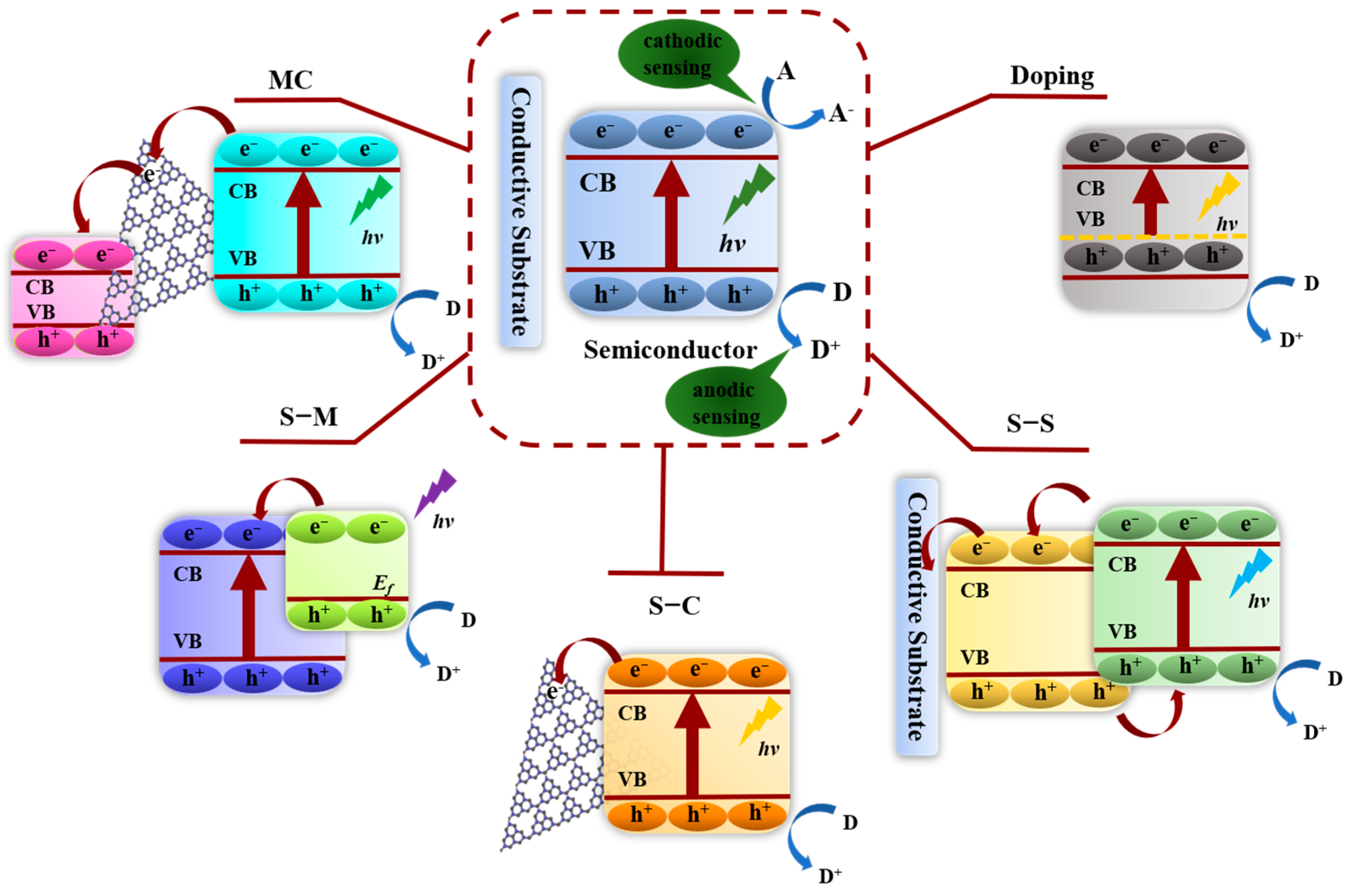
Photoactive materials act as a transducer converting the interactions between the analytes and PEC active materials, which play a crucial role in the PEC sensing system and demonstrate superior light capture capabilities, satisfactory charge separation efficiency, and appropriate energy level [37]. Excellent photoactive nanomaterials with high photoelectric conversion efficiency can greatly contribute to the excellent analytical performance of PEC sensors [48]. According to previous reports, photoactive materials can be divided into semiconductor and semiconductor-based heterojunctions. The semiconductor-based heterojunctions can effectively increase the photoelectric conversion efficiency of photoactive materials by means of heterojunctions, which usually have the following four categories, semiconductor–semiconductor heterojunctions (S–S heterojunctions), semiconductor–carbon heterojunctions (S–C heterojunctions), semiconductor–metal heterojunctions (S–M heterojunctions) and multi-component heterojunctions (MC heterojunctions), as shown in Figure 1. In addition, PEC sensing is based on anodic sensing as an example.

3.1. Semiconductors

Based on their good band gap alignment, controllable size, and excellent biological compatibility, a wide range of semiconductor materials, such as metal oxides [49,50,51,52], metal chalcogenides [53,54], Bi-based semiconductor materials [55,56,57], graphite-like carbon nitride (g-C3N4) [58,59], and organic polymers [60,61], have attracted enormous interest in the construction of PEC sensors for the analysis of toxins and abused drugs in the environment. However, owing to the short absorption threshold, high e-h+ recombination rate, and mismatch of band structure, the photoelectron production efficiency is low, which seriously hinders the feasibility of a single semiconductor. For example, Li et al. developed a photoelectrochemical detector for the determination of organic compounds based on the excellent oxidation power of nanostructured TiO2 photoanodes, which stemmed from the photo-hole generated under UV illumination [62]. Chen et al. fabricated a novel, simple, high-sensitivity, and high-selectivity MC–LR photoelectrochemical sensor on highly ordered and vertically aligned TiO2 nanotubes with molecularly imprinted polypyrrole (PPy) as the recognition element under a UV lamp [49]. Highly ordered and vertically aligned TiO2 nanotubes exhibit a sensitive photo-oxidation current due to their outstanding photocatalytic ability. PPy can recognize the MC–LR targets via the multiple hydrogen bond interactions. Then, the π bond delocalized electron system of PPy facilitates the separation of photogenerated electron–hole pairs and improves the photoelectric conversion efficiency that would enhance the PEC current response. Özcan et al. used commercial TiO2-modified FTO electrodes for the photoelectrochemical determination of paracetamol under three UV fluorescent lamps [63]. As UV light only accounts for about 3–4% of solar energy on earth [64], it is necessary to reduce the band gap of single semiconductor, such as TiO2 (3.2 eV) and ZnO (3.4 eV) [65], to make the photoactive materials exhibit high visible light responsive activity for the useful utilization of solar energy.
Element doping can be regarded as a simple and effective approach to adjusting the PEC performances of semiconductors [66,67], which narrows the bandgap energy of semiconductors and shifts the threshold wavelength to the visible light region because of changes in the nanocrystal electronic structure [68]. Wang et al. successfully grew novel Ti3+ self-doped branched rutile TiO2 nanorod arrays (NRAs) on FTO transparent conductive glass [69]. The Ti3+ defects in the TiO2 nanoparticles, the related oxygen vacancies, as well as the nanobranch structure, expended the optical absorption of the TiO2 NRAs from 400 nm to 510 nm. The doped and branched TiO2 NRAs with larger specific surface areas and stronger light absorption could significantly improve their photoelectrochemical properties. Li et al. constructed co-doped ZnO diluted magnetic semiconductor (DMS) for fabricating PEC aptasensors of acetamiprid (Figure 2a) [70]. On account of the sp-d exchange interactions between the band electron and the localized d electrons of Co2+, Co2+ doping is beneficial in expanding the optical absorption bandwidth of ZnO to the visible region and promoting the separation of photogenerated carriers. The linear range of the fabricated aptasensor for acetamiprid was 0.5–800 nmol L−1, and the detection limit was 0.18 nmol L−1. Zheng et al. fabricated a boron and graphene quantum dots-co-doped g-C3N4 (named GBCN) as a dopamine PEC sensor, which showed a low detection limit of 0.96 nM, a wide detection range of 0.001–800 μM, and good stability (no obvious photocurrent change was found both after 20 light source on/off cycles and 90-day storage), as shown in Figure 2b [71]. The excellent properties of the fabricated PEC-active material might be due to the following reasons: (1) GBCN has more dopamine active adsorption sites with the obviously increased specific surface area of g-C3N4 after co-doping; (2) co-doping makes the adsorption red shift from 470 to 540 nm and increases the absorption intensity; (3) the synergistic effect of boron and graphene quantum dots effectively promotes the migration of photogenerated electrons from the conduction band of g-C3N4 to graphene quantum dots, as well as facilitating charge separation.
Oxygen vacancies [48,72,73,74,75] and nitrogen vacancies [76] have also been reported to increase the charge separation efficiency and broaden the light adsorption range. Naldoni et al. synthesized novel crystalline core/disordered shell black TiO2 nanoparticles (NPs) with a reduced bandgap of only 1.85 eV, which could fit for visible light absorption, as shown in Figure 3 [72]. The experimental data analysis shows that oxygen vacancies are present in the bulk anatase crystalline phase, while the disordered NP surface appears to be nearly stoichiometric. Whereas, the experimental data analysis also proves that the narrowing of the bandgap is caused by the synergistic existence of oxygen vacancies and surface disorder. Cui et al. utilized the synergistic effect between the oxygen vacancies concentration and lattice strain of BiOBr nanosheets in order to improve the photoelectric response [74]. Yan et al. developed a ciprofloxacin (CIP) PEC sensor by using nitrogen-deficient graphitic carbon nitride (ND-g-CN) as a PEC-active material [76]. Nitrogen vacancies can be served as charge traps to effectively suppress the charge recombination, which works in conjunction with the 2D thin-sheet structures of the available charge separation and transfer to increase light capture and enhance PEC performance. The detection range of the designed PEC sensor for CIP is 60 to 19,090 ng L−1, and the detection limit is 20 ng L−1.

3.2. Semiconductor-Based Heterojunctions

Semiconductor-mediated photocatalysis suffers from the drawbacks of short photogenerated electron–hole pair lifetime and the limited visible light absorption. Fabricating semiconductor heterojunctions has been one of the most widely used strategies over the past decade [77]. In general, the sensitivity range of PEC sensors is strongly dependent on the charge transfer of photoactive materials, which benefits from the high carrier density [78]. The formation of semiconductor-based heterojunction materials allows for the incorporation of materials that can adjust the bandgap energy, increase the charge carrier density, and then further suppress the recombination of photogenerated electron–hole pairs and attain their purpose of improving optoelectronic conversion efficiency.

3.2.1. Semiconductor–Semiconductor Heterojunctions

The Bi-based semiconductors, such as BiPO4 [79,80,81,82], BiVO4 [83,84,85], Bi2WO6 [86,87,88], and BiFeO3 [89,90], are linked with the Bi 6s and O 2p hybrid orbitals of the narrowed bandgap as well as the deep valance band, which have received extensive attention in the study of PEC because of their efficient visible light photocatalytic activity [91]. Bi2WO6, as the simplest member of the Aurivillius family of layered perovskites, possesses good photon absorptivity under visible light irradiation [92]. However, unfortunately, it suffers from rapid photorecombination of carriers in a single component system. Adhikari et al. designed Bi2S3-sensitized Bi2WO6 microstructures topotactically via the hydrothermal technique under a controlled hydrothermal atmosphere and synthesized different weight percentage Bi2S3/Bi2WO6 microstructures for the evaluation of ofloxacin photo-oxidation under visible light [93]. In Figure 4b, after Bi2S3 is introduced into the Bi2WO6, we observe that a wide absorption band has been formed in the visible light range. The calculated energy bandgap is estimated to be 2.12 eV for 10 wt% Bi2S3/Bi2WO6. As shown in Figure 4a, both Bi2S3 and Bi2WO6 are excited to form a photogenerated electron–hole pair under the light illumination. The photogenerated electron in Bi2S3 flows to the conduction band of Bi2WO6, associated with the migration of photogenerated holes in the valence band of Bi2WO6 to the valence band of Bi2S3 simultaneously. Due to the favorable band positions of Bi2S3 and Bi2WO6, the electron–hole recombination could be effectively separated, which is helpful to the high photocatalytic degradation of ofloxacin. Therefore, when ofloxacin is added in the system, competition between photogenerated electrons to flow through the system and oxidize ofloxacin occurs, causing the decreasing of the photocurrent. On the basis of the surface plasmon resonance effect of metallic Bi, Li et al. coupled Bi and CuS, a narrow bandgap semiconducting material, with Bi2WO6 to prepare the Bi-CuS/Bi2WO6 heterojunction with enhanced separation of photo-generated charge carriers, charge transfer, and absorption wavelengths under visible light [87]. Then, a sensitive label-free PEC sensor found on Bi-CuS/Bi2WO6 nanocomposite was fabricated for the determination of paracetamol (PA), which is a ubiquitous NSAID. When PA is put into the electrolyte, the isolated holes on Bi2WO6 VB enter CuS to oxidize PA and acquire electrons, thus continuously producing photoelectrons to enhance the photocurrent signals.
TiO2 is the most widely used photoactive material, which has the advantages of strong photoactivity, good biocompatibility, low cost, and strong chemical stability in various environments [94,95]. Nevertheless, the light absorption in the visible region of TiO2 is limited to a great extent because of the wide bandgap. TiO2 can only absorb UV light (λ < 400 nm), which can kill biomolecules [96]. Therefore, the modification of TiO2 with narrow-bandgap semiconductors is crucial for the PEC biosensing applications to not only enhance its absorption in the visible range but also to improve the lifetime of charge carriers. CdS, CuS, MoS2, and Ag3VO4 are sensitive to the visible light excitation due to their narrow bandgap, about 2.27 eV, 2.1 eV, 1.9 eV, and 2.2 eV, respectively [73,97,98,99]. Wei et al. fabricated an ultrasensitive PEC immunoassay for the detection of MC–LR based on a sensitive PEC material of CdS/TiO2 nanorod arrays (CdS/TiO2 NRAs) to immobilize antigens [100]. The photogenerated holes of CdS NPs help to weaken the self-oxidation, limit the recombination of electron–hole pairs, as well as broaden the light absorption of TiO2 NRAs then immobilize antigens as a visible light-driven material. The photocurrent values of the proposed immunosensor are linear from 0.005 to 500 μg L−1 concentration of MC–LR, and the limit of detection is 0.001 μg L−1. Tang et al. prepared a CuS–TiO2 heterojunction composite via dispersedly depositing CuS NPs on TiO2 nanospheres’ surface, and they then constructed a PEC aptasensor for sensitive detection of MC–LR in an aquatic environment with a detection limit of 2.0 × 10⁻⁵ nM and good stability, of whose initial value the photocurrent response at the PEC aptasensor retained ~91.8% after 10 days’ storage in 4℃ refrigerator (Figure 4c) [6]. As shown in Figure 4d, the absorption edge of CuS–TiO2 heterojunction composite (~504 nm) is much longer than that of pure TiO2 (~388.5 nm). The intensified visible light absorption works together with the improved separation of photogenerated charges and the reduced self-aggregation of CuS nanoparticles to obtain the superior analytical performance for MC–LR. Fan et al. developed a highly sensitive atrazine PEC aptasensor by exploring TiO2 nanotubes decorated with MoS2 quantum dots (MoS2 QDs/TiO2 NTs) as a photoelectrode [47]. Because the visible light absorption range of TiO2 was effectively extended from UV to visible light by MoS2 QDs, the bioactivity of the photoelectrode was well maintained. Therefore, the photoelectrode exhibits high binding capacity of aptamer molecules to targets, which helped the designed atrazine PEC aptasensor achieve high sensitivity and specificity. Zhang et al. obtained a heterojunction of Ag3VO4 nanoparticles-deposited TiO2 nanotube arrays (TNTAs) on a Ti foil electrode, which cooperated with oxygen vacancies of TNTAs caused by the electrochemical reduction treatment to greatly improve the visible light absorption of TNTAs, as well as the charge separation of TNTAs [73].
In addition, other new heterojunctions have also been reported as photosensitive material for the construction of PEC sensors for the analysis of toxins and abused drugs in the environment, such as the CuCo2O4@CoO core–shell hybrid rod for selectively sensing diclofenac [37], ZnPc/CN (zinc phthalocyanine/graphitic nitride) nanocomposite for preparing sulfadimethoxine aptasensor [101], hollow ZnS–CdS nanocage-based molecularly imprinted (ZnS–CdS/rMIP) PEC sensor for sensitive measurement of oxytetracycline [54], CdS nanorod arrays (CdS NRs)/Ag2S on FTO for PEC immunoassay of ochratoxins, and so on. The report results show that the semiconductor–semiconductor heterojunctions usually exhibit significantly improved photocurrent response compared with the pure semiconductor, which could help to increase the analytical performance of the PEC sensors remarkably.

3.2.2. Semiconductor–Carbon Heterojunctions

Carbon materials, such as graphene, reduced graphene oxide (rGO), graphene quantum dots (GQDs), graphite-phase carbon nitride (g-C3N4), carbon nanotubes (CNTs), and so on, all show the inherent advantages of strong chemical stability, good biocompatibility, low toxicity, superior electron mobility, and high specific surface area [30,102]. Therefore, they are usually combined with other semiconductor materials by forming nanohybrids to fabricate high performance PEC sensors [103,104]. The dramatic analytical performances of the PEC sensors are achieved in the following ways: the adsorption of more electroactive species, shortening charge transport time and distance, acceleration of the transfer of photogenerated electrons, and suppression the recombination of charge carriers.
Graphene [105], N-doped graphene [3,48,78], and reduced graphene oxide (rGO) [106,107,108] are the most common carbon materials used to design semiconductor–carbon heterojunctions in PEC sensors. Because of the non-photoactivity and zero bandgap, graphene was usually employed as a carrier or electron transfer promoter for fabricating PEC sensors [109,110,111,112]. Yan et al. proposed a novel cathodic “signal-off” strategy for oxytetracycline PEC aptasensing based on BiOI–graphene nanocomposite [113]. In this sensor, p-type semiconductor BiOI was used as photoactive species, while doping BiOI with a certain amount of graphene can not only improve the absorption in the visible light region but also facilitate the electron transfer of the photoanode. Except pure graphene, N-doped graphene is also used in the PEC field according to the increased bandgap of graphene with the doped N element [114,115]. Zhang et al. designed a novel visible light-driven self-powered PEC aptasensor for MC–LR detection based on 3D N-doped graphene hydrogel/hematite nanocomposites (NGH/Fe2O3), as shown in Figure 5a [78]. Three-dimensional NGH could trap the holes to suppress the recombination, thus promoting the transmission of photo-induced electrons and increasing the conductivity. Therefore, the coupling NGH with Fe2O3 could generate a Schottky junction that could promote the separation of charges and increase the performance of the aptasensor with a wide linear range of 1 pM to 5 nM. Ge et al. synthesized Bi2MoO6 nanoparticles-anchored boron and nitrogen co-doped graphene (BNG) nanosheets nanocomposites with oxygen vacancy [48]. The incorporation of BNG nanosheets increased the oxygen vacancies, broadened the light adsorption range, and accelerated the charge transfer. So, a highly efficient PEC aptasensor for sensitive analysis of lincomycin was constructed based on the prepared Bi2MoO6/BNG photoactive nanocomposite. In addition to graphene and doped graphene, rGO has also been applied to PEC sensing for its improved water dispersibility and chemical reactivity. However, rGO is a p-type semiconductor with a narrow bandgap and relatively poor conductivity, so rGO is frequently coupled with other semiconductor materials in PEC sensing field [116,117]. Lu et al. developed a visible light-driven PEC aptasensor for MC–LR based on rGO/Ti–Fe–O nanotubes (NTs) [107]. It benefited from the large π-conjugated structure and unique electrical conductivity of rGO which endows a superhigh carrier migration rate. Therefore, the photo generated e- can transfer to the surface of rGO rapidly, which effectively promotes the separation of photo-generated e-h+ (as shown in Figure 5b). The fabricated aptasensor exhibits outstanding determination performance for MC–LR with a detection limitation of 5 fM.
Besides graphene-class materials, other carbon materials [38,39,40,118,119] are also used to synthesize semiconductor–carbon heterojunctions as photo-responsive materials for constructing PEC sensors. Liu et al. developed a MC–LR PEC sensor on a surface molecular-imprinted TiO2-coated multiwalled carbon nanotubes (MI–TiO2@CNTs) hybrid heterojunction nanostructure [119]. The sensor displayed a superior photocurrent response sensitivity and a wide linear range from 1.0 pM to 3.0 nM, which was mainly attributed to the high specific surface area and excellent photoelectric activity of the TiO2@CNTs heterojunction nanostructure. As shown in Figure 5c, TiO2 incurred charge separation under illumination, resulting in e-h+. When TiO2 NPs chemically bonded to the surface of CNTs, the electrons transferred from the CB of TiO2 to the surface of CNTs due to their unique electrical conductivity and high electron storage performance. Therefore, the residual holes in TiO2 VB could react with MC–LR that was absorbed on the electrode with MI recognition to produce an increased oxidation current. Gao et al. employed an electrophoretic deposition strategy to successfully fabricate a thin-film PEC sensor of functionalized N-doped graphene quantum dots embedded in BiOBr nanosheets that were loaded on ITO substrate (F-NGQDs/BiOBr/ITO) for paracetamol (PA) [38]. It was found that the thin film with NGQDs/BiOBr heterojunction showed enhanced charge transfer and absorption wavelengths under visible light, thus yielding an increase in the observed photocurrent. As shown in Figure 5d, the VB and CB positions of NGQDs are higher than those of BiOBr. The NGQDs are excited and undergo charge separation to generate e-h+. Since BiOBr has different CB energy levels, photogenerated electrons are rapidly transferred to BiOBr, and then the separated holes on the VB of BiOBr can enter NCQDs and further oxidize PA to obtain electrons, thereby continuing to produce photoelectrons. Li et al. fabricated a novel bismuth vanadate/two-dimensional carbon nitride/deoxyribonucleic acid (BiVO4/2D-C3N4/DNA) MC–LR aptamer PEC sensor with a detection sensitivity range of 5 × 10⁻⁷ to 10 μg L−1 [120]. The photogenerated charge transport process schematic diagram is shown in Figure 5e. 2D-C3N4 plays a key role in multifunctional interface reconciliation. To be specific, 2D-C3N4 not only serves as a photogenerated hole-oriented transfer medium from the BiVO4 photoanode to the detective target; it also fixes the DNA aptamer parallelly via the π–π bond to shorten the hole transfer distance from the semiconductor to the target.
Figure 5. (a) and (b) Mechanism diagram of the MC–LR aptasensors based on NGH/Fe2O3 Schottky junctions [78] and rGO/Ti–Fe–O NTs [107], respectively. (c) Photoexcitation and electron-charge transfer process of the MI–TiO2@CNTs/FTO [119]. (d) Electron-transfer mechanism of PA PEC sensor based on F-NGQDs/BiOBr/ITO electrode [38]. (e) Schematic of photogenerated charge transport process on BiVO4/2D-C3N4/DNA aptamer PEC sensor [120].
Chemosensors 11 00412 g005

3.2.3. Semiconductor–Metal Heterojunctions

The surface plasmon resonance (SPR) effect is an optical near-field effect on the localized surface of noble metal nanoparticles (e.g., Au, Ag, and Pd), resulting in enhanced local electromagnetic fields [121]. Thanks to this effect, the light absorption and photoelectric conversion efficiency of semiconductors can be greatly improved according to the excellent electron mediators of the noble metal nanoparticles through the rapid separation and transportation of photogenerated e-h+ pairs at the semiconductor/metal interface [122]. Au NPs are the most commonly used noble metal [123,124]. Xu et al. constructed a facile and sensitive PEC sensing platform based on plasmonic Au/graphitic carbon nitride composites (Au/g-C3N4) for the detection of 4-chlorophenol (4-CP) [125]. As shown in Figure 6a, upon the illumination on the surface of Au/g-C3N4 composites, the photogenerated electrons of g-C3N4 can be quickly injected into Au. Due to the SPR effect of Au NPs, the interaction between Au NPs and incident light can generate a strong local electromagnetic field that induces plasmon oscillation on the local surface of Au NPs. Therefore, the photogenerated electrons are retained on the surface of Au NPs, which contributes to the effective separation of photogenerated e-h+. At last, large number of electrons accumulating on the CB of g-C3N4 and on the surface of Au NPs can flow to the external circuit via the ITO substrate, resulting a significant enhancement in the photocurrent density. Upon 4-CP introduction, holes in the g-C3N4 VB can be sacrificed by 4-CP, which suppresses the e-h+ recombination then further enhances the photocurrent density. Employing ZnCuInSe (Zn-doped CuInSe2 quantum dots)/Au/TiO2 nanowires sandwich structure, Geng et al. developed a facile PEC aptasensor for kanamycin detection with a linear response range from 0.2 to 250 nM [126]. The Au NPs, which were sandwiched between the TiO2 nanowires and the ZnCuInSe QDs layer, play a dual role in enhancing the photocurrent, as shown in Figure 6b. One is to serve as a photosensitizer that extends the light absorption of the electrode to the visible range; the other is to act as an electron relay that promotes charge transfer between the TiO2 nanowires and ZnCuInSe QDs. Zhang et al. fabricated an atrazine aptasensor based on a Au NPs-modified 3D-ordered microporous (3DOM) TiO2 nanostructure frame with high sensitivity, selectivity, and stability [127]. The fabricated PEC aptasensor exhibits the detection limit of 0.167 ng/L, outstanding anti-interference ability in 100-fold concentration of other endocrine disrupting compounds, and stable photocurrent response after over 2000 s visible light illumination. As shown in Figure 6c, under the visible light illumination, the photogenerated electrons are excited to the CB of 3DOM and the holes left. Au NPs can also produce hot electrons and holes via the SPR effect. According to the energy level of hot electrons and hot holes of Au NPs, as well as the CB and VB of TiO2, the hot electrons generated by Au NPs are easily transferred to the CB of TiO2, and then both are transferred to the external circuit through the FTO to generate photocurrent. As a result, visible light absorption is effectively enhanced and the recombination of photogenerated carriers is efficiently decreased.
In addition to Au, Ag [128,129,130], Cu [131,132], Pd [133,134], Bi [135,136], Pt [137] are also applied to construct composites with semiconductors, which are used as the substrates for the PEC sensor. Wang et al. developed a 4-chlorophenol (4-CP) PEC sensor based on ternary composites of Ag NPs, graphitic carbon nitride (GCN), and carbon spheres (CS) (Ag/GCN/CS), which shows that an enhanced PEC response might be attributed to the synergistic effect between the SPR of Ag NPs and the electron-transfer behavior of CS [128]. The plausible PEC mechanism of the Ag/GCN/CS/ITO is discussed in two incident light spectral regions of 420–550 nm and 550–800 nm, considering the absorption edge of pure GCN (about 550 nm), as shown in Figure 6d. Under the illumination of 420–550 nm, Ag NPs, as electron sinks, can capture the electrons generated by the excited GCN, thereby inhibiting e-h+ recombination. Meanwhile, in the wavelength range of 550–800 nm, a strong oscillation of electrons occurs on the surface of Ag NPs according to the SPR effect, resulting in the transfer of hot electrons from the surface of Ag NPs to the CB of GCN, which can effectively reduce the recombination rate of electrons and holes. Xu et al. prepared a nanocomposite of Bi-doped ultrathin polymeric carbon nitride (Bi/CV-PCN) with carbon vacancies (CVs), displaying high PEC activity and stability, to utilize as the photoactive material for fabricating an enrofloxacin (ENR) aptasensor. As shown in Figure 6e, the electrons in the VB of PCN are excited to the CVs under visible light irradiation, following further transition to the CB of PCN. Since the CB of PCN is more negative than the Fermi level of Bi, the electrons are subsequently transferred from PCN to metal Bi. The addition of Bi metal plays two roles in improving the light trapping ability of PCN, besides accelerating the separation of photogenerated charge carries according to the built-in electric field originating from the SPR effect of Bi. At last, the holes left in the VB of PCN directly oxidize the ENR to produce photocurrent. The synergistic effect of the CVs that form an intermediate energy level, and the SPR effect of Bi, effectively boost the visible light harvesting ability and improve charge separation efficiency, finally resulting in a remarkable improvement on the PEC performance.
Figure 6. (a) Photocurrent generation mechanism schematic diagram of Au/g-C3N4/ITO electrode [125]. (b) Dual role of the Au nanoparticles in the ZnCuInSe/Au/TiO2 sandwich structure serving as a plasmonic photosensitizer and an electron relay [126]. (c) The mechanism of atrazine PEC sensor based on Au NPs/3DOM TiO2/FTO [127]. (d) The plausible PEC mechanism of the Ag/GCN/CS/ITO in different spectral regions of (i) 420–550 nm and (ii) 550–800 nm [128]. (e) Electron-transfer mechanism of ENR sensing based on the Bi/CV-PCN composite [136].
Chemosensors 11 00412 g006

3.2.4. Multi-Component Heterojunctions

Considering the synergistic effect, multi-component heterojunctions composed of visible light active components and electron transfer systems are spatially integrated for the purpose of simultaneously achieving an enhanced visible light response and transfer of charge carriers. Semiconductor, carbon material, and noble metal nanoparticles amount to a good combination, which effectively combines the light absorption and electron transfer ability and SPR effect [128]. Ouyang et al. designed a heterojunction based on coupling CeO2 quantum dots (QDs) with graphitic carbon nitride (g-CN) and Au nanoparticles (ACG) for fabricating a self-powered PEC MC–LR aptasensor with a detection limit of 0.01 pM [40]. As shown in Figure 7a, because of the combination of CeO2 QDs and the g-CN nanosheet into a highly dispersed nanocomposite, the matched band potentials and closely contacted interface between them can efficiently suppress the recombination of photo-generated electron–hole pairs and promote charge separation, which benefits the as-obtained PEC aptasensor to exhibit excellent analytical performances. The Au nanoparticles not only provide many binding sites for MC–LR aptamers through Au-S bond; they also greatly increase the light absorption capacity because of the SPR effect. Similarly, the CoO/Au/g-C3N4 Z-scheme heterojunction [138], Au nanoparticles, and graphene-doped CdS composite [139] were designed to fabricate the PEC sensor.
Semiconductorsand dye molecules-co-sensitized TiO2 is also a good combination. The dye molecules can be adsorbed on the surface of semiconductors, increasing their photo response owing to the greater absorption of the light in the visible region [140]. Lima et al. used TiO2 to co-sensitize with N-methylphenazonium methyl sulfate (PMS) and CdTe QDs; the composite was then immobilized in a Nafion film that severed as the photoelectroactive element for the development of a PEC sensor for the detection of tannic acid [141]. PMS was exploited as a co-modifier of the TiO2, aimed to produce a stable materialthat photosensitive to visible LED light in this study. Sousa et al. fabricated a naringin PEC sensor on a zero-biased fluorine-doped tin oxide (FTO) electrode modified with CdS and chloroprotoporphyrin IX iron(III) (CPPI)-sensitized TiO2, designated CPPI–TiO2/CdS/FTO [142]. The proposed mechanism for the naringin PEC detection is shown in Figure 7b. The CPPI adsorbed on TiO2 particles can absorb light to produce photoexcited states (CPPI*) then inject electrons into the conduction band of TiO2, resulting, simultaneously, in an oxidized form of CPPI+. On the other hand, the CdS can also be excited by visible light to produce electrons and holes in the CB and VB, respectively. Therefore, the photoelectron at the CB of TiO2 can react with the holes at the VB of CdS via a mean z-scheme. Eventually, the photoelectrons on the CB of CdS can be collected from the FTO electrode to generate an anodic photocurrent. In addition to the above-mentioned multi-component heterojunctions, other combinations have also been reported for the analysis of toxins and abused drugs in the environment, such as CdS/Fe2O3 co-sensitized TiO2 nanorod arrays [143], TiO2/CNTs/CdTeQDs [144], In2O3−In2S3−Ti3C2 composite [145], and so on.
Figure 7. (a) The mechanism of MC–LR PEC aptasensor based on ACG heterostructure [40]. (b) Proposed mechanism for the naringin photoelectrochemical detection based on the CPPI–TiO2/CdS platform. CPPI* means the photoexcited states of CPPI [142].
Chemosensors 11 00412 g007

4. Summary and Outlook

Due to the high sensitivity and low cost of PEC sensors, numerous PEC sensors have been fabricated for the rapid and accurate monitoring of toxins and abused drugs in the environment. It was found that the photoelectric conversion efficiency was obviously improved via the innovative design and controllable preparation of photoactive materials. This review mainly summarizes the composition of the engineered photoactive materials, the sensing mechanisms, and actual applications of the fabricated PEC sensors.
Although great progress in the PEC sensing of toxins and abused drugs in the environment has been made by far, there is still plenty of room for development and further applications in the future. Firstly, multi-signal amplification strategies and multi-component heterojunctions are usually used to construct PEC sensors, which might increase the fabrication difficulty and cost, as well as reduce the stability of the PEC sensors. Therefore, it is of great importance to develop highly efficient signal amplification strategies and to prepare high-quality and environmentally friendly photoactive heterojunctions because employment of the often-used, traditional QDs (e.g., CdS) would lead to a second pollution. In addition, most of the reported PEC sensors are focused on the quantitative single-target analysis. A broad–specific PEC immunosensor was constructed for the simultaneous detection of ochratoxin A, ochratoxin B, and ochratoxin C because of the electronegativity of the chlorine atom that increases the antibody binding ability. Considering the difficulty of broad-spectrum antibodies, research can focus on developing an aptamer with multiple recognition sites for multiple-target detection simultaneously. Moreover, integrating PEC sensors with microfluidics and chips is also a good strategy by which to increase detection efficiency via multi-channel simultaneous detection. A microfluidic system with a paper working electrode combined with the PEC sensing technique is also an intelligent design that could facilitate miniaturization and assist integration into portable devices. Furthermore, qualitative detection of unknown toxins and abused drugs in the environment is also urgently needed. The designing of photoactive materials with specific sites and coupling PEC sensing with other methods, such as cyclic voltammetry (CV) and differential pulse voltammetry (DPV), have been reported as effective ways. However, a highly stable light source is usually required to excite the PEC sensors, which also greatly limits their practicality. Consequently, solar-powered and self-powered PEC sensors combined with energy conversion devices are being developed.

Author Contributions

Writing—original draft preparation, Y.M.; X.L.; Y.B. and L.N.; writing—review and editing, Y.M.; X.L.; Y.B. and L.N.; supervision, Y.B.; All authors have read and agreed to the published version of the manuscript.

Funding

This research was funded by the National Natural Science Foundation of China (No. 21904025 and No. 21974033), the Science and Technology Project of Guangzhou (No. 202102020598 and No. 202201000002), and the Department of Science and Technology of Guangdong Province (ID:2022A156).

Institutional Review Board Statement

Not applicable.

Informed Consent Statement

Not applicable.

Conflicts of Interest

The authors declare no conflict of interest.

References

  1. Munoz, M.; Cires, S.; de Pedro, Z.M.; Colina, J.A.; Velasquez-Figueroa, Y.; Carmona-Jimenez, J.; Caro-Borrero, A.; Salazar, A.; Fuster, M.C.S.; Contreras, D.; et al. Overview of toxic cyanobacteria and cyanotoxins in Ibero-American freshwaters: Challenges for risk management and opportunities for removal by advanced technologies. Sci. Total Environ. 2021, 761, 143197. [Google Scholar] [CrossRef]
  2. Chen, L.K.; Guo, C.S.; Sun, Z.Y.; Xu, J. Occurrence, bioaccumulation and toxicological effect of drugs of abuse in aquatic ecosystem: A review. Environ. Res. 2021, 200, 111362. [Google Scholar] [CrossRef]
  3. Du, X.; Jiang, D.; Li, H.; Hao, N.; You, T.; Wang, K. An intriguing signal-off responsive photoelectrochemical aptasensor for ultrasensitive detection of microcystin-LR and its mechanism study. Sens. Actuator B-Chem. 2018, 259, 316–324. [Google Scholar] [CrossRef]
  4. Xu, J.Y.; Zhang, W.Y.; Zhong, S.Z.; Xie, X.X.; Che, H.M.; Si, W.R.; Tuo, X.; Xu, D.X.; Zhao, S.J. Microcystin-leucine-arginine affects brain gene expression programs and behaviors of offspring through paternal epigenetic information. Sci. Total Environ. 2023, 857, 159032. [Google Scholar] [CrossRef]
  5. Guo, X.; Meng, R.Y.; Liu, J.J.; Zhang, S.Y.; Liu, H.H.; Du, X.D.; Zhang, H.Z.; Li, Y.S. Microcystin leucine arginine induces human sperm damage: Involvement of the Ca2+/CaMKKβ/AMPK pathway. Ecotoxicol. Environ. Saf. 2023, 256, 114845. [Google Scholar] [CrossRef]
  6. Tang, Y.; Chai, Y.; Liu, X.; Li, L.; Yang, L.; Liu, P.; Zhou, Y.; Ju, H.; Cheng, Y. A photoelectrochemical aptasensor constructed with core-shell CuS-TiO2 heterostructure for detection of microcystin-LR. Biosens. Bioelectron. 2018, 117, 224–231. [Google Scholar] [CrossRef] [PubMed]
  7. Krishnan, R.Y.; Manikandan, S.; Subbaiya, R.; Biruntha, M.; Balachandar, R.; Karmegam, N. Origin, transport and ecological risk assessment of illicit drugs in the environment—A review. Chemosphere 2023, 311, 137091. [Google Scholar] [CrossRef]
  8. Metcalfe, C.D.; Bayen, S.; Desrosiers, M.; Munoz, G.; Sauve, S.; Yargeau, V. An introduction to the sources, fate, occurrence and effects of endocrine disrupting chemicals released into the environment. Environ. Res. 2022, 207, 112658. [Google Scholar] [CrossRef] [PubMed]
  9. Fent, K.; Weston, A.A.; Caminada, D. Ecotoxicology of human pharmaceuticals. Aquat. Toxicol. 2006, 76, 122–159. [Google Scholar] [CrossRef]
  10. Michalaki, A.; Grintzalis, K. Acute and Transgenerational Effects of Non-Steroidal Anti-Inflammatory Drugs on Daphnia magna. Toxics 2023, 11, 320. [Google Scholar] [CrossRef]
  11. Parolini, M. Toxicity of the Non-Steroidal Anti-Inflammatory Drugs (NSAIDs) acetylsalicylic acid, paracetamol, diclofenac, ibuprofen and naproxen towards freshwater invertebrates: A review. Sci. Total Environ. 2020, 740, 140043. [Google Scholar] [CrossRef]
  12. Patel, M.; Kumar, R.; Kishor, K.; Mlsna, T.; Pittman, C.U.; Mohan, D. Pharmaceuticals of Emerging Concern in Aquatic Systems: Chemistry, Occurrence, Effects, and Removal Methods. Chem. Rev. 2019, 119, 3510–3673. [Google Scholar] [CrossRef] [Green Version]
  13. Marmon, P.; Owen, S.F.; Margiotta-Casaluci, L. Pharmacology-informed prediction of the risk posed to fish by mixtures of non-steroidal anti-inflammatory drugs (NSAIDs) in the environment. Environ. Int. 2021, 146, 106222. [Google Scholar] [CrossRef] [PubMed]
  14. Zastepa, A.; Pick, F.R.; Blais, J.M.; Saleem, A. Analysis of intracellular and extracellular microcystin variants in sediments and pore waters by accelerated solvent extraction and high performance liquid chromatography-tandem mass spectrometry. Anal. Chim. Acta 2015, 872, 26–34. [Google Scholar] [CrossRef] [PubMed]
  15. Fresco-Cala, B.; Gálvez-Vergara, A.; Cárdenas, S. Preparation, characterization and evaluation of hydrophilic polymers containing magnetic nanoparticles and amine-modified carbon nanotubes for the determination of anti-inflammatory drugs in urine samples. Talanta 2020, 218, 121124. [Google Scholar] [CrossRef]
  16. Vardali, S.; Papadouli, C.; Rigos, G.; Nengas, I.; Panagiotaki, P.; Golomazou, E. Recent Advances in Mycotoxin Determination in Fish Feed Ingredients. Molecules 2023, 28, 2519. [Google Scholar] [CrossRef] [PubMed]
  17. Foss, A.J.; Aubel, M.T. Using the MMPB technique to confirm microcystin concentrations in water measured by ELISA and HPLC (UV, MS, MS/MS). Toxicon 2015, 104, 91–101. [Google Scholar] [CrossRef]
  18. Liu, J.; Xing, Y.; Lin, Y.; Xie, Y.; Zhou, X. Effect of pretreatment approach on the ELISA-based detection of cyanotoxins in water: Analysis and application. Sci. Total Environ. 2023, 871, 161988. [Google Scholar] [CrossRef]
  19. Garg, K.; Villavicencio-Aguilar, F.; Solano-Rivera, F.; Gilbert, L. Analytical Validation of a Direct Competitive ELISA for Multiple Mycotoxin Detection in Human Serum. Toxins 2022, 14, 727. [Google Scholar] [CrossRef]
  20. Abdelwahab, N.S.; Abdelrahman, M.M. Simultaneous Determination of Methocarbamol and Ibuprofen by First Derivative Synchronous Fluorescence Spectroscopic Method in Their Binary Mixture and Spiked Human Plasma. J. Fluoresc. 2014, 24, 129–135. [Google Scholar] [CrossRef]
  21. Pouyanfar, N.; Harofte, S.Z.; Soltani, M.; Siavashy, S.; Asadian, E.; Ghorbani-Bidkorbeh, F.; Kecili, R.; Hussain, C.M. Artificial intelligence-based microfluidic platforms for the sensitive detection of environmental pollutants: Recent advances and prospects. Trends Environ. Anal. Chem. 2022, 34, e00160. [Google Scholar] [CrossRef]
  22. Zhao, W.-W.; Xu, J.-J.; Chen, H.-Y. Photoelectrochemical bioanalysis: The state of the art. Chem. Soc. Rev. 2015, 44, 729–741. [Google Scholar] [CrossRef] [PubMed]
  23. Ma, X.; Kang, J.; Wu, Y.; Pang, C.; Li, S.; Li, J.; Xiong, Y.; Luo, J.; Wang, M.; Xu, Z. Recent advances in metal/covalent organic framework-based materials for photoelectrochemical sensing applications. Trac-Trends Anal. Chem. 2022, 157, 116793. [Google Scholar] [CrossRef]
  24. Bilge, S.; Sınağ, A. Current trends and strategies in the development of green MXene-based photoelectrochemical sensing application. Trac-Trends Anal. Chem. 2023, 163, 117059. [Google Scholar] [CrossRef]
  25. Qureshi, A.; Shaikh, T.; Niazi, J.H. Semiconductor quantum dots in photoelectrochemical sensors from fabrication to biosensing applications. Analyst 2023, 148, 1633–1652. [Google Scholar] [CrossRef]
  26. Wang, H.-Y.; Xu, Y.-T.; Wang, B.; Yu, S.-Y.; Shi, X.-M.; Zhao, W.-W.; Jiang, D.; Chen, H.-Y.; Xu, J.-J. A Photoelectrochemical Nanoreactor for Single-Cell Sampling and Near Zero-Background Faradaic Detection of Intracellular microRNA. Angew. Chem. Int. Ed. 2022, 61, e202212752. [Google Scholar]
  27. Li, C.-J.; Hu, J.; Gao, G.; Chen, J.-H.; Wang, C.-S.; Zhou, H.; Chen, G.; Qu, P.; Lin, P.; Zhao, W.-W. Biomolecules-Incorporated Metal-Organic Frameworks Gated Light-Sensitive Organic Photoelectrochemical Transistor for Biodetection. Adv. Funct. Mater. 2023, 33, 2211277. [Google Scholar] [CrossRef]
  28. Shi, L.; Yin, Y.; Zhang, L.-C.; Wang, S.; Sillanpää, M.; Sun, H. Design and engineering heterojunctions for the photoelectrochemical monitoring of environmental pollutants: A review. Appl. Catal. B 2019, 248, 405–422. [Google Scholar] [CrossRef]
  29. Yang, L.; Liu, X.; Li, L.; Zhang, S.; Zheng, H.; Tang, Y.; Ju, H. A visible light photoelectrochemical sandwich aptasensor for adenosine triphosphate based on MgIn2S4-TiO2 nanoarray heterojunction. Biosens. Bioelectron. 2019, 142, 111487. [Google Scholar] [CrossRef]
  30. Wang, H.; Xu, Y.; Xu, D.; Chen, L.; Qiu, X.; Zhu, Y. Graphitic Carbon Nitride for Photoelectrochemical Detection of Environmental Pollutants. ACS EST Engg. 2022, 2, 140–157. [Google Scholar] [CrossRef]
  31. Hao, N.; Zhang, Y.; Zhong, H.; Zhou, Z.; Hua, R.; Qian, J.; Liu, Q.; Li, H.; Wang, K. Design of a Dual Channel Self-Reference Photoelectrochemical Biosensor. Anal. Chem. 2017, 89, 10133–10136. [Google Scholar] [CrossRef] [PubMed]
  32. Zhou, Q.; Tang, D. Recent advances in photoelectrochemical biosensors for analysis of mycotoxins in food. Trac-Trends Anal. Chem. 2020, 124, 115814. [Google Scholar] [CrossRef]
  33. Peng, B.; Tang, L.; Zeng, G.M.; Zhou, Y.Y.; Zhang, Y.; Long, B.Q.; Fang, S.Y.; Chen, S.; Yu, J.F. Current Progress in Aptasensors for Heavy Metal Ions Based on Photoelectrochemical Method: A Review. Curr. Anal. Chem. 2018, 14, 4–12. [Google Scholar] [CrossRef]
  34. Wang, H.; Zhang, B.; Tang, Y.; Wang, C.; Zhao, F.; Zeng, B. Recent advances in bismuth oxyhalide-based functional materials for photoelectrochemical sensing. Trac-Trends Anal. Chem. 2020, 131, 116020. [Google Scholar] [CrossRef]
  35. Chen, W.F.; Liu, S.Y.; Fu, Y.K.; Yan, H.C.; Qin, L.; Lai, C.; Zhang, C.; Ye, H.Y.; Chen, W.J.; Qin, F.Z.; et al. Recent advances in photoelectrocatalysis for environmental applications: Sensing, pollutants removal and microbial inactivation. Coord. Chem. Rev. 2022, 454, 214341. [Google Scholar] [CrossRef]
  36. Shi, Y.T.; Zou, Y.T.; Khan, M.S.; Zhang, M.G.; Yan, J.; Zheng, X.H.; Wang, W.Q.; Xie, Z.G. Metal-organic framework-derived photoelectrochemical sensors: Structural design and biosensing technology. J. Mater. Chem. C 2023, 11, 3692–3709. [Google Scholar] [CrossRef]
  37. Wang, Q.; Jiang, M.; Zhang, L. Label-free and visible-light driven photoelectrochemical sensor with CuCo2O4@CoO Core-shell hybrid rod as photoanode for selective sensing diclofenac. Electrochim. Acta 2021, 397, 139239. [Google Scholar] [CrossRef]
  38. Gao, K.; Bai, X.; Zhang, Y.; Ji, Y. N–doped graphene quantum dots embedded in BiOBr nanosheets as hybrid thin film electrode for quantitative photoelectrochemical detection paracetamol. Electrochim. Acta 2019, 318, 422–429. [Google Scholar] [CrossRef]
  39. Shi, T.; Wen, Z.; Ding, L.; Liu, Q.; Guo, Y.; Ding, C.; Wang, K. Visible/near-infrared light response VOPc/carbon nitride nanocomposites: VOPc sensitizing carbon nitride to improve photo-to-current conversion efficiency for fabricating photoelectrochemical diclofenac aptasensor. Sens. Actuator B-Chem. 2019, 299, 126834. [Google Scholar] [CrossRef]
  40. Ouyang, X.; Tang, L.; Feng, C.; Peng, B.; Liu, Y.; Ren, X.; Zhu, X.; Tan, J.; Hu, X. Au/CeO2/g-C3N4 heterostructures: Designing a self-powered aptasensor for ultrasensitive detection of Microcystin-LR by density functional theory. Biosens. Bioelectron. 2020, 164, 112328. [Google Scholar] [CrossRef] [PubMed]
  41. Bostan, H.B.; Taghdisi, S.M.; Bowen, J.L.; Demertzis, N.; Rezaee, R.; Panahi, Y.; Tsatsakis, A.M.; Karimi, G. Determination of microcystin-LR, employing aptasensors. Biosens. Bioelectron. 2018, 119, 110–118. [Google Scholar] [CrossRef]
  42. Fan, L.; Xiao, G.; Wang, M.; Zhao, S.; Yang, Q.; Cheng, L.; Huang, J.J.; Yue, Z. Ultrasensitive photoelectrochemical microcystin-LR immunosensor using carboxyl-functionalized graphene oxide enhanced gold nanoclusters for signal amplification. Anal. Chim. Acta 2021, 1185, 339078. [Google Scholar] [CrossRef] [PubMed]
  43. Qileng, A.; Wei, J.; Lu, N.; Liu, W.; Cai, Y.; Chen, M.; Lei, H.; Liu, Y. Broad-specificity photoelectrochemical immunoassay for the simultaneous detection of ochratoxin A, ochratoxin B and ochratoxin C. Biosens. Bioelectron. 2018, 106, 219–226. [Google Scholar] [CrossRef]
  44. Bai, X.Y.; Gao, W.K.; Zhou, C.H.; Zhao, D.Y.; Zhang, Y.; Jia, N.Q. Photoelectrochemical determination of diclofenac using oriented single-crystalline TiO2 nanoarray modified with molecularly imprinted polypyrrole. Microchim. Acta 2022, 189, 90. [Google Scholar] [CrossRef]
  45. Chen, J.; Gao, P.; Wang, H.; Han, L.; Zhang, Y.; Wang, P.; Jia, N. A PPy/Cu2O molecularly imprinted composite film-based visible light-responsive photoelectrochemical sensor for microcystin-LR. J. Mater. Chem. 2018, 6, 3937–3944. [Google Scholar] [CrossRef]
  46. Wang, H.; Liang, D.; Xu, Y.; Liang, X.; Qiu, X.; Lin, Z. A highly efficient photoelectrochemical sensor for detection of chlorpyrifos based on 2D/2D β-Bi2O3/g-C3N4 heterojunctions. Environ. Sci.-Nano 2021, 8, 773–783. [Google Scholar] [CrossRef]
  47. Fan, L.; Zhang, C.; Liang, G.; Yan, W.; Guo, Y.; Bi, Y.; Dong, C. Highly sensitive photoelectrochemical aptasensor based on MoS2 quantum dots/TiO2 nanotubes for detection of atrazine. Sens. Actuator B-Chem. 2021, 334, 129652. [Google Scholar] [CrossRef]
  48. Ge, L.; Liu, Q.; Jiang, D.; Ding, L.; Wen, Z.; Guo, Y.; Ding, C.; Wang, K. Oxygen vacancy enhanced photoelectrochemical performance of Bi2MoO6/B, N co-doped graphene for fabricating lincomycin aptasensor. Biosens. Bioelectron. 2019, 135, 145–152. [Google Scholar] [CrossRef]
  49. Chen, K.; Liu, M.; Zhao, G.; Shi, H.; Fan, L.; Zhao, S. Fabrication of a Novel and Simple Microcystin-LR Photoelectrochemical Sensor with High Sensitivity and Selectivity. Environ. Sci. Technol. 2012, 46, 11955–11961. [Google Scholar] [CrossRef]
  50. Zhang, Z.; Huang, L.; Sheng, S.; Jiang, C.; Wang, Y. MIL-125(Ti)-derived COOH functionalized TiO2 grafted molecularly imprinted polymers for photoelectrochemical sensing of ofloxacin. Sens. Actuator B-Chem. 2021, 343, 130119. [Google Scholar] [CrossRef]
  51. Zu, M.; Zhou, X.; Zhang, S.; Qian, S.; Li, D.-S.; Liu, X.; Zhang, S. Sustainable engineering of TiO2-based advanced oxidation technologies: From photocatalyst to application devices. J. Mater. Sci. Technol. 2021, 78, 202–222. [Google Scholar] [CrossRef]
  52. Liu, Y.; Gan, X.; Zhou, B.; Xiong, B.; Li, J.; Dong, C.; Bai, J.; Cai, W. Photoelectrocatalytic degradation of tetracycline by highly effective TiO2 nanopore arrays electrode. J. Hazard. Mater. 2009, 171, 678–683. [Google Scholar] [CrossRef] [PubMed]
  53. Shamraiz, U.; Hussain, R.A.; Badshah, A. Fabrication and applications of copper sulfide (CuS) nanostructures. J. Solid State Chem. 2016, 238, 25–40. [Google Scholar] [CrossRef]
  54. Bai, X.; Zhang, Y.; Gao, W.; Zhao, D.; Yang, D.; Jia, N. Hollow ZnS–CdS nanocage based photoelectrochemical sensor combined with molecularly imprinting technology for sensitive detection of oxytetracycline. Biosens. Bioelectron. 2020, 168, 112522. [Google Scholar] [CrossRef] [PubMed]
  55. Yan, P.; Jiang, D.; Li, H.; Bao, J.; Xu, L.; Qian, J.; Chen, C.; Xia, J. BiPO4 nanocrystal/BiOCl nanosheet heterojunction as the basis for a photoelectrochemical 4-chlorophenol sensor. Sens. Actuator B-Chem. 2019, 279, 466–475. [Google Scholar] [CrossRef]
  56. Yu, S.-Y.; Zhang, L.; Zhu, L.-B.; Gao, Y.; Fan, G.-C.; Han, D.-M.; Chen, G.; Zhao, W.-W. Bismuth-containing semiconductors for photoelectrochemical sensing and biosensing. Coord. Chem. Rev. 2019, 393, 9–20. [Google Scholar] [CrossRef]
  57. Zhou, Y.; Yin, H.; Ai, S. Recent advances and applications of Bi2S3-based composites in photoelectrochemical sensors and biosensors. Trac-Trends Anal. Chem. 2023, 158, 116876. [Google Scholar] [CrossRef]
  58. Li, R.; Liu, Y.; Cheng, L.; Yang, C.; Zhang, J. Photoelectrochemical Aptasensing of Kanamycin Using Visible Light-Activated Carbon Nitride and Graphene Oxide Nanocomposites. Anal. Chem. 2014, 86, 9372–9375. [Google Scholar] [CrossRef]
  59. Low, S.S.; Chen, Z.; Li, Y.; Lu, Y.; Liu, Q. Design principle in biosensing: Critical analysis based on graphitic carbon nitride (G-C3N4) photoelectrochemical biosensor. Trac-Trends Anal. Chem. 2021, 145, 116454. [Google Scholar] [CrossRef]
  60. Li, Y.; Zhang, N.; Zhao, W.-W.; Jiang, D.-C.; Xu, J.-J.; Chen, H.-Y. Polymer Dots for Photoelectrochemical Bioanalysis. Anal. Chem. 2017, 89, 4945–4950. [Google Scholar] [CrossRef]
  61. Wang, C.; Wang, H.; Zhang, M.; Zeng, B.; Zhao, F. Molecularly imprinted photoelectrochemical sensor for aflatoxin B1 detection based on organic/inorganic hybrid nanorod arrays. Sens. Actuator B-Chem. 2021, 339, 129900. [Google Scholar] [CrossRef]
  62. Li, L.; Zhang, S.; Zhao, H. A low cost universal photoelectrochemical detector for organic compounds based on photoelectrocatalytic oxidation at a nanostructured TiO2 photoanode. J. Electroanal. Chem. 2011, 656, 211–217. [Google Scholar] [CrossRef] [Green Version]
  63. Özcan, L. Photoelectrochemical determination of paracetamol by using TiO2 modified FTO electrodes. Anal. Bioanal. Electrochem. 2019, 11, 1117–1128. [Google Scholar]
  64. Lin, Y.-M.; Tseng, Y.-H.; Huang, J.-H.; Chao, C.C.; Chen, C.-C.; Wang, I. Photocatalytic Activity for Degradation of Nitrogen Oxides over Visible Light Responsive Titania-Based Photocatalysts. Environ. Sci. Technol. 2006, 40, 1616–1621. [Google Scholar] [CrossRef] [PubMed]
  65. Fan, D.; Wang, H.; Khan, M.S.; Bao, C.; Wang, H.; Wu, D.; Wei, Q.; Du, B. An ultrasensitive photoelectrochemical immunosensor for insulin detection based on BiOBr/Ag2S composite by in-situ growth method with high visible-light activity. Biosens. Bioelectron. 2017, 97, 253–259. [Google Scholar] [CrossRef]
  66. Chava, R.K.; Kang, M. Improving the photovoltaic conversion efficiency of ZnO based dye sensitized solar cells by indium doping. J. Alloys Compd. 2017, 692, 67–76. [Google Scholar] [CrossRef]
  67. Chahrour, K.M.; Ooi, P.C.; Eid, A.M.; Nazeer, A.A.; Madkour, M.; Dee, C.F.; Wee, M.F.M.R.; Hamzah, A.A. Synergistic effect of bi-phased and self-doped Ti3+ on anodic TiO2 nanotubes photoelectrode for photoelectrochemical sensing. J. Alloys Compd. 2022, 900, 163496. [Google Scholar] [CrossRef]
  68. Chen, L.-C.; Tu, Y.-J.; Wang, Y.-S.; Kan, R.-S.; Huang, C.-M. Characterization and photoreactivity of N-, S-, and C-doped ZnO under UV and visible light illumination. J. Photochem. Photobiol. A 2008, 199, 170–178. [Google Scholar] [CrossRef]
  69. Wang, J.; Wang, X.; Yan, J.; Tan, Q.; Liang, G.; Qu, S.; Zhong, Z. Enhanced Photoelectrochemical Properties of Ti3+ Self-Doped Branched TiO2 Nanorod Arrays with Visible Light Absorption. Material 2018, 11, 1791. [Google Scholar] [CrossRef] [Green Version]
  70. Li, H.; Qiao, Y.; Li, J.; Fang, H.; Fan, D.; Wang, W. A sensitive and label-free photoelectrochemical aptasensor using Co-doped ZnO diluted magnetic semiconductor nanoparticles. Biosens. Bioelectron. 2016, 77, 378–384. [Google Scholar] [CrossRef]
  71. Zheng, L.; Zhang, H.; Won, M.; Kim, E.; Li, M.; Kim, J.S. Codoping g-C3N4 with boron and graphene quantum dots: Enhancement of charge transfer for ultrasensitive and selective photoelectrochemical detection of dopamine. Biosens. Bioelectron. 2023, 224, 115050. [Google Scholar] [CrossRef] [PubMed]
  72. Naldoni, A.; Allieta, M.; Santangelo, S.; Marelli, M.; Fabbri, F.; Cappelli, S.; Bianchi, C.L.; Psaro, R.; Dal Santo, V. Effect of Nature and Location of Defects on Bandgap Narrowing in Black TiO2 Nanoparticles. J. Am. Chem. Soc. 2012, 134, 7600–7603. [Google Scholar] [CrossRef]
  73. Zhang, S.; Zheng, H.; Sun, Y.; Li, F.; Li, T.; Liu, X.; Zhou, Y.; Chen, W.; Ju, H. Oxygen vacancies enhanced photoelectrochemical aptasensing of 2, 3’, 5, 5’-tetrachlorobiphenyl amplified with Ag3VO4 nanoparticle-TiO2 nanotube array heterostructure. Biosens. Bioelectron. 2020, 167, 112477. [Google Scholar] [CrossRef] [PubMed]
  74. Cui, Z.; Guo, S.; Yan, J.; Li, F.; He, W. BiOBr nanosheets with oxygen vacancies and lattice strain for enhanced photoelectrochemical sensing of doxycycline. Appl. Surf. Sci. 2020, 512, 145695. [Google Scholar] [CrossRef]
  75. Wang, H.; Zhang, B.; Wang, C.; Xi, J.; Zhao, F.; Zeng, B. Tailoring the Surface Oxygen Vacancies in Nanoporous BiOCl0.8I0.2 Nanoflowers for Photocathodic Sensing. ACS Appl. Nano Mater. 2020, 3, 6423–6431. [Google Scholar] [CrossRef]
  76. Yan, P.; Dong, J.; Mo, Z.; Xu, L.; Qian, J.; Xia, J.; Zhang, J.; Li, H. Enhanced photoelectrochemical sensing performance of graphitic carbon nitride by nitrogen vacancies engineering. Biosens. Bioelectron. 2020, 148, 111802. [Google Scholar] [CrossRef]
  77. Wang, H.; Zhang, L.; Chen, Z.; Hu, J.; Li, S.; Wang, Z.; Liu, J.; Wang, X. Semiconductor heterojunction photocatalysts: Design, construction, and photocatalytic performances. Chem. Soc. Rev. 2014, 43, 5234–5244. [Google Scholar] [CrossRef]
  78. Zhang, L.; Jiang, D.; Shan, X.; Du, X.; Wei, M.; Zhang, Y.; Chen, Z. Visible light-driven self-powered aptasensors for ultrasensitive Microcystin-LR detection based on the carrier density effect of N-doped graphene hydrogel/hematite Schottky junctions. Analyst 2021, 146, 6220–6227. [Google Scholar] [CrossRef]
  79. Li, M.Y.; Zhang, G.X.; Feng, C.Q.; Wu, H.M.; Mei, H. Highly sensitive detection of chromium (VI) by photoelectrochemical sensor under visible light based on Bi SPR-promoted BiPO4/BiOI heterojunction. Sens. Actuator B-Chem. 2020, 305, 127449. [Google Scholar] [CrossRef]
  80. Zhao, M.; Yang, L.; Jiang, J.; Shi, N.; Huo, W.; Zhao, Z.; Yang, R.; Wang, J.; Zhao, Z.; Li, G.; et al. Highly Sensitive Detection of Ciprofloxacin by Photoelectrochemical Sensor Under Visible Light Based on BiPO4/BiOI Heterojunction. J. Electrochem. Soc. 2019, 166, B1742. [Google Scholar] [CrossRef]
  81. Xu, L.; Jiang, D.; Zhao, Y.; Yan, P.; Dong, J.; Qian, J.; Ao, H.; Li, J.; Yan, C.; Li, H. Integrated BiPO4 nanocrystal/BiOBr heterojunction for sensitive photoelectrochemical sensing of 4-chlorophenol. Dalton Trans. 2018, 47, 13353–13359. [Google Scholar] [CrossRef] [PubMed] [Green Version]
  82. Qian, J.; Yang, Z.; Wang, C.; Wang, K.; Liu, Q.; Jiang, D.; Yan, Y.; Wang, K. One-pot synthesis of BiPO4 functionalized reduced graphene oxide with enhanced photoelectrochemical performance for selective and sensitive detection of chlorpyrifos. J. Mater. Chem. A 2015, 3, 13671–13678. [Google Scholar] [CrossRef]
  83. Zhang, Z.X.; Zhai, L.Y.; Li, M.Q.; Wu, J.H.; Li, L. A novel RGO/BiVO4 photoelectrochemical sensor for tetracycline hydrochloride detection. Chem. Phys. Lett. 2023, 814, 140291. [Google Scholar] [CrossRef]
  84. Qi, X.; Tao, S. MWCNT modified Ni–Fe LDH/BiVO4 heterojunction: Boosted visible-light-driven photoelectrochemical aptasensor for ofloxacin detection. RSC Adv. 2022, 12, 24269–24277. [Google Scholar] [CrossRef]
  85. Cheng, Y.; Chen, C.; Hu, S.; Liu, X.; Zhang, L.; Huang, W.; Song, S.; Xia, L. A Facile Photoelectrochemical Sensor for High Sensitive Dopamine and Ascorbic Acid Detection Based on Bi surface Plasmon Resonance-Promoted BiVO4 Microspheres. J. Electrochem. Soc. 2020, 167, 027536. [Google Scholar] [CrossRef]
  86. Hao, N.; Chen, S.B.; Qian, J.; Zhang, Y.; Liu, Q.; Zhang, X.; Wang, K. A sensitive photoelectrochemical (PEC) platform fabricated with nitrogen doped graphene quantum dots decorated Bi2WO6 for detection of pentachlorophenol. J. Electroanal. Chem. 2017, 801, 410–415. [Google Scholar] [CrossRef]
  87. Li, Y.; Yu, X.; Li, R.; Zhao, F.; Liu, G.; Wang, X. Selective and sensitive visible-light-prompt photoelectrochemical sensor of paracetamol based on Bi2WO6 modified with Bi and copper sulfide. RSC Adv. 2021, 11, 2884–2891. [Google Scholar] [CrossRef]
  88. Tong, M.; Zhang, N.; Tan, Z.; Chi, L.; Zhang, K.; Chen, B.; Hu, F.; Guo, C. Oxygen vacancy-rich Bi2WO6 nanocrystals for fast and wide-range photoelectrochemical sensing of hydrogen peroxide. Microchem. J. 2023, 190, 108618. [Google Scholar] [CrossRef]
  89. Ge, L.; Xu, Y.H.; Ding, L.J.; You, F.H.; Liu, Q.; Wang, K. Perovskite-type BiFeO3/ultrathin graphite-like carbon nitride nanosheets p-n heterojunction: Boosted visible-light-driven photoelectrochemical activity for fabricating ampicillin aptasensor. Biosens. Bioelectron. 2019, 124, 33–39. [Google Scholar] [CrossRef]
  90. Zhou, Q.; Lin, Y.; Zhang, K.; Li, M.; Tang, D. Reduced graphene oxide/BiFeO3 nanohybrids-based signal-on photoelectrochemical sensing system for prostate-specific antigen detection coupling with magnetic microfluidic device. Biosens. Bioelectron. 2018, 101, 146–152. [Google Scholar] [CrossRef] [PubMed]
  91. Pang, Y.; Li, Y.; Xu, G.; Hu, Y.; Kou, Z.; Feng, Q.; Lv, J.; Zhang, Y.; Wang, J.; Wu, Y. Z-scheme carbon-bridged Bi2O3/TiO2 nanotube arrays to boost photoelectrochemical detection performance. Appl. Catal. B 2019, 248, 255–263. [Google Scholar] [CrossRef]
  92. Li, G.; Zhang, D.; Yu, J.C.; Leung, M.K.H. An Efficient Bismuth Tungstate Visible-Light-Driven Photocatalyst for Breaking Down Nitric Oxide. Environ. Sci. Technol. 2010, 44, 4276–4281. [Google Scholar] [CrossRef] [PubMed]
  93. Adhikari, S.; Kim, D.-H. Synthesis of Bi2S3/Bi2WO6 hierarchical microstructures for enhanced visible light driven photocatalytic degradation and photoelectrochemical sensing of ofloxacin. Chem. Eng. J. 2018, 354, 692–705. [Google Scholar] [CrossRef]
  94. Wang, Y.; Zu, M.; Zhou, X.; Lin, H.; Peng, F.; Zhang, S. Designing efficient TiO2-based photoelectrocatalysis systems for chemical engineering and sensing. Chem. Eng. J. 2020, 381, 122605. [Google Scholar] [CrossRef]
  95. Shen, S.; Chen, J.; Wang, M.; Sheng, X.; Chen, X.; Feng, X.; Mao, S.S. Titanium dioxide nanostructures for photoelectrochemical applications. Prog. Mater. Sci. 2018, 98, 299–385. [Google Scholar] [CrossRef]
  96. Kang, Q.; Yang, L.; Chen, Y.; Luo, S.; Wen, L.; Cai, Q.; Yao, S. Photoelectrochemical detection of pentachlorophenol with a Multiple Hybrid CdSexTe1−x/TiO2 Nanotube Structure-Based Label-Free Immunosensor. Anal. Chem. 2010, 82, 9749–9754. [Google Scholar] [CrossRef]
  97. Gao, B.; Zhao, X.; Liang, Z.; Wu, Z.; Wang, W.; Han, D.; Niu, L. CdS/TiO2 Nanocomposite-Based Photoelectrochemical Sensor for a Sensitive Determination of Nitrite in Principle of Etching Reaction. Anal. Chem. 2021, 93, 820–827. [Google Scholar] [CrossRef]
  98. Jia, S.; Li, X.; Zhang, B.; Yang, J.; Zhang, S.; Li, S.; Zhang, Z. TiO2/CuS heterostructure nanowire array photoanodes toward water oxidation: The role of CuS. Appl. Surf. Sci. 2019, 463, 829–837. [Google Scholar] [CrossRef]
  99. Jena, A.; Chen, C.-J.; Chang, H.; Hu, S.-F.; Liu, R.-S. Comprehensive view on recent developments in hydrogen evolution using MoS2 on a Si photocathode: From electronic to electrochemical aspects. J. Mater. Chem. A 2021, 9, 3767–3785. [Google Scholar] [CrossRef]
  100. Wei, J.; Qileng, A.; Yan, Y.; Lei, H.; Zhang, S.; Liu, W.; Liu, Y. A novel visible-light driven photoelectrochemical immunosensor based on multi-amplification strategy for ultrasensitive detection of microcystin-LR. Anal. Chim. Acta 2017, 994, 82–91. [Google Scholar] [CrossRef]
  101. Liu, Q.; Shi, T.; Cheng, Y.; Wen, Z.; Ding, C.; Li, Y.; Wang, K. Amplified photocurrent signal for fabricating photoelectrochemical sulfadimethoxine aptasensor based on carbon nitride photosensitization with visible/near-infrared light responsive zinc phthalocyanine. J. Hazard. Mater. 2021, 406, 124749. [Google Scholar] [CrossRef]
  102. Wang, J.; Liu, Z. Recent advances in two-dimensional layered materials for photoelectrochemical sensing. Trac-Trends Anal. Chem. 2020, 133, 116089. [Google Scholar] [CrossRef]
  103. Chen, Y.; Wang, Y.; Yan, P.; Ouyang, Q.; Dong, J.; Qian, J.; Chen, J.; Xu, L.; Li, H. Co3O4 nanoparticles/graphitic carbon nitride heterojunction for photoelectrochemical aptasensor of oxytetracycline. Anal. Chim. Acta 2020, 1125, 299–307. [Google Scholar] [CrossRef]
  104. Liu, J.; Yan, K.; Zhang, J. A biophotoelectrocatalytic system for pollutant removal based on carbon fiber cloth supported TiO2 photoanode with oxygen vacancy defects and CuO/g-C3N4 photocathode. Carbon 2022, 200, 410–421. [Google Scholar] [CrossRef]
  105. Liu, M.; Yu, J.; Ding, X.; Zhao, G. Photoelectrochemical Aptasensor for the Sensitive Detection of Microcystin-LR Based on Graphene Functionalized Vertically-aligned TiO2 Nanotubes. Electroanalysis 2016, 28, 161–168. [Google Scholar] [CrossRef]
  106. do Prado, T.M.; Cincotto, F.H.; Fatibello-Filho, O.; Cruz de Moraes, F. Bismuth Vanadate/Reduced Graphene Oxide Nanocomposite Electrode for Photoelectrochemical Determination of Diclofenac in Urine. Electroanalysis 2018, 30, 2704–2711. [Google Scholar] [CrossRef]
  107. Lu, H.; Wang, G.; Dai, R.; Ding, X.; Liu, M.; Sun, H.; Sun, C.; Zhao, G. Visible-light-driven photoelectrochemical aptasensor based on reduced graphene oxide/Ti–Fe–O nanotube arrays for highly sensitive and selective determination of microcystin-LR. Electrochim. Acta 2019, 324, 134820. [Google Scholar] [CrossRef]
  108. Zhang, Z.; Zhang, M.; Xu, Y.; Wen, Z.; Ding, C.; Guo, Y.; Hao, N.; Wang, K. Bi3+ engineered black anatase titania coupled with graphene for effective tobramycin photoelectrochemical detection. Sens. Actuator B-Chem. 2020, 321, 128464. [Google Scholar] [CrossRef]
  109. Zhou, Y.; Yin, H.; Ai, S. Applications of two-dimensional layered nanomaterials in photoelectrochemical sensors: A comprehensive review. Coordin. Chem. Rev. 2021, 447, 214156. [Google Scholar] [CrossRef]
  110. Seo, D.-B.; Trung, T.N.; Bae, S.-S.; Kim, E.-T. Improved Photoelectrochemical Performance of MoS2 through Morphology-Controlled Chemical Vapor Deposition Growth on Graphene. Nanomaterials 2021, 11, 1585. [Google Scholar] [CrossRef]
  111. Salem, M.; Akir, S.; Massoudi, I.; Litaiem, Y.; Gaidi, M.; Khirouni, K. Photoelectrochemical and optical properties tuning of graphene-ZnO nanocomposites. J. Alloy. Compd. 2018, 767, 982–987. [Google Scholar] [CrossRef]
  112. Wang, B.; Huang, Z.; Tang, P.; Luo, S.; Liu, Y.; Li, J.; Qi, X. One-pot synthesized Bi2Te3/graphene for a self-powered photoelectrochemical-type photodetector. Nanotechnology 2020, 31, 115201. [Google Scholar] [CrossRef] [PubMed]
  113. Yan, K.; Liu, Y.; Yang, Y.; Zhang, J. A Cathodic “Signal-off” Photoelectrochemical Aptasensor for Ultrasensitive and Selective Detection of Oxytetracycline. Anal. Chem. 2015, 87, 12215–12220. [Google Scholar] [CrossRef] [PubMed]
  114. Hao, N.; Zhang, X.; Zhou, Z.; Qian, J.; Liu, Q.; Chen, S.; Zhang, Y.; Wang, K. Three-dimensional nitrogen-doped graphene porous hydrogel fabricated biosensing platform with enhanced photoelectrochemical performance. Sens. Actuator B-Chem. 2017, 250, 476–483. [Google Scholar] [CrossRef]
  115. Jiang, D.; Du, X.; Liu, Q.; Hao, N.; Wang, K. MoS2/nitrogen doped graphene hydrogels p-n heterojunction: Efficient charge transfer property for highly sensitive and selective photoelectrochemical analysis of chloramphenicol. Biosens. Bioelectron. 2019, 126, 463–469. [Google Scholar] [CrossRef] [PubMed]
  116. Peng, J.; Huang, Q.; Zhuge, W.; Liu, Y.; Zhang, C.; Yang, W.; Xiang, G. Blue-light photoelectrochemical sensor based on nickel tetra-amined phthalocyanine-graphene oxide covalent compound for ultrasensitive detection of erythromycin. Biosens. Bioelectron. 2018, 106, 212–218. [Google Scholar] [CrossRef]
  117. Tayebi, M.; Kolaei, M.; Tayyebi, A.; Masoumi, Z.; Belbasi, Z.; Lee, B.-K. Reduced graphene oxide (RGO) on TiO2 for an improved photoelectrochemical (PEC) and photocatalytic activity. Sol. Energy 2019, 190, 185–194. [Google Scholar] [CrossRef]
  118. Tian, J.; Zhao, H.; Quan, X.; Zhang, Y.; Yu, H.; Chen, S. Fabrication of graphene quantum dots/silicon nanowires nanohybrids for photoelectrochemical detection of microcystin-LR. Sens. Actuator B-Chem. 2014, 196, 532–538. [Google Scholar] [CrossRef]
  119. Liu, M.; Ding, X.; Yang, Q.; Wang, Y.; Zhao, G.; Yang, N. A pM leveled photoelectrochemical sensor for microcystin-LR based on surface molecularly imprinted TiO2@CNTs nanostructure. J. Hazard. Mater. 2017, 331, 309–320. [Google Scholar] [CrossRef]
  120. Li, Y.; Bu, Y.; Jiang, F.; Dai, X.; Ao, J.-P. Fabrication of ultra-sensitive photoelectrochemical aptamer biosensor: Based on semiconductor/DNA interfacial multifunctional reconciliation via 2D-C3N4. Biosens. Bioelectron. 2020, 150, 111903. [Google Scholar] [CrossRef]
  121. Hou, W.; Cronin, S.B. A Review of Surface Plasmon Resonance-Enhanced Photocatalysis. Adv. Funct. Mater. 2013, 23, 1612–1619. [Google Scholar] [CrossRef]
  122. Zhou, X.; Liu, G.; Yu, J.; Fan, W. Surface plasmon resonance-mediated photocatalysis by noble metal-based composites under visible light. J. Mater. Chem. 2012, 22, 21337–21354. [Google Scholar] [CrossRef]
  123. Tan, J.; Peng, B.; Tang, L.; Feng, C.; Wang, J.; Yu, J.; Ouyang, X.; Zhu, X. Enhanced photoelectric conversion efficiency: A novel h-BN based self-powered photoelectrochemical aptasensor for ultrasensitive detection of diazinon. Biosens. Bioelectron. 2019, 142, 111546. [Google Scholar] [CrossRef]
  124. Pei, F.; Feng, S.; Zhang, Y.; Wu, Y.; Chen, C.; Sun, Y.; Xie, Z.; Hao, Q.; Cao, Y.; Tong, Z.; et al. A photoelectrochemical immunosensor based on Z-scheme CdS composite heterojunction for aflatoxin B1. Biosens. Bioelectron. 2022, 214, 114500. [Google Scholar] [CrossRef] [PubMed]
  125. Xu, L.; Ling, S.; Li, H.; Yan, P.; Xia, J.; Qiu, J.; Wang, K.; Li, H.; Yuan, S. Photoelectrochemical monitoring of 4-chlorophenol by plasmonic Au/graphitic carbon nitride composites. Sens. Actuator B-Chem. 2017, 240, 308–314. [Google Scholar] [CrossRef]
  126. Geng, H.; Chen, X.; Sun, L.; Qiao, Y.; Song, J.; Shi, S.; Cai, Q. ZnCuInSe/Au/TiO2 sandwich nanowires-based photoelectrochemical biosensor for ultrasensitive detection of kanamycin. Anal. Chim. Acta 2021, 1146, 166–173. [Google Scholar] [CrossRef]
  127. Zhang, Z.; Ding, X.; Lu, G.; Du, B.; Liu, M. A highly sensitive and selective photoelectrochemical aptasensor for atrazine based on Au NPs/3DOM TiO2 photonic crystal electrode. J. Hazard. Mater. 2023, 451, 131132. [Google Scholar] [CrossRef]
  128. Wang, C.; Zhao, Y.; Xu, L.; Yan, P.; Qian, J.; Zhao, L.; Zhang, J.; Li, H. Specific electron-transfer and surface plasmon resonance integrated boosting visible-light photoelectrochemical sensor for 4-chlorophenol. J. Electroanal. Chem. 2019, 833, 251–257. [Google Scholar] [CrossRef]
  129. Yang, S.; Deng, K.; Zhang, J.; Bai, C.; Peng, J.; Fang, Z.; Xu, W. Synergy effect of Ag plasmonic resonance and heterostructure construction enhanced visible-light photoelectrochemical sensing for quercetin. Electrochim. Acta 2021, 371, 137772. [Google Scholar] [CrossRef]
  130. Zhu, J.-H.; Feng, Y.-G.; Wang, A.-J.; Mei, L.-P.; Luo, X.; Feng, J.-J. A signal-on photoelectrochemical aptasensor for chloramphenicol assay based on 3D self-supporting AgI/Ag/BiOI Z-scheme heterojunction arrays. Biosens. Bioelectron. 2021, 181, 113158. [Google Scholar] [CrossRef]
  131. Zhang, J.; Zhang, X.; Gao, Y.; Yan, J.; Song, W. Integrating CuO/g-C3N4 p-n heterojunctioned photocathode with MoS2 QDs@Cu NWs multifunctional signal amplifier for ultrasensitive detection of AβO. Biosens. Bioelectron. 2021, 176, 112945. [Google Scholar] [CrossRef] [PubMed]
  132. Peng, J.; Zhuge, W.; Liu, Y.; Zhang, C.; Yang, W.; Huang, Y. Photoelectrochemical Dopamine Sensor Based on Cu-Doped Bi2WO6 Micro-Flowers Sensitized Cobalt Tetraaminophthalocyanine Functionalized Graphene Oxide. J. Electrochem. Soc. 2019, 166, B1612–B1619. [Google Scholar] [CrossRef]
  133. Wen, Z.; Zhu, W.; You, F.; Yuan, R.; Ding, L.; Hao, N.; Wei, J.; Wang, K. Ultrasensitive photoelectrochemical aptasensor for carbendazim detection based on in-situ constructing Schottky junction via photoreducing Pd nanoparticles onto CdS microsphere. Biosens. Bioelectron. 2022, 203, 114036. [Google Scholar] [CrossRef] [PubMed]
  134. Zhang, C.; Zhou, L.; Peng, J. Blue-light photoelectrochemical aptasensor for kanamycin based on synergistic strategy by Schottky junction and sensitization. Sens. Actuator B-Chem. 2021, 340, 129898. [Google Scholar] [CrossRef]
  135. Zhang, Y.; Zhu, Y.; Zeng, T.; Qiao, L.; Zhang, M.; Song, K.; Yin, N.; Tao, Y.; Zhao, Y.; Zhang, C.; et al. Self-powered photoelectrochemical aptasensor based on hollow tubular g-C3N4/Bi/BiVO4 for tobramycin detection. Anal. Chim. Acta 2023, 1250, 340951. [Google Scholar] [CrossRef]
  136. Xu, Y.; Jiang, D.; Zhang, M.; Zhang, Z.; Qian, J.; Hao, N.; Ding, C.; Wang, K. High-performance photoelectrochemical aptasensor for enrofloxacin based on Bi-doped ultrathin polymeric carbon nitride nanocomposites with SPR effect and carbon vacancies. Sens. Actuator B-Chem. 2020, 316, 128142. [Google Scholar] [CrossRef]
  137. Tian, Y.; Cui, Q.; Xu, L.; Jiao, A.; Ma, H.; Wang, C.; Zhang, M.; Wang, X.; Li, S.; Chen, M. Alloyed AuPt nanoframes loaded on h-BN nanosheets as an ingenious ultrasensitive near-infrared photoelectrochemical biosensor for accurate monitoring glucose in human tears. Biosens. Bioelectron. 2021, 192, 113490. [Google Scholar] [CrossRef]
  138. Tang, L.; Ouyang, X.; Peng, B.; Zeng, G.; Zhu, Y.; Yu, J.; Feng, C.; Fang, S.; Zhu, X.; Tan, J. Highly sensitive detection of microcystin-LR under visible light using a self-powered photoelectrochemical aptasensor based on a CoO/Au/g-C3N4 Z-scheme heterojunction. Nanoscale 2019, 11, 12198–12209. [Google Scholar] [CrossRef] [PubMed]
  139. Okoth, O.K.; Yan, K.; Feng, J.; Zhang, J. Label-free photoelectrochemical aptasensing of diclofenac based on gold nanoparticles and graphene-doped CdS. Sens. Actuator B-Chem. 2018, 256, 334–341. [Google Scholar] [CrossRef]
  140. Altın, İ.; Sökmen, M.; Bıyıklıoğlu, Z. Sol gel synthesis of cobalt doped TiO2 and its dye sensitization for efficient pollutant removal. Mater. Sci. Semicond. Process. 2016, 45, 36–44. [Google Scholar] [CrossRef]
  141. Lima, F.M.D.; Freires, A.D.; Pereira, N.D.; Silva, G.G.; da Rocha, C.Q.; Damos, F.S.; Luz, R.D.S. Photoelectrochemical sensing of tannic acid based on the use of TiO2 sensitized with 5-methylphenazinium methosulfate and carboxy-functionalized CdTe quantum dots. Microchim. Acta 2018, 185, 521. [Google Scholar] [CrossRef] [PubMed]
  142. Sousa, C.S.; Lima, K.; Botelho, C.N.; Pereira, N.M.; Fernandes, R.N.; Silva, G.G.; Damos, F.S.; Luz, R.C.S. Photoelectrochemical sensor for determination of naringin at low oxidation potential using a modified FTO electrode with cadmium sulfide and titanium dioxide sensitized with chloroprotoporphyrin IX iron(III). J. Solid State Electrochem. 2020, 24, 1715–1726. [Google Scholar] [CrossRef]
  143. Wei, J.; Xie, X.; Chang, W.; Yang, Z.; Liu, Y. Ultrasensitive photoelectrochemical detection of microcystin-LR based on hybridization chain reaction assisted exciton-plasmon interaction and enzymatic biocatalytic precipitation. Sens. Actuator B-Chem. 2018, 276, 180–188. [Google Scholar] [CrossRef]
  144. Botelho, C.N.; Pereira, N.D.M.; Silva, G.G.; Silva de Menezes, A.; Brito Bezerra, C.W.; Damos, F.S.; Luz, R.D.C.S. Photoelectrochemical-assisted determination of caffeic acid exploiting a composite based on carbon nanotubes, cadmium telluride quantum dots, and titanium dioxide. Anal. Method 2019, 11, 4775–4784. [Google Scholar] [CrossRef]
  145. Yan, T.; Ding, H.; Feng, R.; Yuan, R.; Zhao, Y.; Sun, M.; Yan, L.; Wei, Q. Self-powered Aptasensors Made with the In2O3–In2S3–Ti3C2 Composite for Dual-mode Detection of Microcystin-LR. ACS Appl. Mater. Interfaces 2022, 14, 25308–25316. [Google Scholar] [CrossRef] [PubMed]
Figure 1. Schematic diagram of a typical PEC sensor based on semiconductor photoactive materials.
Chemosensors 11 00412 g001
Figure 2. (a) The schematic diagram of the fabricated PEC aptasensor based on co-doped ZnO DMS coupled with acetamiprid aptamer [70]. (b) The boron and graphene quantum dots co-doped g-C3N4 as dopamine PEC sensor [71].
Chemosensors 11 00412 g002
Figure 3. The density of states structure of black TiO2 NPs with a crystalline and defective core, and a disordered shell [72].
Chemosensors 11 00412 g003
Figure 4. (a) The possible mechanism of photogenerated electron–hole transfer in Bi2S3/Bi2WO6 under light irradiation; (b) the UV–vis absorbance spectra and Tauc plot [93]. (c) Schematic diagram of the assembly and PEC mechanism of the fabricated aptasensor based on CdS/TiO2 NRAs; (d) the diffuse reflectance spectroscopy of TiO2 nanospheres, CuS NPs, and CuS–TiO2 composite [6].
Chemosensors 11 00412 g004
Disclaimer/Publisher’s Note: The statements, opinions and data contained in all publications are solely those of the individual author(s) and contributor(s) and not of MDPI and/or the editor(s). MDPI and/or the editor(s) disclaim responsibility for any injury to people or property resulting from any ideas, methods, instructions or products referred to in the content.

