Article

Preparation of Thermo-Sensitive Molecular Imprinted SERS Substrate with Robust Recyclability for Detection of Ofloxacin

1
Department of Engineering and Technology, Jiangsu Engineering and Research Center of Food Safety, Jiangsu Vocational Institute of Commerce, Nanjing 210007, China
2
College of Chemistry and Materials Science, Nanjing Normal University, Nanjing 210023, China
3
State Key Laboratory of Analytical Chemistry for Life Science, School of Chemistry & Chemical Engineering and Center of Materials Analysis, Nanjing University, Nanjing 210023, China
*
Authors to whom correspondence should be addressed.
These authors contributed equally to this work.
Chemosensors 2022, 10(11), 437; https://doi.org/10.3390/chemosensors10110437
Submission received: 9 September 2022 / Revised: 14 October 2022 / Accepted: 17 October 2022 / Published: 24 October 2022

Abstract

:
To this day, the preparation of surface-enhanced Raman spectroscopy (SERS) substrates with high sensitivity, selectivity, and stability has been the bottleneck to realizing SERS-based quantitative analysis in practical applications. In this paper, a thermo-sensitive imprinting SERS substrate material (TM@TiO2@Ag) is developed with a uniform structure and morphology, a controllable “hot spot” and photocatalytic regeneration. The as-prepared TM@TiO2@Ag nanocomposite is characterized by scanning electron microscopy, transmission electron microscopy, Fourier transform infrared spectroscopy, X-ray diffraction, dynamic light scattering, ultraviolet–visible (UV-Vis) spectroscopy, etc. After the effects of its thermo-sensitive property on localized surface plasmon resonance (LSPR) and SERS signals are investigated, this nanomaterial is used as the Raman-enhanced substrate for rapid and trace detection of ofloxacin (OFL) in water. It is found that, with the aid of unique structure and composition, temperature sensitivity, and molecule imprinting, the SERS sensor possesses considerably strong anti-interference ability not only to structure-unlike but also to structure-like co-existing substances, extremely low detectable concentration of 1.1 × 10¹¹ M for OFL at 1397 cm−1, as well as excellent reusability due to its photocatalytic degradation to target analytes.

1. Introduction

Since the 21st century, the abuse of antibiotics has become more and more serious all over the world [1,2,3]. Although relevant documents have been issued to restrict or ban the use of certain antibiotics, the abuse of these pharmaceuticals is still rampant due to their high efficiency and low cost, coupled with a lack of supervision [4,5,6]. Owing to the slow decomposition, most antibiotics not only remain in the human body but also in the environment and contaminate food through excretion and other means, leading to the increase in antibiotic resistance of bacteria in nature [7,8,9,10,11]. Therefore, it is imperative to develop rapid, selective, and sensitive antibiotic trace detection technology.
Ofloxacin (OFL) is a represent fluoroquinolone antibiotic widely used in human and veterinary medicine for therapeutic purposes against both gram-positive and gram-negative bacteria [12]. At the moment, the analytical methods for OFL residue mainly include immunoassay and instrumental analysis [13]. Immunoassays such as enzyme-linked immunosorbent assays are a kind of high sensitivity, specificity, rapidity, and low-cost method, but they have the defect of a high false positive rate [14,15,16]. High-performance liquid chromatography-mass spectrometry (HPLC-MS) is the most commonly used instrumental analysis method, which can accurately detect antibiotics [17]. However, it requires a very expensive and sophisticated apparatus, which cannot satisfy the needs for rapid detection. In addition, complex sample pretreatment methods, such as liquid-phase extraction [18] and solid-phase extraction [19], are carried out prior to the HPLC-MS analysis. Such pretreatment operations are inevitable to consume considerable amounts of organic solvents as well as labor and time.
Surface-enhanced Raman spectroscopy (SERS) has the merits of low cost, fast speed, and convenient operation so that it can realize in-situ and real-time detection in a short time [20,21]. The development of SERS technology strongly depends on the design and preparation of new active substrates, especially the regulation of the gap of noble metal nanoparticles in the substrates. Recent studies have revealed that the combination of SERS and thermo-sensitive hydrogels can adjust the gap between nanoparticles and generate hot spots so as to further improve the detection sensitivity of SERS [22,23]. Zhou et al. [24] prepared a compact battery-controlled nanostructure-assembled SERS system for the capture and detection of trace small-molecule pollutants in water. Switching on the battery, the system was heated and the thermo-responsive microgels shrank, which immobilized the analyte and drove the Au-nanorods close to each other and close to the Ag-ZnO nanotapers. Then, the “hot spots” were created, and the SERS signals were amplified. In our previous research, gold nanorods (GNRs) were in situ grown within poly(N-isopropylacrylamide-N-vinyl-2-pyrrolidone), P(NIPAM-NVP), and hydrogel films to prepare a P(NIPAM-NVP)/GNRs composite. It was used as the SERS substrate to sensitively determine diquat with a limit of detection (LOD) of 2.7 × 10⁻¹³ M. Diquat could be rapidly detected with an enrichment factor of about four after three times of swelling-shrinking processes of the hydrogel film [25]. Although the combination of SERS and thermo-sensitive hydrogel can increase the detection sensitivity, it is still necessary to involve the laboring sample pretreatment procedures in SERS detection [26]. As the conventional sample pretreatment methods can only be selective for a class of substances and lack specificity for the molecule of interest; thus, it is critical to fabricate a highly specific SERS substrate for the target analyte.
Molecularly imprinted polymers (MIPs) have been widely utilized in many areas because they are artificially template-made recognition materials with high affinity for the target molecule. Compared with traditional pretreatment methods used in the analysis, MIPs have many advantages, such as high specificity, good chemical stability, low cost, ease of preparation, and reversible adsorption/release of the target molecules [27], which have attracted much attention for analyte preconcentration. Therefore, sensors fabricated by combining SERS, thermo-sensitive hydrogel with MIPs can achieve not only high sensitivity but also high selectivity, which will help to promote practical applications of SERS in detection. However, so far, research in this direction has not attracted much attention. The only study was carried out by Li et al. to detect the Raman probe molecule (R6G) using thermo-sensitive ZnO/Ag/MIPs [28]. However, it is difficult to ensure the stability and reproducibility of the SERS signal because of the poor controllability of the preparation and anisotropy of the rod-shaped nanostructure of the substrate. Furthermore, to our best knowledge, no works were reported on the introduction of the thermo-sensitive molecular imprinting SERS substrate for pharmaceutical detection.
In consideration of the above situation, herein, a thermo-sensitive molecular imprinting SERS substrate material with uniform morphology and a controllable “hot spot”, and capable of realizing photocatalytic regeneration was designed and synthesized for more rapid, selective, sensitive, and stable detection of OFL. In the experiment, hollow TiO2 was prepared using SiO2 as a sacrificial template and then coated with (3-aminopropyl) triethoxysilane (APTES). Next, the TiO2 modified by the amino group was combined with Ag modified by CO32− (TiO2@Ag). Finally, a TiO2@Ag grafted thermo-sensitive imprinting material (TM@TiO2@Ag) was formed by free radical polymerization with acrylic acid (AA) as the functional monomer, hydroxyethyl methacrylate (HEMA) as the auxiliary functional monomer, OFL as the template molecule, and N-isopropylacrylamide (NIPAM) as the thermo-sensitive monomer. The effect of the thermo-sensitive property of the substrate hydrogel on the SERS signal of OFL was studied, and eventually, the TM@TiO2@Ag was used as the Raman-enhanced sensor for rapid and trace detection of OFL. Furthermore, the photocatalytic regeneration property of the substrate was also studied with the involvement of hollow TiO2 nanospheres as photocatalysts. The whole procedure of preparing TM@TiO2@Ag nanocomposites and the application of OFL SERS detection is shown in Scheme 1.

2. Materials and Methods

2.1. Materials

Monodispersed SiO2 colloids (~290 nm in diameter) were purchased from Nissan Chemical Ind., Ltd. (Tokyo, Japan). Tetrabutyl titanate (TBOT, 99%), triethanolamine (TEA, 76%), ammonium hydroxide (NH3·H2O, 25~28%), Sodium borohydride (NaBH4, 96%), anhydrous ethanol and sodium hydroxide (NaOH, 96%) were all purchased from Sinopharm Chemical Reagent Co., Ltd. (Shanghai, China). Ofloxacin (OFL, 98%) was supplied by McLean Biochemical Technology Co., Ltd. (Shanghai, China). Ciprofloxacin (CIP, 98%), N-isopropylacrylamide (NIPAM, 98%), (3-aminopropyl) triethoxysilane (APTES, 98%), 3-(trimethoxysilyl) propyl methacrylate (MPS, 97%), azodiisobutyronitrile (AIBN, 99%), hydroxyethyl methacrylate (HEMA, 99%), sulfadimethylpyrimidine (SM2, 99%) and acrylic acid (AA, 99%) were all purchased from Aladdin Reagent Co., Ltd. (Shanghai, China). N,N-methylene bisacrylamide (BIS, 98%) was supplied by Senbeijia Biology Technology Co., Ltd. (Nanjing, China). 1,2-Propanediol (99%) and potassium carbonate (K2CO3, 99%) were offered by Lingfeng Chemical Reagent Co., Ltd. (Shanghai, China). Silver nitrate (AgNO3, 99%) was supplied by Shanghai Chemical Reagent Co., Ltd. (Shanghai, China). 2,2′-Azoisobutyronitrile (99%) was offered by Beijing J&K Scientific Ltd. (Beijing, China). Pure water was produced with SAGA-10TY Water Purifier from Nanjing Yipuyida Technology Development Co., Ltd. (Nanjing, China). The portable drinking water was bought from local supermarkets in Nanjing, China.

2.2. Instruments and Measurements

An S4800 high-resolution thermal field emission scanning electron microscope (FESEM, Hitachi Corporation, Tokyo, Japan) was used to characterize the particle shape and surface morphology of the prepared nanocomposites. The working voltage and distance were 15 KV and 7.5 mm, respectively. The surface morphology and chemical composition of TM@TiO2@Ag were investigated using a JEM-2100F transmission electron microscope (TEM) equipped with energy-dispersive X-ray spectroscopy (EDX) (JEOL, Tokyo, Japan). Dynamic light scattering measurement of TM@TiO2@Ag was conducted using a ZEN3590 particle size and zeta potentiometer (DLS, Malvern Corporation, Malvern, UK). The crystal structures of the prepared nanocomposites were characterized by a D/max 2500 VL/PC powder X-ray diffraction (XRD, Rigaku Corporation, Tokyo, Japan) analysis using CuKα (λ = 1.5406 Å) radiation. The chemical structures of TM@TiO2@Ag and non-imprinted composite material (TN@TiO2@Ag) were characterized by Cary 5000 Fourier transform infrared spectroscopy (FT-IR, Agilent Technologies, Santa Clara, CA, USA). The sample was prepared by mixing the potassium bromide in a certain proportion (generally 30~100/1), and the scanning range was 400~4000 cm−1. The optical properties of the prepared nanoparticles were carried out by a TU-1901 double-beam ultraviolet–visible (UV-Vis) spectrophotometer (Purkinje Corporation, Beijing, China). The spectral scanning wavelength range was 300~500 nm. The SERS property of TM@TiO2@Ag was characterized by an in Via confocal micro-Raman spectroscopy (Renishaw Corporation, London, UK). The scanning range was 200~2000 cm−1, the excitation wavelength was 785 nm, the laser power was 17 mW, and the magnification was 50 times.

2.3. Preparation of Materials

2.3.1. Preparation of TiO2

Titanium dioxide hollow spheres (TiO2) were prepared according to [29] with minor modifications. In brief, SiO2 colloids with a particle size of about 290 nm were transferred to a centrifuge tube, centrifuged, and then dried at 60 °C for 12 h to obtain SiO2 nanoparticles by centrifugation. In total, 0.2 g SiO2 was dispersed into 56 mL anhydrous ethanol and 0.4 mL ammonia was added dropwise and dispersed by ultrasound for 10 min. Subsequently, 0.75 mL of TBOT was added dropwise, and stirred at room temperature for 3 h. The product was gathered by centrifugation and washed repeatedly with anhydrous ethanol before it was dried at 50 °C for 24 h. Then it was placed in a muffle furnace and calcined at 500 °C for 2 h to obtain TiO2-wrapped SiO2 (SiO2/TiO2). Afterward, 0.2 g SiO2/TiO2 was dispersed in 10 mL NaOH (0.5 M) by ultrasonic dispersion for 10 min, and then stirred at 40 °C for 40 min to erode SiO2. Finally, the hollow TiO2 was obtained by centrifugation, washed with anhydrous ethanol 3 times, and dried at 50 °C for 24 h.

2.3.2. Preparation of Ag Nanoparticles

Silver nanoparticles (AgNPs) were prepared according to [30] with minor modification. In brief, 3 mL of 1% AgNO3 were mixed with 200 mL water under vigorous stirring, followed by the addition of 1 mL of 0.2 M aqueous solution of K2CO3. Then, 9 mL of 0.5 mg/mL NaBH4 were added quickly to the mixture and stirred for 5 min. The obtained solution was stored at 4 °C before used.

2.3.3. Preparation of TiO2@Ag

Titanium dioxide@silver nanocomposites (TiO2@Ag) were prepared according to [31] with minor modifications. In brief, 1 g TiO2 obtained above was dispersed in 50 mL anhydrous ethanol, and a drop of TEA was added for ultrasonic dispersion. Afterward, 0.6 mL APTES and 1 mL pure water were mixed and stirred for 5 min and added to the above solution. After the pH was adjusted to 9–10 with NH3·H2O, the reaction was conducted at 65 °C for 12 h. The product was centrifuged at a rotational speed of 6000 rpm for 3 min, and isolated. In total, 0.1 g amino-silica-coated TiO2 obtained above was diluted with 20 mL anhydrous ethanol and placed in a divider funnel. Then the solution was added drop by drop to 180 mL AgNPs solution under continuous stirring for 30 min and sequentially stirred for 2 h after dropping. During the trickling process, AgNPs were adsorbed to the surface of TiO2 by electrostatic interaction of protonated amino groups of TiO2 and anionic carbonate groups of AgNPs. The formed TiO2@Ag were centrifuged under 6000 rpm for 3 min and then collected.

2.3.4. Preparation of TM@TiO2@Ag and TN@TiO2@Ag

Thermo-sensitive molecule imprinting polymer coated TiO2@Ag (TM@TiO2@Ag) was prepared according to [32] with minor modification. In brief, 10 mL of MPS was mixed with 100 mL of anhydrous ethanol, and 2 g TiO2@Ag was added. The temperature of the mixture was maintained at 40 °C for 12 h. The vinyl grafted TiO2@Ag was obtained after centrifugation, washing with anhydrous ethanol 3 times, and drying at 50 °C for 24 h. Then, 0.6 g NIPAM, 0.03 g OFL, 30 µL AA, 90 µL HEMA, and 0.5 g above surface-modified TiO2@Ag were mixed with 3 mL 1,2-propanediol, and then the mixture was transferred to a Schlenk bottle with magnetic rotor. After 30 min of N2 filling, 600 µL BIS was added to the dispersion solution under a sonication treatment for 10 min, and 0.03 g AIBN was added. Subsequently, the mixed solution was placed under 60 °C under an N2 atmosphere for 12 h. The products were taken out by centrifugation under 8000 rpm and dispersed in water after thoroughly washed with anhydrous ethanol and pure water. After the template molecule OFL was removed by 300 W mercury lamp irradiation for 2 h, the precipitate TM@TiO2@Ag was obtained and dried at 40 °C for 24 h. According to the above procedure, non-imprinted composite materials (TN@TiO2@Ag) were prepared without OFL.

2.4. Raman Measurement

The Raman spectrum measurements were conducted for SERS signal acquisition according to the following process: 1 mL solution containing OFL with an individual concentration was mixed with 0.005 g TM@TiO2@Ag. After ultrasonic dispersion at certain temperature for 20 min, the sample solution was dropped on the glass slide and dried at certain temperature to collect Raman spectrum. The detection excitation wavelength was 785 nm, exposure time was 10 s, and laser power was 17 mW. The images were observed with a 50× Nikon objective. The Raman spectra were collected in 3 locations and the average intensity value of 3 times of measurements was taken.
Linear calibration curves were determined by monitoring SERS intensities of characteristic peaks as a function of the logarithm of the concentrations of the analyte. The limit of detection (LOD) and limit of quantification (LOQ) were decided by the following formula [33]:
LOD = 3 S k
LOQ = 3.3   LOD
where k is the slope of the calibration curve, S is the standard deviation of Raman intensity of pure water.

2.5. Photocatalytic Experiments

The photocatalytic degradation of OFL was carried out under irradiation of 300 W mercury lamp. In the experiment, 40 mg of TM@TiO2@Ag was dispersed in 40 mL OFL solution (10 mg/L) by stirring for 60 min in the dark. Then, 4 mL sample solutions were taken every 5 min and centrifuged, and the supernatants were filtered with 0.45 µm polytetrafluoroethylene filter membrane. The concentration of OFL was detected by UV-Vis spectrophotometer at the detection wavelength of 289 nm.

3. Results

3.1. Characterization of TM@TiO2@Ag

TM@TiO2@Ag was prepared through a multistep procedure as illustrated in Scheme 1. SEM and TEM were used to grasp the morphology and composition of the as-prepared SiO2/TiO2, TiO2@Ag, and TM@TiO2@Ag during the preparation of TM@TiO2@Ag (Figure 1). SiO2/TiO2 presented a uniform microsphere (Figure 1a,d) as the SiO2 nanosphere was used as the template. After coating the hollow TiO2 with Ag, the product TiO2@Ag was still of spherical structure, but the surfaces of microspheres became rough (Figure 1b,e). This is because a large number of AgNPs were distributed on the surface of TiO2. The SEM image of TM@Ag@TiO2 is shown in Figure 1c. There was no significant change in morphology between TM@Ag@TiO2 and Ag@TiO2 in the SEM image. However, it can be seen from the TEM image that a layer of shade emerged around Ag@TiO2, indicating the successful synthesis of MIP on the surface of Ag@TiO2 (Figure 1f). At the same time, TM@Ag@TiO2 also shows a uniform dispersion, and the average particle size was about 381.24 nm by DLS (Figure 1g). Further investigation on element mapping of TM@Ag@TiO2 (Figure 1h–m) displayed the uniform distribution of C, N, Ti, Ag, O, and Si after the profile of these main elements as well as Cu (from copper mesh) was recorded by EDX (Figure 1n).
XRD patterns of SiO2/TiO2, TiO2@Ag, and TM@TiO2@Ag are shown in Figure 2a. The diffraction peaks with the center at 2θ values of 25.2°, 37.8°, 48.0°, 53.9°, 55.1°, and 62.7° in all three nanocomposites, were attributed to the (101), (004), (200), (105), (211) and (204) crystallographic planes of anatase-phase TiO2, respectively [34]. In contrast, the (111), (200), (220), and (311) lattice planes of the face-centered cubic phase of AgNPs of TiO2@Ag nanocomposites contributed four significant peaks with the center at 2θ values of 38.2°, 44.3°, 64.5°, and 77.5°, respectively [35]. This indicated that AgNPs had been dispersed on the surface of TiO2. Moreover, it was also found that the XRD patterns of TM@TiO2@Ag presented identical diffraction peaks compared to the TiO2@Ag but the intensities were relatively weaker. This was caused by the formation of an organic molecular imprinting layer on the surface of TiO2@Ag. Meanwhile, it was shown that the graft of the polymer layer did not affect the crystal structure of TiO2@Ag, which was consistent with the literature [36]. The XRD results further demonstrated that SiO2/TiO2, TiO2@Ag, and TM@TiO2@Ag were prepared.
Figure 2b is the result of FT-IR characterization of TM@TiO2@Ag and TN@TiO2@Ag. It can be seen that their FT-IR spectra were basically the same without any obvious difference. The characteristic peak at 1631 cm−1 was contributed by the stretching vibration of C=O [37]. The absorption around 3442 cm−1 corresponded to the bending vibration of –OH. The peak at 1384 cm−1 was attributed to the bending vibration of -CH(CH3)2 [37]. In addition, the peak at 1121 cm−1 was ascribed to the stretching vibrations of C-O-C [38]. Hence, the experimental results confirmed that the material was successfully prepared.

3.2. Optical Property of TM@TiO2@Ag

The optical properties of the TM@TiO2@Ag nanocomposite were investigated by UV-Vis spectroscopy. The UV-Vis absorption spectra of AgNPs, TiO2@Ag, and TM@TiO2@Ag nanospheres are shown in Figure 3. The dispersed AgNPs had a maximum absorption band at 389 nm, while the surface plasmon absorption peak of the composite TiO2@Ag nanoparticle was wider and the maximum redshifted to 409 nm. Moreover, compared to TiO2@Ag, the absorption band of TM@TiO2@Ag became wider and redshifted obviously after grafting the thermo-sensitive molecular imprinting layer. Both the redshifts of the surface plasmon absorption band and the broadening of the peaks indicated that the interparticle coupling of AgNPs was enhanced [39].

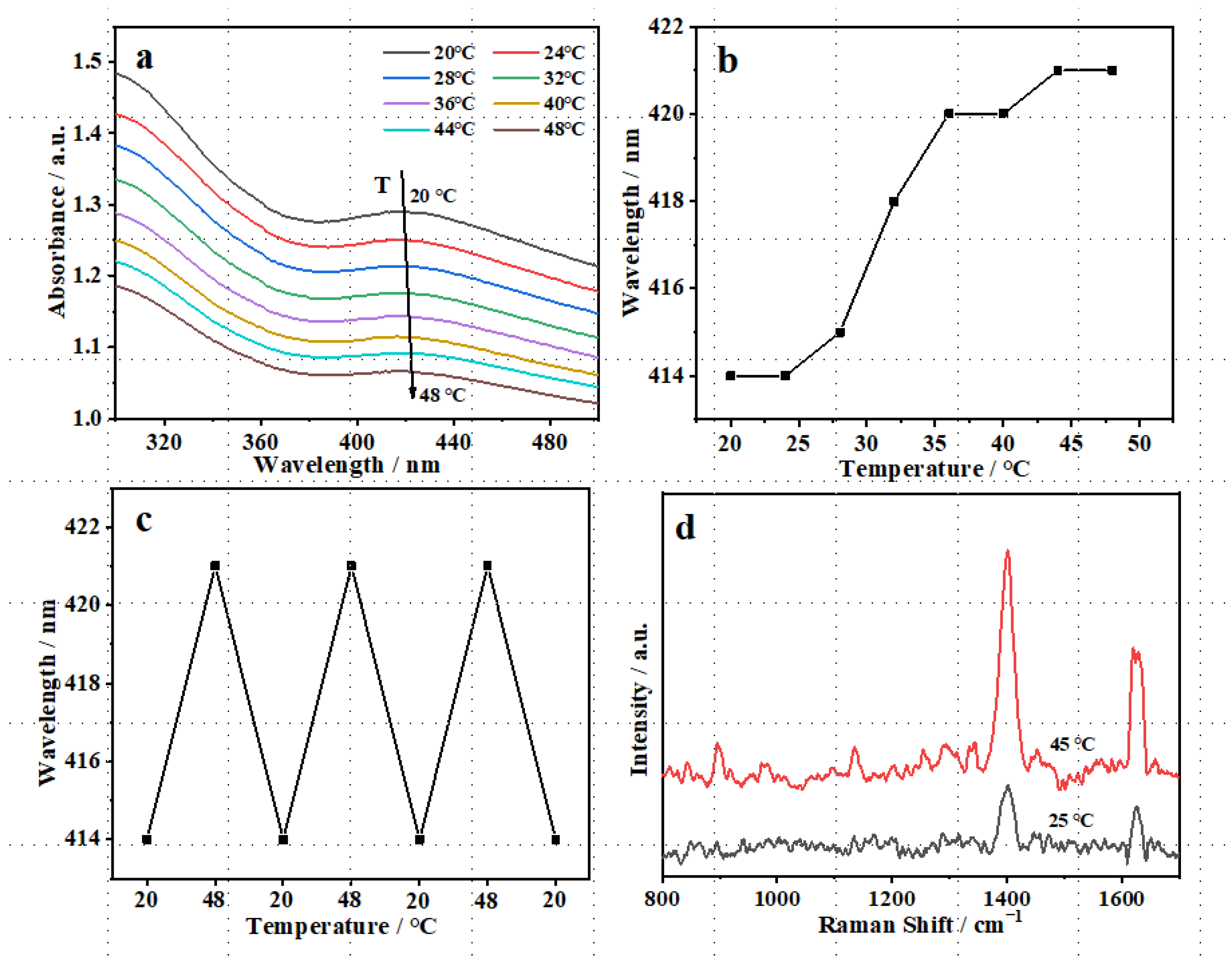
3.3. Thermo-Sensitivity of TM@TiO2@Ag

The localized surface plasmon resonance (LSPR) property of TM@TiO2@Ag was recorded by an UV-Vis spectrophotometer. Figure 4a shows the UV-Vis absorption spectra of TM@TiO2@Ag dispersed in water with the temperature changed from 20 to 48 °C. It was seen that the intensity of the LSPR of TM@TiO2@Ag was reduced from 1.29 to 1.07. The LSPR peak position exhibited a trend of gradual red shift with the displacement value of 7 nm (Figure 4b) because the volume of MIPs hydrogel around TM@TiO2@Ag decreased with the rising temperature. This volume contraction led to the aggregation of TM@TiO2@Ag due to the increasing density, the increase in the refractive and dielectric constant of the environment, and then the red shift of LSPR peak position. Meanwhile, due to the aggregation, the electrostatic interaction between TM@TiO2@Ag nanoparticles increased and the resonance coupling occurred, which resulted in the redshift of the LSPR peak with the decrease in peak intensity and the broadening of the peak shape [23,40]. In this present study, the TM@TiO2@Ag nanocomposite exactly showed an invertible displacement of the LSPR peak during cooling or heating. As observed from Figure 4c, when the temperature was altered, the LSPR of TM@TiO2@Ag varied accordingly. As the temperature increased from 20 to 48 °C, the LSPR peak position changed from 414 to 421 nm. However, when the temperature dropped from 48 to 20 °C, the LSPR peak position changed back to 414 nm again, indicating that the volume of the TM@TiO2@Ag had distinct invertibility. These further demonstrated that TM@TiO2@Ag combined the optical characteristics of AgNPs with the thermo-sensitive characteristics of hydrogel, as well as possessed excellent stability. Figure 4d shows the variation in the SERS spectra of 10−7 M OFL at different temperatures. When the temperature rose from 25 to 45 °C, the intensity of the SERS characteristic peak of OFL increased. The dominant reasons for this change were as follows [25]: (1) The hydrogel shrunk with the increase in temperature and the number of AgNPs per unit area increased, and then the detectable molecular number of OFL also increased; (2) The refractive index and dielectric constant of the medium were enhanced as the volume of MIPs hydrogel decreased; (3) The contraction of the volume and the aggregation of TM@TiO2@Ag produced a stronger “hot spot” effect and more enhancement effect. Based on this phenomenon, the thermo-sensitive property is beneficial to improving the sensitivity of SERS detection using TM@TiO2@Ag as a substrate.

3.4. Photocatalytic Performance of TM@TiO2@Ag

Figure 5 exhibits the photocatalytic degradation property of TM@TiO2@Ag, as well as AgNPs, TiO2, and TiO2@Ag for OFL, where C0 and C were the concentrations of OFL at initial and different times, respectively. Due to the synergistic enhancement of Ag photocatalysis on TiO2, the addition of silver significantly improved the photocatalytic effect of TiO2. After grafting the thermo-sensitive imprinted layer, the photocatalytic efficiency of TM@TiO2@Ag was the same as that of TiO2@Ag, but the photocatalytic rate of TM@TiO2@Ag decreased slightly. This is because the thermo-sensitive imprinted layer has little influence on the light absorption of the TiO2@Ag.

3.5. Selectivity, Sensitivity, and Stability of TM@TiO2@Ag in OFL SERS Detection

The selective SERS detection was significant because it reflects the specific recognition characteristics of the synthesized TM@TiO2@Ag. The structural simulation CIP and the non-structural simulation SM2 of OFL were voted as comparison objects. As shown in Figure 6, the characteristic Raman peak intensity of OFL detected by TM@TiO2@Ag was the strongest, while the Raman peaks of CIP and SM2 were much lower than that of OFL. However, the Raman signals of the three compounds adsorbed on non-imprinted TN@TiO2@Ag were all inconspicuous because there were hardly any specific recognition sites formed in the TN@TiO2@Ag. CIP and OFL were alike in the partial structure of fluoroquinolones, but their stereochemical structures and specific molecular sizes were different. TM@TiO2@Ag can specifically recognize the target molecule and display low Raman intensity for co-existing substances. These results obviously propose that TM@TiO2@Ag could be served as a preeminent SERS substrate for highly selectively identifying OFL.
The enhancement factor of the TM@TiO2@Ag composite structure can be calculated as 8.71 × 10⁶ for OFL, referring to the calculation method in our previous work [25]. The SERS spectra of OFL at different concentrations (10−5~10−12 M) are shown in Figure 7a. It can be found that, with the decrease in OFL concentration, the SERS intensity progressively weakened, while it was still obvious at the concentration of 10−11 M. When the OFL concentration was 10−12 M, the Raman signal disappeared. In order to prove the quantitative detection ability of the presented sensor, the intensity specified at 1397 and 1626 cm−1 was plotted as a function of the logarithm of OFL concentration (Figure 7b). The fitted equations were I = 278.84 logC + 3665.47 (R2 = 0.9554) and I = 190.11 logC + 2497.10 (R2 = 0.9966), respectively, at 1397 and 1626 cm−1. The LOD and LOQ for OFL detected at 1397 cm−1 by the TM@TiO2@Ag substrate were calculated to be 1.1 × 10¹¹ M and 3.7 × 10¹¹ M, respectively. This result demonstrated that it is considerably sensitive to make TM@TiO2@Ag as the SERS substrate to detect trace amounts of OFL.
For the purpose of investigating the stability of the TM@TiO2@Ag substrate, a series of 10−5 M OFL SERS spectra were obtained from 8 different points at random, as shown in Figure 7c. There was no obvious difference in SERS intensity at 1397 and 1626 cm−1, indicating excellent stability. As displayed in Figure 7d, the relative standard deviation (RSD) value of the intensity at 1397 cm−1 was calculated to be 12.6%.

3.6. The Recyclability of TM@TiO2@Ag

It is very essential to design and construct a recyclable SERS sensor. Delightedly, due to its good photocatalytic activity for OFL, the TM@TiO2@Ag nanocomposite developed in this study had self-cleanable properties. In Figure 8, the photocatalytic performance of TM@TiO2@Ag was investigated, and three cyclic SERS detections were performed to inspect the reusability of TM@TiO2@Ag. It can be clearly seen from Figure 8a that the Raman peak intensity was very obvious when the OFL with 10−5 M was adsorbed on the surface of TM@TiO2@Ag. After UV irradiation, however, the Raman signals disappeared completely. The results undoubtedly illustrated the self-cleaning ability of TM@TiO2@Ag. At the same time, the relative intensity of the first adsorption OFL was 100%, while reduced to 0 after UV light was applied as shown in Figure 8b. Once OFL was adsorbed again, the SERS signal also recovered to nearly 90%. It was interesting to observe that TM@TiO2@Ag still maintained comparable SERS performance (78.8%) after three cycles. The results proved the robust recyclability of TM@TiO2@Ag. In addition, compared with the traditional elution method with solvent, photocatalytic degradation is an easier, cleaner, and more time-saving protocol to remove target analytes/template molecules.

3.7. Detection of OFL in Actual Sample

The portable drinking water sample was analyzed and no OFL was detected, implying that the concentration of OFL in the water was lower than the LOD of this method. In order to examine the applicability of the TM@TiO2@Ag for the SERS determination of OFL in actual samples, the recovery of OFL in drinking water was investigated by adding certain amounts of standard to the water sample. The results are shown in Table 1. The spiking recoveries were from 88.2% to 107.5% with the RSDs from 8.2% to 12.5%, indicating good reliability of the present analytical protocol.

3.8. Comparison with Other Methods for OFL

Several studies were performed for OFL determination, such as spectrofluorimetry, colorimetry, electrochemistry, photoelectrochemical methods, and SERS [41,42,43,44,45,46]. The selectivity, linear range, LOD, and regeneration method of every detection method are listed in Table 2. Compared with the previously reported detection methods of OFL, our proposed SERS method based on TM@TiO2@Ag nanocomposites presented several advantages for the detection of OFL, such as superior selectivity, high sensitivity, and a wide linear range. Its superior selectivity mainly comes from the imprinting function, and the high sensitivity benefits from its thermo-sensitive property, which leads to “hot spots” and significantly enhances the Raman signal. Moreover, this molecularly imprinted material has the exceptional self-cleaning ability and good reusability due to its photocatalytical performance of TiO2, broadening the field of SERS analysis.

3.9. Mechanism of SERS Detection

It is generally believed that the enhancement mechanism includes charge transfer enhancement and electromagnetic enhancement. The mechanism is regarded as an electromagnetic enhancement of TM@TiO2@Ag when served as the substrate in this research. This is due to the plasmon resonance of TiO2@Ag and Ag. In addition, there are gaps between Ag and Ag or Ag and TiO2, which could generate plenty of “hot spots” and significantly improve the Raman signal. When the thermo-sensitive imprinted layer is decorated on the surface of TiO2@Ag composites, it can make OFL closer to TM@TiO2@Ag, which is also helpful to SERS detection [47]. Moreover, as a photocatalyst, TiO2 as the photoactive center can photodegrade organic molecules, and Ag can be used as the electron trap in this experiment. The OFL is availably photodegraded by TiO2@Ag when adsorbed on the surface of TM@TiO2@Ag, which can be used for the recovery of SERS substrates. Compared with the traditional imprinted molecular elution, photocatalytic degradation saves the elution solvent and elution time, and is easier, cleaner, and more environmentally friendly.

4. Conclusions

In summary, a novel thermo-sensitive molecular imprinted photocatalytic nanomaterial (TM@TiO2@Ag) was designed and prepared successfully. After well characterized, the LSPR property of TM@TiO2@Ag with its unique structure and the SERS effect were studied in association with the thermo-sensitive and molecular imprinting properties, based on which the signal recognition and enhancement mechanisms were elucidated. TM@TiO2@Ag showed an outstanding SERS behavior with respect to the selectivity, sensitivity, and stability for OFL detection in the water sample, suggesting its applicability as a quantitative SERS detection substrate. More interestingly, the photocatalytic degradation feature attributed to nano-TiO2 endowed this SERS substrate with excellent self-cleaning ability for facile reuse. Therefore, TM@TiO2@Ag is expected to become a superior recyclable SERS sensor for detecting fluoroquinolone antibiotics in environmental and other scenes.

Author Contributions

Conceptualization, C.J.; Methodology, T.W. and C.J.; Literature searching and original draft preparation, T.W., Y.W. and X.H.; Writing—review and editing: C.J. and H.-z.L.; Funding acquisition: H.-z.L., Y.W. and C.J. All authors have read and agreed to the published version of the manuscript.

Funding

This research was funded by the National Key R&D Program of China (2019YFC1605400), the National Natural Science Foundation of China (51578295, 21874065, 22176085), the Excellent Scientific and Technological Innovation Team of Jiangsu Universities, Educational Commission of Jiangsu Province (21KJB430005), the Leading Talents Project of Jiangsu Vocational Institute of Commerce and the Key Projects of of Jiangsu Vocational Institute of Commerce (JSJM2022001).

Institutional Review Board Statement

Not applicable.

Informed Consent Statement

Not applicable.

Data Availability Statement

The data presented in this study are available in article.

Acknowledgments

The authors were also grateful to Weiping Qian and Jian Dong, State Key Laboratory of Bioelectronics, Southeast University, for their kind help.

Conflicts of Interest

The authors declare no conflict of interest.

References

  1. Gai, S.; Zhang, J.; Fan, R.Q.; Xing, K.; Chen, W.; Zhu, K.; Zheng, X.B.; Wang, P.; Fang, X.K.; Yang, Y.L. Highly stable zinc-based metal-organic frameworks and corresponding flexible composites for removal and detection of antibiotics in water. ACS Appl. Mater. Interfaces 2020, 12, 8650–8662. [Google Scholar] [CrossRef] [PubMed]
  2. Malik, A.H.; Iyer, P.K. Conjugated polyelectrolyte based sensitive detection and removal of antibiotics tetracycline from water. ACS Appl. Mater. Interfaces 2017, 9, 4433–4439. [Google Scholar] [CrossRef] [PubMed]
  3. Yang, Y.; Yin, S.; Li, Y.X.; Lu, D.; Zhang, J.; Sun, C.J. Application of aptamers in detection and chromatographic purification of antibiotics in different matrices. TrAC Trends Anal. Chem. 2017, 95, 1–22. [Google Scholar] [CrossRef]
  4. Zhou, Y.; Yang, Q.; Zhang, D.N.; Gan, N.; Li, Q.; Cuan, J. Detection and removal of antibiotic tetracycline in water with a highly stable luminescent MOF. Sens. Actuators B Chem. 2018, 262, 137–143. [Google Scholar] [CrossRef]
  5. Galvan, D.D.; Yu, Q.M. Surface-enhanced Raman scattering for rapid detection and characterization of antibiotic-resistant bacteria. Adv. Healthc. Mater. 2018, 7, 1701335. [Google Scholar] [CrossRef] [PubMed]
  6. Fang, Q.Q.; Li, Y.Y.; Miao, X.X.; Zhang, Y.Q.; Yan, J.; Yu, T.Y.; Liu, J. Sensitive detection of antibiotics using aptamer conformation cooperated enzyme assisted SERS technology. Analyst 2019, 144, 3649–3658. [Google Scholar] [CrossRef] [PubMed]
  7. Lan, L.Y.; Yao, Y.; Ping, J.F.; Ying, Y.B. Recent advances in nanomaterial-based biosensors for antibiotics detection. Biosens. Bioelectron. 2017, 91, 504–514. [Google Scholar] [CrossRef]
  8. Ahmed, S.; Ning, J.N.; Peng, D.P.; Chen, T.; Ahmad, I.; Ali, A.; Lei, Z.X.; Shabbir, M.A.; Cheng, G.Y.; Yuan, Z.H. Current advances in immunoassays for the detection of antibiotics residues: A Review. Food Agric. Immunol. 2020, 31, 268–290. [Google Scholar] [CrossRef]
  9. Wang, W.E.; Sang, Q.Q.; Yang, M.; Du, J.; Yang, L.B.; Jiang, X.; Han, X.X.; Zhao, B. Detection of several quinolone antibiotic residues in water based on Ag-TiO2 SERS strategy. Sci. Total Environ. 2020, 702, 134956. [Google Scholar] [CrossRef]
  10. Wu, Y.Y.; Huang, P.C.; Wu, F.Y. A Label-free colorimetric aptasensor based on controllable aggregation of AuNPs for the detection of multiplex antibiotics. Food Chem. 2020, 304, 125377. [Google Scholar] [CrossRef]
  11. Pawar, M.K.; Tayade, K.C.; Sahoo, S.K.; Mahulikar, P.P.; Kuwar, A.S.; Chaudhari, B.L. Selective ciprofloxacin antibiotic detection by fluorescent siderophore pyoverdin. Biosens. Bioelectron. 2016, 81, 274–279. [Google Scholar] [CrossRef]
  12. Kaur, K.; Kumar, A.; Kumar, A.; Singh, M.B.; Rao, A.L.J. Spectrophotometric methods for the determination of fluoroquinolones: A Review. Crit. Rev. Anal. Chem. 2008, 38, 2–18. [Google Scholar] [CrossRef]
  13. Wang, Q.; Zhao, W.-M. Optical methods of antibiotic residues detections: A comprehensive review. Sens. Actuators B Chem. 2018, 269, 238–256. [Google Scholar] [CrossRef]
  14. Peng, J.; Kong, D.Z.; Liu, L.Q.; Song, S.S.; Kuang, H.; Xu, C.L. Determination of quinoxaline antibiotics in fish feed by enzyme-linked immunosorbent assay using a monoclonal antibody. Anal. Methods 2015, 7, 5204–5209. [Google Scholar] [CrossRef]
  15. Shanin, I.A.; Zvereva, E.A.; Eremin, S.A.; Sviridov, O.V.; Zherdev, A.V.; Dzantiev, B.B. Development of an immunoenzyme assay to control the total content of antibiotics of the fluoroquinolone group in milk. Appl. Biochem. Microbiol. 2020, 55, 563–569. [Google Scholar] [CrossRef]
  16. Wang, Z.H.; Zhu, Y.; Ding, S.Y.; He, F.Y.; Beier, R.C.; Li, J.C.; Jiang, H.Y.; Feng, C.W.; Wan, Y.P.; Zhang, S.X.; et al. Development of a monoclonal antibody-based broad-specificity ELISA for fluoroquinolone antibiotics in foods and molecular modeling studies of cross-reactive compounds. Anal. Chem. 2007, 79, 4471–4483. [Google Scholar] [CrossRef]
  17. Piatkowska, M.; Jedziniak, P.; Zmudzki, J. Multiresidue Method for the simultaneous determination of veterinary medicinal products, feed additives and illegal dyes in eggs using liquid chromatography-tandem mass spectrometry. Food Chem. 2016, 197, 571–580. [Google Scholar] [CrossRef]
  18. Yan, H.Y.; Wang, H.; Qin, X.Y.; Liu, B.M.; Du, J.J. Ultrasound-assisted dispersive liquid-liquid microextraction for determination of fluoroquinolones in pharmaceutical wastewater. J. Pharm. Biomed. Anal. 2011, 54, 53–57. [Google Scholar] [CrossRef]
  19. Shen, C.-Y.; Shen, D.-X.; Ding, T.; Xu, J.-Z.; Jiang, Y.; Wu, B.; Lian, H.-Z. Simultaneous determination of 14 quinolones in royal jelly by liquid chromatography-tandem mass spectrometry using anion-exchange solid-phase extraction. J. Liq. Chromatogr. Relat. Technol. 2011, 34, 1415–1430. [Google Scholar] [CrossRef]
  20. He, S.; Chua, J.; Tan, E.K.M.; Kah, J.C.Y. Optimizing the SERS enhancement of a facile gold nanostar immobilized paper-based SERS substrate. RSC Adv. 2017, 7, 16264–16272. [Google Scholar] [CrossRef]
  21. Li, J.J.; Yan, H.; Tan, X.C.; Lu, Z.C.; Han, H.Y. Cauliflower-inspired 3D SERS substrate for multiple mycotoxins detection. Anal. Chem. 2019, 91, 3885–3892. [Google Scholar] [CrossRef] [PubMed]
  22. Alvarez-Puebla, R.A.; Contreras-Caceres, R.; Pastoriza-Santos, I.; Perez-Juste, J.; Liz-Marzan, L.M. Au@pNIPAM colloids as molecular traps for surface-enhanced, spectroscopic, ultra-sensitive analysis. Angew. Chem. 2009, 48, 138–143. [Google Scholar] [CrossRef] [PubMed]
  23. Jiang, C.Y.; Qian, Y.; Gao, Q.; Dong, J.; Qian, W.P. In situ controllable preparation of gold nanorods in thermo-responsive hydrogels and their application in surface enhanced Raman scattering. J. Mater. Chem. 2010, 20, 8711–8716. [Google Scholar] [CrossRef]
  24. Zhou, Q.T.; Meng, G.W.; Zheng, P.; Cushing, S.; Wu, N.Q.; Huang, Q.; Zhu, C.H.; Zhang, Z.; Wang, Z.W. A surface-enhanced Raman scattering sensor integrated with battery-controlled fluidic device for capture and detection of trace small molecules. Sci. Rep. 2015, 5, 12865. [Google Scholar] [CrossRef] [PubMed] [Green Version]
  25. Jiang, C.Y.; Ma, X.Y.; Xue, M.Y.; Lian, H.-Z. Application of thermoresponsive hydrogel/gold nanorods composites in the detection of diquat. Talanta 2017, 174, 192–197. [Google Scholar] [CrossRef]
  26. Hu, B.X.; Sun, D.W.; Wei, Q.Y. A dynamically optical and highly stable pNIPAM@ Au NRs nanohybrid substrate for sensitive SERS detection of malachite green in fish fillet. Talanta 2020, 218, 121188. [Google Scholar] [CrossRef]
  27. Zhu, Y.Y.; Wu, L.; Yan, H.; Lu, Z.C.; Yin, W.M.; Han, H.Y. Enzyme induced molecularly imprinted polymer on SERS substrate for ultrasensitive detection of patulin. Anal. Chim. Acta 2020, 1101, 111–119. [Google Scholar] [CrossRef]
  28. Li, H.J.; Wang, X.N.; Wang, Z.R.; Jiang, J.Q.; Wei, M.B.; Zheng, J.H.; Yan, Y.S.; Li, C.X. Thermo-responsive molecularly imprinted sensor based on the surface-enhanced Raman scattering for selective detection of R6G in the water. Dalton Trans. 2017, 46, 11282–11290. [Google Scholar] [CrossRef]
  29. Li, H.B.; Li, J.; Zhu, Y.Y.; Xie, W.Y.; Shao, R.; Yao, X.X.; Gao, A.Q.; Yin, Y.D. Cd2+-doped amorphous TiO2 hollow spheres for robust and ultrasensitive photoelectrochemical sensing of hydrogen sulfide. Anal. Chem. 2018, 90, 5496–5502. [Google Scholar] [CrossRef]
  30. Phadtare, S.; Kumar, A.; Vinod, V.P.; Dash, C.; Palaskar, D.V.; Rao, M.; Shukla, P.G.; Sivaram, S.; Sastry, M. Direct assembly of gold nanoparticle “shells” on polyurethane microsphere “cores” and their application as enzyme immobilization templates. Chem. Mater. 2003, 15, 1944–1949. [Google Scholar] [CrossRef]
  31. Feng, Q.A.; Tang, D.Y.; Lv, H.T.; Zhang, W.L.; Li, W.B. Surface-initiated ATRP to modify ZnO nanoparticles with poly(N-isopropylacrylamide): Temperature-controlled switching of photocatalysis. J. Alloys Compd. 2017, 691, 185–194. [Google Scholar] [CrossRef]
  32. Jiang, C.Y.; Wu, T.; Liu, J.X.; Wang, Y.P. Application of a thermo-sensitive imprinted SERS substrate to the rapid trace detection of ofloxacin. Anal. Methods 2020, 39, 4783–4788. [Google Scholar] [CrossRef] [PubMed]
  33. Wang, K.Q.; Sun, D.W.; Pu, H.B.; Wei, Q.Y. Surface-enhanced Raman scattering of core-shell Au@ Ag nanoparticles aggregates for rapid detection of difenoconazole in grapes. Talanta 2019, 191, 449–456. [Google Scholar] [CrossRef] [PubMed]
  34. An, T.C.; Chen, J.Y.; Nie, X.; Li, G.Y.; Zhang, H.M.; Liu, X.L.; Zhao, H.J. Synthesis of carbon nanotube-anatase TiO2 sub-micrometer-sized sphere composite photocatalyst for synergistic degradation of gaseous styrene. ACS Appl. Mater. Interfaces 2012, 4, 5988–5996. [Google Scholar] [CrossRef]
  35. Chen, D.H.; Chen, C.J. Formation and characterization of Au-Ag bimetallic nanoparticles in water-in-oil microemulsions. J. Mater. Chem. 2002, 12, 1557–1562. [Google Scholar] [CrossRef]
  36. Feng, Q.; Tang, D.Y.; Lv, H.T.; Zhang, W.L.; Li, W.B. Temperature-responsive zinc oxide nanorods arrays grafted with poly(N-isopropylacrylamide) via SI-ATRP. RSC Adv. 2015, 5, 62024–62032. [Google Scholar] [CrossRef]
  37. Li, G.Y.; Yu, N.N.; Gao, Y.R.; Tao, Q.; Liu, X.Y. Polymeric hollow spheres assembled from ALG-g-PNIPAM and β-cyclodextrin for controlled drug release. Int. J. Biol. Macromol. 2016, 82, 381–386. [Google Scholar] [CrossRef]
  38. Sun, R.; Hughes, S. Fractional extraction and physico-chemical characterization of hemicelluloses and cellulose from sugar beet pulp. Carbohydr. Polym. 1998, 36, 293–299. [Google Scholar] [CrossRef]
  39. Yang, Z.Q.; Ma, C.C.; Wang, W.; Zhang, M.T.; Hao, X.P.; Chen, S.G. Fabrication of Cu2O-Ag nanocomposites with enhanced durability and bactericidal activity. J. Colloid Interface Sci. 2019, 557, 156–167. [Google Scholar] [CrossRef]
  40. Jiang, C.Y.; Weng, X.L.; Qian, W.P. Preparation and thermoresponsive properties of AuNPs/PNIPAM composite particle. Acta Phys.-Chim. Sin. 2008, 24, 2159–2164. [Google Scholar]
  41. Zhu, Y.R.; Lu, Y.G.; Shi, L.Y.; Yang, Y.L. β-Cyclodextrin functionalized N,Zn codoped carbon dots for specific fluorescence detection of fluoroquinolones in milk samples. Microchem. J. 2020, 153, 104517. [Google Scholar] [CrossRef]
  42. Mirzajani, R.; Pourreza, N.; Burromandpiroze, J. Fabrication of magnetic Fe3O4@nSiO2@mSiO2-NH2 core-shell mesoporous nanocomposite and its application for highly efficient ultrasound assisted dispersive SPE-spectrofluorimetric detection of ofloxacin in urine and plasma samples. Ultrason. Sonochemistry 2018, 40, 101–112. [Google Scholar] [CrossRef] [PubMed]
  43. Zhou, X.T.; Wang, L.M.; Shen, G.Q.; Zhang, D.W.; Xie, J.L.; Mamut, A.; Huang, W.W.; Zhou, S.S. Colorimetric determination of ofloxacin using unmodified aptamers and the aggregation of gold nanoparticles. Microchim. Acta 2018, 185, 355. [Google Scholar] [CrossRef] [PubMed]
  44. Adhikari, S.; Kim, D.H. Synthesis of Bi2S3/Bi2WO6 hierarchical microstructures for enhanced visible light driven photocatalytic degradation and photoelectrochemical sensing of ofloxacin. Chem. Eng. J. 2018, 354, 692–705. [Google Scholar] [CrossRef]
  45. Feng, L.M.; Xue, Q.; Liu, F.; Cao, Q.P.; Feng, J.J.; Yang, L.; Zhang, F.L. Voltammetric determination of ofloxacin by using a laser-modified carbon glassy electrode. Microchim. Acta 2020, 187, 86. [Google Scholar] [CrossRef]
  46. El-Zahry, M.R.; Lendl, B. Structure elucidation and degradation kinetic study of ofloxacin using surface enhanced Raman spectroscopy. Spectrochim. Acta Part A Mol. Biomol. Spectrosc. 2018, 193, 63–70. [Google Scholar] [CrossRef] [PubMed]
  47. Chen, Y.; O’Donoghue, M.B.; Huang, Y.F.; Kang, H.Z.; Phillips, J.A.; Chen, X.L.; Estevez, M.C.; Yang, C.Y.J.; Tan, W.H. A surface energy transfer nanoruler for measuring binding site distances on live cell surfaces. J. Am. Chem. Soc. 2010, 132, 16559–16570. [Google Scholar] [CrossRef]
Scheme 1. Schematic illustration of preparation of TM@TiO2@Ag and application on OFL SERS detection.
Chemosensors 10 00437 sch001
Figure 1. SEM (ac) and TEM images (df) of SiO2/TiO2 (a,d), TiO2@Ag (b,e), and TM@TiO2@Ag (c,f); diameter distribution presented in histogram of TM@TiO2@Ag (g); element mapping images of TM@TiO2@Ag with ‘C’ (h), ‘N’ (i), ‘Ti’ (j), ‘Ag’ (k), ‘O’ (l), and ‘Si’ (m); EDX spectrum of TM@TiO2@Ag (n).
Chemosensors 10 00437 g001
Figure 2. XRD patterns of SiO2/TiO2, TiO2@Ag, and TM@TiO2@Ag (a); FT-IR spectra of TM@TiO2@Ag and TN@TiO2@Ag (b). Inset: zoom in of local FT-IR spectra of TM@TiO2@Ag and TN@TiO2@Ag from 1300~1800 cm1.
Chemosensors 10 00437 g002
Figure 3. UV-Vis adsorption spectra recorded for the aqueous dispersion of AgNPs, TiO2@Ag, and TM@TiO2@Ag.
Chemosensors 10 00437 g003
Figure 4. UV-Vis absorption spectra of TM@TiO2@Ag with temperature change (a); the relationship between LSPR peak wavelength of TM@TiO2@Ag and temperature (b); the relationship between LSPR peak position of TM@TiO2@Ag and temperature cycle (c); SERS spectra of 10−7 M OFL at different temperatures (d).
Chemosensors 10 00437 g004
Figure 5. Photocatalytic degradation property of AgNPs, TiO2, TiO2@Ag, and TM@TiO2@Ag for OFL.
Chemosensors 10 00437 g005
Figure 6. Selectivity of TM@TiO2@Ag and TN@TiO2@Ag for different compounds at the same concentration of 10−5 M.
Chemosensors 10 00437 g006
Figure 7. SERS spectra of different concentrations of OFL obtained from TM@TiO2@Ag (a); linear relationship between Raman intensity of OFL at the peak of 1397 and 1626 cm−1 and the concentrations range of 10−5~10−11 M (b); a series of SERS spectra of OFL at 10−5 M from 8 different random spots (c); the intensity of OFL at 10−5 M at 1397 cm−1 in the SERS spectra (d).
Chemosensors 10 00437 g007
Figure 8. The Raman spectra of OFL at 10−5 M with the TM@TiO2@Ag before and after irradiation with UV light (a); SERS responses of OFL upon three repeated detections and UV irradiations (b).
Chemosensors 10 00437 g008
Table 1. Analytical results of OFL detection in drinking water (n = 3).
SampleSpiked/10−12 MFound/10−12 MRecovery/%RSD/%
Drinking water100107.5107.512.5
1000955.395.58.2
10,000881988.29.6
Table 2. Comparison of the present method with those reported in recent literature.
MaterialDetection MethodSelectivityLinear RangeLOD/nMRegeneration MethodRef.
β-Cyclodextrin functionalized N,Zn codoped carbon dotsSpectrofluorimetricN.A. *0.075–3.75 μM50N.A. *[41]
Fe3O4@nSiO2@mSiO2–NH2SpectrofluorimetricGood1.0–500.0 µg/L0.58Elution with nitric acid[42]
Aptamers and AuNPsColorimetricGood20–400 nM3.38N.A. *[43]
Bi2S3/Bi2WO6PhotoelectrochemicalN.A. *1–100 μM906N.A. *[44]
Laser-modified glassy carbon electrodesElectrochemicalGood0.25–200 μM75N.A. *[45]
Ag NPsSERSN.A. *100–500 ng/mL0.65N.A. *[46]
TM@TiO2@AgSERSGood10−5–10 μM0.011Photocatalytic degradationThis work
* Not available.
Publisher’s Note: MDPI stays neutral with regard to jurisdictional claims in published maps and institutional affiliations.

