
Does Artificial Intelligence Make Clinical Decision Better? A Review of Artificial Intelligence and Machine Learning in Acute Kidney Injury Prediction


Abstract
:1. Introduction
1.1. AKI Definition
1.2. AKI Risk Factors and Risk Scores
1.3. From Automated Electronic Alerts to AI
2. Methods
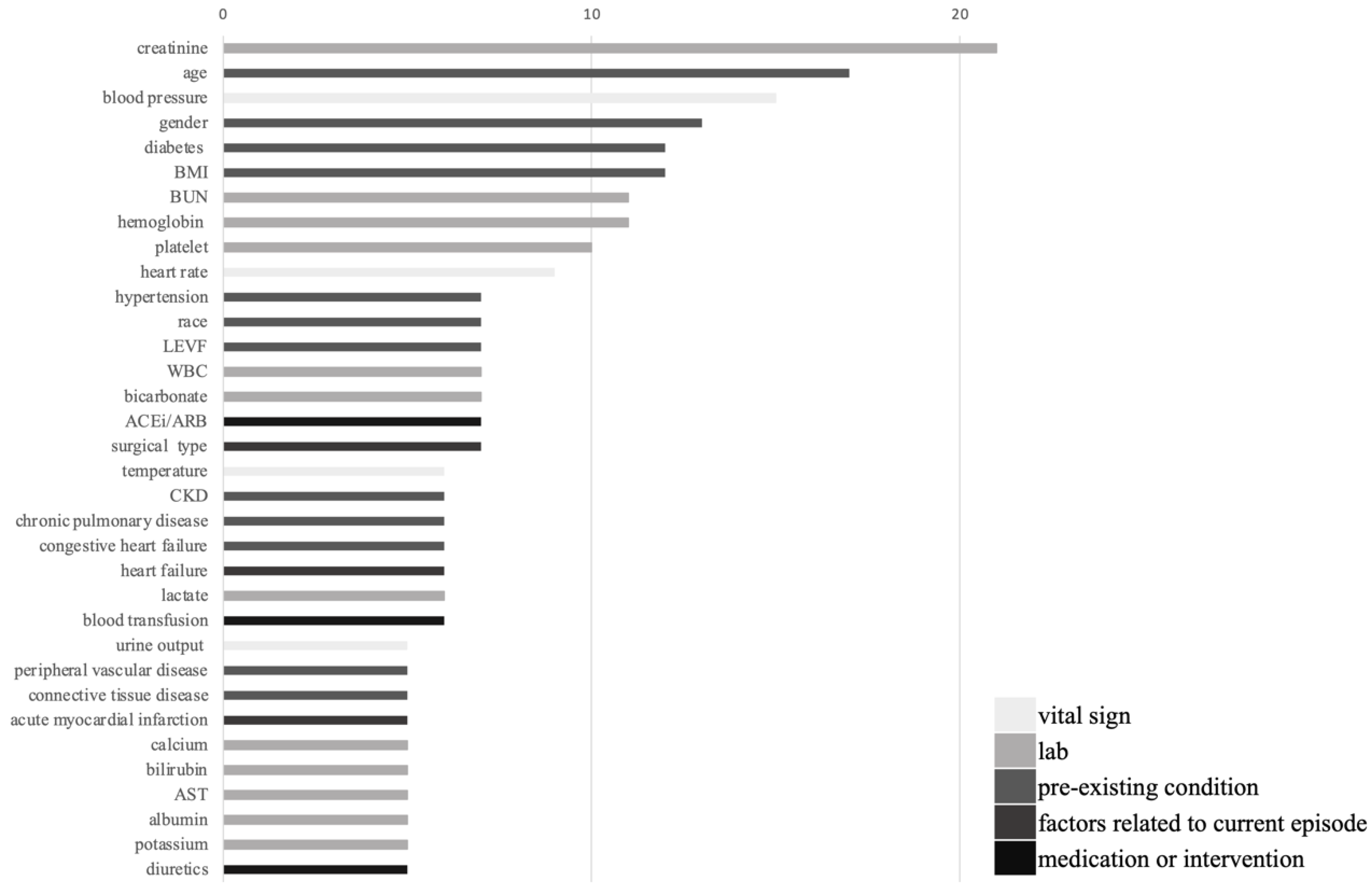
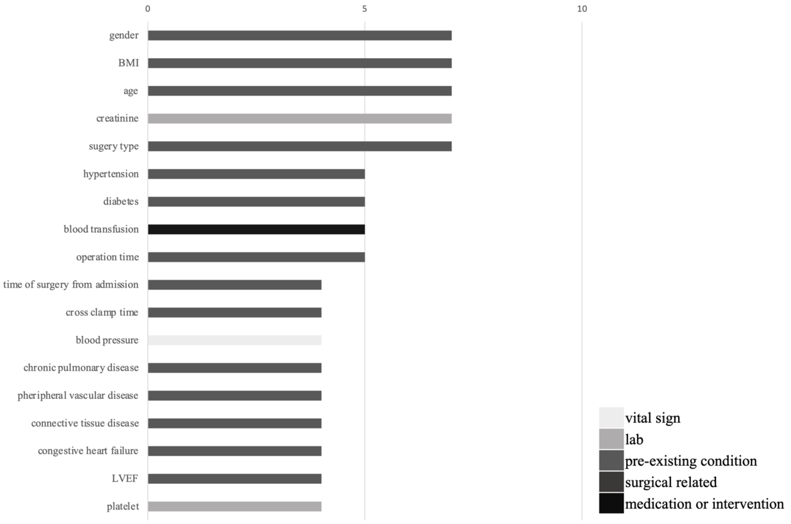
3. Results
4. Discussion
5. Conclusions
Supplementary Materials
Author Contributions
Funding
Institutional Review Board Statement
Informed Consent Statement
Data Availability Statement
Conflicts of Interest
References
- Section 2: AKI Definition. Kidney Int. Suppl. 2012, 2, 19–36. [CrossRef] [Green Version]
- Kuo, G.; Yang, S.Y.; Chuang, S.S.; Fan, P.C.; Chang, C.H.; Hsiao, Y.C.; Chen, Y.C. Using acute kidney injury severity and scoring systems to predict outcome in patients with burn injury. J. Formos. Med. Assoc. 2016, 115, 1046–1052. [Google Scholar] [CrossRef] [PubMed]
- Kim, G.H.; Oh, K.H.; Yoon, J.W.; Koo, J.W.; Kim, H.J.; Chae, D.W.; Noh, J.W.; Kim, J.H.; Park, Y.K. Impact of burn size and initial serum albumin level on acute renal failure occurring in major burn. Am. J. Nephrol. 2003, 23, 55–60. [Google Scholar] [CrossRef]
- Jenq, C.C.; Tsai, M.H.; Tian, Y.C.; Lin, C.Y.; Yang, C.; Liu, N.J.; Lien, J.M.; Chen, Y.C.; Fang, J.T.; Chen, P.C.; et al. RIFLE classification can predict short-term prognosis in critically ill cirrhotic patients. Intensive Care Med. 2007, 33, 1921–1930. [Google Scholar] [CrossRef]
- Waikar, S.S.; Curhan, G.C.; Wald, R.; McCarthy, E.P.; Chertow, G.M. Declining mortality in patients with acute renal failure, 1988 to 2002. J. Am. Soc. Nephrol. 2006, 17, 1143–1150. [Google Scholar] [CrossRef] [PubMed] [Green Version]
- Hobson, C.; Lysak, N.; Huber, M.; Scali, S.; Bihorac, A. Epidemiology, outcomes, and management of acute kidney injury in the vascular surgery patient. J. Vasc. Surg. 2018, 68, 916–928. [Google Scholar] [CrossRef]
- Hobson, C.; Singhania, G.; Bihorac, A. Acute Kidney Injury in the Surgical Patient. Crit. Care Clin. 2015, 31, 705–723. [Google Scholar] [CrossRef] [Green Version]
- Amdur, R.L.; Chawla, L.S.; Amodeo, S.; Kimmel, P.L.; Palant, C.E. Outcomes following diagnosis of acute renal failure in U.S. veterans: Focus on acute tubular necrosis. Kidney Int. 2009, 76, 1089–1097. [Google Scholar] [CrossRef] [Green Version]
- Ishani, A.; Xue, J.L.; Himmelfarb, J.; Eggers, P.W.; Kimmel, P.L.; Molitoris, B.A.; Collins, A.J. Acute kidney injury increases risk of ESRD among elderly. J. Am. Soc. Nephrol. 2009, 20, 223–228. [Google Scholar] [CrossRef] [PubMed] [Green Version]
- Coca, S.G.; Singanamala, S.; Parikh, C.R. Chronic kidney disease after acute kidney injury: A systematic review and meta-analysis. Kidney Int. 2012, 81, 442–448. [Google Scholar] [CrossRef] [Green Version]
- Nakasone, H.; Sakugawa, H.; Fukuchi, J.; Miyagi, T.; Sugama, R.; Hokama, A.; Nakayoshi, T.; Kawakami, Y.; Yamashiro, T.; Kinjo, F.; et al. A patient with primary biliary cirrhosis associated with autoimmune hemolytic anemia. J. Gastroenterol. 2000, 35, 245–249. [Google Scholar] [CrossRef] [PubMed]
- Lo, L.J.; Go, A.S.; Chertow, G.M.; McCulloch, C.E.; Fan, D.; Ordonez, J.D.; Hsu, C.Y. Dialysis-requiring acute renal failure increases the risk of progressive chronic kidney disease. Kidney Int. 2009, 76, 893–899. [Google Scholar] [CrossRef] [PubMed] [Green Version]
- Pannu, N.; James, M.; Hemmelgarn, B.; Klarenbach, S.; Alberta Kidney Disease Network. Association between AKI, recovery of renal function, and long-term outcomes after hospital discharge. Clin. J. Am. Soc. Nephrol. 2013, 8, 194–202. [Google Scholar] [CrossRef]
- Gammelager, H.; Christiansen, C.F.; Johansen, M.B.; Tonnesen, E.; Jespersen, B.; Sorensen, H.T. Five-year risk of end-stage renal disease among intensive care patients surviving dialysis-requiring acute kidney injury: A nationwide cohort study. Crit. Care 2013, 17, R145. [Google Scholar] [CrossRef] [Green Version]
- Forni, L.G.; Darmon, M.; Ostermann, M.; Oudemans-van Straaten, H.M.; Pettila, V.; Prowle, J.R.; Schetz, M.; Joannidis, M. Renal recovery after acute kidney injury. Intensive Care Med. 2017, 43, 855–866. [Google Scholar] [CrossRef] [PubMed]
- Hoste, E.A.; Bagshaw, S.M.; Bellomo, R.; Cely, C.M.; Colman, R.; Cruz, D.N.; Edipidis, K.; Forni, L.G.; Gomersall, C.D.; Govil, D.; et al. Epidemiology of acute kidney injury in critically ill patients: The multinational AKI-EPI study. Intensive Care Med. 2015, 41, 1411–1423. [Google Scholar] [CrossRef] [PubMed]
- Singbartl, K.; Kellum, J.A. AKI in the ICU: Definition, epidemiology, risk stratification, and outcomes. Kidney Int. 2012, 81, 819–825. [Google Scholar] [CrossRef] [PubMed] [Green Version]
- Perazella, M.A. Drug use and nephrotoxicity in the intensive care unit. Kidney Int. 2012, 81, 1172–1178. [Google Scholar] [CrossRef] [Green Version]
- Gameiro, J.; Branco, T.; Lopes, J.A. Artificial Intelligence in Acute Kidney Injury Risk Prediction. J. Clin. Med. 2020, 9, 678. [Google Scholar] [CrossRef] [PubMed] [Green Version]
- Bellomo, R.; Ronco, C.; Kellum, J.A.; Mehta, R.L.; Palevsky, P.; Acute Dialysis Quality Initiative Workgroup. Acute renal failure—Definition, outcome measures, animal models, fluid therapy and information technology needs: The Second International Consensus Conference of the Acute Dialysis Quality Initiative (ADQI) Group. Crit. Care 2004, 8, R204–R212. [Google Scholar] [CrossRef] [Green Version]
- Mehta, R.L.; Kellum, J.A.; Shah, S.V.; Molitoris, B.A.; Ronco, C.; Warnock, D.G.; Levin, A.; Acute Kidney Injury, N. Acute Kidney Injury Network: Report of an initiative to improve outcomes in acute kidney injury. Crit. Care 2007, 11, R31. [Google Scholar] [CrossRef] [Green Version]
- Perrone, R.D.; Madias, N.E.; Levey, A.S. Serum creatinine as an index of renal function: New insights into old concepts. Clin. Chem. 1992, 38, 1933–1953. [Google Scholar] [CrossRef] [PubMed]
- Bauer, J.H.; Brooks, C.S.; Burch, R.N. Clinical appraisal of creatinine clearance as a measurement of glomerular filtration rate. Am. J. Kidney Dis. 1982, 2, 337–346. [Google Scholar] [CrossRef]
- Van Acker, B.A.; Koomen, G.C.; Koopman, M.G.; de Waart, D.R.; Arisz, L. Creatinine clearance during cimetidine administration for measurement of glomerular filtration rate. Lancet 1992, 340, 1326–1329. [Google Scholar] [CrossRef]
- Delanaye, P.; Cavalier, E.; Pottel, H. Serum Creatinine: Not So Simple! Nephron 2017, 136, 302–308. [Google Scholar] [CrossRef]
- Papadakis, M.A.; Arieff, A.I. Unpredictability of clinical evaluation of renal function in cirrhosis. Prospective study. Am. J. Med. 1987, 82, 945–952. [Google Scholar] [CrossRef]
- Green, T.P.; Mirkin, B.L. Furosemide disposition in normal and proteinuric rats: Urinary drug-protein binding as a determinant of drug excretion. J. Pharmacol. Exp. Ther. 1981, 218, 122–127. [Google Scholar] [PubMed]
- McIlroy, D.R.; Wagener, G.; Lee, H.T. Biomarkers of acute kidney injury: An evolving domain. Anesthesiology 2010, 112, 998–1004. [Google Scholar] [CrossRef] [Green Version]
- Chen, T.H.; Chang, C.H.; Lin, C.Y.; Jenq, C.C.; Chang, M.Y.; Tian, Y.C.; Hung, C.C.; Fang, J.T.; Yang, C.W.; Wen, M.S.; et al. Acute kidney injury biomarkers for patients in a coronary care unit: A prospective cohort study. PLoS ONE 2012, 7, e32328. [Google Scholar] [CrossRef] [PubMed]
- Yui, S.; Nakatani, Y.; Mikami, M. Calprotectin (S100A8/S100A9), an inflammatory protein complex from neutrophils with a broad apoptosis-inducing activity. Biol. Pharm. Bull. 2003, 26, 753–760. [Google Scholar] [CrossRef] [Green Version]
- Pepper, R.J.; Wang, H.H.; Rajakaruna, G.K.; Papakrivopoulou, E.; Vogl, T.; Pusey, C.D.; Cook, H.T.; Salama, A.D. S100A8/A9 (calprotectin) is critical for development of glomerulonephritis and promotes inflammatory leukocyte-renal cell interactions. Am. J. Pathol. 2015, 185, 1264–1274. [Google Scholar] [CrossRef] [Green Version]
- Chang, C.H.; Yang, C.H.; Yang, H.Y.; Chen, T.H.; Lin, C.Y.; Chang, S.W.; Chen, Y.T.; Hung, C.C.; Fang, J.T.; Yang, C.W.; et al. Urinary Biomarkers Improve the Diagnosis of Intrinsic Acute Kidney Injury in Coronary Care Units. Medicine 2015, 94, e1703. [Google Scholar] [CrossRef] [PubMed]
- Vanmassenhove, J.; Vanholder, R.; Nagler, E.; Van Biesen, W. Urinary and serum biomarkers for the diagnosis of acute kidney injury: An in-depth review of the literature. Nephrol. Dial. Transplant. 2013, 28, 254–273. [Google Scholar] [CrossRef] [PubMed] [Green Version]
- Marx, D.; Metzger, J.; Pejchinovski, M.; Gil, R.B.; Frantzi, M.; Latosinska, A.; Belczacka, I.; Heinzmann, S.S.; Husi, H.; Zoidakis, J.; et al. Proteomics and Metabolomics for AKI Diagnosis. Semin. Nephrol. 2018, 38, 63–87. [Google Scholar] [CrossRef] [Green Version]
- Kashani, K.; Cheungpasitporn, W.; Ronco, C. Biomarkers of acute kidney injury: The pathway from discovery to clinical adoption. Clin. Chem. Lab. Med. 2017, 55, 1074–1089. [Google Scholar] [CrossRef] [PubMed]
- Lameire, N.H.; Bagga, A.; Cruz, D.; De Maeseneer, J.; Endre, Z.; Kellum, J.A.; Liu, K.D.; Mehta, R.L.; Pannu, N.; Van Biesen, W.; et al. Acute kidney injury: An increasing global concern. Lancet 2013, 382, 170–179. [Google Scholar] [CrossRef]
- Hahn, K.; Kanbay, M.; Lanaspa, M.A.; Johnson, R.J.; Ejaz, A.A. Serum uric acid and acute kidney injury: A mini review. J. Adv. Res. 2017, 8, 529–536. [Google Scholar] [CrossRef] [Green Version]
- Sgura, F.A.; Bertelli, L.; Monopoli, D.; Leuzzi, C.; Guerri, E.; Sparta, I.; Politi, L.; Aprile, A.; Amato, A.; Rossi, R.; et al. Mehran contrast-induced nephropathy risk score predicts short- and long-term clinical outcomes in patients with ST-elevation-myocardial infarction. Circ. Cardiovasc. Interv. 2010, 3, 491–498. [Google Scholar] [CrossRef] [Green Version]
- Abellas-Sequeiros, R.A.; Raposeiras-Roubin, S.; Abu-Assi, E.; Gonzalez-Salvado, V.; Iglesias-Alvarez, D.; Redondo-Dieguez, A.; Gonzalez-Ferreiro, R.; Ocaranza-Sanchez, R.; Pena-Gil, C.; Garcia-Acuna, J.M.; et al. Mehran contrast nephropathy risk score: Is it still useful 10 years later? J. Cardiol. 2016, 67, 262–267. [Google Scholar] [CrossRef] [Green Version]
- Uchino, S.; Kellum, J.A.; Bellomo, R.; Doig, G.S.; Morimatsu, H.; Morgera, S.; Schetz, M.; Tan, I.; Bouman, C.; Macedo, E.; et al. Acute renal failure in critically ill patients: A multinational, multicenter study. JAMA 2005, 294, 813–818. [Google Scholar] [CrossRef] [Green Version]
- Chang, C.H.; Lee, C.C.; Chen, S.W.; Fan, P.C.; Chen, Y.C.; Chang, S.W.; Chen, T.H.; Wu, V.C.; Lin, P.J.; Tsai, F.C. Predicting Acute Kidney Injury Following Mitral Valve Repair. Int. J. Med. Sci. 2016, 13, 19–24. [Google Scholar] [CrossRef] [Green Version]
- Wang, Y.; Bellomo, R. Cardiac surgery-associated acute kidney injury: Risk factors, pathophysiology and treatment. Nat. Rev. Nephrol. 2017, 13, 697–711. [Google Scholar] [CrossRef]
- Nashef, S.A.; Roques, F.; Michel, P.; Gauducheau, E.; Lemeshow, S.; Salamon, R. European system for cardiac operative risk evaluation (EuroSCORE). Eur. J. Cardiothorac. Surg. 1999, 16, 9–13. [Google Scholar] [CrossRef]
- Wykrzykowska, J.J.; Garg, S.; Onuma, Y.; de Vries, T.; Goedhart, D.; Morel, M.A.; van Es, G.A.; Buszman, P.; Linke, A.; Ischinger, T.; et al. Value of age, creatinine, and ejection fraction (ACEF score) in assessing risk in patients undergoing percutaneous coronary interventions in the ‘All-Comers’ LEADERS trial. Circ. Cardiovasc. Interv. 2011, 4, 47–56. [Google Scholar] [CrossRef] [Green Version]
- Shahian, D.M.; Jacobs, J.P.; Badhwar, V.; Kurlansky, P.A.; Furnary, A.P.; Cleveland, J.C., Jr.; Lobdell, K.W.; Vassileva, C.; Wyler von Ballmoos, M.C.; Thourani, V.H.; et al. The Society of Thoracic Surgeons 2018 Adult Cardiac Surgery Risk Models: Part 1-Background, Design Considerations, and Model Development. Ann. Thorac. Surg. 2018, 105, 1411–1418. [Google Scholar] [CrossRef] [Green Version]
- O’Brien, S.M.; Feng, L.; He, X.; Xian, Y.; Jacobs, J.P.; Badhwar, V.; Kurlansky, P.A.; Furnary, A.P.; Cleveland, J.C., Jr.; Lobdell, K.W.; et al. The Society of Thoracic Surgeons 2018 Adult Cardiac Surgery Risk Models: Part 2-Statistical Methods and Results. Ann. Thorac. Surg. 2018, 105, 1419–1428. [Google Scholar] [CrossRef] [Green Version]
- Wendt, D.; Thielmann, M.; Kahlert, P.; Kastner, S.; Price, V.; Al-Rashid, F.; Patsalis, P.; Erbel, R.; Jakob, H. Comparison between different risk scoring algorithms on isolated conventional or transcatheter aortic valve replacement. Ann. Thorac. Surg. 2014, 97, 796–802. [Google Scholar] [CrossRef] [PubMed]
- Mathioudakis, N.N.; Giles, M.; Yeh, H.C.; Haywood, C., Jr.; Greer, R.C.; Golden, S.H. Racial differences in acute kidney injury of hospitalized adults with diabetes. J. Diabetes Complicat. 2016, 30, 1129–1136. [Google Scholar] [CrossRef] [PubMed] [Green Version]
- Susantitaphong, P.; Cruz, D.N.; Cerda, J.; Abulfaraj, M.; Alqahtani, F.; Koulouridis, I.; Jaber, B.L.; Acute Kidney Injury Advisory Group of the American Society of Nephrology. World incidence of AKI: A meta-analysis. Clin. J. Am. Soc. Nephrol. 2013, 8, 1482–1493. [Google Scholar] [CrossRef] [PubMed] [Green Version]
- Fan, P.C.; Chen, T.H.; Lee, C.C.; Tsai, T.Y.; Chen, Y.C.; Chang, C.H. ADVANCIS Score Predicts Acute Kidney Injury After Percutaneous Coronary Intervention for Acute Coronary Syndrome. Int. J. Med. Sci. 2018, 15, 528–535. [Google Scholar] [CrossRef] [PubMed] [Green Version]
- Zhou, L.Z.; Yang, X.B.; Guan, Y.; Xu, X.; Tan, M.T.; Hou, F.F.; Chen, P.Y. Development and Validation of a Risk Score for Prediction of Acute Kidney Injury in Patients With Acute Decompensated Heart Failure: A Prospective Cohort Study in China. J. Am. Heart Assoc. 2016, 5, e004035. [Google Scholar] [CrossRef] [PubMed] [Green Version]
- Cheungpasitporn, W.; Kashani, K. Electronic Data Systems and Acute Kidney Injury. Contrib. Nephrol. 2016, 187, 73–83. [Google Scholar] [CrossRef] [PubMed]
- Park, S.; Baek, S.H.; Ahn, S.; Lee, K.H.; Hwang, H.; Ryu, J.; Ahn, S.Y.; Chin, H.J.; Na, K.Y.; Chae, D.W.; et al. Impact of Electronic Acute Kidney Injury (AKI) Alerts With Automated Nephrologist Consultation on Detection and Severity of AKI: A Quality Improvement Study. Am. J. Kidney Dis. 2018, 71, 9–19. [Google Scholar] [CrossRef] [PubMed] [Green Version]
- Wu, Y.; Chen, Y.; Li, S.; Dong, W.; Liang, H.; Deng, M.; Chen, Y.; Chen, S.; Liang, X. Value of electronic alerts for acute kidney injury in high-risk wards: A pilot randomized controlled trial. Int. Urol. Nephrol. 2018, 50, 1483–1488. [Google Scholar] [CrossRef] [Green Version]
- Lachance, P.; Villeneuve, P.M.; Rewa, O.G.; Wilson, F.P.; Selby, N.M.; Featherstone, R.M.; Bagshaw, S.M. Association between e-alert implementation for detection of acute kidney injury and outcomes: A systematic review. Nephrol. Dial. Transplant. 2017, 32, 265–272. [Google Scholar] [CrossRef]
- Lachance, P.; Villeneuve, P.M.; Wilson, F.P.; Selby, N.M.; Featherstone, R.; Rewa, O.; Bagshaw, S.M. Impact of e-alert for detection of acute kidney injury on processes of care and outcomes: Protocol for a systematic review and meta-analysis. BMJ Open 2016, 6, e011152. [Google Scholar] [CrossRef] [Green Version]
- Kolhe, N.V.; Reilly, T.; Leung, J.; Fluck, R.J.; Swinscoe, K.E.; Selby, N.M.; Taal, M.W. A simple care bundle for use in acute kidney injury: A propensity score-matched cohort study. Nephrol. Dial. Transplant. 2016, 31, 1846–1854. [Google Scholar] [CrossRef] [Green Version]
- Kolhe, N.V.; Staples, D.; Reilly, T.; Merrison, D.; McIntyre, C.W.; Fluck, R.J.; Selby, N.M.; Taal, M.W. Impact of Compliance with a Care Bundle on Acute Kidney Injury Outcomes: A Prospective Observational Study. PLoS ONE 2015, 10, e0132279. [Google Scholar] [CrossRef] [Green Version]
- Hodgson, L.E.; Roderick, P.J.; Venn, R.M.; Yao, G.L.; Dimitrov, B.D.; Forni, L.G. The ICE-AKI study: Impact analysis of a Clinical prediction rule and Electronic AKI alert in general medical patients. PLoS ONE 2018, 13, e0200584. [Google Scholar] [CrossRef]
- Kate, R.J.; Perez, R.M.; Mazumdar, D.; Pasupathy, K.S.; Nilakantan, V. Prediction and detection models for acute kidney injury in hospitalized older adults. BMC Med. Inform. Decis. Mak. 2016, 16, 39. [Google Scholar] [CrossRef] [PubMed] [Green Version]
- Thottakkara, P.; Ozrazgat-Baslanti, T.; Hupf, B.B.; Rashidi, P.; Pardalos, P.; Momcilovic, P.; Bihorac, A. Application of Machine Learning Techniques to High-Dimensional Clinical Data to Forecast Postoperative Complications. PLoS ONE 2016, 11, e0155705. [Google Scholar] [CrossRef] [Green Version]
- Davis, S.E.; Lasko, T.A.; Chen, G.; Siew, E.D.; Matheny, M.E. Calibration drift in regression and machine learning models for acute kidney injury. J. Am. Med. Inform. Assoc. 2017, 24, 1052–1061. [Google Scholar] [CrossRef] [PubMed]
- Cheng, P.; Waitman, L.R.; Hu, Y.; Liu, M. Predicting Inpatient Acute Kidney Injury over Different Time Horizons: How Early and Accurate? AMIA Annu. Symp. Proc. 2017, 2017, 565–574. [Google Scholar]
- Ibrahim, N.E.; McCarthy, C.P.; Shrestha, S.; Gaggin, H.K.; Mukai, R.; Magaret, C.A.; Rhyne, R.F.; Januzzi, J.L., Jr. A clinical, proteomics, and artificial intelligence-driven model to predict acute kidney injury in patients undergoing coronary angiography. Clin. Cardiol. 2019, 42, 292–298. [Google Scholar] [CrossRef] [Green Version]
- Koola, J.D.; Davis, S.E.; Al-Nimri, O.; Parr, S.K.; Fabbri, D.; Malin, B.A.; Ho, S.B.; Matheny, M.E. Development of an automated phenotyping algorithm for hepatorenal syndrome. J. Biomed. Inform. 2018, 80, 87–95. [Google Scholar] [CrossRef] [PubMed]
- Koyner, J.L.; Carey, K.A.; Edelson, D.P.; Churpek, M.M. The Development of a Machine Learning Inpatient Acute Kidney Injury Prediction Model. Crit. Care Med. 2018, 46, 1070–1077. [Google Scholar] [CrossRef]
- Huang, C.; Murugiah, K.; Mahajan, S.; Li, S.X.; Dhruva, S.S.; Haimovich, J.S.; Wang, Y.; Schulz, W.L.; Testani, J.M.; Wilson, F.P.; et al. Enhancing the prediction of acute kidney injury risk after percutaneous coronary intervention using machine learning techniques: A retrospective cohort study. PLoS Med. 2018, 15, e1002703. [Google Scholar] [CrossRef] [PubMed]
- Lin, K.; Hu, Y.; Kong, G. Predicting in-hospital mortality of patients with acute kidney injury in the ICU using random forest model. Int. J. Med. Inform. 2019, 125, 55–61. [Google Scholar] [CrossRef]
- Simonov, M.; Ugwuowo, U.; Moreira, E.; Yamamoto, Y.; Biswas, A.; Martin, M.; Testani, J.; Wilson, F.P. A simple real-time model for predicting acute kidney injury in hospitalized patients in the US: A descriptive modeling study. PLoS Med. 2019, 16, e1002861. [Google Scholar] [CrossRef] [Green Version]
- Huang, C.; Li, S.X.; Mahajan, S.; Testani, J.M.; Wilson, F.P.; Mena, C.I.; Masoudi, F.A.; Rumsfeld, J.S.; Spertus, J.A.; Mortazavi, B.J.; et al. Development and Validation of a Model for Predicting the Risk of Acute Kidney Injury Associated With Contrast Volume Levels During Percutaneous Coronary Intervention. JAMA Netw. Open 2019, 2, e1916021. [Google Scholar] [CrossRef] [PubMed] [Green Version]
- Tomasev, N.; Glorot, X.; Rae, J.W.; Zielinski, M.; Askham, H.; Saraiva, A.; Mottram, A.; Meyer, C.; Ravuri, S.; Protsyuk, I.; et al. A clinically applicable approach to continuous prediction of future acute kidney injury. Nature 2019, 572, 116–119. [Google Scholar] [CrossRef] [PubMed]
- Adhikari, L.; Ozrazgat-Baslanti, T.; Ruppert, M.; Madushani, R.; Paliwal, S.; Hashemighouchani, H.; Zheng, F.; Tao, M.; Lopes, J.M.; Li, X.; et al. Improved predictive models for acute kidney injury with IDEA: Intraoperative Data Embedded Analytics. PLoS ONE 2019, 14, e0214904. [Google Scholar] [CrossRef] [Green Version]
- Flechet, M.; Falini, S.; Bonetti, C.; Guiza, F.; Schetz, M.; Van den Berghe, G.; Meyfroidt, G. Machine learning versus physicians’ prediction of acute kidney injury in critically ill adults: A prospective evaluation of the AKIpredictor. Crit. Care 2019, 23, 282. [Google Scholar] [CrossRef] [PubMed] [Green Version]
- Parreco, J.; Soe-Lin, H.; Parks, J.J.; Byerly, S.; Chatoor, M.; Buicko, J.L.; Namias, N.; Rattan, R. Comparing Machine Learning Algorithms for Predicting Acute Kidney Injury. Am. Surg. 2019, 85, 725–729. [Google Scholar] [CrossRef]
- Xu, Z.; Luo, Y.; Adekkanattu, P.; Ancker, J.S.; Jiang, G.; Kiefer, R.C.; Pacheco, J.A.; Rasmussen, L.V.; Pathak, J.; Wang, F. Stratified Mortality Prediction of Patients with Acute Kidney Injury in Critical Care. Stud. Health Technol. Inform. 2019, 264, 462–466. [Google Scholar] [CrossRef] [PubMed]
- Tran, N.K.; Sen, S.; Palmieri, T.L.; Lima, K.; Falwell, S.; Wajda, J.; Rashidi, H.H. Artificial intelligence and machine learning for predicting acute kidney injury in severely burned patients: A proof of concept. Burns 2019, 45, 1350–1358. [Google Scholar] [CrossRef] [PubMed]
- Zhang, Z.; Ho, K.M.; Hong, Y. Machine learning for the prediction of volume responsiveness in patients with oliguric acute kidney injury in critical care. Crit. Care 2019, 23, 112. [Google Scholar] [CrossRef] [Green Version]
- Zimmerman, L.P.; Reyfman, P.A.; Smith, A.D.R.; Zeng, Z.; Kho, A.; Sanchez-Pinto, L.N.; Luo, Y. Early prediction of acute kidney injury following ICU admission using a multivariate panel of physiological measurements. BMC Med. Inform. Decis. Mak. 2019, 19, 16. [Google Scholar] [CrossRef]
- Rashidi, H.H.; Sen, S.; Palmieri, T.L.; Blackmon, T.; Wajda, J.; Tran, N.K. Early Recognition of Burn- and Trauma-Related Acute Kidney Injury: A Pilot Comparison of Machine Learning Techniques. Sci. Rep. 2020, 10, 205. [Google Scholar] [CrossRef] [PubMed]
- Zhou, C.; Wang, R.; Jiang, W.; Zhu, J.; Liu, Y.; Zheng, J.; Wang, X.; Shang, W.; Sun, L. Machine learning for the prediction of acute kidney injury and paraplegia after thoracoabdominal aortic aneurysm repair. J. Card. Surg. 2020, 35, 89–99. [Google Scholar] [CrossRef]
- Martinez, D.A.; Levin, S.R.; Klein, E.Y.; Parikh, C.R.; Menez, S.; Taylor, R.A.; Hinson, J.S. Early Prediction of Acute Kidney Injury in the Emergency Department With Machine-Learning Methods Applied to Electronic Health Record Data. Ann. Emerg. Med. 2020, 76, 501–514. [Google Scholar] [CrossRef]
- Lei, G.; Wang, G.; Zhang, C.; Chen, Y.; Yang, X. Using Machine Learning to Predict Acute Kidney Injury After Aortic Arch Surgery. J. Cardiothorac. Vasc. Anesth. 2020, 34, 3321–3328. [Google Scholar] [CrossRef] [PubMed]
- Lei, L.; Wang, Y.; Xue, Q.; Tong, J.; Zhou, C.M.; Yang, J.J. A comparative study of machine learning algorithms for predicting acute kidney injury after liver cancer resection. PeerJ 2020, 8, e8583. [Google Scholar] [CrossRef]
- Qu, C.; Gao, L.; Yu, X.Q.; Wei, M.; Fang, G.Q.; He, J.; Cao, L.X.; Ke, L.; Tong, Z.H.; Li, W.Q. Machine Learning Models of Acute Kidney Injury Prediction in Acute Pancreatitis Patients. Gastroenterol. Res. Pract. 2020, 2020, 3431290. [Google Scholar] [CrossRef] [PubMed]
- Tseng, P.Y.; Chen, Y.T.; Wang, C.H.; Chiu, K.M.; Peng, Y.S.; Hsu, S.P.; Chen, K.L.; Yang, C.Y.; Lee, O.K. Prediction of the development of acute kidney injury following cardiac surgery by machine learning. Crit. Care 2020, 24, 478. [Google Scholar] [CrossRef]
- Sun, L.; Zhu, W.; Chen, X.; Jiang, J.; Ji, Y.; Liu, N.; Xu, Y.; Zhuang, Y.; Sun, Z.; Wang, Q.; et al. Machine Learning to Predict Contrast-Induced Acute Kidney Injury in Patients With Acute Myocardial Infarction. Front. Med. 2020, 7, 592007. [Google Scholar] [CrossRef]
- Churpek, M.M.; Carey, K.A.; Edelson, D.P.; Singh, T.; Astor, B.C.; Gilbert, E.R.; Winslow, C.; Shah, N.; Afshar, M.; Koyner, J.L. Internal and External Validation of a Machine Learning Risk Score for Acute Kidney Injury. JAMA Netw. Open 2020, 3, e2012892. [Google Scholar] [CrossRef]
- Hsu, C.N.; Liu, C.L.; Tain, Y.L.; Kuo, C.Y.; Lin, Y.C. Machine Learning Model for Risk Prediction of Community-Acquired Acute Kidney Injury Hospitalization From Electronic Health Records: Development and Validation Study. J. Med. Internet Res. 2020, 22, e16903. [Google Scholar] [CrossRef] [PubMed]
- Penny-Dimri, J.C.; Bergmeir, C.; Reid, C.M.; Williams-Spence, J.; Cochrane, A.D.; Smith, J.A. Machine Learning Algorithms for Predicting and Risk Profiling of Cardiac Surgery-Associated Acute Kidney Injury. Semin. Thorac. Cardiovasc. Surg. 2020. [Google Scholar] [CrossRef]
- Li, Y.; Xu, J.; Wang, Y.; Zhang, Y.; Jiang, W.; Shen, B.; Ding, X. A novel machine learning algorithm, Bayesian networks model, to predict the high-risk patients with cardiac surgery-associated acute kidney injury. Clin. Cardiol. 2020, 43, 752–761. [Google Scholar] [CrossRef]
- Ugwuowo, U.; Yamamoto, Y.; Arora, T.; Saran, I.; Partridge, C.; Biswas, A.; Martin, M.; Moledina, D.G.; Greenberg, J.H.; Simonov, M.; et al. Real-Time Prediction of Acute Kidney Injury in Hospitalized Adults: Implementation and Proof of Concept. Am. J. Kidney Dis. 2020, 76, 806–814.e801. [Google Scholar] [CrossRef] [PubMed]


| Scheme | Year | Design | Population | AKI Definition | Timing of AKI | AKI Incidence (%) | Patient Number | External Validation | Continuous Prediction |
|---|---|---|---|---|---|---|---|---|---|
| Kate et al. [60] | 2016 | retrospective | medical and surgical | AKIN | during hospitalization | 8.9% | 25,521 | no | no |
| Thottakkara et al. [61] | 2016 | retrospective | surgical | KDIGO | post operation | 36.0% | 50,318 | no | no |
| Davis et al. [62] | 2017 | retrospective | medical and surgical | KDIGO | during hospitalization | 6.8% | 2003 | no | no |
| Cheng et al. [63] | 2018 | retrospective | medical and surgical | KDIGO | during hospitalization | 9.0% | 60,534 | no | no |
| Ibrahim et al. [64] | 2018 | prospective | PCI | KDIGO | pre and post intervention | 4.8% | 889 | no | no |
| Koola et al. [65] | 2018 | retrospective | medical and surgical | KDIGO | during hospitalization | NR (41.6% HRS) | 504 | no | no |
| Koyner et al. [66] | 2018 | retrospective | medical and surgical | KDIGO | 24 h post admission | 14.4% | 121,158 | no | no |
| Huang et al. [67] | 2018 | retrospective | PCI | KDIGO | during hospitalization | 7.4% | 947,091 | no | no |
| Lin et al. [68] | 2019 | retrospective | ICU | KDIGO | during hospitalization | 14% | 19,044 | no | no |
| Simonov et al. [69] | 2019 | retrospective | medical and surgical | KDIGO | 24 h post admission | 11.4–19.1% | 169,859 | yes | no |
| Huang et al. [70] | 2019 | retrospective | PCI | AKIN | pre and post intervention | 6.4% | 2,076,694 | no | no |
| Tomašev et al. [71] | 2019 | retrospective | medical and surgical | KDIGO | during hospitalization | 13.4% | 703,782 | no | yes |
| Adhikari et al. [72] | 2019 | retrospective | surgical | KDIGO | post operation | 46.0% | 2901 | no | no |
| Flechet et al. [73] | 2019 | prospective | ICU | KDIGO | during hospitalization | 12% † | 252 | no | no |
| Parreco et al. [74] | 2019 | retrospective | medical and surgical | KDIGO | during hospitalization | 5.6% | 151,098 | no | no |
| Xu et al. [75] | 2019 | retrospective | medical and surgical | KDIGO | during hospitalization | NR | 58,976 | no | no |
| Tran et al. [76] | 2019 | prospective | burn | KDIGO | during hospitalization | 50.0% | 50 | no | no |
| Zhang et al. [77] | 2019 | retrospective | ICU | KDIGO | 24 h post admission | 58.1% | 6682 | no | no |
| Zimmerman et al. [78] | 2019 | retrospective | ICU | KDIGO | 72 h post admission | 16.5% | 46,000 | no | no |
| Rashidi et al. [79] | 2020 | retrospective and prospective | burn and trauma | KDIGO | 1st week post ICU admission | 50.0% | 101 | no | no |
| Zhou et al. [80] | 2020 | retrospective | TAAAR | NR | post operation | 12.7% | 212 | no | no |
| Martinez et al. [81] | 2020 | retrospective | medical and surgical | KDIGO | emergency department | 7.9% | 59,792 | no | no |
| Lei et al. [82] | 2020 | retrospective | TAAR | KDIGO | post operation | 72.6% | 897 | no | no |
| Lei et al. [83] | 2020 | retrospective | hepatectomy | KDIGO | post operation | 6.6% | 1173 | no | no |
| Qu et al. [84] | 2020 | retrospective | acute pancreatitis | KDIGO | during hospitalization | 24.0% | 334 | no | no |
| Tseng et al. [85] | 2020 | retrospective | Cardiac surgery | KDIGO | post operation | 24.3% | 671 | no | no |
| Sun et al. [86] | 2020 | retrospective | PCI | KDIGO | during hospitalization | 15.1% | 1495 | no | no |
| Churpek et al. [87] | 2020 | retrospective | medical and surgical | KDIGO | during hospitalization | 14.3% | 495,971 | yes | no |
| Hsu et al. [88] | 2020 | retrospective | medical and surgical | KDIGO | Community acquired AKI | 8.4% | 234,867 | no | no |
| Penny-Dimri et al. [89] | 2020 | retrospective | Cardiac surgery | Other * | post operation | 6.5% | 97,964 | no | no |
| Li et al. [90] | 2020 | retrospective | Cardiac surgery | KDIGO | post operation | 37.5% | 5533 | no | no |
| Study | Feature Selection Algorithm | Feature Selection Method | Data Splitting | Machine Learning Algorithm | AUROC |
|---|---|---|---|---|---|
| Kate et al. [60] | NR | NR | ten-fold cross-validation | naïve Bayes | 0.654 |
| SVM | 0.621 | ||||
| decision trees | 0.639 | ||||
| logistic regression | 0.660 | ||||
| Thottakkara et al. [61] | LASSO | embedded method | training data (70%); validation (30%) | naïve Bayes | 0.819 |
| generalized additive model | 0.858 | ||||
| logistic regression | 0.853 | ||||
| support vector machine | 0.857 | ||||
| Davis et al. [62] | according to clinical experience or previous report | NR | five-fold cross-validation | random forest | 0.73 |
| neural network | 0.72 | ||||
| naïve Bayes | 0.69 | ||||
| logistic regression | 0.78 | ||||
| Cheng et al. [63] | according to clinical experience or previous report | NR | ten-fold cross-validation | random forest | 0.765 |
| AdaBoostM1 | 0.751 | ||||
| logistic regression | 0.763 | ||||
| Ibrahim et al. [64] | LASSO | embedded method | Monte Carlo cross-validation | logistic regression | 0.79 |
| Koola et al. [65] | LASSO | embedded method | five-fold cross-validation | logistic regression | 0.93 |
| naïve Bayes; | 0.73 | ||||
| support vector machines; | 0.90 | ||||
| random forest; | 0.91 | ||||
| gradient boosting | 0.88 | ||||
| Koyner et al. [66] | tree-based method | embedded method | ten-fold cross-validation | gradient boosting | 0.9 |
| Huang et al. [67] | XGBoost and LASSO | embedded method | training data (70%); validation (30%) | gradient boost; | 0.728 |
| logistic regression | 0.717 | ||||
| Lin et al. [68] | according to clinical experience or previous report | NR | five-fold cross-validation | SVM | 0.86 |
| Simonov et al. [69] | according to clinical experience or previous report | NR | training data (67%); validation (33%) | discrete-time logistic regression | 0.74 |
| Huang et al. [70] | stepwise backward selection, LASSO, premutation-based selection | embedded method | training (50%); validation (50%) | generalized additive model | 0.777 |
| Tomašev et al. [71] | L1 regularization | embedded method | training (80%); validation (5%); calibration (5%); test (10%) | recurrent neural network | 0.934 |
| Adhikari et al. [72] | F-test | filter method | five-fold cross-validation | random forest | 0.86 |
| Flechet et al. [73] | according to clinical experience or previous report | NR | NR | random forest | 0.78 |
| Parreco et al. [74] | NR | NR | NR | gradient boosting; | 0.834 |
| logistic regression; | 0.827 | ||||
| deep learning | 0.817 | ||||
| Xu et al. [75] | gradient boosting | embedded method | five-fold cross-validation | gradient boosting | 0.749 |
| Tran et al. [76] | NR | NR | Scikit-learn cross validation | k-nearest neighbor | 0.92 |
| Zhang et al. [77] | XGBoost | embedded method | bootstrap validation | gradient boosting | 0.86 |
| Zimmerman et al. [78] | logistic regression | embedded method | five-fold cross-validation | logistic regression | 0.783 |
| random forest | 0.779 | ||||
| neural network | 0.796 | ||||
| Rashidi et al. [79] | according to clinical experience or previous report | NR | Scikit-learn cross validation | recurrent neural network | 0.92 |
| Zhou et al. [80] | NR | NR | five-fold cross-validation | logistic regression | 0.73 |
| linear kernel SVM | 0.84 | ||||
| Gaussian kernel SVM | 0.77 | ||||
| random forest | 0.89 | ||||
| Martinez et al. [81] | LASSO | embedded method | ten-fold cross-validation | random forest | not provided |
| Lei et al. [82] | NR | NR | training data (70%); validation (30%) | Gradient boosting | 0.8 |
| Lei et al. [82] | NR | NR | training data (70%); validation (30%) | Gradient boosting | 0.772 |
| Light gradient boosted machine | 0.725 | ||||
| random forest | 0.662 | ||||
| DecisionTree | 0.628 | ||||
| Qu et al. [84] | NR | NR | ten-fold cross-validation | random forest | 0.821 |
| classification and regression tree | 0.8033 | ||||
| logistic regression | 0.8728 | ||||
| extreme gradient boosting | 0.9193 | ||||
| Tseng et al. [85] | tree-based method | embedded method | five-fold cross-validation | random forest | 0.839 |
| random forest with extreme gradient boosting | 0.843 | ||||
| Sun et al. [86] | Boruta algorithm | wrapper method | ten-fold cross-validation | random forest | 0.82 |
| logistic regression; | 0.69 | ||||
| Churpek et al. [87] | gradient boosting | embedded method | ten-fold cross-validation | gradient boosted machine | 0.72 |
| Hsu et al. [88] | XGBoost and LASSO | embedded method | five-fold cross-validation | logistic regression; | 0.767 |
| Penny-Dimri et al. [89] | tree-based method | embedded method | five-fold cross-validation | logistic regression; | 0.77 |
| gradient boosted machine | 0.78 | ||||
| neural networks | 0.77 | ||||
| Li et al. [90] | LASSO | embedded method | ten-fold cross-validation | Bayesian networks | 0.736 |
Publisher’s Note: MDPI stays neutral with regard to jurisdictional claims in published maps and institutional affiliations. |
© 2021 by the authors. Licensee MDPI, Basel, Switzerland. This article is an open access article distributed under the terms and conditions of the Creative Commons Attribution (CC BY) license (https://creativecommons.org/licenses/by/4.0/).
Share and Cite
Lee, T.H.; Chen, J.-J.; Cheng, C.-T.; Chang, C.-H. Does Artificial Intelligence Make Clinical Decision Better? A Review of Artificial Intelligence and Machine Learning in Acute Kidney Injury Prediction. Healthcare 2021, 9, 1662. https://doi.org/10.3390/healthcare9121662
Lee TH, Chen J-J, Cheng C-T, Chang C-H. Does Artificial Intelligence Make Clinical Decision Better? A Review of Artificial Intelligence and Machine Learning in Acute Kidney Injury Prediction. Healthcare. 2021; 9(12):1662. https://doi.org/10.3390/healthcare9121662
Chicago/Turabian StyleLee, Tao Han, Jia-Jin Chen, Chi-Tung Cheng, and Chih-Hsiang Chang. 2021. "Does Artificial Intelligence Make Clinical Decision Better? A Review of Artificial Intelligence and Machine Learning in Acute Kidney Injury Prediction" Healthcare 9, no. 12: 1662. https://doi.org/10.3390/healthcare9121662