Article

Somatic Embryogenesis and Plant Regeneration from Sugi (Japanese Cedar, Cryptomeria japonica D. Don, Cupressaceae) Seed Families by Marker Assisted Selection for the Male Sterility Allele ms1

1
Department of Research Planning and Coordination, Forestry and Forest Products Research Institute, Matsunosato 1, Tsukuba 305-8687, Japan
2
Department of Forest Molecular Genetics and Biotechnology, Forestry and Forest Products Research Institute, Matsunosato 1, Tsukuba 305-8687, Japan
3
Niigata Prefecture Niigata Regional Promotion Bureau, Hodojima 2009, Niigata 956-8635, Japan
4
Graduate School of Science and Technology, Niigata University, Ikarashi 8050, Niigata 950-2181, Japan
*
Author to whom correspondence should be addressed.
Plants 2020, 9(8), 1029; https://doi.org/10.3390/plants9081029
Submission received: 15 July 2020 / Revised: 7 August 2020 / Accepted: 10 August 2020 / Published: 13 August 2020
(This article belongs to the Special Issue Advances in Plant Regeneration)

Abstract

:
One of the possible countermeasures for pollinosis caused by sugi (Cryptomeria japonica), a serious public health problem in Japan, is the use of male sterile plants (MSPs; pollen-free plants). However, the production efficiencies of MSPs raised by conventional methods are extremely poor, time consuming, and resulting in a high seedling cost. Here, we report the development of a novel technique for efficient production of MSPs, which combines marker-assisted selection (MAS) and somatic embryogenesis (SE). SE from four full sib seed families of sugi, carrying the male sterility gene MS1, was initiated using megagametophyte explants that originated from four seed collections taken at one-week intervals during the month of July 2017. Embryogenic cell lines (ECLs) were achieved in all families, with initiation rates varying from 0.6% to 59%. Somatic embryos were produced from genetic marker-selected male sterile ECLs on medium containing maltose, abscisic acid (ABA), polyethylene glycol (PEG), and activated charcoal (AC). Subsequently, high frequencies of germination and plant conversion (≥76%) were obtained on plant growth regulator-free medium. Regenerated plantlets were acclimatized successfully, and the initial growth of male sterile somatic plants was monitored in the field.

1. Introduction

Sugi, which accounts for 44% of Japan’s planted forest area, is the most important tree species in forestry. However, over 30% of the total population in Japan (and about 50% of the residents of Tokyo) suffer from sugi pollinosis, an allergic reaction, resulting in an estimated economic loss of more than 600 billion yens per year, which represents a serious social and public health problem. One possible countermeasure against sugi pollinosis is to use male sterile plants (MSPs), which produce no pollen. The first natural male sterile sugi was discovered in Toyama Prefecture in 1992 [1], and its frequency in the planted forest area is estimated to be one in several thousand [2]. As a result of vigorous selection across the country, 23 male sterile sugi individuals have now been discovered [3]. These male sterile trees have a mutant allele of one of four recessive male sterility genes, MS1, MS2, MS3, and MS4 [4], which have been identified based on the results of test crossings [5,6,7]. In order to accelerate the molecular breeding of C. japonica, a number of DNA markers have been developed and a high-density linkage map was constructed [8]. Based on these sources of information, the male-sterile genes, MS1, MS2, MS3, and MS4, have been mapped onto the 9th, 5th, 1st, and 4th linkage groups, respectively [8,9,10]. In addition, markers tightly linked to the MS1 gene or derived from a putative MS1 gene have been developed [4,10,11,12,13]. These studies enabled marker-assisted selection (MAS) to select trees with ms1 [14].
Since the first tree found possesses ms1 (mutant allele in MS1) and the majority of others have also been ms1 (with only one tree representing each of the ms2, ms3, and ms4 mutant alleles), trees with ms1 have generally been used for tree improvement and seedling production. At present, MSPs of sugi are obtained by artificial crossing between a male sterile tree (ms1/ms1) and a tree heterozygous for MS1 (Ms1/ms1) [15]. MSPs amongst the resulting seedlings are identified after inducing male flowering by the application of gibberellin, a plant growth hormone which induces flowering in sugi [16,17]. Using this method, about half (or more) seedlings that do not become male sterile due to the law of segregation are discarded, making production efficiency extremely poor. For seed production, usage of superior trees is ideal (i.e., growth performance and morphological traits). Generally, the superior trees with ms1 are selected from the resulting seedlings in a simple design without repetition. The ideal selection form is a trial in repetition, but it takes time to propagate clones by cutting from seedlings produced by artificial crossing. Tissue culture as a tool for clonal propagation is an option to accelerate the breeding process for MSPs of sugi. Studies on micropropagation of sugi by tissue and organ culture have been reported since the 1970s [18,19,20,21,22,23,24,25,26,27,28,29], and recently reports on somatic embryogenesis (SE) as a plant regeneration system (including studies on the influence of plant material, explant collection time, explant genotype, and culture conditions as the main factors affecting SE) have been published [30,31,32,33,34,35,36,37,38,39]. However, tissue culture studies for male sterile sugi are limited to reports on micropropagation through shoot culture published by Fujisawa et al. [40] and Ishii et al. [41], and via SE reported by Maruyama et al. [42,43,44]. In addition, according to our knowledge this is the first detailed report on regeneration of MSPs of sugi by means of SE and MAS.
Here, we examined whether use of a DNA marker for MAS to achieve early selection of male sterile embryogenic cell lines (ECLs) at the undifferentiated cell stage can be combined with large-scale somatic embryo propagation to produce a possible 100% MSP production rate. The technique could produce multiple clones arising from artificial crossing in a considerably shorter time than from cuttings. We thus report the SE initiation efficiency and plant regeneration achieved from ECLs carrying the male sterility allele ms1.

2. Results and Discussion

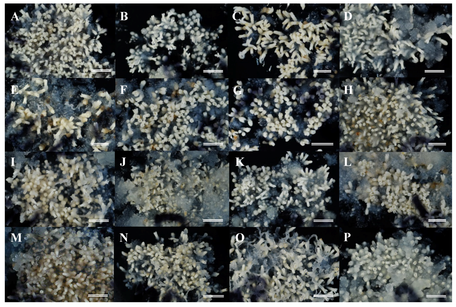
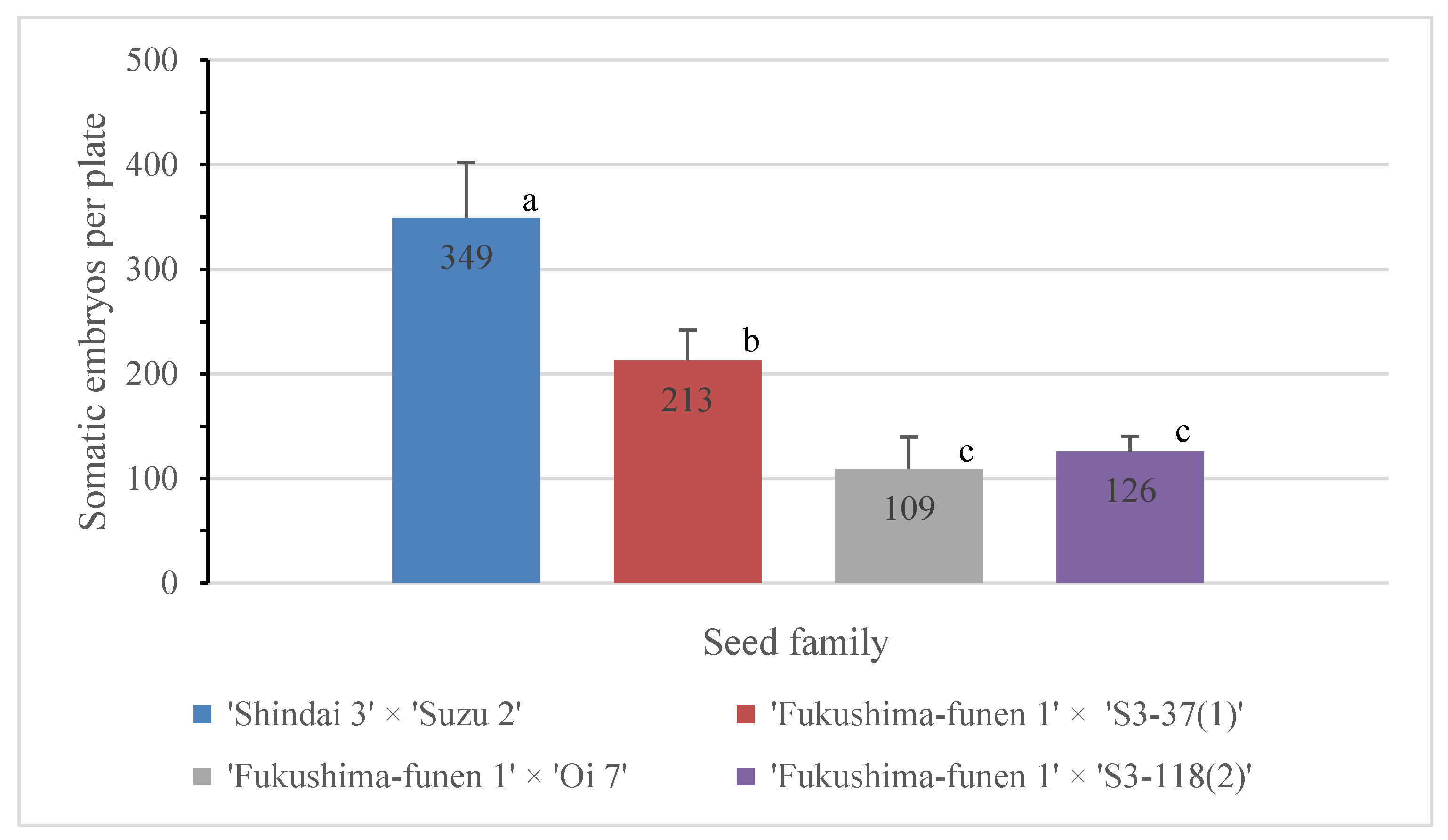
2.1. Somatic Embryogenesis Initiation

Our strategy was to produce successful MSPs by SE from selected male sterile ECLs. The first part of our experimental approach was to assess initiation of SE from the different seed families carrying the male sterility gene. The whole megagametophyte containing the zygotic embryo was used as the initial explant for induction and culture of ECLs to be used for SE. Although extrusion of embryogenic cells in a number of explants could be observed about 2 weeks after the start of culture, the establishment of stable lines with evident embryogenic cell proliferation was achieved most frequently after 4–6 weeks of culture (Figure 1).
As shown in Table 1, despite observed differences among seed families and variations due to collection date, SE was initiated in all families using megagametophyte explants from the seeds collected in early to late July 2017. The highest SE initiation frequency (59.03%) was recorded using explants from seeds of the ‘Fukushima-funen 1’×’Oi 7’ family collected on July 24. This result was similar to those reported for some male fertile sugi families, such as Gujo 4 (62.5%), Kofu-sho 2 (52.5%), and Minamitama 5 (65.9%) [32]. In contrast, the lowest SE initiation frequency (0.62% for the July 03 collection), and the lowest overall average frequency (across all collections), was recorded for the ‘Fukushima-funen 1’×’S3-37(1)’ seed family (7.67%). The lowest average frequency, taking data for all four seed families into account, was recorded for the collection of July 03 (12.70%), this value increasing to 30.11% and 33.84% for collections taken on July 10 and July 24, respectively, and reaching the maximum value (42.24%) for seeds collected on July 18. An influence of seed collection date on the induction efficiency of embryogenic cells has been previously reported for male fertile sugi families [32,33] and other conifers [45,46,47,48,49,50,51,52,53,54,55,56]. The results of statistical analysis indicated that the proportion of the explants with SE initiation response significantly differed among families (χ2 = 366.6, df = 3, p < 0.001) and among seed collection dates (χ2 = 177.9, df = 3, p < 0.001). Frequencies of each family and seed collection date with SE initiation response were all significantly differentiated with the exception for the collections of July 10 and 24 (p > 0.05) (Table 1).
Maintenance/proliferation medium was able to support the growth of initiated ECLs by subculture routines carried out at intervals of 2–3 weeks (Figure 2). Stable ECLs have been maintained for more than 2 years without loss of their initial morphological characteristics and proliferation potential. Stable embryogenic cultures showed early stage of somatic embryos characterized by a densely embryonal head with distinct suspensor system (elongated cells), as described in Maruyama et al. [33].

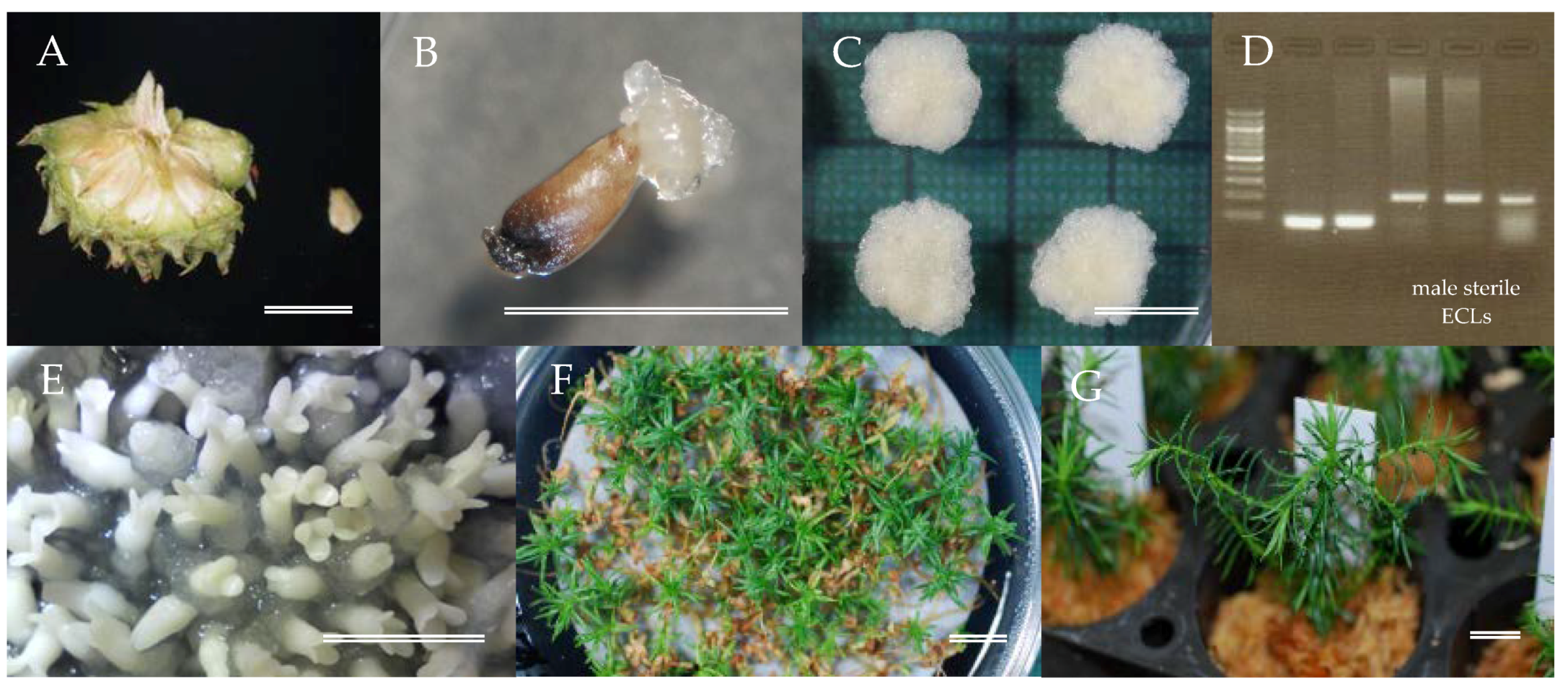
2.2. Selection of Male Sterile ECLs

After establishing the ECLs, it was necessary to identify and select those lines that were male sterile prior to further propagation. As shown in Table 2, over the 616 ECLs analyzed from four seed families, we selected 236 as male sterile lines (pollen-free lines) using MAS. Where PCR for detection of the marker DNA did not produce a clear positive band on agarose gels, ECLs were described as “doubted lines”. The lack of a clear PCR product could be due to failure of the PCR amplification or poor DNA extraction. Since this is the first step of the screening process, from which significant numbers of male sterile lines are identified, we did not repeat the experiments in order to eliminate this doubted line category and thereby determine the exact numbers in each of the male fertile or sterile lines. The preciseness of the numbers in each category was also affected by our cautious assumption that if the allele-specific PCR assay for male sterility did not produce a clear strong amplification product on agarose gels, that particular ECL was not male sterile. Using the numbers in Table 2, the ratio of male sterile lines to fertile lines was 1:1.4 (chi-square = 16.85 and p = 4.05 × 10⁻⁵ with df = 1). For a back-crossed pedigree (a seed parent (ms1/ms1) crossed with a pollen parent (Ms1/ms1)), Mendelian inheritance would predict a 1:1 ratio of male fertile and sterile progeny. An additional note of caution to be applied here is that the marker used is not the MS1 gene itself but a closely linked marker to MS1 (0.58 cM to MS1) [12]. This genetic distance indicates that at least one individual out of 200 offspring had a recombination event between MS1 and the marker dD_Contig_3995-165. Such offspring would not give a positive result for presence of a mutant allele of MS1. Hence, for the present study, of the analyzed total of 616 lines, at least three individuals were not classified correctly as male fertile or sterile line. Although the marker used in the current study was a closely linked marker to MS1 (0.58 cM to MS1) [12], the MAS of the ECL was effective. In our previous studies on MAS for ms1, we also developed linked markers to MS1 [10,57]. These markers were distantly located to MS1 (3.1 cM) and/or unable to be used in the current families due to the lack of suitable SNPs among parents (Ueno et al. unpublished). More recently, since completion of the above analysis, we have identified a candidate gene for MS1 [58] and developed diagnostic markers [59]. These markers will be useful to verify the result of the current study.

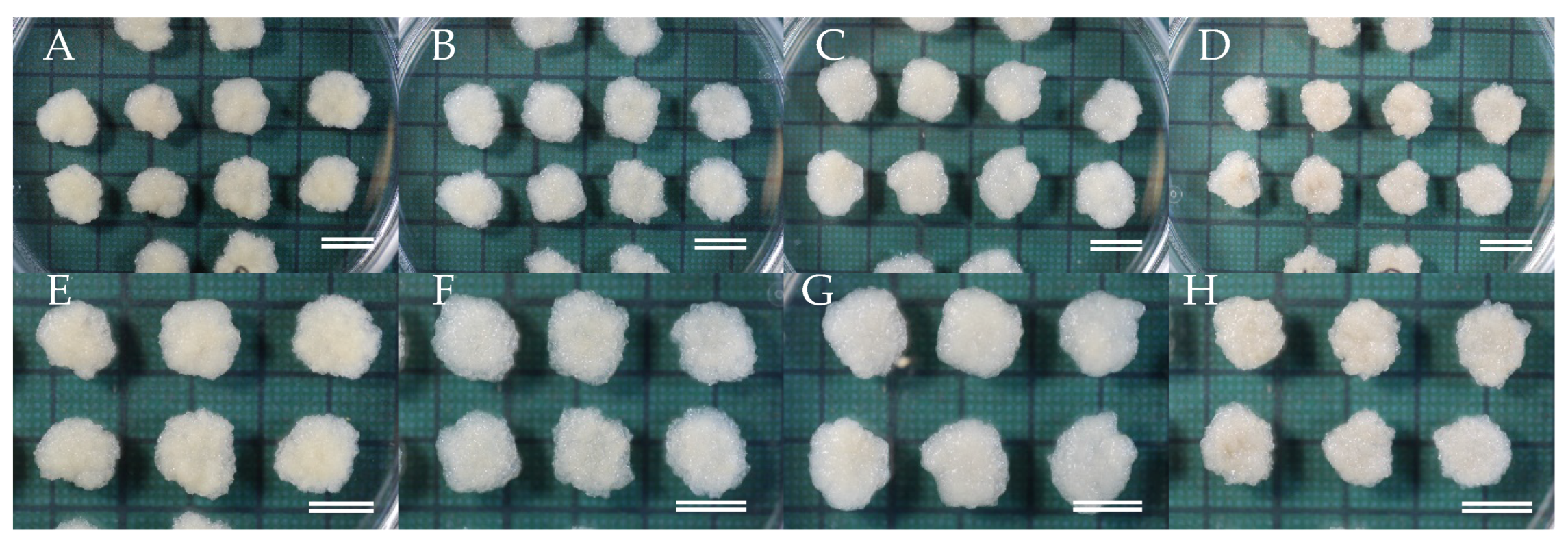
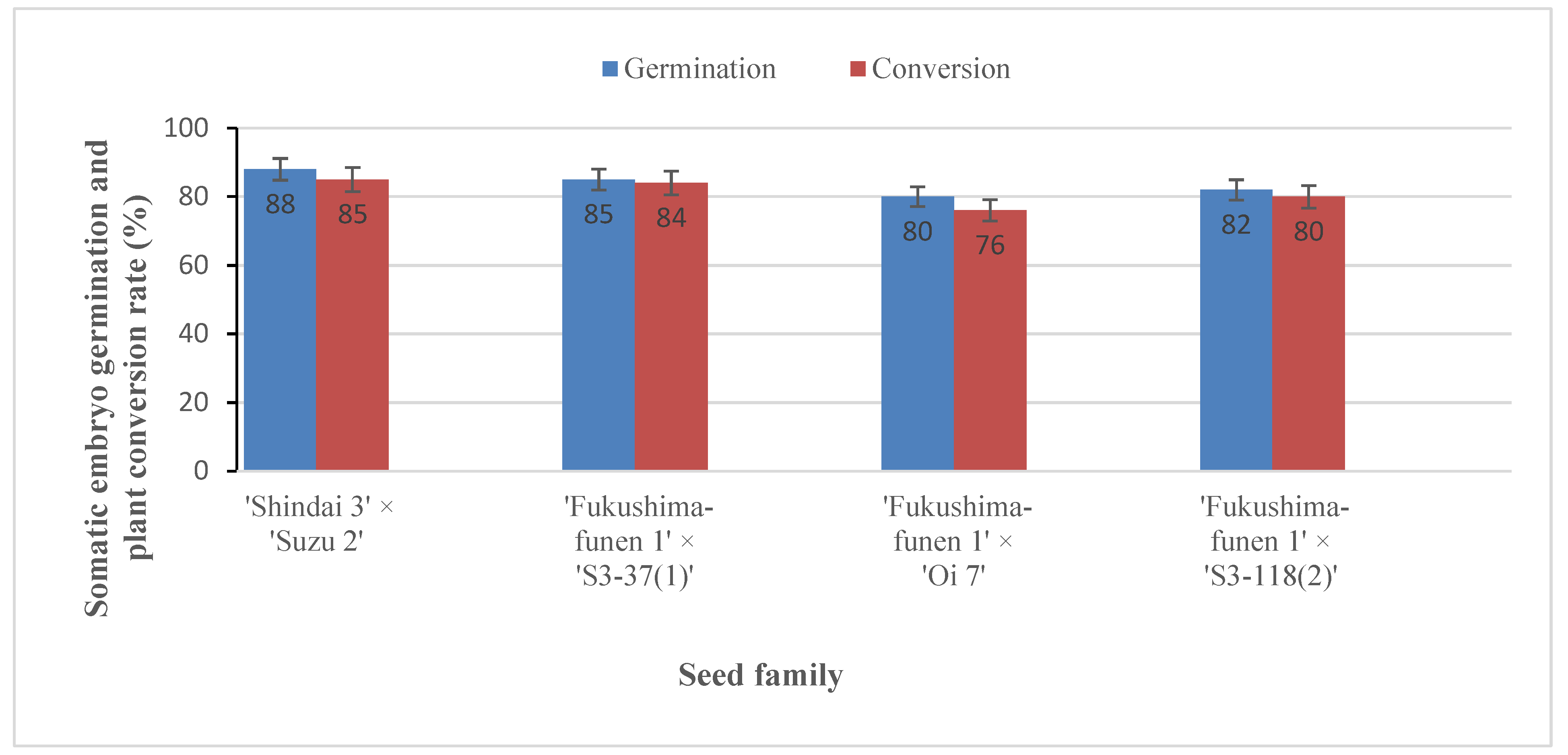
2.3. Maturation of Somatic Embryos

The next challenge was to develop the selected male sterile ECLs into mature somatic embryos that could be used for regeneration and conversion into somatic plants. Formation of cotyledonary somatic embryos from most of the ECLs was observed about 6 weeks after the transfer of the embryogenic cells to maturation medium (Figure 3). Ten male sterile ECLs showing the best somatic embryo maturation efficiencies from each seed family were selected for production of somatic plants. As shown in Figure 4, the seed family ‘Shindai 3’×’Suzu 2’ produced the highest average number of somatic embryos per plate (349 cotyledonary embryos per 0.5 g), whereas the lowest number (109 embryos) was recorded for the ‘Fukushima-funen 1’×’Oi 7’ seed family. The families ‘Fukushima-funen 1’×’S3-37(1)’ and ‘Fukushima-funen 1’×’S3-118(2)’ produced intermediate values of 213 and 126 embryos per plate, respectively. Somatic embryo production obtained in this study was higher than those previously reported for sugi in 2000 (up to 67 embryos per plate) [33] and 2003 (up to 46 embryos per plate) [36], but similar to the results published in 2007 (up to 361 embryos per plate) [39]. For comparison, the numbers of cotyledonary embryos produced per gram of embryogenic cells in studies of other conifer trees have been reported as 68–147 for Pinus strobus [46], 2–441 for P. sylvestris [60], 10–1550 for P. radiata [61], 0–798 for P. densiflora [62], 67–551 for Larix leptolepis [63], 8–1566 for hybrid Larix × eurolepis [64], 80–200 for Picea abies [65], and 22–925 for Abies fraseri [66].
Although the number of mature somatic embryos produced varied among the seed families, the induction of somatic embryos was confirmed in all families, with an average production of at least 100 cotyledonary embryos per plate (equivalent to 200 embryos per gram). This value compares well with those listed earlier for other conifers. Differences in successful somatic embryo production among seed families, as observed here for the male sterile lines, were also found in male fertile sugi families [33,36] and in other Japanese conifers such as Chamaecyparis obtusa [67,68], Pinus thunberghii [69], and P. armandii var. amamiana [70].

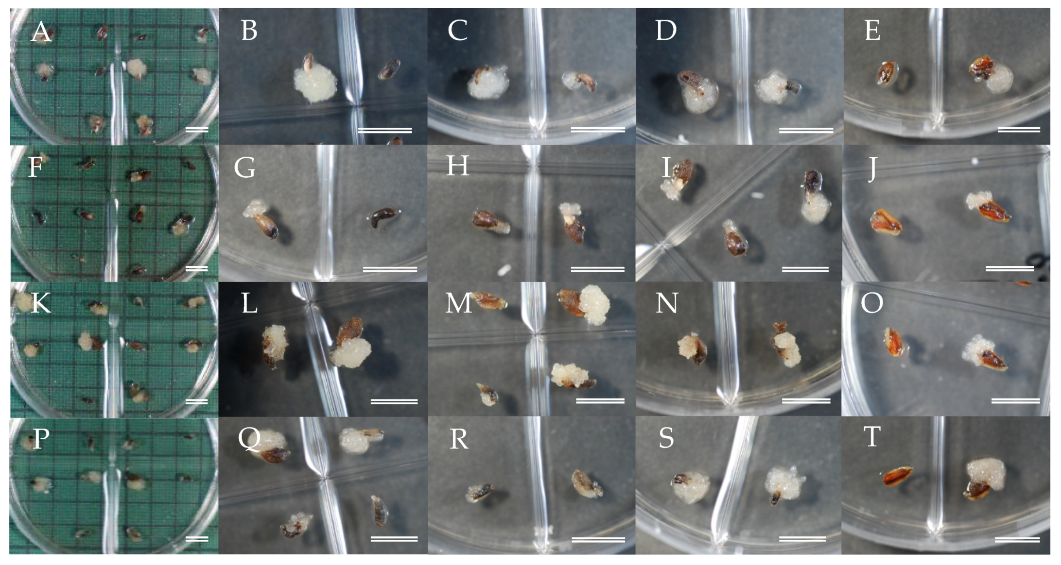
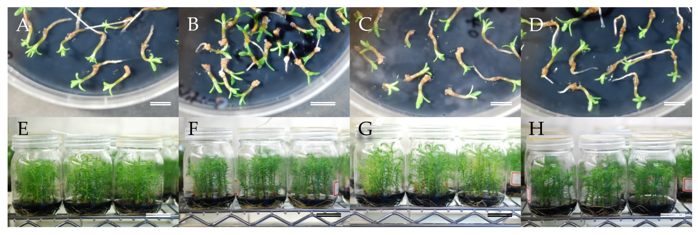
2.4. Regeneration of Somatic Plants

At this point in our process, cotyledonary somatic embryos were now available from all four seed families and could be transferred to germination medium as the first stage of regeneration of somatic plants. The embryos germinated readily, with the start of germination observed at about 1–2 weeks after transfer (Figure 5A–D) and conversion in the majority of cases achieved after 3–6 weeks of culture. The percentage of successful germination and plant conversion that was achieved varied from 80–88% and 76–85%, respectively (Figure 6). Although no significant differences in these values were detected among the seed families, the best result for both germination (88%) and conversion rate (85%) was obtained with somatic embryos from ‘Shindai 3’×’Suzu 2’ family.
The high germination and conversion frequencies obtained in all families demonstrated that the somatic embryos produced were of high quality. Germination frequencies achieved in this study were higher than those previously reported by Maruyama et al. [33] and Igasaki et al. [36], who recorded germination frequencies in the ranges of 12–57% and 33–63%, respectively. Maturation efficiency and the quality of the somatic embryos produced are two of the most important criteria for the optimization of a SE protocol for practical use [71]. Notwithstanding the fact that cotyledonary embryos were produced on medium containing a high concentration (17.5%) of polyethylene glycol (PEG) they readily germinated after the transfer to a plant growth regulator-free medium, without any post-maturation treatment. This is in contrast to studies of somatic embryo maturation of Picea abies, in which PEG is reported to stimulate embryogenesis but inhibit the subsequent germination process [72]. Partial desiccation and/or cold treatments after maturation on medium containing PEG have been reported necessary to improve somatic embryo germination and conversion in a number of conifer species, including some pines [73,74,75,76,77,78,79], spruces [80,81,82,83], hybrid larch [84], Fraser fir [66], and Chinese fir [85].
Somatic male sterile plants developed in vitro (Figure 5E–H) were successfully acclimatized in plant containers (Figure 7A–D) and grew well with no signs of abnormal appearance (Figure 7E,F), and the subsequent growth of male sterile somatic plants in the field was monitored [86]. A previous comparison of traits between male sterile and fertile sugi trees in selected stands indicated that no marked differences were observed in any of the physical characteristics examined (tree height, diameter at breast height, basal bending, modulus of elasticity of tree trunk, and types of snow damage) [87]. The indications are, therefore, that the somatic MSPs produced in this study will grow normally, and pollen production will be their only deficiency. However, in addition to growth performance in the field, it will be essential to monitor the genetic stability of the somatic plants using molecular marker technology in order to confirm that a practical and efficient protocol for sugi MSP propagation has been established.

3. Materials and Methods

3.1. Initial Explant, Medium, and Culture Conditions

Four seed collections were carried out at 1-week intervals during the month of July (2017) from four full sib seed families of sugi carrying the male sterility gene MS1 (Table 1). At each collection date, samples of zygotic embryos were observed to determine their developmental stage according to the scale used to classify zygotic embryo development in loblolly pine [88]. The developmental stage of explants collected on July 03 was pre-embryo stage equivalent to stages 1–2. Collections on July 10 and 18 were mostly represented by early embryo stages equivalent to stages 3–4 and 5–6, respectively. Seeds collected on July 28 showed pre-cotyledonary stages equivalent to stages 7–8 in the scale of Pullman and Buchanan [88].
The whole megagametophyte (about 3–4 mm long) containing the zygotic embryo was used as the initial explant for SE initiation. Seeds were surface sterilized with 1% (w/v available chlorine) sodium hypochlorite solution for 15 min and then rinsed 3 times with sterile distilled water for 5 min each time before isolation of megagametophyte explants. For induction of embryogenic cells, explants were placed horizontally onto initiation medium contained in 90 × 15 mm quad-plates (3 explants per well, 12 per plate) and cultured in darkness at 25 °C. Initiation medium containing basal salts reduced to half concentration from the standard EM medium [33] was supplemented with 10 g L−1 sucrose, 10 μM 2,4-dichlorophenoxyacetic acid (2,4-D), 5 μM 6-benzylaminopurine (BA), 0.5 g L−1 casein acid hydrolysate, 0.5 g L−1 glutamine, and solidified with 3 g L−1 gellan gum (Gelrite®; Wako Pure Chemical, Osaka, Japan). The pH was adjusted to 5.8 prior to autoclaving the medium for 15 min at 121 °C.

3.2. Maintenance and Proliferation of ECLs

Induced ECLs were subcultured every 2–3 weeks on maintenance/proliferation medium containing basal salts reduced to half concentration from the standard EM medium [33] was supplemented with 3 μM 2,4-D, 1 μM BA, 30 g L−1 sucrose, 1.5 g L−1 glutamine, and 3 g L−1 gellan gum. Clumps of embryogenic cells (12 per plate) were cultured in darkness at 25 °C.

3.3. Selection of Male Sterile ECLs

Male sterile ECLs were selected according to the methodology described elsewhere [12]. Embryogenic cells for DNA extraction were sampled from each culture line using a microspatula. One spoonful of sampled tissue (30–40 mg under wet conditions) was dispersed into 200 μL of 2 × CTAB buffer [59] and stored at −30 °C until DNA extraction. The freeze-thawed samples were disrupted using a TissueLyser II (Qiagen) at a frequency of 30 Hz for 30 s. Samples were then incubated at 65 °C for 10 min. Chloroform (50 μL) was then added, the samples emulsified, and centrifuged at 12,000 rpm at room temperature for 10 min. The aqueous phase was transferred to a new 1.5 mL tube and a two-thirds volume of isopropanol was added to precipitate DNA, which was collected by centrifugation at 12,000 rpm at 4 °C for 15 min. The pellet was washed by 70% ethanol and vacuum-dried for 5 min, before finally dissolving the dried nucleic acid pellet in 100 μL of TE buffer. One microliter of RNase solution (2 mg/mL) was added and incubated at 37 °C for 2 h with a little agitation on a shaker. To identify the marker (dD_Contig_3995-165) for male sterility [12] by PCR, a 1 μL sample of each DNA solution was used as a template. The reaction mixture, totaling 10 μL, contained 3 μL of 2 × Multiplex (Qiagen), 0.2 μM of each forward and reverse primers, and 1 μL of DNA, which was then amplified in a GeneAmp PCR System 9700 (Applied Biosystems, Foster City, CA, USA) with the following thermal conditions: initial denaturation at 95 °C for 15 min, 4 cycles of 95 °C for 30 s, 64 °C for 90 s with −1 °C per cycle and 72 °C for 30 s, and 34 cycles of 95 °C for 30 s, 60 °C for 90 s, and 72 °C for 30 s. The PCR products were visualized by 2% agarose gel electrophoresis and ethidium bromide staining.

3.4. Maturation of Somatic Embryos

For development and maturation of somatic embryos, proliferated ECLs (early stage of somatic embryos characterized by an embryonal head with suspensor system) [33] were cultured in clumps (5 masses per 90 × 20 mm plate, 100 mg each) on maturation medium for 8 weeks. Maturation medium contained the basal salt concentration of the standard EM medium [33], supplemented with 30 g L−1 maltose, 2 g L−1 activated charcoal (AC), 100 µM abscisic acid (ABA), amino acids (in g L−1: glutamine 2, asparagine 1, arginine 0.5, citrulline 0.079, ornithine 0.076, lysine 0.055, alanine 0.04, and proline 0.035), 175 g L−1 PEG (Av. Mol. Wt.: 7300–9300; Wako Pure Chemical, Osaka, Japan), and 3.3 g L−1 gellan gum. The plates were sealed with Parafilm® and kept in darkness at 25 °C.

3.5. Germination and Plant Conversion

Cotyledonary embryos collected from the maturation medium were laid horizontally onto the germination medium (maintenance/proliferation medium containing 20 g L−1 sucrose, 2 g L−1 AC, and 10 g L−1 agar but without addition of plant growth regulators) and cultured at 25 °C under a photon flux density of 45–65 µmol m−2 s−1 provided by 100 V, 40 W white fluorescent lamps. The photoperiod was 16 h. Germination, taken as the emergence of the root, and plant conversion, taken as the emergence of both root and epicotyl, were recorded after 8 weeks of culturing.

3.6. Growth In Vitro and Acclimatization of Somatic Plants

The growth of emblings was promoted by transferring them to culture flasks containing germination medium supplemented with 30 g L−1 sucrose and 5 g L−1 AC, and culturing under the same conditions described above for about 10–12 weeks before ex vitro acclimatization. Developed somatic plants removed from the culture flasks were transplanted into plant containers filled with spagmoss (Sphagnum moss) and kept inside plastic boxes with transparent covers. Plant containers were irrigated with tap water as needed during the first 2 weeks. After this initial 2-week period, the covers were opened gradually and the plant containers were fertilized with Nagao’s nutrient solution [17]. The covers were completely removed 4 weeks after transplanting. Acclimatized somatic plants were grown in a greenhouse (about 3 months) until an approximate height of 30 cm was attained, after which they were transplanted to the field.

3.7. Statistical Analysis

The differentiation of the proportion of the explants with SE initiation response among families and seed collection dates were examined using Pearson’s Chi-squared test. To further elucidate which part of the data was causing the significant differentiation, the residuals of the Chi-squared test were used to conduct the post hoc analysis and the p-values were adjusted with a Bonferroni correction [89]. Pearson’s Chi-squared test was performed using R version 3.6.2 [90] and the post hoc analysis based on the residuals of the Chi-squared test was done using R package “chisq.posthoc.test” [91]. The data for somatic embryo production efficiency, somatic embryo germination, and plant conversion were analyzed using one-way analysis of variance, followed by Tukey’s multiple-range test.

4. Conclusions

An efficient protocol to propagate male sterile somatic plants of sugi combining selection of ECLs with MAS and propagation via SE has been established (Figure 8). Using four different seed families of sugi carrying the male sterility gene MS1, collected during the month of July, initiation of SE was demonstrated. Despite some differences in the initiation rate, the numbers of male sterile ECLs selected, and somatic embryo maturation among the four seed families, large numbers of stable culture lines were established, and we were able to produce pollen-free somatic plants arising from all families. By selecting for male sterility at the embryogenic cell stage, SE can ultimately generate multiple somatic MSPs in a fraction of the time taken by existing conventional methods. We believe that the methodology developed in this study will serve as a powerful tool to establish an efficient breeding technology for MSPs of sugi.

Author Contributions

Conceptualization and methodology, T.E.M., S.U., S.H., and Y.M.; funding acquisition and project administration, Y.M.; plant material preparation, S.H.; data curation, T.E.M. and S.U.; experiments and data analysis, T.E.M., T.K., and S.U., writing—original draft, T.E.M. and S.U.; writing—review and editing, T.E.M., S.U., and Y.M. All authors have read and agreed to the published version of the manuscript.

Funding

This research was supported by the grants from Ministry of Agriculture, Forestry and Fisheries of Japan (MAFF) and NARO Bio-oriented Technology Research Advancement Institution (BRAIN) (the Science and technology research promotion program for agriculture, forestry, fisheries and food industry (No.28013B)) and the grants from NARO Bio-oriented Technology Research Advancement Institution (BRAIN) (Research program on development of innovative technology (No.28013BC)).

Acknowledgments

We thank the Niigata Prefecture Forestry Research Institute for the logistic support in the plant material preparation. Authors also thank Hideki Mori for assistance with the statistical analysis.

Conflicts of Interest

The authors declare no conflict of interest.

References

  1. Taira, H.; Teranishi, H.; Kenda, Y. A case study of male sterility in sugi (Cryptomeria japonica). J. Jpn. For. Soc. 1993, 75, 377–379, (In Japanese with English Abstract). [Google Scholar]
  2. Igarashi, M.; Watanabe, J.; Saito, Y.; Ozawa, H.; Saito, N.; Furukawa, S.; Ishii, Y. Breeding of Cryptomeria japonica for the countermeasure of pollinosis. Bull. Fukushima Prefect. For. Res. Centre. 2006, 39, 1–9. (In Japanese) [Google Scholar]
  3. Saito, M. Development status and future prospect of non-pollen cedar. For. Sci. 2008, 54, 17–20. (In Japanese) [Google Scholar]
  4. Hasegawa, Y.; Ueno, S.; Matsumoto, A.; Ujino-Ihara, T.; Uchiyama, K.; Totsuka, S.; Iwai, J.; Hakamata, T.; Moriguchi, Y. Fine mapping of the male-sterile genes (MS1, MS2, MS3, and MS4) and development of SNP markers for marker-assisted selection in Japanese cedar (Cryptomeria japonica D. Don). PLoS ONE 2018, 13, e0206695. [Google Scholar] [CrossRef] [PubMed]
  5. Taira, H.; Saito, M.; Furuta, Y. Inheritance of the trait of male sterility in Cryptomeria japonica. J. For. Res. 1999, 4, 271–273. [Google Scholar] [CrossRef]
  6. Yoshii, E. Study for the Characteristics and Applications of Nuclear Male Sterility in Cryptomeria Japonica D. Don. Ph.D. Thesis, Niigata University, Niigata, Japan, March 2007. (In Japanese). [Google Scholar]
  7. Miyajima, D.; Yoshii, E.; Hosoo, Y.; Taira, H. Cytological and genetic studies on male sterility in Cryptomeria japonica D. Don (Shindai 8). J. Jpn. For. Soc. 2010, 92, 106–109, (In Japanese with English Summary). [Google Scholar] [CrossRef] [Green Version]
  8. Moriguchi, Y.; Ujino-Ihara, T.; Uchiyama, K.; Futamura, N.; Saito, M.; Ueno, S.; Matsumoto, A.; Tani, N.; Taira, H.; Shinohara, K.; et al. The construction of a high-density linkage map for identifying SNP markers that are tightly linked to a nuclear-recessive major gene for male sterility in Cryptomeria japonica D. Don. BMC Genom. 2012, 13, 95. [Google Scholar] [CrossRef] [Green Version]
  9. Moriguchi, Y.; Ueno, S.; Higuchi, Y.; Miyajima, D.; Itoo, S.; Futamura, N.; Tsumura, Y. Establishment of a microsatellite panel covering the sugi (Cryptomeria japonica) genome, and its application for localization of a male sterile gene (ms-2). Mol. Breed. 2014, 33, 315–325. [Google Scholar] [CrossRef]
  10. Moriguchi, Y.; Uchiyama, K.; Ueno, S.; Ujino-Ihara, T.; Matsumoto, A.; Iwai, J.; Miyajima, D.; Saito, M.; Sato, M.; Tsumura, Y.; et al. A high-density linkage map with 2,560 markers and its application for the localization of the male-sterile genes ms3 and ms4 in Cryptomeria japonica D. Don. Tree Genet. Genom. 2016, 12, 57. [Google Scholar] [CrossRef]
  11. Mishima, K.; Hirao, T.; Tsubomura, M.; Tamura, M.; Kurita, M.; Nose, M.; Hanaoka, S.; Takahashi, M.; Watanabe, A. Identification of novel putative causative genes and genetic marker for male sterility in Japanese cedar (Cryptomeria japonica D. Don). BMC Genom. 2018, 19, 277. [Google Scholar] [CrossRef]
  12. Ueno, S.; Uchiyama, K.; Moriguchi, Y.; Ujino-Ihara, T.; Matsumoto, A.; Wei, F.J.; Saito, M.; Higuchi, Y.; Futamura, N.; Kanamori, H.; et al. Scanning RNA-Seq and RAD-Seq approach to develop SNP markers closely linked to MALE STERILITY 1 (MS1) in Cryptomeria japonica D. Don. Breed. Sci. 2019, 69, 19–29. [Google Scholar] [CrossRef] [Green Version]
  13. Hasegawa, Y.; Ueno, S.; Fu-Jin, W.; Matsumoto, A.; Ujino-Ihara, T.; Uchiyama, K.; Moriguchi, Y.; Kasahara, M.; Fujino, T.; Shigenobu, S.; et al. Development of diagnostic PCR and LAMP markers for MALE STERILITY 1 (MS1) in Cryptomeria japonica D Don. BMC Res. Note 2020. (under review). [Google Scholar]
  14. Moriguchi, Y.; Ueno, S.; Hasegawa, Y.; Tadama, T.; Watanabe, M.; Saito, R.; Hirayama, S.; Iwai, J.; Konno, Y. Marker-assisted selection of trees with MALE STERILITY 1 in Cryptomeria japonica D. Don. Forests 2020, 11, 734. [Google Scholar] [CrossRef]
  15. Saito, M.; Teranishi, H. A breeding strategy of male sterile Cryptomeria japonica D. Don cultivars. Jpn. J. Palynol. 2014, 60, 27–35, (In Japanese with English Abstract). [Google Scholar]
  16. Shidei, T.; Akai, T.; Ichikawa, S. Flower bud formation on Sugi (Cryptomeria japonica) and Metasequoia (Metasequoia glyptosytoboides) by gibberellic acid treatment. J. Jpn. For. Soc. 1959, 41, 312–315. (In Japanese) [Google Scholar]
  17. Nagao, A. Differences of flower initiation of Cryptomeria japonica under various alternating temperatures. J. Jap. For. Soc. 1983, 65, 335–338, (In Japanese with English Abstract). [Google Scholar]
  18. Isikawa, H. In vitro formation of adventitious bud and root on the hypocotyl of Cryptomeria japonica. Bot. Mag. Tokyo 1974, 87, 73–77. [Google Scholar] [CrossRef]
  19. Sato, T. Search for the synthetic media suitable for embryo culture of Cryptomeria and Japanese black pine. J. Jpn. For. Soc. 1978, 60, 81–86, (In Japanese with English Abstract). [Google Scholar] [CrossRef]
  20. Fukuda, K.; Morohoshi, N. Tissue culture of sugi (Cryptomeria japonica) and properties of callus-lignin. Bull. Exp. For. Tokyo Univ. Agricult. Technol. 1983, 19, 39–46. (In Japanese) [Google Scholar]
  21. Isikawa, H. In vitro culture of forest tree calluses and organs. JARQ 1984, 18, 131–141. [Google Scholar]
  22. Sato, T. The in vitro plantlet regeneration following induction of adventitious buds on various tissues from small sugi seedlings. J. Jpn. For. Soc. 1986, 68, 389–392. (In Japanese) [Google Scholar] [CrossRef]
  23. Isikawa, H. Generation of adventitious plant organs by tissue culture methods in forest trees. Bull. For. For. Prod. Res. Inst. 1987, 343, 119–153. (In Japanese) [Google Scholar]
  24. Isikawa, H. In vitro culture of Cryptomeria callus and organs. In Cell and Tissue Culture in Forestry, Case Histories: Gymnosperms, Angiosperms and Palms; Bonga, J.M., Durzan, D.J., Eds.; Martinus Nijhoff Publishers: Dordrecht, The Netherlands, 1987; Volume 3, pp. 109–113. [Google Scholar]
  25. Sato, T. Basic studies of organ and callus culture in woody plants. Bull. For. Prod. Res. Inst. 1991, 360, 35–119, (In Japanese with English Summary). [Google Scholar]
  26. Fukushima, T. Tissue culture of Cryptomeria japonica, Chamaecyparis obtusa, Pinus densiflora and P. thunbergii. Bull. Shimane Pref. For. Res. Cen. 1993, 44, 13–26. (In Japanese) [Google Scholar]
  27. Mikawa, K. Tissue culture of sugi. Bull. Yamagata Pref. For. Res. Cen. 1994, 24, 19–24. (In Japanese) [Google Scholar]
  28. Taniguchi, T.; Kondo, K. In vitro clonal propagation of Cryptomeria japonica from current stem segments. J. Jpn. For. Soc. 1997, 79, 246–248. (In Japanese) [Google Scholar]
  29. Hine-Gómez, A.; Valverde-Cerdas, L. Establecimiento in vitro de Cryptomeria japonica (Taxocidaceae). Rev. Biol. Trop. 2003, 51, 683–690, (In Espanish with English Abstract). [Google Scholar]
  30. Ogita, S.; Ishikawa, H.; Kubo, T.; Sasamoto, H. Somatic embryogenesis from immature and mature zygotic embryos of Cryptomeria japonica I: Embryogenic cell induction and its morphological characteristics. J. Wood. Sci. 1999, 45, 87–91. [Google Scholar] [CrossRef]
  31. Ogita, S.; Sasamoto, H.; Kubo, T. Selection and microculture of single embryogenic cell clusters in Japanese conifers: Picea jezoensis, Larix leptolepis and Cryptomeria japonica. In Vitro Cell. Dev. Biol. Plant 1999, 35, 428–431. [Google Scholar] [CrossRef]
  32. Taniguchi, T.; Kondo, T. Difference in ability of initiation and maintenance of embryogenic cultures among sugi (Cryptomeria japonica D. Don) seed families. Plant Biotechnol. 2000, 17, 159–162. [Google Scholar] [CrossRef]
  33. Maruyama, E.; Tanaka, T.; Hosoi, Y.; Ishii, K.; Morohoshi, N. Embryogenic cell culture, protoplast regeneration, cryopreservation, biolistic gene transfer and plant regeneration in Japanese cedar (Cryptomeria japonica D. Don). Plant Biotechnol. 2000, 17, 281–296. [Google Scholar] [CrossRef]
  34. Ishii, K.; Maruyama, E.; Hosoi, Y. Somatic embryogenesis of Japanese conifers. In Molecular Breeding of Woody Plants; Morohoshi, N., Komamine, A., Eds.; Elsevier: Amsterdam, The Netherlands, 2001; pp. 297–304. [Google Scholar]
  35. Ogita, S.; Sasamoto, H.; Yeung, E.C.; Thorpe, T. The effects of glutamine on the maintenance of embryogenic cultures of Cryptomeria japonica. In Vitro Cell. Dev. Biol. Plant 2001, 37, 268–273. [Google Scholar] [CrossRef]
  36. Igasaki, T.; Sato, T.; Akashi, N.; Mohri, T.; Maruyama, E.; Kinoshita, I.; Walter, C.; Shinohara, K. Somatic embryogenesis and plant regeneration from immature zygotic embryos of Cryptomeria japonica D. Don. Plant Cell Rep. 2003, 22, 239–243. [Google Scholar] [CrossRef]
  37. Igasaki, T.; Akashi, N.; Ujino-Ihara, T.; Matsubayashi, Y.; Sakagami, Y.; Shinohara, K. Phytosulfokine stimulates somatic embryogenesis in Cryptomeria japonica. Plant Cell Physiol. 2003, 44, 1412–1416. [Google Scholar] [CrossRef] [Green Version]
  38. Nakagawa, R.; Ogita, S.; Kubo, T.; Funada, R. Effect of polyamines and L-ornithine on the development of proembryogenic masses of Cryptomeria japonica. Plant Cell Tiss. Organ Cult. 2006, 85, 229–234. [Google Scholar] [CrossRef]
  39. Maruyama, E.; Hosoi, Y. Polyethylene glycol enhance somatic embryo production in Japanese cedar (Cryptomeria japonica D. Don). Prop. Ornam. Plants 2007, 7, 57–61. [Google Scholar]
  40. Fujisawa, T.; Tsubomura, M.; Taniguchi, T. Development of mass production technology for non-pollen cedar. For. Prod. Res. Inst. Select. Res. Results 2010, 2010, 64–65. (In Japanese) [Google Scholar]
  41. Ishii, K.; Hosoi, Y.; Taniguchi, T.; Tsubomura, M.; Kondo, T.; Yamada, H.; Saito, M.; Suda, T.; Fukisawa, T.; Tanaka, K.; et al. In vitro culture of various genotypes of male sterile Japanese cedar (Cryptomeria japonica D. Don). Plant Biotechnol. 2011, 28, 103–106. [Google Scholar] [CrossRef]
  42. Maruyama, E.T.; Hosoi, Y.; Futamura, N.; Saito, M. Initiation of embryogenic cultures from immature seeds of pollen-free sugi (Cryptomeria japonica). Kanto Shinrin Kenkyu 2014, 65, 107–110, (In Japanese with English abstract). [Google Scholar]
  43. Maruyama, E.T.; Miyazawa, S.; Ueno, S.; Onishi, N.; Totsuka, S.; Iwai, J.; Moriguchi, Y. Differences among families on embryogenic cell induction from seed of pollen-free sugi (Cryptomeria japonica) produced at the Niigata prefecture. Kanto Shinrin Kenkyu 2018, 69, 1–2, (In Japanese with English abstract). [Google Scholar]
  44. Maruyama, E.T.; Hosoi, Y.; Miyazawa, S.; Ueno, S.; Onishi, N.; Totsuka, S.; Iwai, J.; Moriguchi, Y. Pollen-free plant regeneration from embryogenic cells derived from sugi (Cryptomeria japonica). Kanto Shinrin Kenkyu 2019, 70, 37–40, (In Japanese with English abstract). [Google Scholar]
  45. Finer, J.J.; Kriebel, H.B.; Becwar, M.R. Initiation of embryogenic callus and suspension cultures of eastern white pine (Pinus strobus L.). Plant Cell Rep. 1989, 8, 203–206. [Google Scholar] [CrossRef] [PubMed]
  46. Klimaszewska, K.; Park, Y.S.; Overton, C.; MacEacheron, I.; Bonga, J.M. Optimized somatic embryogenesis in Pinus strobus L. In Vitro Cell. Dev. Biol. Plant. 2001, 37, 392–399. [Google Scholar] [CrossRef]
  47. Miguel, C.; Goncalves, S.; Tereso, S.; Marum, L.; Maroco, J.; Oliveira, M. Somatic embryogenesis from 20 open-pollinated families of Portuguese plus trees of maritime pine. Plant Cell Tiss. Organ Cult. 2004, 76, 121–130. [Google Scholar] [CrossRef]
  48. Hargreaves, C.L.; Reeves, C.B.; Find, J.I.; Gough, K.; Josekutty, P.; Skudder, D.B.; van der Maas, S.A.; Sigley, M.R.; Menzies, M.I.; Low, C.B.; et al. Improving initiation, genotype capture, and family representation in somatic embryogenesis of Pinus radiata by a combination of zygotic embryo maturity, media, and explant preparation. Can. J. For. Res. 2009, 39, 1566–1574. [Google Scholar] [CrossRef]
  49. Montalbán, I.A.; de Diego, N.; Moncaleán, P. Enhancing initiation and proliferation in radiata pine (Pinus radiata D. Don) somatic embryogenesis through seed family screening, zygotic embryo staging and media adjustments. Acta Physiol. Plant. 2012, 34, 451–460. [Google Scholar] [CrossRef]
  50. Montalbán, I.A.; Setien-Olarra, A.; Hargreaves, C.L.; Moncaleán, P. Somatic embryogenesis in Pinus halepensis Mill.: An important ecological species from the Mediterranean forest. Trees 2013, 27, 1339–1351. [Google Scholar] [CrossRef]
  51. Find, J.I.; Hargreaves, C.L.; Reeves, C.B. Progress towards initiation of somatic embryogenesis from differentiated tissues of radiata pine (Pinus radiata D. Don) using cotyledonary embryos. In Vitro Cell. Dev. Biol. Plant. 2014, 50, 190–198. [Google Scholar] [CrossRef]
  52. Barberini, S.; Danti, R.; Lambardi, M. Somatic plant regeneration from selected common cypress (Cupressus sempervirens L.) clones resistant to the bark canker disease. Plant Cell Tiss. Organ Cult. 2016, 124, 393–403. [Google Scholar] [CrossRef]
  53. Ahn, C.H.; Tull, R.A.; Montello, P.M.; Merkle, S.A. A clonal propagation system for Atlantic white cedar (Chamaecyparis thyoides) via somatic embryogenesis without the use of plant growth regulators. Plant Cell Tiss. Organ Cult. 2017, 130, 91–101. [Google Scholar] [CrossRef]
  54. Hu, R.; Sun, Y.; Wu, B.; Duan, H.; Zheng, H.; Hu, D.; Lin, H.; Tong, Z.; Xu, J.; Li, Y.; et al. Somatic embryogenesis of immature Cunninghamia lanceolata (Lamb.) Hook zygotic embryos. Sci. Rep. 2017, 7, 56. [Google Scholar] [CrossRef] [PubMed] [Green Version]
  55. Ahn, C.H.; Choi, Y.E. In vitro clonal propagation and stable cryopreservation system for Platycladus orientalis via somatic embryogenesis. Plant Cell Tiss. Organ Cult. 2017, 131, 513–523. [Google Scholar] [CrossRef]
  56. Reeves, C.; Hargreaves, C.; Trotin, J.F.; Lelu-Walter, M.A. Simple and efficient protocols for the initiation and proliferation of embryogenic tissue of Douglas-fir. Trees 2018, 32, 175–190. [Google Scholar] [CrossRef] [Green Version]
  57. Moriguchi, Y.; Ueno, S.; Saito, M.; Higuchi, Y.; Miyajima, D.; Itoo, S.; Tsumura, Y. A simple allele-specific PCR marker for identifying male-sterile trees: Towards DNA marker-assisted selection in the Cryptomeria japonica breeding program. Tree Genet. Genom. 2014, 10, 1069–1077. [Google Scholar] [CrossRef]
  58. Hasegawa, Y.; Ueno, S.; Wei, F.J.; Matsumoto, A.; Uchiyama, K.; Ujino-Ihara, T.; Hakamata, T.; Fujino, T.; Kasahara, M.; Bino, T.; et al. Identification and genetic diversity analysis of a male-sterile gene MS1 in Japanese cedar (Cryptomeria japonica D. Don). BioRxiv 2020, 2020, 2005–2009. [Google Scholar]
  59. Hasegawa, Y.; Ueno, S.; Wei, F.J.; Matsumoto, A.; Ujino-Ihara, T.; Uchiyama, K.; Moriguchi, Y.; Kasahara, M.; Fujino, T.; Shigenobu, S.; et al. Development of diagnostic PCR and LAMP markers for MALE STERILITY 1 (MS1) in Cryptomeria japonica D. Don. BioRxiv 2020, 2020, 2005–2019. [Google Scholar]
  60. Lelu-Walter, M.A.; Bernier-Cardou, M.; Klimaszewska, K. Simplified and improved somatic embryogenesis for clonal propagation of Pinus Pinaster. Plant Cell Rep. 2006, 25, 767–776. [Google Scholar] [CrossRef]
  61. Montalbán, I.A.; De Diego, N.; Moncaleán, P. Bottlenecks in Pinus radiata somatic embryogenesis: Improving maturation and germination. Trees 2010, 24, 1061–1071. [Google Scholar] [CrossRef]
  62. Kim, Y.W.; Moon, H.K. Enhancement of somatic embryogenesis and plant regeneration in Japanese red pine. Plant Biotechnol. Rep. 2014, 8, 259–266. [Google Scholar] [CrossRef]
  63. Kim, Y.W.; Moon, H.K. Enhancement of somatic embryogenesis and plant regeneration in Japanese larch (Larix leptolepis). Plant Cell Tiss. Organ Cult. 2007, 88, 241–245. [Google Scholar] [CrossRef]
  64. Lelu-Walter, M.A.; Pãques, L.E. Simplified and improved somatic embryogenesis of hybrid larches (Larix x eurolepis and Larix × marschlinsii). Perspectives for breeding. Ann. For. Sci. 2009, 66, 104. [Google Scholar] [CrossRef]
  65. Szczygiel, K.; Hazubska-Przybyl, T.; Bojarczuk, K. Somatic embryogenesis of selected coniferous tree species of the genera Picea, Abies and Larix. Acta Soc. Botan. Polon. 2007, 76, 7–15. [Google Scholar]
  66. Pullman, G.S.; Olson, K.; Fisher, T.; Egertdotter, U.; Frampton, J.; Bucalo, K. Fraser fir somatic embryogenesis: High frequency initiation, maintenance, embryo development, germination and cryopreservation. New For. 2016, 47, 453–480. [Google Scholar] [CrossRef]
  67. Taniguchi, T.; Kurita, M.; Itahana, N.; Kondo, T. Somatic embryogenesis and plant regeneration from immature zygotic embryos of Hinoki cypress (Chamaecyparis obtusa Sieb. et Zucc.). Plant Cell Rep. 2004, 23, 26–31. [Google Scholar] [CrossRef]
  68. Maruyama, E.; Ishii, K.; Hosoi, Y. Efficient plant regeneration of Hinoki cypress (Chamaecyparis obtusa Sieb. et Zucc.) via somatic embryogenesis. J. For. Res. 2005, 10, 73–77. [Google Scholar] [CrossRef]
  69. Maruyama, E.; Hosoi, Y.; Ishii, K. Somatic embryo production and plant regeneration of Japanese black pine (Pinus thunbergii). J. For. Res. 2005, 10, 403–407. [Google Scholar] [CrossRef]
  70. Maruyama, E.; Hosoi, Y.; Ishii, K. Somatic embryogenesis and plant regeration in Yakutanegoyou, Pinus armandii Franch. var. amamiana (Koidz.) Hatusima, an endemic and endangered species in Japan. In Vitro Cell. Dev. Biol. Plant. 2007, 43, 28–34. [Google Scholar] [CrossRef]
  71. Maruyama, E.; Hosoi, Y.; Ishii, K. Somatic embryogenesis in Sawara cypress (Chamaecyparis pisifera Sieb. et Zucc.) for stable and efficient plant regeneration, propagation and protoplast culture. J. For. Res. 2002, 7, 23–34. [Google Scholar] [CrossRef]
  72. Bozhkov, P.V.; von Arnold, S. Polyethylene glycol promotes maturation but inhibits further development of Picea abies somatic embryos. Physiol. Plant. 1998, 104, 211–224. [Google Scholar] [CrossRef]
  73. Maruyama, E.T.; Hosoi, Y. Progress in somatic embryogenesis of Japanese pines. Front. Plant Sci. 2019, 10, 31. [Google Scholar] [CrossRef]
  74. Hosoi, Y.; Maruyama, T.E. Plant regeneration from embryogenic tissue of Pinus luchuensis Mayr, and endemic species in Ryukyu island, Japan. Plant Biotechnol. 2012, 29, 401–406. [Google Scholar] [CrossRef] [Green Version]
  75. Maruyama, T.E.; Hosoi, Y. Post-maturation treatments improves and synchronizes somatic embryo germination of three species of Japanese pines. Plant Cell Tiss. Organ. Cult. 2012, 110, 45–52. [Google Scholar] [CrossRef]
  76. Lara-Chavez, A.; Flinn, B.S.; Egertsdotter, U. Initiation of somatic embryogenesis from immature zygotic embryos of Oocarpa pine (Pinus oocarpa Schiede ex Schlectendal). Tree Physiol. 2011, 31, 539–554. [Google Scholar] [CrossRef] [PubMed]
  77. Klimaszewska, K.; Trontin, J.F.; Becwar, M.R.; Devillard, C.; Park, Y.S.; Lelu-Walter, M.A. Recent progress in somatic embryogenesis of four Pinus spp. Tree For. Sci. Biotechol. 2007, 1, 11–25. [Google Scholar]
  78. Jones, N.B.; van Staden, J. Improved somatic embryo production from embryogenic tissue of Pinus patula. In Vitro Cell. Dev. Biol. Plant. 2001, 37, 543–549. [Google Scholar] [CrossRef]
  79. Liao, Y.K.; Amerson, H.V. Slash pine (Pinus elliottii Engelm.) somatic embryogenesis II. Maturation of somatic embryos and plant regeneration. New For. 1995, 10, 165–182. [Google Scholar]
  80. Liao, Y.K.; Juan, I.P. Improving the germination of somatic embryos of Picea morrisonicola Hayata: Effects of cold storage and partial drying. J. For. Res. 2015, 20, 114–124. [Google Scholar] [CrossRef]
  81. Pond, S.E.; von Aderkas, P.; Bonga, J.M. Improving tolerance of somatic embryos of Picea glauca to flash desiccation with a cold treatment (desiccation after cold acclimation). In Vitro Cell. Dev. Biol. Plant. 2002, 38, 334–341. [Google Scholar] [CrossRef]
  82. Attree, S.M.; Pomeroy, M.K.; Fowke, L.C. Development of white spruce (Picea glauca (Moench.) Voss) somatic embryos during culture with abscisic acid and osmoticum, and their tolerance to drying and frozen storage. J. Exp. Bot. 1995, 46, 433–439. [Google Scholar] [CrossRef]
  83. Roberts, D.R.; Sutton, B.C.S.; Flinn, B.S. Synchronous and high frequency germination of interior spruce somatic embryos following partial drying at high relative humidity. Can. J. Bot. 1990, 68, 1086–1090. [Google Scholar] [CrossRef]
  84. Lelu, M.A.; Klimaszewska, K.; Pflaum, G.; Bastien, C. Effect of maturation duration on desiccation tolerance in hybrid larch (Larix x leptoeuropaea Dengler) somatic embryos. In Vitro Cell. Dev. Biol. Plant. 1995, 31, 15–20. [Google Scholar] [CrossRef]
  85. Zhou, X.; Zheng, R.; Liu, G.; Xu, Y.; Zhou, Y.; Laux, T.; Zhen, Y.; Harding, S.A.; Shi, J.; Chen, J.; et al. Desiccation treatment and endogenous IAA levels are key factors influencing high frequency somatic embryogenesis in Cunninghamia lanceolata (Lamb.) Hook. Front. Plant Sci. 2017, 8, 2054. [Google Scholar] [CrossRef] [PubMed] [Green Version]
  86. Kaneeda, T.; Maruyama, T.; Onishi, N.; Totsuka, S.; Moriguchi, Y. Trait evaluation of pollen-free sugi plants produced by tissue culture. In Proceedings of the 130th Japan Forest Society Conference, Niigata University, Niigata, Japan, 20–23 March 2019; p. 132. [Google Scholar]
  87. Miura, S.; Moriguchi, Y.; Taira, H. Comparison of traits between male sterile and fertile Cryptomeria japonica D. Don trees in selected stands. J. Jpn. For. Soc. 2009, 91, 290–294. [Google Scholar] [CrossRef]
  88. Pullman, G.S.; Buchanan, M. Loblolly pine (Pinus taeda L.): Stage-specific elemental analysis of zygotic embryo and female gametophyte tissue. Plant Sci. 2003, 164, 943–954. [Google Scholar] [CrossRef]
  89. Wright, S.P. Adjusted p-values for simultaneous inference. Biometrics 1992, 48, 1005–1013. [Google Scholar] [CrossRef]
  90. R Core Team. R. A Language and Environment for Statistical Computing; R Foundation for Statistical Computing: Vienna, Austria, 2019. [Google Scholar]
  91. Ebbert, D. Chisq.posthoc.test: A Post Hoc Analysis for Pearson’s Chi-Squared Test for Count Data. R Package Vers. 0.1.2. Available online: https://CRAN.R-project.org/package=chisq.posthoc.test (accessed on 23 July 2020).
Figure 1. Somatic embryogenesis (SE) initiation from different seed families, (AE) lines from ‘Shindai 3’ × ’Suzu 2’ family, (FJ) lines from ‘Fukushima-funen 1’ × ’S3-37(1)’ family, (KO) lines from ‘Fukushima-funen 1’ × ’Oi 7’ family, (PT) lines from ‘Fukushima-funen 1’ × ’S3-118(2)’ family. Bars 1 cm.
Plants 09 01029 g001
Figure 2. Embryogenic cell proliferation from different seed families: (A,E) line from ‘Shindai 3’ × ’Suzu 2’ family, (B,F) line from ‘Fukushima-funen 1’ × ’S3-37(1)’ family, (C,G) line from ‘Fukushima-funen 1’ × ’Oi 7’ family, (D,H) line from ‘Fukushima-funen 1’ × ’S3-118(2)’ family. Bars 1 cm.
Plants 09 01029 g002
Figure 3. Somatic embryo maturation from embryogenic cells originated from different seed families, (AD) lines from ‘Shindai 3’ × ’Suzu 2’ family, (EH) lines from ‘Fukushima-funen 1’ × ’S3-37(1)’ family, (IL) lines from ‘Fukushima-funen 1’ × ’Oi 7’ family, (MP) lines from ‘Fukushima-funen 1’ × ’S3-118(2)’ family. Bars 1 cm.
Plants 09 01029 g003
Figure 4. Somatic embryo production efficiency of sugi seed families carrying the male sterility gene MS1. Data represent the mean ± SE of somatic embryos per plate (cotyledonary embryos per 0.5 g) from ten male sterile lines per seed family. The lower case letters indicate significant differences according to Tukey’s multiple range test at p < 0.05.
Plants 09 01029 g004
Figure 5. Regeneration and in vitro growth of somatic plants originating from embryogenic cells of different seed families, (AD) somatic embryo germination, (EH) plants growing in vitro before acclimatization. (A,E) line from ‘Shindai 3’ × ’Suzu 2’ family, (B,F) line from ‘Fukushima-funen 1’ × ’S3-37(1)’ family, (C,G) line from ‘Fukushima-funen 1’ × ’Oi 7’ family, (DH) line from ‘Fukushima-funen 1’ × ’S3-118(2)’ family. Bars (AD) 1 cm, (EH) 5 cm.
Plants 09 01029 g005
Figure 6. Germination and plant conversion of somatic embryos from sugi seed families carrying the male sterility gene MS1. Data represent the mean germination and plant conversion rate ± SE of somatic embryos from ten ECLs per seed family. No significant differences in respective percentage values were detected among the seed families according to Tukey’s multiple range test at p < 0.05.
Plants 09 01029 g006
Figure 7. Acclimatization and ex vitro growth of somatic male sterile plants, (A,B) acclimatization in plant containers, (C,D) plastic boxes used for acclimatization, (E,F) acclimatized plants growing in a greenhouse before transplanting to the field. Bars (A,B) 5 cm, (CF) 10 cm.
Plants 09 01029 g007
Figure 8. Somatic embryogenesis (SE) and male sterile plants (MSPs) production from embryogenic cells originated from sugi seed families carrying the male sterility gene MS1, (A) cone and isolated seed, (B) SE initiation, (C) proliferation of embryogenic cells, (D) selection of male sterile ECLs by MAS, (E) somatic embryo maturation from selected lines, (F) somatic embryo germination and plant conversion, (G) acclimatized somatic plants. Bars 1 cm.
Plants 09 01029 g008
Table 1. SE initiation frequency of sugi seed families carrying the male sterility gene MS1. Data represent the explants with SE initiation response/total number of explants tested; and the numbers in the parentheses represent the initiation frequency (%) for each family at four different seed collection dates.
Seed FamilySE Initiation Frequency by Seed Collection Date
July 03July 10July 18July 24All Collections
♀ ‘Shindai 3’
♂ ‘Suzu 2’
54/156
(34.62)
90/191
(47.12)
101/192
(52.60)
39/168
(23.21)
284/707
(40.17) ***
♀ ‘Fukushima-funen 1’
♂ ‘S3-37(1)’
2/324
(0.62)
25/240
(10.42)
37/276
(13.41)
17/216
(7.87)
81/1056
(7.67) ***
♀ ‘Fukushima-funen 1’
♂ ‘Oi 7’
11/156
(7.05)
144/432
(33.33)
115/204
(56.37)
85/144 (
59.03)
355/936
(37.93) ***
♀ ‘Fukushima-funen 1’
♂ ‘S3-118(2)’
29/120
(24.17)
55/180
(30.56)
136/249
(54.62)
196/468
(41.88)
416/1017
(40.90) ***
All families96/756
(12.70) ***
314/1043
(30.11) ns
389/921
(42.24) ***
337/996
(33.84) ns
1136/3716
(30.57)
ns: No significant differentiation at p > 0.05 by Pearson’s Chi-squared test; ***: Significantly different at p < 0.001 by Pearson’s Chi-squared test.
Table 2. Summary of marker-assisted selection (MAS) for the male sterility allele ms1 in four sugi seed families. Data represent the overall result of analyzed embryogenic cell lines (ECLs) derived from four collection dates for each family.
FamilyAnalyzed
ECLs
Male Fertile
ECLs
Male Sterile
ECLs
Doubted
ECLs
‘Shindai 3’ בSuzu 2’16082717
‘Fukushima-funen 1’ × ‘S3-37(1)’ 136834310
‘Fukushima-funen 1’ × ‘Oi 7’ 160697615
‘Fukushima-funen 1’ × ‘S3-118(2)’ 1601004614
Total61633423646

