Article

LHCSR3-Type NPQ Prevents Photoinhibition and Slowed Growth under Fluctuating Light in Chlamydomonas reinhardtii

Department of Botany and Centre for Molecular Biosciences Innsbruck, University of Innsbruck, Sternwartestraße 15, 6020 Innsbruck, Austria
Plants 2020, 9(11), 1604; https://doi.org/10.3390/plants9111604
Submission received: 16 October 2020 / Revised: 5 November 2020 / Accepted: 16 November 2020 / Published: 18 November 2020
(This article belongs to the Special Issue High Light Stresses in Photosynthetic Organisms)

Abstract

:
Natural light intensities can rise several orders of magnitude over subsecond time spans, posing a major challenge for photosynthesis. Fluctuating light tolerance in the green alga Chlamydomonas reinhardtii requires alternative electron pathways, but the role of nonphotochemical quenching (NPQ) is not known. Here, fluctuating light (10 min actinic light followed by 10 min darkness) led to significant increase in NPQ/qE-related proteins, LHCSR1 and LHCSR3, relative to constant light of the same subsaturating or saturating intensity. Elevated levels of LHCSR1/3 increased the ability of cells to safely dissipate excess light energy to heat (i.e., qE-type NPQ) during dark to light transition, as measured with chlorophyll fluorescence. The low qE phenotype of the npq4 mutant, which is unable to produce LHCSR3, was abolished under fluctuating light, showing that LHCSR1 alone enables very high levels of qE. Photosystem (PS) levels were also affected by light treatments; constant light led to lower PsbA levels and Fv/Fm values, while fluctuating light led to lower PsaA and maximum P700+ levels, indicating that constant and fluctuating light induced PSII and PSI photoinhibition, respectively. Under fluctuating light, npq4 suffered more PSI photoinhibition and significantly slower growth rates than parental wild type, whereas npq1 and npq2 mutants affected in xanthophyll carotenoid compositions had identical growth under fluctuating and constant light. Overall, LHCSR3 rather than total qE capacity or zeaxanthin is shown to be important in C. reinhardtii in tolerating fluctuating light, potentially via preventing PSI photoinhibition.

1. Introduction

Photosynthetic bacteria, algae, and plants are able to cope with rapid fluctuations in light intensity, although this requires significantly more regulation of photosynthesis than light of constant intensity [1,2]. Rates of CO2 assimilation are limited and cannot always keep pace with rapid changes in light intensity that happen, e.g., as clouds pass across the sun or in sun spots under canopies. Nonphotochemical quenching (NPQ) is a general term for mechanisms that regulate how much light energy is available for photosynthesis [3]. NPQ is considered important in preventing elevated formation of reactive oxygen species (ROS) that otherwise could occur during excess light absorption [4,5]. The so-called qE component of NPQ, which reduces quantum yields of chlorophyll fluorescence in the light-harvesting antennae, is the dominant thermal dissipation pathway driven by pH changes in the thylakoid lumen [6,7,8]. In contrast to the constitutively high qE capacity found in many land plants, the fresh water alga Chlamydomonas reinhardtii has a flexible qE capacity that responds to the environment [9]. Under low light or high light when supplemented with organic carbon (both conditions that do not lead to excess absorption of light energy), the qE capacity of C. reinhardtii is minimal. Only under energetic imbalances, resulting from low CO2 availability and excess light or exposure or photo-oxidative stress, qE capacity is increased to safely mitigate excess-absorbed light energy [10,11,12].
In C. reinhardtii, thermal dissipation of excess light energy via qE is intricately linked to Light-Harvesting Complex (LHC)-like Stress-Related (LHCSR) thylakoid membrane proteins, LHCSR1 and LHCSR3 [13], which have been lost in plants through evolution. LHCSR1-mediated quenching was shown to occur in LHCII [14,15], or energy transfer via LHCII to PSI [16], while LHCSR3-mediated qE occurs in PSII-LHCII-LHCSR3 supercomplexes [17,18] or in LHCII-LHCSR3 associated to PSI [14]. The pH-regulated xanthophyll cycle (e.g., de-epoxidation of violaxanthin to zeaxanthin) is a ubiquitous response of photosynthetic organisms to high light. Zeaxanthin in proximity to photosynthetic complexes is involved in the qE of plants and some algae, but is not required for high qE in C. reinhardtii [19,20,21]. Further to its role in qE, zeaxanthin has several other stress-associated roles (e.g., an antioxidant [5,21]). Tobacco mutants with an altered xanthophyll cycle and accelerated relaxation of qE had elevated photosynthetic efficiency in the field under naturally fluctuating light conditions, relative to WT, which was attributed to prevention of lost photosynthesis by excess dissipation during a decrease in light intensity [22]. Increasing photosynthetic efficiency via tweaking NPQ, and specifically qE, could be one way to elevate growth rates of lipid-rich algae, which are considered one of the most promising future sources of renewable biofuels [23].
Previously, tolerance to fluctuating light in C. reinhardtii has focused on cyclic electron flow via PGR5 or PGRL1 proteins, and the importance of maintaining the donor side of PSI oxidized via flavodiirons to prevent PSI photoinhibition [24,25]. In the diatom Phaeodactylum tricornutum, qE-type NPQ is part of the fluctuating light acclimation [26], but so far, the role of qE in fluctuating light tolerance in C. reinhardtii has not been elucidated. Here, I investigated how NPQ mechanisms may protect against fluctuating light, by comparing the growth and photosynthetic response of the npq4 mutant deficient in LHCSR3 to wild type (WT) and the npq1 and npq2 mutants with disrupted xanthophyll compositions.

2. Results

2.1. Light Fluctuations, but Not Constant Light, Slowed npq4 Growth

Colony density of npq4 was visually much less than WT-4A when grown under repeated light fluctuations of 10 min exposure and 10 min darkness (Figure 1). This agreed with increases in fresh weight of WT-4A being 2.5-fold and 6-fold higher than npq4 when fluctuating light intensities were 100 and 500 µmoL quanta m−2 s−1, respectively, after 6–8 day growth (Figure S1). Colony growth rates of the two genotypes were very similar under constant saturating (500 µmoL quanta m−2 s−1) and subsaturating (100 µmoL quanta m−2 s−1) light intensities (Figure 1 and Figure S1) and also when darkness was replaced with low light intensity (50 µmoL quanta m−2 s−1; Figure S1).

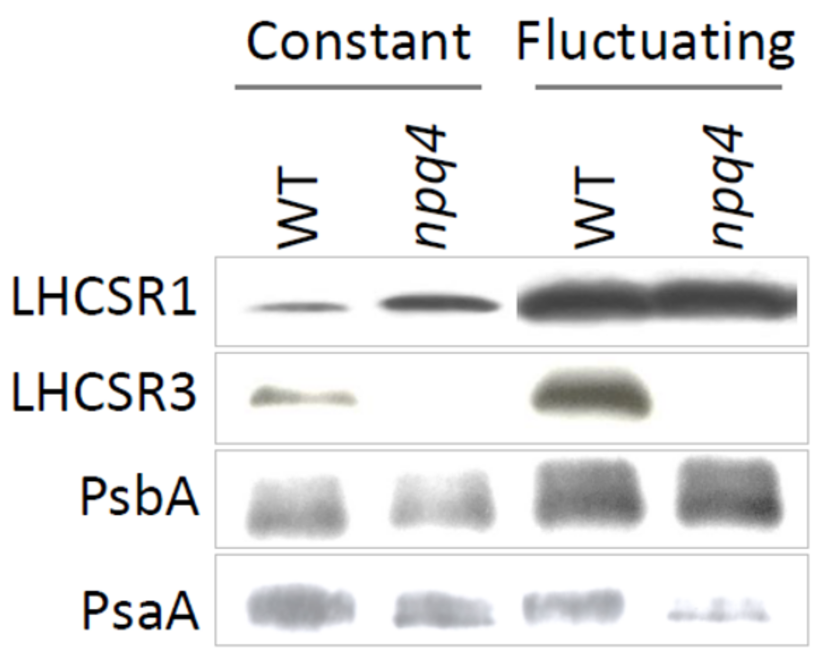
2.2. Light Fluctuations Induced Accumulation of LHCSR1, LHCSR3, and qE Capacity

Levels of LHCSR proteins, and therefore qE capacity (Figure 2), were clearly different under fluctuating and constant light. The level of LHCSR3, in WT, and LHCSR1, in WT and npq4, increased several folds under fluctuating light (Figure 3). This agreed with the higher Y(NPQ) values relative to those found in cells grown in constant light at the same intensity (Figure 2B). Even at 100 µmoL quanta m−2 s−1 fluctuating light, Y(NPQ) of npq4 increased so much that it equaled WT-4A (Figure 1 and Figure 2B). There was no consistent influence of initial culture dilution on Y(NPQ) values.

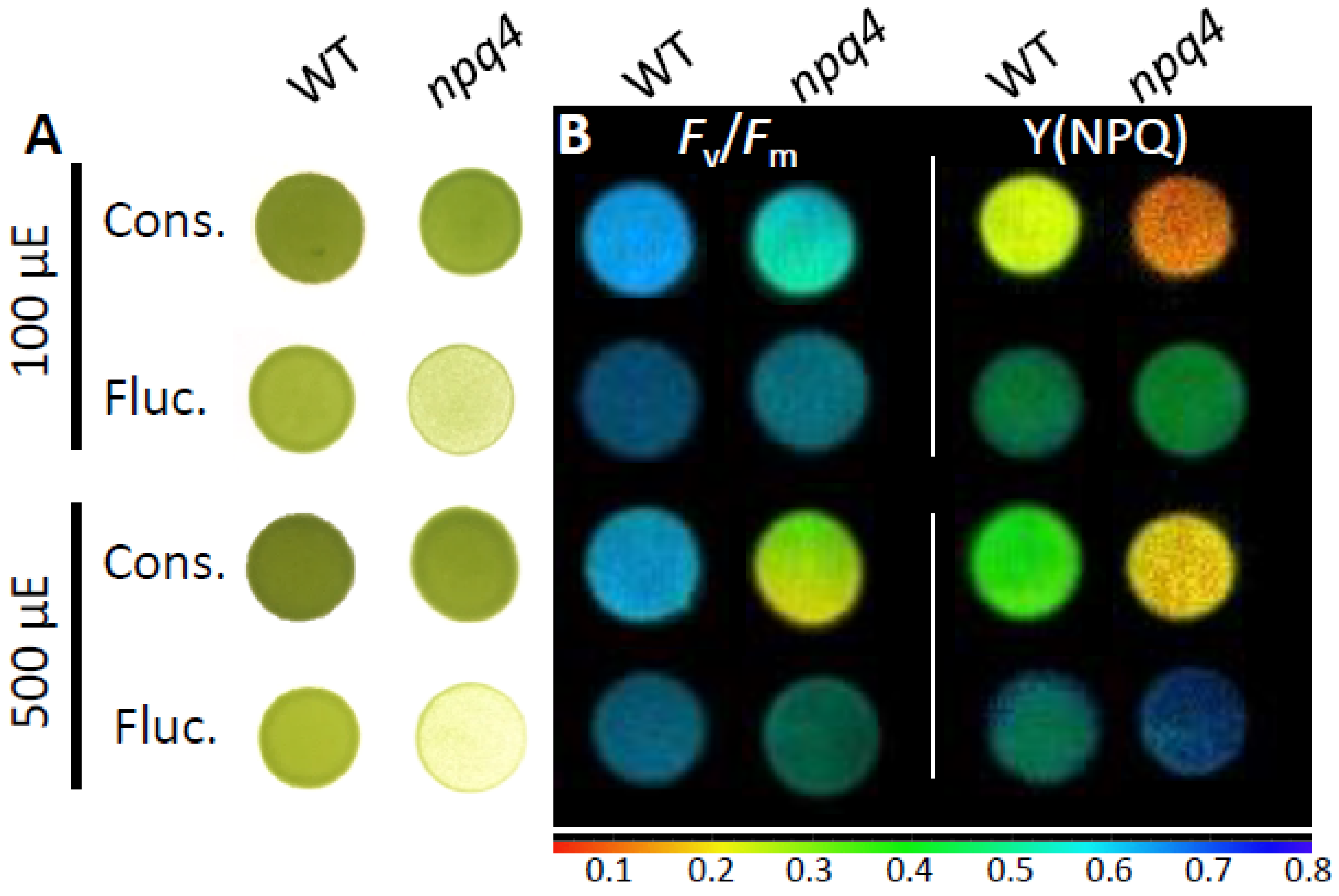
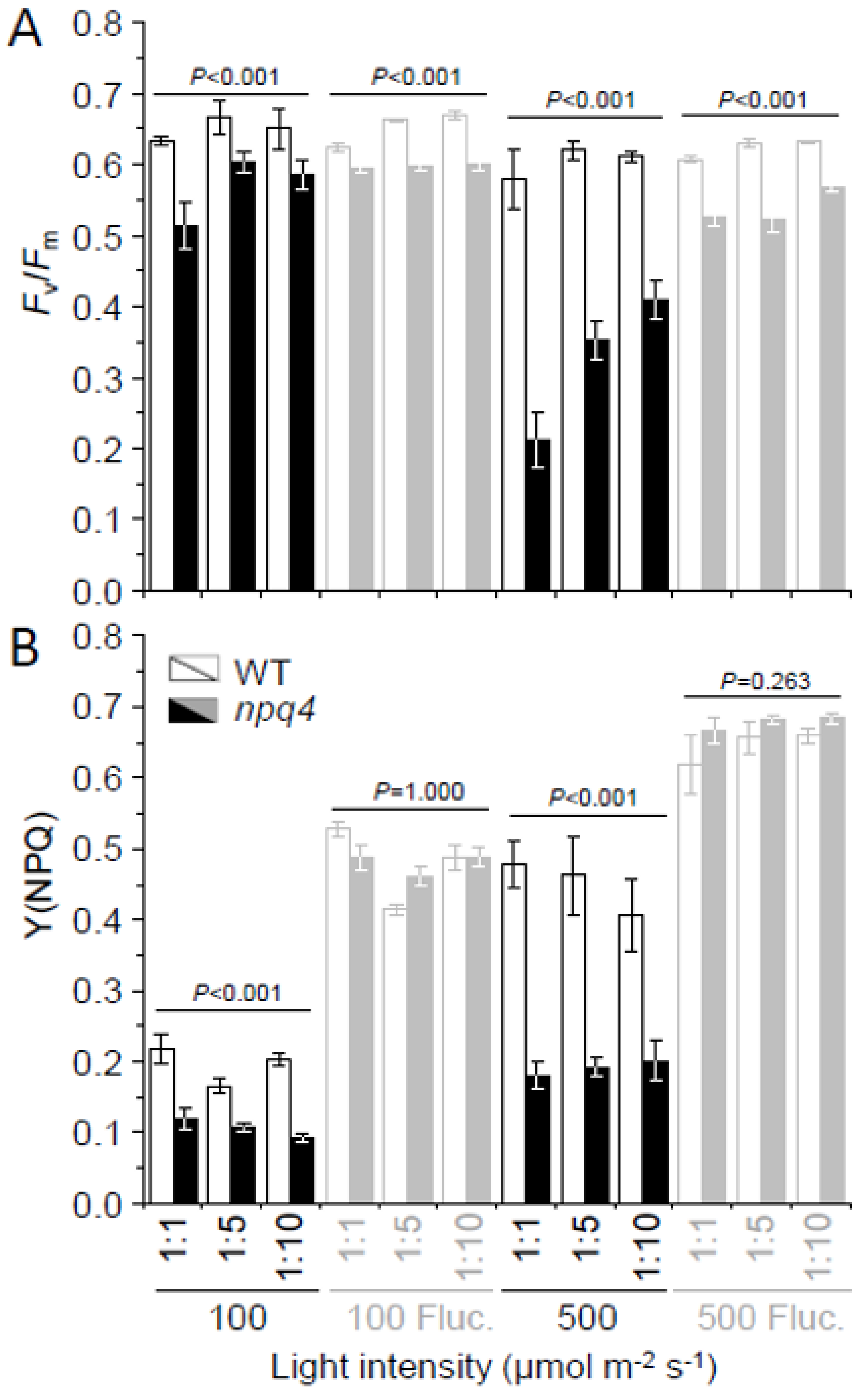
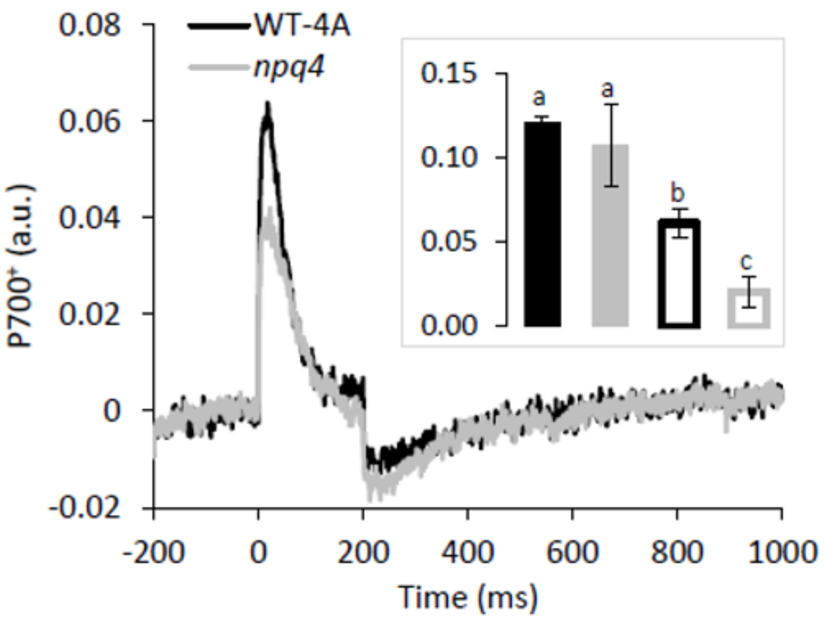
2.3. Enhanced PSII and PSI Photoinhibition Occurred in npq4 Under Constant and Fluctuating Light, Respectively

Values of Fv/Fm, here used as a proxy for PSII photoinhibition, were generally higher in WT-4A than npq4 under all light treatments. Notably, at 500 µmoL quanta m−2 s−1, Fv/Fm values of npq4 were much lower under constant than fluctuating light (Figure 1 and Figure 2A). This agreed with the lowest PsbA levels (PSII reaction center) found in npq4 under constant light and much higher levels in cells grown under fluctuating light (Figure 3). However, under fluctuating light WT-4A and npq4 had lower levels of PsaA (PSI reaction center), relative to constant light-treated cells (Figure 3 and Figure S2). This agrees with significantly altered maximum P700 redox changes (Pm), here used as a proxy for PSI photoinhibition. Lowered Pm values were found in both genotypes under fluctuating light, with npq4 significantly lower than WT-4A (Figure 4).

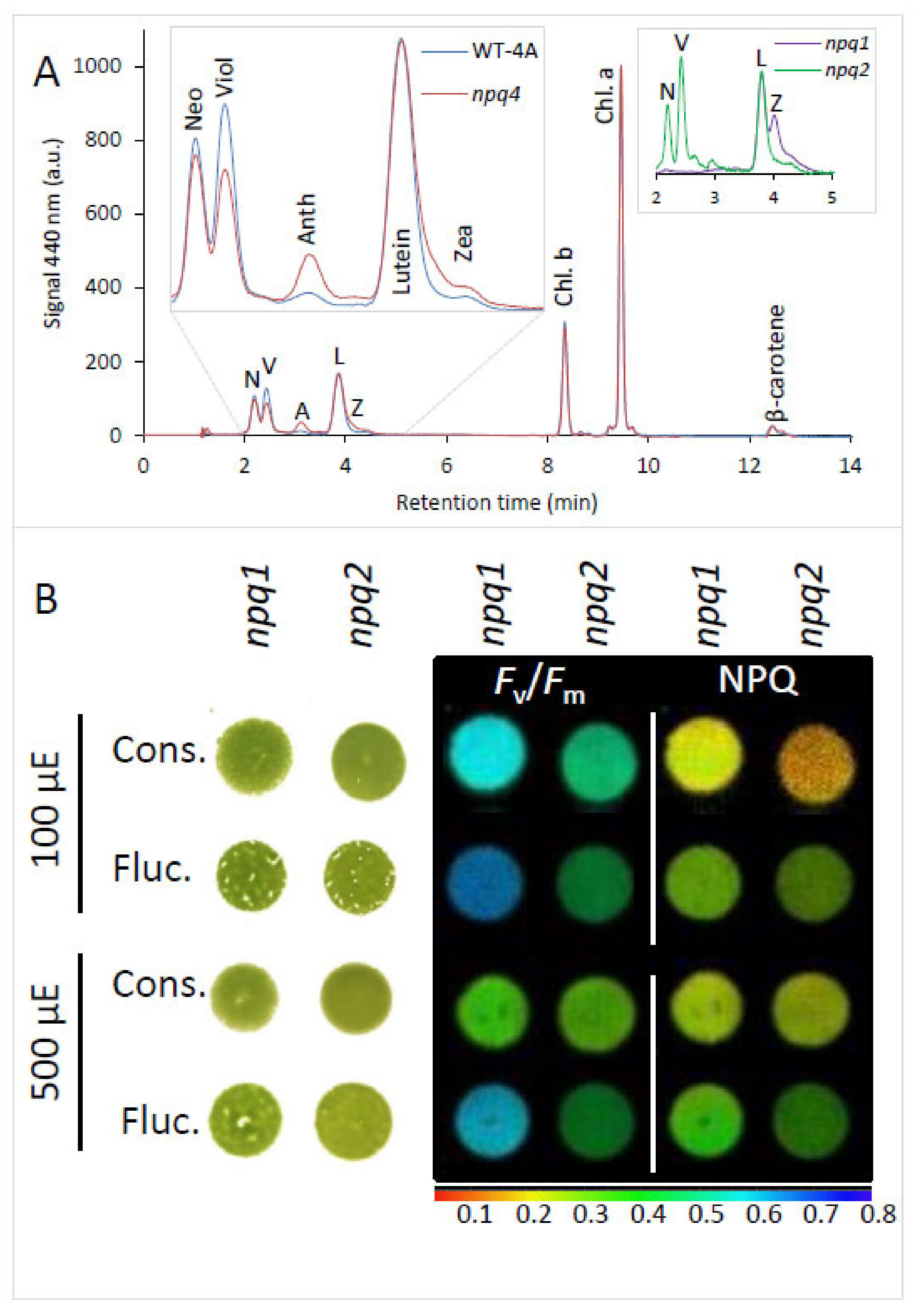
2.4. Xanthophyll Composition Did Not Affect Tolerance to Fluctuating Light

Under fluctuating light, npq4 contained significantly more (p = 0.02) antheraxanthin and slightly more zeaxanthin, and significantly less (p < 0.01) violaxanthin, showing higher activation of the xanthophyll cycle than in WT-4A (Figure 5A). While it is known that de-epoxidized xanthophylls do not play a significant contribution to qE in C. reinhardtii [19], the xanthophyll cycle is still tightly coupled to high light exposure [27]. To investigate if differences in xanthophyll composition also influenced tolerance to fluctuating light, the NPQ mutants npq1 (no zeaxanthin) and npq2 (only zeaxanthin, and neither violaxanthin nor neoxanthin [27]) were included in the study. No differences in colony growth between npq1 and npq2 were observed under constant or fluctuating light (Figure 5B), but similar to the other genotypes, fluctuating light increased qE capacity and led to higher Fv/Fm values relative to cells under constant light of the same intensity (Figure 5B).

3. Discussion

Regulation of electron transport is of general importance for efficient photosynthesis and to prevent photoinhibition, especially under fluctuating light [1,28]. NPQ influences electron transport rates [20] and in plants has been shown to have both positive and negative impacts on plant growth, under constant and fluctuating light [22,29]. Growth of npq4 was only mildly affected under constant light (Figure 1), which is in agreement with other studies using LHCSR-deficient mutants at similar light intensities, at least under ambient oxygen tensions [10,30]. C. reinhardtii responded to fluctuating light by increasing levels of LHCSR1 and LHCSR3 (Figure 3), inferring that these qE proteins are important in the dynamic regulation of photosynthesis under fluctuating light. Surprisingly, Y(NPQ) values of npq4 were as high as WT under fluctuating light (Figure 2B), which must have been strictly mediated by LHCSR1, due to the absence of LHCSR3 in this mutant [13].
LHCSR-mediated qE is dependent on pH [20] and becomes active in response to protonation of luminal-exposed part of the protein [7,8] that naturally occurs under high light. The xanthophyll cycle is another high light-associated process regulated by pH, but has limited contribution to qE of C. reinhardtii and other Chlorophyta alga [19,31]. Absence of an active xanthophyll cycle in the npq1 and npq2 mutants did not affect tolerance to fluctuating light (Figure 5B). Therefore, higher levels of de-epoxidized xanthophylls in npq4 under fluctuating light compared to WT (Figure 5A) is unlikely to have contributed to any change in fluctuating light tolerance. Nonetheless, a greater shift in the de-epoxidation ratio of npq4 is indicative of a lower luminal pH than occurred in WT. Since the level of LHCSR-mediated qE is directly associated to pH [20], low pH values may provide an explanation of the particularly high Y(NPQ) values in npq4 in response to fluctuating light (Figure 2B). We can also be confident from the behavior of npq4 that LHCSR1 is able to induce a large, fast, and reversible pH-dependent quenching of LHCII [15] in response to fluctuating light.
Under fluctuating light, a lower luminal pH and elevated electric field across the thylakoid membrane (ΔΨ) increases incidences of charge recombination, ROS production, and PSII photoinhibition in Arabidopsis thaliana [32]. In contrast, here, Fv/Fm values of npq4 were higher under fluctuating light, relative to constant light (Figure 2A), indicating that fluctuating light did not induce PSII photoinhibition as much as under constant light. Indeed, higher levels of PsbA, the D1 reaction center of PSII, were likewise found under fluctuating than under constant light (Figure 3 and Figure S2). PSII photoinhibition directly impacts linear electron flow, potentially influencing PSI photoinhibition [28].
So far, it has been shown that PGR5/PGRL1-mediated cyclic electron flow contributes to fluctuating light tolerance in photosynthetic organisms [25,33], but in C. reinhardtii, at least, the role of flavodiirons in preventing acceptor side limitation of PSI is more critical. In the absence of flavodiirons, fluctuating light leads to PSI photoinhibition [24,25]. This highlights that PSI can be rendered vulnerable under fluctuating light. Under constant light, enhanced PSI photoinhibition has been observed in npq4, but only under elevated oxygen tensions [10], or in cells also deficient in PGRL1-mediated cyclic electron flow [34,35]. Western blotting (Figure 3) and P700 absorbance measurements (Figure 4) revealed that repeated 10 min light–dark treatments led to decrease in PSI levels, particularly in npq4. Photodamage of PSI can be exacerbated by high rates of electron flow from PSII [36]. Therefore, a lack of LHCSR3 and potentially low qE in npq4 could contribute to enhanced PSI photoinhibition. However, in fluctuating light, under which PSI photoinhibition of npq4 occurred, Y(NPQ) values were the same as in WT (Figure 2B), indicating that the influence of qE on linear electron flow would have been equal in both genotypes.
Another explanation to enhanced PSI photoinhibition in npq4 under fluctuating light would concern state transitions. This phosphorylation-mediated NPQ mechanism [37] is particularly active in C. reinhardtii during the first few minutes of light-to-dark and dark-to-light acclimation and is affected in npq4 [38]. High light-treated cells are in state 1, but when subjected to darkness they transit to state 2 due to chlororespiration-induced phosphorylation of specific components of the antenna, including LHCII and LHCSR3 [39,40]. During transition to state 2, the majority of LHCII migrates energy transfer to PSI [41], and LHCSR3 also migrates as part of the LHCSR3-LHCII antenna of PSI [40]. Since LHCSR3 can quench LHCII associated with PSI [14], it is possible that LHCSR3 directly protects PSI from photodamage during fluctuating light, when cells are repeatedly exposed to high light in state 2 [38]. Unlike LHCSR3, LHCSR1 is not phosphorylated [34], and therefore unlikely to be in the mobile LHCII fraction during transition to state II. While the large increase in LHCSR1 levels (Figure 3) suggests that LHCSR1-mediated qE is important under fluctuating light, without LHCSR3, as in npq4, LHCSR1 alone could not prevent PSI photoinhibition (Figure 4 and Figure 5). High LHCSR1 levels may have even promoted PSI photoinhibition, since LHCSR1 has been reported to quench LHCII via PSI [16]. Finally, npq4 was not growth-sensitive to repeated 10-fold increases in light intensity (i.e., when darkness was replaced by low light; Figure S1). Therefore, deactivation of the Calvin–Benson cycle during 10 min of darkness [42] was likely important to reveal the sensitivity of npq4 to fluctuating light.

4. Conclusions

Overall, LHCSR3-mediated NPQ rather than overall qE capacity is important in tolerating fluctuating light that involves darkness. In agreement with previous studies, PSI showed vulnerability to fluctuating light, and with the use of npq4, it was possible to show that LHCSR3 can protect PSI, a role that LHCSR1 seems not to be able to fulfill. In contrast to the efficient repair cycle of PSII, photoinhibition of PSI is much more costly due to very slow PSI repair rates [43]. Therefore, enhanced photoinhibition of PSI due to absence of LHCSR3 can explain the growth phenotype of npq4 under fluctuating light. Interactions between LHCSR1/3 and LHCII with PSI in energy dissipation in response to dynamic light exposure require further elucidation.

5. Material and Methods

5.1. C. reinhardtii Strains and Growth Conditions

The C. reinhardtii LHCSR3-deficient strain npq4 (CC-4614) and its parental WT-4A (CC-4051) were purchased from the Chlamydomonas Centre (www.chlamycollection.org). Liquid cultures were initiated in photoautotrophic media (THP), which was identical to TAP except acetic acid was replaced by HCl. Cultures were adjusted to 5.0, 1.0, and 0.5 µg chlorophyll mL−1 or 12,500, 2500, and 1250 cells (from here-on referred to as 1:1, 1:5, and 1:10 dilutions, respectively), in the 10 µL starting culture pipetted onto THP media containing 1.5% agar. Each Petri dish contained four replicates of each strain placed in an alternating order, with different plates hosting each dilution. The Petri dish lids were placed on, but not sealed with any film or tape to allow gas exchange, before placing in a growth chamber (Percival, PGC-6HO) at 25 °C under subsaturating or saturating light intensity (100 or 500 µmoL photons m−2 s−1, respectively) with a 24 h time span composed of either constant 12 h illumination or repeated light fluctuations of 10 min illumination and 10 min darkness (see Figure S3 for a profile of the light intensity during one light fluctuation cycle). This light cycle was chosen to enable activation and relaxation of LHCSR3-associated NPQ processes [35]. For comparing growth rates, all colonies of the same genotype were carefully scraped, using a flat-ended spatula, together from the agar and the fresh weight was divided by the time colonies had been growing, which were 6, 7, and 8 days for 1:1, 1:5, and 1:10 dilutions, respectively. Additional cultures initiated from 1:1, 1:5, 1:10, and 1:25 dilutions were treated with fluctuating light as above, except that darkness was replaced by low light (50 µmoL photons m−2 s−1) to prevent deactivation of the Calvin–Benson cycle. All dilutions under this treatment were weighed after 7 days growth.

5.2. Chlorophyll Fluorescence

At the end of the 12 h constant light treatment on day 6, 7, and 8 for 1:1, 1:5, and 1:10 dilutions, respectively, cultures from both light treatments were moved to darkness for 1 h. After removing the Petri dish lid, chlorophyll fluorescence during a 600 ms saturating pulse was measured with a Maxi Imaging PAM M-series (WALZ). Minimum (F) and maximum fluorescence (Fm) was used to calculate maximum PSII quantum yields (Fv/Fm) via (FmF)/Fm. Thereafter, Y(NPQ), as an indicator of the fraction of light energy dissipated to heat via qE, was measured after 30 s at 396 µmoL photons m−2 s−1 with a subsequent saturating pulse and calculated by (F/Fm′)−(F/Fm), with Fm and Fm′ measured before and during light, respectively. Immediately after, cultures were frozen in liquid nitrogen.

5.3. Western Blotting of Proteins

For detecting specific proteins via Western blotting, the frozen samples were thawed and extracted in 50–150 µL (depending upon culture amount) of 2% SDS in 50 mM TRIS-HCl, pH 6.8, containing protease inhibitor cocktail (Roche). After centrifugation for 10 min at 4 °C and 16,000 × g, supernatants were measured for protein content using the BCA assay for loading on 12% acrylamide gels at equal protein level (1 µg for PsbA and LHCSR3 and 10 µg for PsaA and LHCSR1; see Figure S2 for showing blots were below saturation point), before running for 1.5 h at 150 V, semi-dry transfer to nitrocellulose membranes, and incubation with antibodies (Agrisera), according to [10].

5.4. HPLC of Photosynthetic Pigments

Photosynthetic pigments were measured by HPLC in cultures grown from 1:10 dilution as above, except cells grown across the whole agar plate rather than from individual 10 µL spots (n = 3 plates/genotype). Pigments from ca. 2 mg of lyophilized cultures were extracted in 0.5 mL of ice-cold acetone and measured by absorbance at 440 nm, after separation with an Agilent 1100 HPLC system equipped with a LiChrospher 100 RP-18 column (125 mm × 4 mm, 5 µm), according to [11].

5.5. Maximum Photo-Oxidizable P700 Level

Maximum photo-oxidizable P700 levels (Pm) were measured during a 200 ms saturating pulse using a DUAL-PAM (Walz), of cultures grown as for pigment measurements (see Section 2.4). Cells were scraped off the agar and suspended in THP liquid media (see Section 2.1) at equal total chlorophyll concentration of 30 µg mL−1, and measured according to Roach, Na, Stöggl, and Krieger-Liszkay [10].

5.6. Statistics

For chlorophyll fluorescence measurements, four individual colonies were used as replicates for each culture dilution. Data were analyzed by two-way ANOVA in SPSS Statistics 25 (IBM) to reveal p-values between WT and npq4, considering all dilutions collectively (n = 12 colonies) for each light treatment. For comparing colony growth, average fold-difference in fresh weight accumulation for the three culture dilutions (1:1, 1:5, and 1:10) was compared with a Students t-test under each light treatment. Significant differences were considered when p < 0.05.

Supplementary Materials

The following are available online at https://www.mdpi.com/2223-7747/9/11/1604/s1. Figure S1: Colony growth comparison of LHCSR3 deficient nqq4 mutant relative to wild type (WT 4A) under various light treatments. (Cells were cultured under repeated 10 minute fluctuations (fl.) of light intensity between 0 (moon) and 100 or 500 µmol photons m−2 s−1 (sun), or diurnal (12/12h) constant light c on.) at the same two intensities. Data is shown as fold difference between fresh weight of all WT 4A and npq4 colonies at each initial culture dilution and after 6-8 days culturing (see methods). The average fold difference is depicted by a dashed line. (Growth of nqq4 and WT 4A under light fluctuating between 50 and 500 µmol photons m−2 s−1 (small sun, big sun). Representative images of colonies growing on agar are shown to the left of average colony fresh weight after 7 days of each initial culture dilution. Figure S2: Western blots of proteins from WT and npq4 cells under constant or fluctuating light at 500 µmol photons m−2 s−1. For PsaA, proteins were loaded at 20 µg (200%) or 10 µg (100%) total protein, and for PsbA and LHCSR1, proteins were loaded at 2 µg (200%) or 1 µg (100%) total protein. All bands are from the same blot and transferred from the same gel. Figure S3: The change in light intensity during 10 min of fluctuating light, as measured with SQ 520 PAR sensor (Apogee Instruments).

Funding

This research received no external funding.

Acknowledgments

I thank Bettina Lehr and Birgit Stenzel (University of Innsbruck) for excellent technical assistance.

Conflicts of Interest

The author declares no conflict of interest.

References

  1. Allahverdiyeva, Y.; Suorsa, M.; Tikkanen, M.; Aro, E.M. Photoprotection of photosystems in fluctuating light intensities. J. Exp. Bot. 2015, 66, 2427–2436. [Google Scholar] [CrossRef]
  2. Alboresi, A.; Storti, M.; Morosinotto, T. Balancing protection and efficiency in the regulation of photosynthetic electron transport across plant evolution. New Phytol. 2019, 221, 105–109. [Google Scholar] [CrossRef] [PubMed] [Green Version]
  3. Müller, P.; Li, X.-P.; Niyogi, K.K. Update on photosynthesis non-photochemical quenching. A response to excess light energy. Plant Physiol. 2001, 125, 1558–1566. [Google Scholar] [CrossRef] [PubMed] [Green Version]
  4. Roach, T.; Krieger-Liszkay, A. Photosynthetic regulatory mechanisms for efficiency and prevention of photo-oxidative stress. Ann. Plant Rev. Online 2019, 273–306. [Google Scholar] [CrossRef]
  5. Erickson, E.; Wakao, S.; Niyogi, K.K. Light stress and photoprotection in Chlamydomonas reinhardtii. Plant J. 2015, 82, 449–465. [Google Scholar] [CrossRef] [PubMed]
  6. Li, X.-P.; Gilmore, A.M.; Caffarri, S.; Bassi, R.; Golan, T.; Kramer, D.; Niyogi, K.K. Regulation of photosynthetic light harvesting involves intrathylakoid lumen pH sensing by the PsbS protein. J. Biol. Chem. 2004, 279, 22866–22874. [Google Scholar] [CrossRef] [Green Version]
  7. Liguori, N.; Roy, L.M.; Opacic, M.; Durand, G.; Croce, R. Regulation of light harvesting in the green alga Chlamydomonas reinhardtii: The c-terminus of LHCSR is the knob of a dimmer switch. J. Am. Chem. Soc. 2013, 135, 18339–18342. [Google Scholar] [CrossRef]
  8. Ballottari, M.; Truong, T.B.; De Re, E.; Erickson, E.; Stella, G.R.; Fleming, G.R.; Bassi, R.; Niyogi, K.K. Identification of pH-sensing sites in the light harvesting complex stress-related 3 protein essential for triggering non-photochemical quenching in Chlamydomonas reinhardtii. J. Biol. Chem. 2016, 291, 7334–7346. [Google Scholar] [CrossRef] [Green Version]
  9. Niyogi, K.K.; Truong, T.B. Evolution of flexible non-photochemical quenching mechanisms that regulate light harvesting in oxygenic photosynthesis. Curr. Opin. Plant Biol. 2013, 16, 307–314. [Google Scholar] [CrossRef]
  10. Roach, T.; Na, C.S.; Stöggl, W.; Krieger-Liszkay, A. The non-photochemical quenching protein LHCSR3 prevents oxygen-dependent photoinhibition in Chlamydomonas reinhardtii. J. Exp. Bot. 2020, 71, 2650–2660. [Google Scholar] [CrossRef]
  11. Roach, T.; Baur, T.; Stoggl, W.; Krieger-Liszkay, A. Chlamydomonas reinhardtii responding to high light: A role for 2-propenal (acrolein). Physiol. Plant. 2017, 161, 75–87. [Google Scholar] [CrossRef] [PubMed]
  12. Polukhina, I.; Fristedt, R.; Dinc, E.; Cardol, P.; Croce, R. Carbon supply and photoacclimation crosstalk in the green alga Chlamydomonas reinhardtii. Plant Physiol. 2016, 172, 1494–1505. [Google Scholar] [CrossRef] [PubMed] [Green Version]
  13. Peers, G.; Truong, T.B.; Ostendorf, E.; Busch, A.; Elrad, D.; Grossman, A.R.; Hippler, M.; Niyogi, K.K. An ancient light-harvesting protein is critical for the regulation of algal photosynthesis. Nature 2009, 462, 518–521. [Google Scholar] [CrossRef] [PubMed]
  14. Girolomoni, L.; Cazzaniga, S.; Pinnola, A.; Perozeni, F.; Ballottari, M.; Bassi, R. LHCSR3 is a nonphotochemical quencher of both photosystems in Chlamydomonas reinhardtii. Proc. Natl. Acad. Sci. USA 2019, 116, 4212–4217. [Google Scholar] [CrossRef] [Green Version]
  15. Dinc, E.; Tian, L.; Roy, L.M.; Roth, R.; Goodenough, U.; Croce, R. LHCSR1 induces a fast and reversible ph-dependent fluorescence quenching in LHCII in Chlamydomonas reinhardtii cells. Proc. Natl. Acad. Sci. USA 2016, 113, 7673–7678. [Google Scholar] [CrossRef] [Green Version]
  16. Kosuge, K.; Tokutsu, R.; Kim, E.; Akimoto, S.; Yokono, M.; Ueno, Y.; Minagawa, J. LHCSR1-dependent fluorescence quenching is mediated by excitation energy transfer from LHCII to photosystem I in Chlamydomonas reinhardtii. Proc. Natl. Acad. Sci. USA 2018, 115, 3722–3727. [Google Scholar] [CrossRef] [Green Version]
  17. Tokutsu, R.; Minagawa, J. Energy-dissipative supercomplex of photosystem II associated with LHCSR3 in Chlamydomonas reinhardtii. Proc. Natl. Acad. Sci. USA 2013, 110, 10016–10021. [Google Scholar] [CrossRef] [Green Version]
  18. Semchonok, D.A.; Yadav, K.N.S.; Xu, P.Q.; Drop, B.; Croce, R.; Boekema, E.J. Interaction between the photoprotective protein LHCSR3 and c2s2 photosystem II supercomplex in Chlamydomonas reinhardtii. Biochim. Biophys. Acta Bioenerg. 2017, 1858, 379–385. [Google Scholar] [CrossRef]
  19. Quaas, T.; Berteotti, S.; Ballottari, M.; Flieger, K.; Bassi, R.; Wilhelm, C.; Goss, R. Non-photochemical quenching and xanthophyll cycle activities in six green algal species suggest mechanistic differences in the process of excess energy dissipation. J. Plant Physiol. 2015, 172, 92–103. [Google Scholar] [CrossRef]
  20. Tian, L.J.; Nawrocki, W.J.; Liu, X.; Polukhina, I.; van Stokkum, I.H.M.; Croce, R. pH dependence, kinetics and light-harvesting regulation of nonphotochemical quenching in Chlamydomonas. Proc. Natl. Acad. Sci. USA 2019, 116, 8320–8325. [Google Scholar] [CrossRef] [Green Version]
  21. Garcia-Plazaola, J.I.; Esteban, R.; Fernandez-Marin, B.; Kranner, I.; Porcar-Castell, A. Thermal energy dissipation and xanthophyll cycles beyond the arabidopsis model. Photosynth. Res. 2012, 113, 89–103. [Google Scholar] [CrossRef] [PubMed]
  22. Kromdijk, J.; Głowacka, K.; Leonelli, L.; Gabilly, S.T.; Iwai, M.; Niyogi, K.K.; Long, S.P. Improving photosynthesis and crop productivity by accelerating recovery from photoprotection. Science 2016, 354, 857–861. [Google Scholar] [CrossRef] [PubMed] [Green Version]
  23. Enamala, M.K.; Enamala, S.; Chavali, M.; Donepudi, J.; Yadavalli, R.; Kolapalli, B.; Aradhyula, T.V.; Velpuri, J.; Kuppam, C. Production of biofuels from microalgae—A review on cultivation, harvesting, lipid extraction, and numerous applications of microalgae. Renew. Sustain. Energy Rev. 2018, 94, 49–68. [Google Scholar] [CrossRef]
  24. Chaux, F.; Burlacot, A.; Mekhalfi, M.; Auroy, P.; Blangy, S.; Richaud, P.; Peltier, G. Flavodiiron proteins promote fast and transient o2 photoreduction in Chlamydomonas. Plant Physiol. 2017, 174, 1825–1836. [Google Scholar] [CrossRef] [Green Version]
  25. Jokel, M.; Johnson, X.; Peltier, G.; Aro, E.M.; Allahverdiyeva, Y. Hunting the main player enabling Chlamydomonas reinhardtii growth under fluctuating light. Plant J. 2018, 94, 822–835. [Google Scholar] [CrossRef] [Green Version]
  26. Lepetit, B.; Gelin, G.; Lepetit, M.; Sturm, S.; Vugrinec, S.; Rogato, A.; Kroth, P.G.; Falciatore, A.; Lavaud, J. The diatom phaeodactylum tricornutum adjusts nonphotochemical fluorescence quenching capacity in response to dynamic light via fine-tuned lhcx and xanthophyll cycle pigment synthesis. New Phytol. 2017, 214, 205–218. [Google Scholar] [CrossRef]
  27. Niyogi, K.K.; Bjorkman, O.; Grossman, A.R. Chlamydomonas xanthophyll cycle mutants identified by video imaging of chlorophyll fluorescence quenching. Plant Cell 1997, 9, 1369–1380. [Google Scholar] [CrossRef] [Green Version]
  28. Roach, T.; Krieger-Liszkay, A. Regulation of photosynthetic electron transport and photoinhibition. Curr. Protein Pept. Sci. 2014, 15, 351–362. [Google Scholar] [CrossRef] [Green Version]
  29. Roach, T.; Krieger-Liszkay, A. The role of the PsbS protein in the protection of photosystems I and II against high light in Arabidopsis thaliana. BBA Bioenerg. 2012, 1817, 2158–2165. [Google Scholar] [CrossRef] [Green Version]
  30. Cantrell, M.; Peers, G. A mutant of Chlamydomonas without LHCSR maintains high rates of photosynthesis, but has reduced cell division rates in sinusoidal light conditions. PLoS ONE 2017, 12, e0179395. [Google Scholar] [CrossRef] [Green Version]
  31. Masojídek, J.; Kopecká, J.; Koblížek, M.; Torzillo, G. The xanthophyll cycle in green algae (chlorophyta): Its role in the photosynthetic apparatus. Plant Biol. 2004, 6, 342–349. [Google Scholar] [CrossRef] [PubMed]
  32. Davis, G.A.; Kanazawa, A.; Schottler, M.A.; Kohzuma, K.; Froehlich, J.E.; Rutherford, A.W.; Satoh-Cruz, M.; Minhas, D.; Tietz, S.; Dhingra, A.; et al. Limitations to photosynthesis by proton motive force-induced photosystem II photodamage. eLife 2016, 5, e16921. [Google Scholar] [CrossRef] [PubMed] [Green Version]
  33. Suorsa, M.; Jarvi, S.; Grieco, M.; Nurmi, M.; Pietrzykowska, M.; Rantala, M.; Kangasjarvi, S.; Paakkarinen, V.; Tikkanen, M.; Jansson, S.; et al. Proton gradient regulation5 is essential for proper acclimation of arabidopsis photosystem I to naturally and artificially fluctuating light conditions. Plant Cell 2012, 24, 2934–2948. [Google Scholar] [CrossRef] [PubMed] [Green Version]
  34. Bergner, S.V.; Scholz, M.; Trompelt, K.; Barth, J.; Gabelein, P.; Steinbeck, J.; Xue, H.; Clowez, S.; Fucile, G.; Goldschmidt-Clermont, M.; et al. State transition7-dependent phosphorylation is modulated by changing environmental conditions, and its absence triggers remodeling of photosynthetic protein complexes. Plant Physiol. 2015, 168, 615–634. [Google Scholar] [CrossRef] [Green Version]
  35. Chaux, F.; Johnson, X.; Auroy, P.; Beyly-Adriano, A.; Te, I.; Cuine, S.; Peltier, G. Pgrl1 and LHCSR3 compensate for each other in controlling photosynthesis and avoiding photosystem I photoinhibition during high light acclimation of Chlamydomonas cells. Mol. Plant 2017, 10, 216–218. [Google Scholar] [CrossRef] [Green Version]
  36. Li, L.; Aro, E.-M.; Millar, A.H. Mechanisms of photodamage and protein turnover in photoinhibition. Trends Plant Sci. 2018, 23, 667–676. [Google Scholar] [CrossRef]
  37. Depege, N.; Bellafiore, S.; Rochaix, J.D. Role of chloroplast protein kinase stt7 in LHCII phosphorylation and state transition in Chlamydomonas. Science 2003, 299, 1572–1575. [Google Scholar] [CrossRef] [Green Version]
  38. Roach, T.; Na, C.S. LHCSR3 affects de-coupling and re-coupling of LHCII to PSII during state transitions in Chlamydomonas reinhardtii. Sci. Rep. 2017, 7, 43145. [Google Scholar] [CrossRef] [Green Version]
  39. Bennoun, P. The present model for chlororespiration. Photosynth. Res. 2002, 73, 273–277. [Google Scholar] [CrossRef]
  40. Allorent, G.; Tokutsu, R.; Roach, T.; Peers, G.; Cardol, P.; Girard-Bascou, J.; Seigneurin-Berny, D.; Petroutsos, D.; Kuntz, M.; Breyton, C.; et al. A dual strategy to cope with high light in Chlamydomonas reinhardtii. Plant Cell 2013, 25, 545–557. [Google Scholar] [CrossRef] [Green Version]
  41. Goldschmidt-Clermont, M.; Bassi, R. Sharing light between two photosystems: Mechanism of state transitions. Curr. Opin. Plant Biol. 2015, 25, 71–78. [Google Scholar] [CrossRef] [PubMed] [Green Version]
  42. Jacquot, J.-P. Dark deactivation of chloroplast enzymes finally comes to light. Proc. Natl. Acad. Sci. USA 2018, 115, 9334–9335. [Google Scholar] [CrossRef] [PubMed] [Green Version]
  43. Sonoike, K. Photoinhibition of photosystem I. Physiol. Plant. 2011, 142, 56–64. [Google Scholar] [CrossRef] [PubMed]
Figure 1. Phenotype of Light-Harvesting Complex (LHC)-like Stress-Related 1 (LHCSR3)-deficient nqq4 relative to wild type (WT-4A) under photoautotrophic conditions on agar and either diurnal constant (12/12 h, on/off) or fluctuating (10/10 min, on/off) light treatments. Representative images of (A) colonies and (B) Fv/Fm and Y (non-photochemical quenching (NPQ)) after 8 day growth (starting dilution 1:10, see Materials and Methods) under diurnal/constant (Cons.) or fluctuating (Fluc.) light of 100 (upper) or 500 (lower) µmoL photons m−2 s−1. Chlorophyll fluorescence parameters are given on a false-color scale shown below.
Plants 09 01604 g001
Figure 2. Effect of either diurnal constant or fluctuating light treatments on (A) Fv/Fm and (B) Y(NPQ) values of photoautotrophic agar-grown cells. Colonies were cultured under 100 or 500 µmoL photons m−2 s−1, of either diurnal constant (12/12 h on/off; black filled or black-outlined bars) or fluctuating light (10/10 min on/off, gray filled or gray-outlined bars). Wild type (WT-4A; open bars) and LHCSR3-deficient npq4 (closed bars) are shown side by side (n = 4 replicate colonies ±SD). The p-values from two-way ANOVA are shown for comparisons of npq4 to WT-4A for each light treatment when considering all differing initial culture dilutions together (n = 12 colonies).
Plants 09 01604 g002
Figure 3. Effect of 500 µmoL photons m−2 s−1 fluctuating (10/10 min on/off) or constant (12/12 h on/off) light treatment on qE proteins and photosystem reaction center levels of photoautotrophic agar-grown cells. Levels of LHCSR1, LHCSR3, PsbA, and PsaA are shown, via Western blotting, in LHCSR3-deficient npq4 and wild type (WT-4A) grown under constant (left) or fluctuating light (right). In this Figure, exactly the same sample from each treatment was loaded for all four blots. Loading controls for saturation are shown in Figure S2.
Plants 09 01604 g003
Figure 4. Effect of 500 µmoL photons m−2 s−1 fluctuating (10/10 min on/off) or constant (12/12 h on/off) light treatment on maximum light-induced P700+ values of photoautotrophic agar-grown cells. Wild type (WT-4A; black) and LHCSR3-deficient npq4 (gray) agar-grown cells were suspended in photoautotrophic media to a chlorophyll concentration of 30 µg mL−1, and P700+ was recorded during a 200 ms saturating pulse, starting at 0 ms, with typical traces shown after averaging the three technical replicates of fluctuating light-treated cells. The inset shows average Pm values for npq4 and WT under constant (filled) or fluctuating light (open), n = 4 replicate cultures ± SD, with different letters denoting significant difference, p < 0.05.
Plants 09 01604 g004
Figure 5. Xanthophyll levels under fluctuating light and lack of influence of xanthophyll compositions on tolerance to fluctuating light. (A) Typical HPLC chromatograms showing the relative amounts of neoxanthin (Neo), violaxanthin (viol), antheraxanthin (anth), Lutein, and zeaxanthin (zea) of wild type (WT-4A) and npq4 under 500 µmoL photons m−2 s−1 fluctuating light. The insets are an expansion of 2–5 min, with left WT-4A (blue) and npq4 (red) and right zeaxanthin epoxidase-deficient (npq1; purple) and violaxanthin de-epoxidase-deficient (npq2; green) mutants. (B) Colonies of npq1 and npq2 are shown side by side after growth under constant (cons.) or fluctuating (fluc.) light at 100 (upper) or 500 (lower) µmoL photons m−2 s−1. Chlorophyll fluorescence parameters, Fv/Fm and qE-NPQ are shown on a false-color scale (below).
Plants 09 01604 g005
Publisher’s Note: MDPI stays neutral with regard to jurisdictional claims in published maps and institutional affiliations.

