Natural Surfactant Saponin from Tissue of Litsea glutinosa and Its Alternative Sustainable Production


 , and
, and
Abstract
:
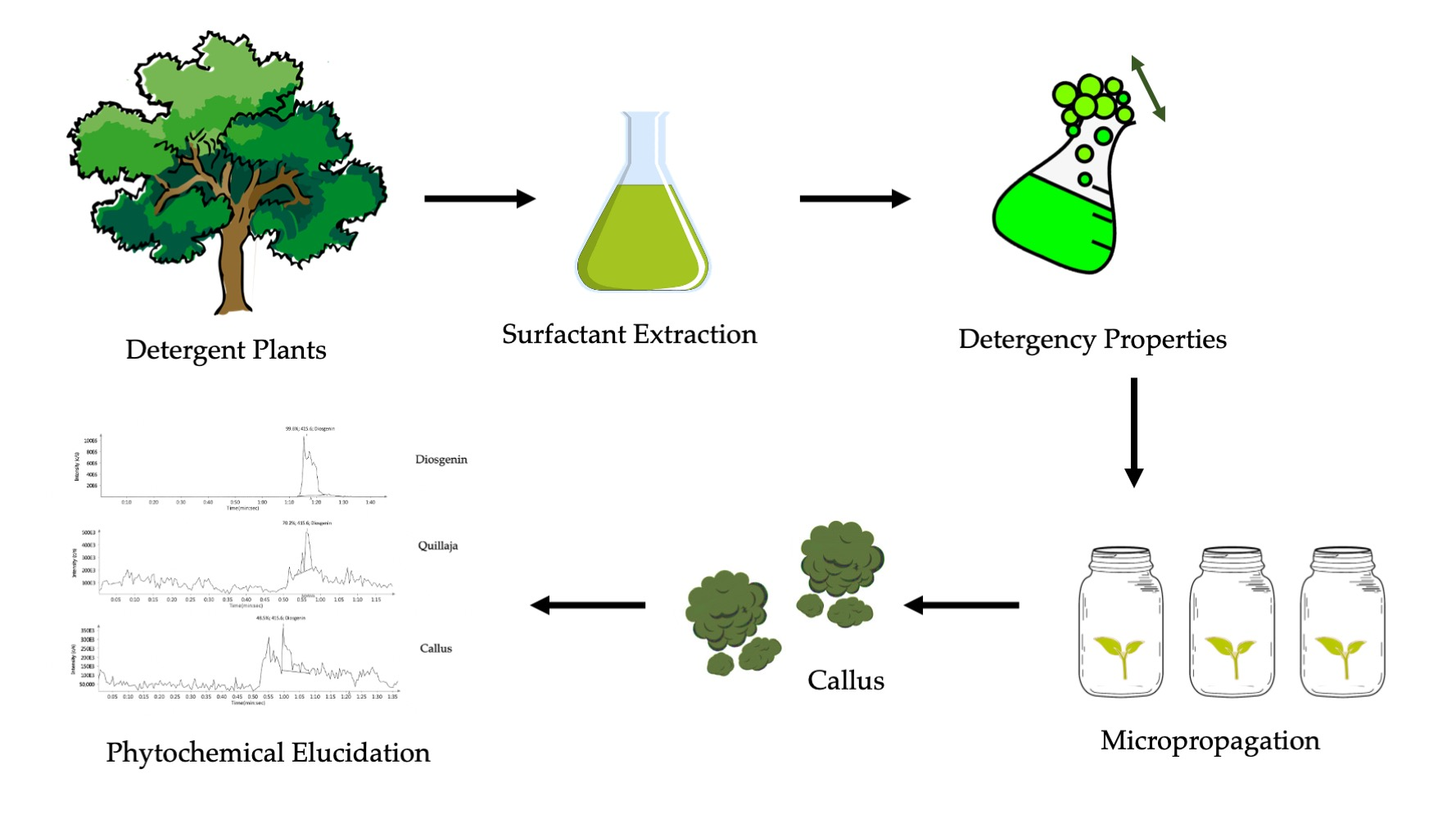
1. Introduction
2. Results and Discussion
2.1. Foaming Ability, Stability, and Detergency Ability
2.2. Chemical Properties
2.2.1. Total Saponin Content
2.2.2. Fourier Transform Infrared Spectroscopy (FTIR)
2.3. In Vitro Plant Material Propagation
2.4. High Performance Thin Layer Chromatography (HPTLC)
2.5. Compact Mass Spectrometry (CMS)
3. Materials and Methods
3.1. Chemicals and Standards
3.2. Collection and Authentication of Plant Materials
3.3. Determination of Detergent Properties
3.3.1. Foaming Ability and Stability
3.3.2. Detergency Ability
3.4. Chemical Analyses
3.4.1. Determination Saponin Contents
3.4.2. Fourier Transform Infrared Spectroscopy (FTIR)
3.5. In Vitro Plant Material Propagation
3.6. High Performance Thin Layer Chromatography (HPTLC)
3.7. Compact Mass Spectrometry (CMS)
3.8. Statistical Analysis
4. Conclusions
Supplementary Materials
Author Contributions
Funding
Acknowledgments
Conflicts of Interest
References
- Akbari, S.; Abdurahman, N.H.; Yunus, R.M.; Fayaz, F.; Alara, O.R. Biosurfactants—A new frontier for social and environmental safety: A mini review. Biotechnol. Res. Innov. 2018, 2, 81–90. [Google Scholar] [CrossRef]
- Fracchia, L.; Ceresa, C.; Franzetti, A.; Cavallo, M.; Gandolfi, I.; Van Hamme, J.; Gkorezis, P.; Marchant, R.; Banat, I.M. Industrial applications of biosurfactants. In Biosurfactants: Production and Utilization—Processes, Technologies, and Economics; CRC Press Taylor & Francis Group: Boca Raton, FL, USA, 2014; pp. 245–260. [Google Scholar]
- Garai, S. Triterpenoid saponins. Nat. Prod. Chem. Res. 2014, 2, 1000148. [Google Scholar]
- Zhou, W.; Wang, X.; Chen, C.; Zhu, L. Enhanced soil washing of phenanthrene by a plant-derived natural biosurfactant, Sapindus saponin. Colloids Surf. A Physicochem. Eng. Asp. 2013, 425, 122–128. [Google Scholar] [CrossRef]
- Holmberg, K. Natural surfactants. Curr. Opin. Colloid Interface Sci. 2001, 6, 148–159. [Google Scholar] [CrossRef]
- Oleszek, W.; Hamed, A. Saponin-based surfactants. In Surfactants Renew. Resour; John Wiley & Sons, Ltd.: Chichester, UK, 2010; Volume 239. [Google Scholar] [CrossRef]
- Kregiel, D.; Berlowska, J.; Witonska, I.; Antolak, H.; Proestos, C.; Babic, M.; Babic, L.; Zhang, B. Saponin-based, biological-active surfactants from plants. In Application and Characterization of Surfactants; InTech: Rijeka, Croatia, 2017; pp. 183–205. [Google Scholar]
- Samal, K.; Das, C.; Mohanty, K. Eco-friendly biosurfactant saponin for the solubilization of cationic and anionic dyes in aqueous system. Dye. Pigment. 2017, 140, 100–108. [Google Scholar] [CrossRef]
- Sahu, S.S.; Gandhi, I.S.R.; Khwairakpam, S. State-of-the-art review on the characteristics of surfactants and foam from foam concrete perspective. J. Inst. Eng. (India) Ser. A 2018, 99, 391–405. [Google Scholar] [CrossRef]
- Basu, A.; Basu, S.; Bandyopadhyay, S.; Chowdhury, R. Optimization of evaporative extraction of natural emulsifier cum surfactant from Sapindus mukorossi—Characterization and cost analysis. Ind. Crops Prod. 2015, 77, 920–931. [Google Scholar] [CrossRef]
- Sparg, S.; Light, M.; Van Staden, J. Biological activities and distribution of plant saponins. J. Ethnopharmacol. 2004, 94, 219–243. [Google Scholar] [CrossRef]
- Faizal, A.; Geelen, D. Saponins and their role in biological processes in plants. Phytochem. Rev. 2013, 12, 877–893. [Google Scholar] [CrossRef]
- Osbourn, A. Saponins and plant defence—A soap story. Trends Plant Sci. 1996, 1, 4–9. [Google Scholar] [CrossRef]
- Augustin, J.M.; Kuzina, V.; Andersen, S.B.; Bak, S. Molecular activities, biosynthesis and evolution of triterpenoid saponins. Phytochemistry 2011, 72, 435–457. [Google Scholar] [CrossRef] [PubMed]
- Tippel, J.; Gies, K.; Harbaum-Piayda, B.; Steffen-Heins, A.; Drusch, S. Composition of Quillaja saponin extract affects lipid oxidation in oil-in-water emulsions. Food Chem. 2017, 221, 386–394. [Google Scholar] [CrossRef] [PubMed]
- El Aziz, M.; Ashour, A.; Melad, A. A review on saponins from medicinal plants: Chemistry, isolation, and determination. J. Nanomed. Res. 2019, 8, 6–12. [Google Scholar]
- Le, A.V.; Parks, S.; Nguyen, M.; Roach, P. Improving the vanillin-sulphuric acid method for quantifying total saponins. Technologies 2018, 6, 84. [Google Scholar] [CrossRef] [Green Version]
- Arivalagan, M.; Gangopadhyay, K.; Kumar, G. Determination of steroidal saponins and fixed oil content in fenugreek (Trigonella foenum-graecum) genotypes. Indian J. Pharm. Sci. 2013, 75, 110. [Google Scholar] [CrossRef] [Green Version]
- Yang, D.-J.; Lu, T.-J.; Hwang, L.S. Isolation and identification of steroidal saponins in Taiwanese yam cultivar (Dioscorea pseudojaponica Yamamoto). J. Agric. Food Chem. 2003, 51, 6438–6444. [Google Scholar] [CrossRef]
- Inalegwu, B.; Sodipo, O. Antimicrobial and foam forming activities of extracts and purified saponins of leaves of Tephrosia vogelii. Eur. J. Exp. Biol. 2015, 5, 49–53. [Google Scholar]
- Sarkhel, S. Evaluation of the anti-inflammatory activities of Quillaja saponaria Mol. saponin extract in mice. Toxicol. Rep. 2016, 3, 1–3. [Google Scholar] [CrossRef] [Green Version]
- San Martín, R.; Briones, R. Industrial uses and sustainable supply of Quillaja saponaria (Rosaceae) saponins. Econ. Bot. 1999, 53, 302–311. [Google Scholar] [CrossRef]
- Koike, K.; Jia, Z.; Nikaido, T. New triterpenoid saponins and sapogenins from Saponaria officinalis. J. Nat. Prod. 1999, 62, 1655–1659. [Google Scholar] [CrossRef]
- Albiero, A.L.M.; Sertié, J.A.A.; Bacchi, E.M. Antiulcer activity of Sapindus saponaria L. in the rat. J. Ethnopharmacol. 2002, 82, 41–44. [Google Scholar] [CrossRef]
- Tsuzuki, J.K.; Svidzinski, T.I.; Shinobu, C.S.; Silva, L.F.; Rodrigues-Filho, E.; Cortez, D.A.; Ferreira, I.C. Antifungal activity of the extracts and saponins from Sapindus saponaria L. An. Acad. Bras. Cienc. 2007, 79, 577–583. [Google Scholar] [CrossRef]
- Damke, E.; Tsuzuki, J.K.; Chassot, F.; Cortez, D.A.; Ferreira, I.C.; Mesquita, C.S.; da-Silva, V.R.; Svidzinski, T.I.; Consolaro, M.E. Spermicidal and anti-Trichomonas vaginalis activity of Brazilian Sapindus saponaria. BMC Complement. Altern. Med. 2013, 13, 196. [Google Scholar] [CrossRef] [Green Version]
- Chen, S.-L.; Yu, H.; Luo, H.-M.; Wu, Q.; Li, C.-F.; Steinmetz, A. Conservation and sustainable use of medicinal plants: Problems, progress, and prospects. Chin. Med. 2016, 11, 37. [Google Scholar] [CrossRef] [PubMed] [Green Version]
- Ghaderi, S.; Ebrahimi, S.N.; Ahadi, H.; Moghadam, S.E.; Mirjalili, M.H. In vitro propagation and phytochemical assessment of Perovskia abrotanoides Karel. (Lamiaceae)—A medicinally important source of phenolic compounds. Biocatal. Agric. Biotechnol. 2019, 19, 101113. [Google Scholar] [CrossRef]
- Vanisree, M.; Lee, C.-Y.; Lo, S.-F.; Nalawade, S.M.; Lin, C.Y.; Tsay, H.-S. Studies on the production of some important secondary metabolites from medicinal plants by plant tissue cultures. Bot. Bull. Acad. Sin. Taipei 2004, 45, 1–22. [Google Scholar]
- Veraplakorn, V. Micropropagation and callus induction of Lantana camara L.—A medicinal plant. Agric. Nat. Resour. 2016, 50, 338–344. [Google Scholar] [CrossRef]
- Wisetkomolmat, J.; Suppakittpaisarn, P.; Sommano, S.R. Detergent plants of Northern Thailand: Potential sources of natural saponins. Resources 2019, 8, 10. [Google Scholar] [CrossRef] [Green Version]
- Wisetkomolmat, J.; Inta, A.; Krongchai, C.; Kittiwachan, S.; Jantanasakulwong, K.; Rachtanapun, P.; Sommano, S.R. Ethnochemometric of plants traditionally utilised as local detergents in the forest dependent culture. Saudi J. Biol. Sci. 2020, Submitted (unpublished). [Google Scholar]
- Chowdhury, J.U.; Bhuiyan, M.N.I.; Nandi, N.C. Aromatic plants of Bangladesh: Essential oils of leaves and fruits of Litsea glutinosa (Lour.) CB Robinson. Bangladesh J. Bot. 2008, 37, 81–83. [Google Scholar] [CrossRef]
- Sommano, S.; Sirikum, P.; Suksathan, R. Phytochemical screening and ethnobotanical record of some medicinal plants found in Huai Hong Krai royal development study centre, Chiang Mai Thailand. Med. Plants-Int. J. Phytomedicines Relat. Ind. 2016, 8, 213–218. [Google Scholar] [CrossRef]
- Wang, Y.-S.; Liao, Z.; Li, Y.; Huang, R.; Zhang, H.-B.; Yang, J.-H. A new megastigmane diglycoside from Litsea glutinosa (Lour.) CB Rob. J. Braz. Chem. Soc. 2011, 22, 2234–2238. [Google Scholar] [CrossRef] [Green Version]
- Wang, Y.-S.; Huang, R.; Lu, H.; Li, F.-Y.; Yang, J.-H. A new 2′-oxygenated flavone glycoside from Litsea glutinosa (Lour.) CB Rob. Biosci. Biotechnol. Biochem. 2010, 74, 652–654. [Google Scholar] [CrossRef]
- Pradeepa, K.; Krishna, V.; Santosh, K.; Girish, K.K. Antinociceptive property of leaves extract of Litsea glutinosa. Asian J. Pharm. Clin. Res. 2013, 6, 182–184. [Google Scholar]
- Das, D.; Maiti, S.; Maiti, T.K.; Islam, S.S. A new arabinoxylan from green leaves of Litsea glutinosa (Lauraeae): Structural and biological studies. Carbohydr. Polym. 2013, 92, 1243–1248. [Google Scholar] [CrossRef]
- Ramana, K.V.; Raju, A.J.S. Pollination ecology of Litsea glutinosa (Lour.) CB Robinson (Lauraceae): A commercially and medicinally important semi-evergreen tree species. Songklanakarin J. Sci. Technol. 2019, 41, 30–36. [Google Scholar]
- Chen, Y.-F.; Yang, C.-H.; Chang, M.-S.; Ciou, Y.-P.; Huang, Y.-C. Foam properties and detergent abilities of the saponins from Camellia oleifera. Int. J. Mol. Sci. 2010, 11, 4417–4425. [Google Scholar] [CrossRef] [Green Version]
- Yang, C.-H.; Huang, Y.-C.; Chen, Y.-F.; Chang, M.-H. Foam properties, detergent abilities and long-term preservative efficacy of the saponins from Sapindus mukorossi. J. Food Drug Anal. 2010, 18, 155–222. [Google Scholar]
- Böttcher, S.; Drusch, S. Interfacial properties of saponin extracts and their impact on foam characteristics. Food Biophys. 2016, 11, 91–100. [Google Scholar] [CrossRef]
- Zayas, J.F. Functionality of Proteins in Food; Springer Science & Business Media: Berlin, Germany, 2012. [Google Scholar]
- Patel, I.; Talathi, A. Use of traditional Indian herbs for the formulation of shampoo and their comparative analysis. Int. J. Pharm. Pharm. Sci. 2016, 8, 28–32. [Google Scholar]
- Murgu, M.; Rodrigues-Filho, E. Dereplication of glycosides from Sapindus saponaria using liquid chromatography-mass spectrometry. J. Braz. Chem. Soc. 2006, 17, 1281–1290. [Google Scholar] [CrossRef] [Green Version]
- Gaikwad, D.; Undale, K.; Kalel, R.; Patil, D. Acacia concinna pods: A natural and new bioreductant for palladium nanoparticles and its application to Suzuki–Miyaura coupling. J. Iran. Chem. Soc. 2019, 16, 2135–2141. [Google Scholar] [CrossRef]
- Tenon, M.; Feuillère, N.; Roller, M.; Birtić, S. Rapid, cost-effective and accurate quantification of Yucca schidigera Roezl. steroidal saponins using HPLC-ELSD method. Food Chem. 2017, 221, 1245–1252. [Google Scholar] [CrossRef] [PubMed] [Green Version]
- Khanpara, K.; Renuka, V.; Harisha, C. A detailed investigation on shikakai (Acacia concinna Linn.) fruit. J. Curr. Pharm. Res. 2012, 9, 6–10. [Google Scholar]
- Chavan, H.V.; Bandgar, B.P. Aqueous extract of Acacia concinna pods: An efficient surfactant type catalyst for synthesis of 3-carboxycoumarins and cinnamic acids via Knoevenagel condensation. ACS Sustain. Chem. Eng. 2013, 1, 929–936. [Google Scholar] [CrossRef]
- Penfold, J.; Thomas, R.; Tucker, I.; Petkov, J.; Stoyanov, S.; Denkov, N.; Golemanov, K.; Tcholakova, S.; Webster, J. Saponin adsorption at the air–water interface—Neutron reflectivity and surface tension study. Langmuir 2018, 34, 9540–9547. [Google Scholar] [CrossRef]
- De Lourdes Contreras-Pacheco, M.; Santacruz-Ruvalcaba, F.; García-Fajardo, J.A.; de Jesús Sánchez, G.J.; Ruíz, L.M.A.; Estarrón-Espinosa, M.; Castro-Castro, A. Diosgenin quantification, characterisation and chemical composition in a tuber collection of Dioscorea spp. in the state of J. alisco, M exico. Int. J. Food Sci. Technol. 2013, 48, 2111–2118. [Google Scholar]
- Magedans, Y.V.; Yendo, A.C.; Costa, F.D.; Gosmann, G.; Fett-Neto, A.G. Foamy matters: An update on Quillaja saponins and their use as immunoadjuvants. Future Med. Chem. 2019, 11, 1485–1499. [Google Scholar] [CrossRef]
- Ashokkumar, R.; Ramaswamy, M. Phytochemical screening by FTIR spectroscopic analysis of leaf extracts of selected Indian medicinal plants. Int. J. Curr. Microbiol. Appl. Sci. 2014, 3, 395–406. [Google Scholar]
- Kareru, P.; Keriko, J.; Gachanja, A.; Kenji, G. Direct detection of triterpenoid saponins in medicinal plants. Afr. J. Tradit. Complement. Altern. Med. 2008, 5, 56–60. [Google Scholar] [CrossRef] [Green Version]
- Almutairi, M.S.; Ali, M. Direct detection of saponins in crude extracts of soapnuts by FTIR. Nat. Prod. Res. 2015, 29, 1271–1275. [Google Scholar] [CrossRef]
- Li, R.; Wu, Z.L.; Wang, Y.J.; Li, L.L. Separation of total saponins from the pericarp of Sapindus mukorossi Gaerten. by foam fractionation. Ind. Crops Prod. 2013, 51, 163–170. [Google Scholar] [CrossRef]
- Haque, T.; Uddin, M.Z.; Saha, M.L.; Mazid, M.A.; Hassan, M.A. Propagation, antibacterial activity and phytochemical profiles of Litsea glutinosa (Lour.) CB Robinson. Dhaka Univ. J. Biol. Sci. 2014, 23, 165–171. [Google Scholar] [CrossRef] [Green Version]
- Devi, P.; Meera, R. Study of antioxdant, antiinflammatory and woundhealing activity of extracts of Litsea glutinosa. J. Pharm. Sci. Res. 2010, 2, 155. [Google Scholar]
- Agrawal, N.; Pareek, D.; Dobhal, S.; Sharma, M.C.; Joshi, Y.C.; Dobhal, M.P. Butanolides from methanolic extract of Litsea glutinosa. Chem. Biodivers. 2013, 10, 394–400. [Google Scholar] [CrossRef]
- Wahyuni, D.K.; Huda, A.; Faizah, S.; Purnobasuki, H.; Wardoyo, B.P.E. Effects of Light, Sucrose concentration and repetitive subculture on callus growth and medically important production in Justicia gendarussa Burm. f. Biotechnol. Rep. 2020, 27, e00473. [Google Scholar] [CrossRef]
- Chen, Y.-M.; Huang, J.-Z.; Hou, T.-W.; Pan, I.-C. Effects of light intensity and plant growth regulators on callus proliferation and shoot regeneration in the ornamental succulent Haworthia. Bot. Stud. 2019, 60, 1–8. [Google Scholar] [CrossRef]
- Vanneste, S.; Friml, J. Auxin: A trigger for change in plant development. Cell 2009, 136, 1005–1016. [Google Scholar] [CrossRef]
- D’Agostino, I.B.; Kieber, J.J. Molecular mechanisms of cytokinin action. Curr. Opin. Plant Biol. 1999, 2, 359–364. [Google Scholar] [CrossRef]
- Afshari, R.T.; Angoshtari, R.; Kalantari, S. Effects of light and different plant growth regulators on induction of callus growth in rapeseed (Brassica napus L.) genotypes. Plant Omics J. 2011, 4, 60–67. [Google Scholar]
- Jawahar, M.; Ravipaul, S.; Jeyaseelan, M. In vitro regeneration of Vitex negundo L.—A multipurpose woody aromatic medicinal shrub. Plant Tissue Cult. Biotechnol. 2008, 18, 37–42. [Google Scholar] [CrossRef]
- Hanafy, M.; Abou-Setta, L.M. Saponins production in shoot and callus cultures of Gypsophila paniculata. J. Appl. Sci. Res. 2007, 3, 1045–1049. [Google Scholar]
- Karthika, K.; Jamuna, S.; Paulsamy, S. TLC and HPTLC fingerprint profiles of different bioactive components from the tuber of Solena amplexicaulis. J. Pharmacogn. Phytochem. 2014, 3, 198–206. [Google Scholar]
- Senguttuvan, J.; Subramaniam, P. HPTLC Fingerprints of Various Secondary Metabolites in the Traditional Medicinal Herb Hypochaeris radicata L. J. Bot. 2016, 2016, 5429625. [Google Scholar] [CrossRef] [Green Version]
- Das, B.; Paul, T.; Apte, K.G.; Chauhan, R.; Saxena, R.C. Evaluation of antioxidant potential & quantification of polyphenols of Diplazium esculentum Retz. with emphasis on its HPTLC chromatography. J. Pharm. Res. 2013, 6, 93–100. [Google Scholar]
- Todd, J.F. Recommendations for nomenclature and symbolism for mass spectroscopy (including an appendix of terms used in vacuum technology). (Recommendations 1991). Pure Appl. Chem. 1991, 63, 1541–1566. [Google Scholar] [CrossRef] [Green Version]
- Dass, C. Fundamentals of Contemporary Mass Spectrometry; John Wiley & Sons: Hoboken, NJ, USA, 2007; Volume 16. [Google Scholar]
- Li, R.; Zhou, Y.; Wu, Z.; Ding, L. ESI-QqTOF-MS/MS and APCI-IT-MS/MS analysis of steroid saponins from the rhizomes of Dioscorea panthaica. J. Mass Spectrom. 2006, 41, 1–22. [Google Scholar] [CrossRef]
- Zheng, W.; Wang, F.; Zhao, Y.; Sun, X.; Kang, L.; Fan, Z.; Qiao, L.; Yan, R.; Liu, S.; Ma, B. Rapid characterization of constituents in Tribulus terrestris from different habitats by UHPLC/Q-TOF MS. J. Am. Soc. Mass Spectrom. 2017, 28, 2302–2318. [Google Scholar] [CrossRef]
- Pradhan, A.; Bhattacharyya, A. Quest for an eco-friendly alternative surfactant: Surface and foam characteristics of natural surfactants. J. Clean. Prod. 2017, 150, 127–134. [Google Scholar] [CrossRef]
- Makkar, H.P.; Siddhuraju, P.; Becker, K. Saponins. In Plant Secondary Metabolites; Springer: Berlin, Germany, 2007; pp. 93–100. [Google Scholar]
- Osman, N.I.; Sidik, N.J.; Awal, A. Effects of variations in culture media and hormonal treatments upon callus induction potential in endosperm explant of Barringtonia racemosa L. Asian Pac. J. Trop. Biomed. 2016, 6, 143–147. [Google Scholar] [CrossRef] [Green Version]
- Avula, B.; Wang, Y.-H.; Rumalla, C.S.; Ali, Z.; Smillie, T.J.; Khan, I.A. Analytical methods for determination of magnoflorine and saponins from roots of Caulophyllum thalictroides (L.) Michx. Using UPLC, HPLC and HPTLC. J. Pharm. Biomed. Anal. 2011, 56, 895–903. [Google Scholar] [CrossRef] [PubMed]
- Perez-Hurtado, P.; Palmer, E.; Owen, T.; Aldcroft, C.; Allen, M.; Jones, J.; Creaser, C.S.; Lindley, M.R.; Turner, M.A.; Reynolds, J.C. Direct analysis of volatile organic compounds in foods by headspace extraction atmospheric pressure chemical ionisation mass spectrometry. Rapid Commun. Mass Spectrom. 2017, 31, 1947–1956. [Google Scholar] [CrossRef] [Green Version]


| Extracts | Type of Extracts | Concentration (% v/v) | Foam Height (cm) | Detergency Ability (%) | |
|---|---|---|---|---|---|
| 30 s | 5 min | ||||
| Control | SLS | 0.5 | 7.93 ± 0.06 a | 7.03 ± 0.06 a | 88.98 ± 1.92 a |
| Acacia concinna | Methanol | 5 | 2.30 ± 0.10 g | 1.03 ± 0.21 f | 71.52 ± 2.70 d |
| Methanol | 10 | 3.10 ± 0.10 f | 1.13 ± 0.12 f | 82.43 ± 1.33 b | |
| Water | 5 | 5.43 ± 0.21 e | 4.47 ± 0.15 e | 59.65 ± 2.67 fg | |
| Water | 10 | 6.10 ± 0.20 d | 5.13 ± 0.15 d | 78.42 ± 1.10 c | |
| Sapindus rarak | Methanol | 5 | 5.93 ± 0.12 d | 5.30 ± 0.10 d | 66.60 ± 1.69 f |
| Methanol | 10 | 7.10 ± 0.10 b | 6.57 ± 0.25 b | 74.44 ± 1.90 d | |
| Water | 5 | 6.87 ± 0.06 c | 5.77 ± 0.15 c | 57.38 ± 0.66 fg | |
| Water | 10 | 7.13 ± 0.15 b | 6.74 ± 0.17 b | 64.29 ± 3.41 f | |
| Litsea glutinosa | Methanol | 5 | 1.13 ± 0.06 i | 0.60 ± 0.10 g | 54.26 ± 2.96 g |
| Methanol | 10 | 1.13 ± 0.15 i | 0.60 ± 0.10 g | 65.69 ± 2.81 f | |
| Water | 5 | 1.37 ± 0.12 h | 0.47 ± 0.06 g | 48.21 ± 1.71 h | |
| Water | 10 | 1.48 ± 0.08 h | 0.50 ± 0.10 g | 56.06 ± 1.69 fg | |
| Hormone | Concentration (mg/L) | % Callus Induction | Fresh Weight (g) | Dry Weight (g) | TSC |
|---|---|---|---|---|---|
| 2,4-D | 0.5 | 70 ± 14.14 c | 0.53 ± 0.24 b | 0.03 ± 0.01 e | 1.91 ± 0.03 de |
| 1.0 | 76 ± 16.97 bc | 0.62 ± 0.25 b | 0.09 ± 0.05 b | 2.01 ± 0.04 d | |
| 2.0 | 48 ± 5.66 de | 0.61 ± 0.13 b | 0.13 ± 0.03 a | 2.00 ± 0.16 d | |
| IAA | 0.5 | 90 ± 8.49 ab | 0.84 ± 0.47 a | 0.07 ± 0.03 c | 2.56 ± 0.01 c |
| 1.0 | 92 ± 5.66 ab | 0.56 ± 0.36 b | 0.07 ± 0.05 c | 2.73 ± 0.07 b | |
| 2.0 | 100 ± 0.00 a | 0.62 ± 0.29 b | 0.05 ± 0.03 d | 3.12 ± 0.10 a | |
| KIN | 0.5 | 34 ± 8.49 ef | - | - | - |
| 1.0 | 32 ± 5.66 ef | - | - | - | |
| 2.0 | 20 ± 5.66 f | - | - | - | |
| BA | 0.5 | 64 ± 5.66 cd | 0.06 ± 0.03 c | 0.01 ± 0.00 f | 1.85 ± 0.01 e |
| 1.0 | 66 ± 2.83 cd | 0.06 ± 0.03 c | 0.01 ± 0.00 f | 1.79 ± 0.08 e | |
| 2.0 | 38 ± 2.83 ef | 0.05 ± 0.01 c | 0.01 ± 0.00 f | 2.68 ± 0.04 bc |
| Spot No. | Diosgenin | Quillaja | S. rarak | A. concinna | L. glutinosa | LG Callus |
|---|---|---|---|---|---|---|
| 1 | 0.07 | |||||
| 2 | 0.10 | 0.12 | 0.12 | |||
| 3 | 0.15 | 0.14 | ||||
| 4 | 0.20 | 0.19 | 0.19 | 0.20 | 0.21 | |
| 5 | 0.26 | |||||
| 6 | 0.29 | 0.32 | 0.32 | |||
| 7 | 0.38 | 0.40 | 0.40 | 0.38 | 0.39 | |
| 8 | 0.46 | 0.44 | 0.44 | 0.45 | 0.45 | |
| 9 | 0.48 | |||||
| 10 | 0.52 | 0.54 | 0.54 | 0.53 | 0.53 | |
| 11 | 0.58 | 0.59 | 0.62 | 0.64 | ||
| 12 | 0.68 | 0.69 | ||||
| 13 | 0.72 | 0.74 | 0.70 | |||
| 14 | 0.84 | 0.84 | 0.84 | 0.84 | 0.84 | 0.84 |
| 15 | 0.87 | 0.90 |
| Label | Maximum Intensity (c/s) | % Peak Area | Base Peak Mass (m/z) |
|---|---|---|---|
| Diosgenin | 1.1 × 10⁸ | 99.6 | 415.6 |
| Quillaja | 3.1 × 10⁵ | 70.2 | 415.6 |
| A. concinna | 1.4 × 10⁶ | 81.1 | 415.6 |
| S. rarak | 1.7 × 10⁵ | 14 | 415.6 |
| L. glutinosa | 1.5 × 10⁶ | 86.8 | 415.6 |
| Callus | 2.4 × 10⁵ | 48.5 | 415.6 |
Publisher’s Note: MDPI stays neutral with regard to jurisdictional claims in published maps and institutional affiliations. |
© 2020 by the authors. Licensee MDPI, Basel, Switzerland. This article is an open access article distributed under the terms and conditions of the Creative Commons Attribution (CC BY) license (http://creativecommons.org/licenses/by/4.0/).
Share and Cite
Wisetkomolmat, J.; Suksathan, R.; Puangpradab, R.; Kunasakdakul, K.; Jantanasakulwong, K.; Rachtanapun, P.; Sommano, S.R. Natural Surfactant Saponin from Tissue of Litsea glutinosa and Its Alternative Sustainable Production. Plants 2020, 9, 1521. https://doi.org/10.3390/plants9111521
Wisetkomolmat J, Suksathan R, Puangpradab R, Kunasakdakul K, Jantanasakulwong K, Rachtanapun P, Sommano SR. Natural Surfactant Saponin from Tissue of Litsea glutinosa and Its Alternative Sustainable Production. Plants. 2020; 9(11):1521. https://doi.org/10.3390/plants9111521
Chicago/Turabian StyleWisetkomolmat, Jiratchaya, Ratchuporn Suksathan, Ratchadawan Puangpradab, Keawalin Kunasakdakul, Kittisak Jantanasakulwong, Pornchai Rachtanapun, and Sarana Rose Sommano. 2020. "Natural Surfactant Saponin from Tissue of Litsea glutinosa and Its Alternative Sustainable Production" Plants 9, no. 11: 1521. https://doi.org/10.3390/plants9111521