Article

Metabolomic Profiling, Antibacterial, and Molluscicidal Properties of the Medicinal Plants Calotropis procera and Atriplex halimus: In Silico Molecular Docking Study

1
Zoology and Entomology Department, Faculty of Science, Helwan University, Helwan 11795, Egypt
2
Botany and Microbiology Department, Faculty of Science, Helwan University, Helwan 11795, Egypt
3
Department of Biology, College of Science, Princess Nourah bint Abdulrahman University, P.O. Box 84428, Riyadh 11671, Saudi Arabia
4
Medical Malacology Department, Theodor Bilharz Research Institute, Giza 12411, Egypt
*
Author to whom correspondence should be addressed.
Plants 2023, 12(3), 477; https://doi.org/10.3390/plants12030477
Submission received: 11 December 2022 / Revised: 11 January 2023 / Accepted: 14 January 2023 / Published: 19 January 2023
(This article belongs to the Special Issue Plant Extracts as Biological Protective Agents)

Abstract

:
The potential of plant-based natural compounds in the creation of new molluscicidal and antimicrobial medications has gained attention in recent years. The current study compared the metabolic profiles, antibacterial, and molluscicidal properties of the medicinal plants Calotropis procera (C. procera) and Atriplex halimus (A. halimus). In both plants, 118 metabolites were identified using gas chromatography-mass spectrometry. Palmitic acid, stigmasterol, and campesterol were the most prevalent constituents. C. procera extract showed stronger antibacterial activity than A. halimus against Escherichia coli and Proteus mirabilis. Both extracts exhibited molluscicidal activity against Biomphalaria alexandrina, with LC50 values of C. procera (135 mg/L) and A. halimus (223.8 mg/L). Survival rates of snails exposed to sub-lethal concentrations (LC25) of C. procera and A. halimus extracts were 5% and 20%, respectively. The hatchability of snail eggs exposed to both extracts has been dramatically reduced. Both extracts significantly decreased the levels of alkaline phosphatase, acid phosphatase, total protein, and albumin in snails, as well as causing DNA damage and resulting in numerous hermaphrodite and digestive gland damages and distortions. Molecular docking showed palmitic acid binding with acid, alkaline, and alanine aminotransferases in treated digestive gland snails. In conclusion, C. procera and A. halimus have antibacterial and molluscicidal properties.

1. Introduction

Schistosomiasis is a devastating infection that affects millions of humans and animals across the world [1]. According to a report by the World Health Organization published in January 2022, approximately 236.6 million people worldwide required treatment for schistosomiasis in 2019 (https://www.who.int/news-room/fact-sheets/detail/schistosomiasis (accessed on 10 November 2022)). Schistosoma mansoni is a parasitic trematode species that inhabits several African and South American countries and is regarded as the species that causes schistosomiasis. The intermediate host of this parasite is a freshwater snail, Biomphalaria alexandrina (phylum Mollusca, class Gastropoda) [2]. Snails have risen to prominence in both the medical and economic fields as a result of their participation in the spread of diseases that afflict a wide variety of animals [3]. Chemical methods of snail population control have several drawbacks, including high costs, toxicity to non-target organisms, and environmental accumulation [4], whereas biological snail population management can be inexpensive, safe, and more effective [5].
Because of the alarming spread of multidrug-resistant bacteria and the fact that microbiological infections are often fatal, the rapid development of new antibacterial metabolites is crucial [6]. The biological activities of natural chemicals originating from plants have led to the discovery and development of a substantial number of distinct medications; as a result, most of the current pharmacopeia’s effective medicines started as extracts from plants [7].
Calotropis procera is a member of the family Apocynaceae [7]. It is used traditionally to treat a variety of illnesses, such as diarrhea, leprosy, fever, and skin irritations such as eczema [8]. The plant’s leaf extract showed antidiabetic and antioxidant properties [9], and is used in treatments of rheumatoid arthritis disease [10]. Phytochemical constituents of plant leaves showed the presence of flavonoids such as soquercitrin, quercetin, and isorhamnetin [11].
Atriplex halimus, or saltbush, is a halophytic shrub of the family Amaranthaceae. This plant can survive in extreme environments, including salt, drought, and high temperatures [12]. Additionally, the plant is able to flourish in heavily metal-contaminated soil [13] and has been utilized in phytoremediation [14]. In folk medicine, the plant is utilized to treat cardiovascular disease, diabetes, and arthritis [12]. Chemical constituents of plants containing bioactive metabolites belong to various chemical classes such as flavonoids [15] and simple phenols [16].
Calotropis procera and Atriplex halimus have been used for traditional medicine for decades, and their capacity to thrive in challenging environments suggests that they may be rich in metabolites with yet-to-be-described activities.
In the current study, the chemical profiling, molluscicidal activity against Biomphalaria alexandrina snails, and antibacterial activities of the methanol extract of two medicinal shrubs, Calotropis procera and Atriplex halimus, were investigated.

2. Results

2.1. Metabolic Profiling of the C. procera and A. halimus Methanol Extracts

C. procera and A. halimus metabolic profiling was performed using a GC/MS apparatus. Fifty-two different metabolites have been successfully identified in C. procera; these metabolites include amino acids, sugars, sesquiterpenes, phenols, sesquiterpenoids, glucosides, saturated and unsaturated fatty acids, and sterols. Palmitic acid, campesterol, stigmasterol, oleic acid, and stearic acid were the predominant metabolites, accounting for 10.74, 8.48, 8.13, 8.04, and 4.24% of the plant extract, respectively. The amount of phytol compound was 3.91% of the total. In terms of sugars, sucrose (3.29%) and trehalose (3.23%) were the most abundant. L-proline was the most abundant amino acid in the extract, at 1.21%. Butanedioic acid and malic acid were the major carboxylic acids found in the extract. Regarding vitamins, α-tocopherol (vitamin E) and α-carotene (precursor of vitamin A) represent 1.34 and 1.76% of the total, respectively (Table 1). Both sesquiterpenoids and phenolic compounds made up 0.71 and 0.23 percent of the total.
Sixty-six compounds have been identified from the methanolic extract of A. halimus. Fatty acids, amino acids, sugars, and sugar alcohols are all represented among the metabolites. The fatty acids palmitic acid (6.47%), oleic acid (5.25%), and stearic acid (4.01%) constitute the majority of methanolic extracts. The second-most abundant component of the plant extract is sugars and sugar alcohols, including myo-inositol (5.14), glycerol (3.43), sucrose (2.24%), D-Fructofuranose (2.74%), and D-Pinitol (1.63%). The entire extract also contains a substantial number of organic acids, such as citric acid, which constitutes 4.05% of the total extract. Alanine was the most common amino acid among those identified, representing 1.18%. On the other hand, only trace amounts of sesquiterpenes have been found (Table 2).

2.2. Antibacterial Activity

Five distinct species of potentially pathogenic bacteria were used to investigate the antibacterial activity of the methanol extracts. Three different bacterial strains (Escherichia coli ATCC 25923, Pseudomonas aeruginosa ATCC 7853, and Proteus mirabilis ATCC 29906) were inhibited by an extract of C. procera, but only Pseudomonas aeruginosa ATCC 7853 was inhibited by an extract of A. halimus (Table 3). C. procera was more effective than gentamycin against Pseudomonas aeruginosa and Proteus mirabilis. However, neither extract inhibited the growth of Staphylococcus aureus or Klebsiella pneumoniae.

2.3. Molluscicidal Activity

The plant methanol extracts were tested for their efficacy against B. alexandrina snails, and both extracts demonstrated molluscicidal activity against the snails. According to the sublethal concentration LC50 (135 and 223.8 mg/L, respectively), C. procera extract exhibited higher activity than A. halimus extract (Table 4).
Over the course of four weeks, data on the survival rates of B. alexandrina snails exposed to sublethal concentrations (LC25) of C. procera methanolic extract (127.8 mg/L) or A. halimus extract (204.5 mg/L) were collected weekly (Table 5). Both extracts dramatically reduced snail survival when compared to the control group. After four weeks of exposure to C. procera and A. halimus extracts, the survival rate of snails was reduced to 5% and 20%, respectively.
C. procera extract was more toxic to snails than A. halimus extract. Similar results were recorded with the hatchability rates of eggs exposed to these sub-lethal doses of LC25 from plant extracts. The data showed that, compared to the control and A. halimus samples, the extract from C. procera considerably decreased the hatchability rate to 30% and increased the mortality of the snail’s eggs (Table 6).
The exposure of B. alexandrina snails to sub-lethal concentrations of C. procera or A. halimus methanolic extracts caused obvious DNA breaks, as revealed by the percentage of the comet, tail length, percent DNA in tail, and tail moment, which were increased (p < 0.05 and 0.01) compared to control snails (Figure 1 and Table 7).
Sub-lethal doses (LC25) of methanol extracts of the studied plants had a biochemical effect against B. alexandrina snails. Alkaline phosphatase concentration was decreased to 75.4 ± 0.1 and 60.5 ± 0.3 μmoles/mg following exposure to A. halimus and C. procera extracts, respectively. Comparatively, acid phosphatase concentrations were dramatically decreased following exposure to LC25 plant extracts compared to the control group. In addition, the concentrations of total protein and albumin have reduced, and the level of alanine aminotransferase has increased dramatically to 88.5 ± 0.6 and 107.2 ± 0.4 U/L, with sub-lethal quantities of A. halimus and C. procera, respectively (Table 8).
Inspecting the histological sections of B. Alexandrina demonstrated that the digestive and hermaphrodite characteristics of the control sample were distinct from those of the experimental sample. The digestive gland of control snails displayed normally distributed digestive tubules with digestive cells composing each follicle (Figure 2A). Degeneration and vacuolation of many digestive cells as well as high expression of cyclin D1 were observed in snails that were exposed to a methanolic extract of C. procera (Figure 2B). Whereas the hermaphrodite gland revealed the presence of a male acinus with numerous spermatozoa in the center and a female acinus with mature ovum in the follicle center (Figure 2D). Additionally, the hermaphrodite gland was severely damaged, and both the male and female acini represented cyclin D1 on spermatozoa and mature ova (Figure 2E). Whereas cyclin D1 expression was undetectable in both glands in the control groups, 70% and 40% expression were found in the interstitial cells after exposure to C. procera and A. halimus extracts, respectively. Compared to snails exposed to C. procera, those treated with a methanolic extract of A. halimus had less impact on the digestive and hermaphrodite glands. However, in the case of exposure to A. halimus degeneration of digestive cells, the presence of vacuolated types and low expression of cyclin D1 in the digestive and hermaphrodite follicles were observed (Figure 2C,F).

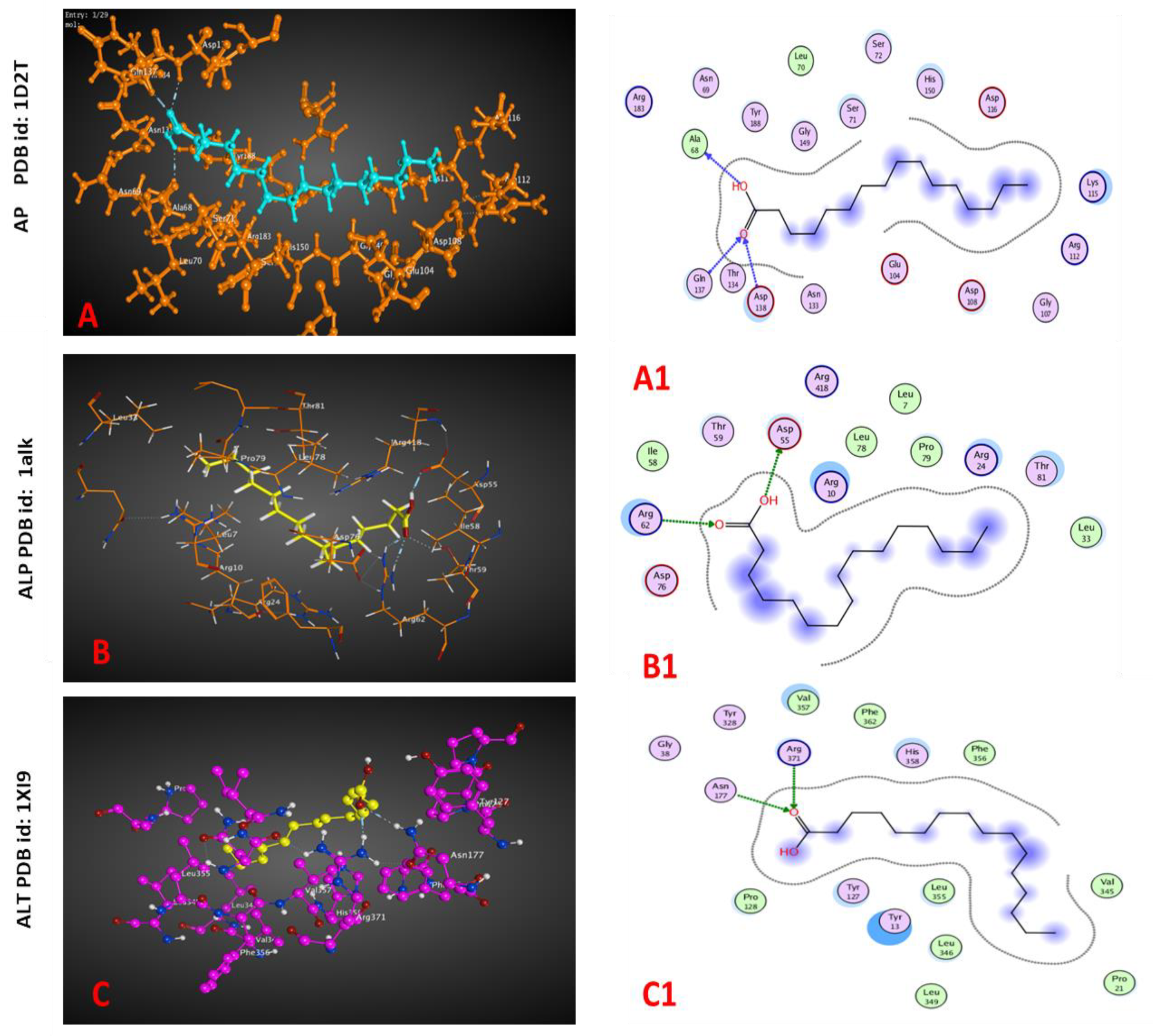
2.4. Molecular Docking

The molecular docking revealed a potential interaction between the ligand molecule, palmitic acid, with acid, alkaline phosphatases, and ALT (Figure 3). There was an inhibitory action for the palmitic acid extracted from both plants against acid and alkaline phosphatases, while a reverse effect was detected with the ALT enzyme, as revealed by interaction-free energy, the docking score. In silico interaction ability was detected through H-acceptor scores (−4.3, −2.6, and −3.1 Kcal/mol) against acid and alkaline phosphatases and ALT enzymes, respectively (Table 9).

3. Discussion

Calotropis procera and Atriplex halimus are medicinal shrubs that can withstand harsh environmental conditions such as extreme heat, drought, and salinity [11,12]. They are also used in phytoremediation to remove heavy metals from soil [11,14]. Because of these factors, these plants are promising in terms of bioactive metabolite production as well as antimicrobial and anti-molluscicidal activity. The plants are from the genera Calotropis and Atriplex, both of which have demonstrated several biological activities.
The metabolic profiling of the methanolic extracts of C. procera and A. halimus was conducted using GC/MS. According to the present data, C. procera extract consists primarily of fatty acids and sterol metabolites, including palmitic acid, campesterol, stigmasterol, oleic acid, and stearic acid. Previous research reported by [17] found that the essential oil of the plant was predominantly composed of the metabolite’s phytol and linoleic acid. According to [18], the two most abundant fatty acids in the ethanolic leaf extract are palmitic acid and linoleic acid. A stigmasterol metabolite has been previously identified in the plant latex [19]. The biological activity and nutritional values of the metabolites identified in the plant extract have been previously characterized. Palmitic acid has been demonstrated to be cytotoxic to human leukemic cells while having no effect on healthy cells [20] and to possess antiviral activity [21,22]. Stigmasterol has been shown to reduce cholesterol levels and has additional health benefits, including protection against cancer, inflammation, and osteoarthritis [23]. It also proved to have larvicidal and antimicrobial properties [24,25]. Oleic acid has antioxidant properties, as reported by [26,27].
Herein, the extract of C. procera has a considerable concentration of the terpenoids phytol and spathulenol. The accumulation of terpenoid compounds in the plant oil extract has been previously reported [17]. Phytol and its derivatives were found to have anti-cancer, antioxidant, anti-pain, anti-inflammatory, immune-modulating, and antibacterial effects [28,29]. Spathulenol possesses an anti-inflammatory effect [30].
Our analysis of the metabolic profile of A. halimus confirmed the majority of methanolic extracts are fatty acids such as palmitic acid, oleic acid, and stearic acid, as well as sugar alcohols and sugars. Terpenoid metabolites made up a minor portion of the total. The qualitative study of the plant’s aerial part revealed the presence of flavonoids, polyphenols, and tannins [31]. Flavonol glycosides, phenolic glucosides, and methoxylated flavonoid glycosides were all isolated from the butanol extract from shoots [32]. The aqueous extract of the plant showed the presence of flavonoids [33]. LC/MS analysis of the plant ethanolic extract showed an abundance of phenolic compounds such as gallic acids and caffeic acid.
This study demonstrated that myo-inositol, D-pinitol, and xylonic acid are found in A. halimus extract and that these carbohydrates have biological and commercial value. Myo-inositol has been linked to numerous positive health effects in humans, including anti-diabetic and antioxidant properties, suppression of liver carcinogenesis, and alterations in mood state as a function of increased or decreased levels in the brain [34]. D-pinitol has been shown to protect the liver from lipid peroxidation, lower blood sugar, fight cancer, reduce inflammation, and function as an antioxidant [35]. Many different applications exist for xylonic acid, including chelation and use as a food additive. In addition to being a plasticizer and exhibiting high thermal stability, it also possesses a number of other useful properties [36].
In this study, five pathogenic bacterial species were tested for susceptibility to the antibacterial activity of methanolic extracts of C. procera and A. halimus. C. procera exhibited greater antibacterial activity than A. halimus in inhibiting the growth of Escherichia coli, Pseudomonas aeruginosa, and Proteus mirabilis. Moreover, C. procera was more effective than gentamycin against Pseudomonas aeruginosa and Proteus mirabilis. Both bacterial species are human pathogens: Pseudomonas aeruginosa is a major cause of illness in patients with cystic fibrosis, and it may cause persistent infections, largely due to its remarkable adaptability [37]; Proteus mirabilis is responsible for some urinary tract infections [38]. Proteus mirabilis has been shown to be resistant to various medicines, including colistin, in addition to showing decreased sensitivity to imipenem [39,40]. Studies on C. procera have shown that a methanol extract of the plant’s leaves has antibacterial properties against both Gram-positive and Gram-negative bacteria, including Staphylococcus aureus, Bacillus subtilis, Escherichia coli, and Pseudomonas aeruginosa [41]. The ethanolic extract of leaves showed antibacterial activity against Escherichia coli [18]. The development of Salmonella typhi, Staphylococcus aureus, and Pseudomonas aeruginosa has been shown to be inhibited by essential oils extracted from the plant’s leaves [18].
In the present study, both C. procera and A. halimus methanol extracts showed molluscicidal activity toward B. alexandrina snails, with the C. procera extract being more effective. The molluscicidal activities of C. procera extract against Biomphalaria alexandrina [42], Biomphalaria arabica snails [43], and Schistosoma mansoni [44] have been reported. Moreover, the plant oil showed antiparasitic activity against Blastocystis spp. [45]. Molluscicidal activities have been attributed to various members of the genus Atriplex. Methanolic extracts of Atriplex glauca, for instance, were found to significantly reduce the survival and growth rates of B. alexandrina snails. Meanwhile, Atriplex inflata demonstrated activity against Galba truncatula [46].
In the present investigation, the extracts from C. procera and A. halimus were toxic to adult B. alexandrina snails, lowered egg hatching success, and increased egg mortality. Additionally, these extracts caused high levels of DNA damage in the snails’ digestive glands. Snail DNA can be used as an indicator of toxicity, and the comet test is a highly accurate technique for identifying DNA damages, including single-strand breaks [47]. Both the oxidation of DNA nucleotides and the covalent bonding that forms with DNA are potential pathways for this genotoxicity; both of these mechanisms can contribute to strand breaks in DNA. Breaks in the DNA of aquatic species have been shown in previous studies to be connected with detrimental effects on a range of biological processes, including fertilization, the immunological response, development, and population dynamics [48].
Immunohistochemistry is a histological technique that determines the presence or absence of specific antigens in tissues by the use of specific immunological antibodies. Cyclin D1 is widely used as a histopathological marker in many tissues as it an important regulator of the cell cycle progression from G1 to S phase. In normal tissues, its expression is well regulated, while the highly expressed cyclin D1 might be a good surrogate to genotoxicity as it refers to the deregulation of the cell cycle [49,50]. In this research, adult snails that were exposed to sublethal concentrations of C. procera or A. halimus had anomalies in their digestive and hermaphrodite glands. These abnormalities included degenerations in digestive cells, ova, and sperm. Immunohistochemical analysis using cyclin D1 as a marker validated these changes. There was no expression of cyclin D1 in both glands of the control group, while 70% and 40% expression of cyclin D1 were observed in the interstitial cells after exposure to C. procera and A. halimus methanolic extracts, respectively. The molluscicidal activity of C. procera and A. halimus may be related to the presence of high levels of fatty acids, particularly palmitic acid, which has been reported to be responsible for the killing of Pomacea canaliculata snails [51]. The immunohistochemical analysis relies on specific interactions between antibodies and their target antigens [49,50]. Cyclin D1 controls how far along the cell cycle it is allowed to go. Increased cyclin D1 expression could enhance tumor growth by disrupting cell cycle control [52,53]. Bartkova et al. [54] proved that normal tissues have relatively low levels of cyclin D1 based on immunohistochemistry, and breast carcinomas exhibited overexpression and upregulation of cyclin D1.
Here, B. alexandrina snails were affected biochemically using sub-lethal doses (LC25) of methanol extracts of the plants that were studied. Both alkaline and acid phosphatase levels have gone down. The levels of total protein and albumin have also been lowered. The amount of alanine aminotransferase has been significantly increased. These findings are consistent with those of [55], in which they also recorded similar observations following chlorophyllin exposure to B. alexandrina snails. It has been reported that the toxicity of the snail species Lanistes varicus is correlated with decreased levels of protein, alkaline, and acid phosphatase [56]. It has been reported that the toxic effect of Casimiroa edulis and Cestrum diurnum plants on B. alexandrina is responsible for the modification in alanine aminotransferase activity [57].
Molecular docking is a potential method for investigating the activity of ligand compounds against the effects of some proteins via receptor–ligand interactions. Acid phosphatase is an enzyme incorporated in lysosomes and involved in autolysis and necrosis, while alkaline phosphatase has an important role in protein synthesis in gastropods [55]. The enzymatic mechanisms included acid phosphatase, alkaline phosphatase, and ALT. Our study showed that there were interactions between palmitic acid and acid, alkaline phosphatase, and ALT that might cause the inhibition of acid and alkaline phosphatase levels and lead to a decrease in the total protein concentration.

4. Materials and Methods

4.1. Plants

The healthy leaves of Calotropis procera and Atriplex halimus plants were collected from Wadi Degla Protectorate in Cairo, Egypt. Atriplex halimus was collected during the vegetative stage, while Calotropis procera was at the flowering stage in the summer of 2021 (June), the plants were collected early in the morning. Drought and extreme high temperatures characterize the summer season in Wadi Degla Protectorate. Three replicates were randomly obtained from three separate individuals for each species. The identification of the plants has been carried out according to [58].
A voucher plant specimen was kept at the herbarium of Helwan University’s Faculty of Science in Egypt.

4.1.1. Metabolites Extraction

Fresh leaves of Calotropis procera and Atriplex halimus were allowed to air-dry in the shade. The leaves were ground into a fine powder. Briefly, five grams of powdered materials were extracted using 100 mL of 80% methanol at 50 °C for an hour while being stirred constantly. The samples were completely dried by evaporating the filtrate solutions at 40 °C until they were completely dry. The extract yield was estimated using the following formula: 100 (V/W), where V is the volume of dry extract and W is the weight of the plant material extracted. The yield of Calotropis procera was 0.70 g, while the yield of Atriplex halimus was 0.42 g.

4.1.2. Gas Chromatography-Mass Spectroscopy (GC-MS) Analysis

Derivatization and sample preparation
Each dried polar residue from C. procera and A. halimus (three replicates from each plant) was combined with 80 µL of N, O-bis (trimethylsilyl) trifluoroacetamide silylation reagent (BFSTA) and 20 µL of trimethylchlorosilane (TMCS), and the mixture was then incubated for 1 h at 65 °C.
GC/MS data collection and compounds identification
The analysis of the metabolites was performed using a TRACE-GC ultra-gas chromatograph (Thermal Scientific Corp., Alvarado, TX, USA) coupled to a thermos mass spectrometer detector (ISQ single quadrupole mass spectrometer) and a 30 m × 0.32 mm i.d., 0.25 m film thickness, TR-5 MS column. With a split ratio of 1:10 and a flow rate of 1.0 mL/min, helium gas was used as the carrier gas. First, the temperature was adjusted to 60 °C for one min, and then it was gradually increased to 240 °C at a rate of 4.0 °C/min. Both the injector and the detector were maintained at a temperature of 210 °C. During the injection phase, 1 µL of the plant extract was diluted with hexane at a ratio of 1:10 hexane, v/v. Electron ionization (EI) at 70 eV was utilized to get mass spectra-spanning m/z ranges of 40–450. AMDIS, open-source software (www.amdis.net, accessed on 9 August 2022), Wiley’s spectrum library, and NSIT’s library databases were used to determine the identities of the metabolites.

4.2. The Antibacterial Activities

The antibacterial effect of plants methanol extracts was evaluated against one pathogen Gram-positive bacterium (Staphylococcus aureus ATCC 25923) and four Gram-negative pathogenic bacterial species, namely Escherichia coli ATCC 25922, Pseudomonas aeruginosa ATCC 7853, Proteus mirabilis ATCC 29906, and Klebsiella pneumoniae ATCC 700721. First, the nutrient agar (Diffco) medium was inoculated with the bacterial strains and placed in a 37 °C incubator. Each bacterium was then inoculated with a single colony and cultivated for 24 h at 37 °C in a nutrient broth medium. The antibacterial activities of plants’ extracts were studied using the well diffusion technique, as explained by [59]. Plates (9 mm) containing 20 mL of nutritional agar medium were inoculated with 100 µL of the bacterial suspension (1 × 10⁶ CFU/mL). The agar plates were drilled into using a 6-mm cork borer to create wells. Each well had 100 µL of the plant extract at a concentration of 20 mg/mL. The plates were then incubated at 4 °C for 8 h, followed by 24 h at 37 °C. The well containing 100 µL of ethyl acetate was utilized as a negative control, while gentamycin (10 g/disc) was employed as a positive control in the experiment. Inhibition zones formed around the wells served as an indicator for the antibacterial efficacy. Inhibitory zone widths were then assessed in mm.

4.3. Molluscicidal Activity

4.3.1. Snails

The methanolic extracts of A. halimus and C. procera leaves were tested for their molluscicidal properties against B. alexandrina snails. Snails’ average size was 8–10 mm, and they were acclimatized at the Laboratory of Medical Malacology at the Theodor Bilharz Research Institute (TBRI) in Giza, Egypt.

4.3.2. Assessment of the Molluscicidal Activity of the Plant’s Methanol Extracts

Plants’ methanol extracts at different concentrations (95, 80, 65, 50, 35, and 25 mg/L) were prepared in order to determine the LC 10, 25, 50, and LC 90 at room temperature (22–25 °C) with a photoperiodicity of 12-h light/12-h dark [60]. Thirty replica snails of consistent size were utilized for each plant concentration, and another thirty identically sized snails were treated with dechlorinated water as a control. The snails were exposed to either plant extracts or dechlorinated water (for control) for 96 h. Then the snails were removed, rinsed with dechlorinated water, and given 24 h to recover. Dead snails were recorded as the average of the three replicates. Death of snails was distinguished by the immersion of snails in a small amount of 15–20% sodium hydroxide solution; if bubbles and blood come out of the snail, it is recorded as alive, and if not, it is recorded as dead [61].

4.3.3. Effect of Plants Extract on Survival Rate of Snails

The snails, all of which measured between 8 and 10 mm in length, were randomly assigned to one of three groups: the first was exposed to the sub-lethal concentrations (LC25) of C. procera extract (127.8 mg/L), the second to the LC25 of A. halimus extract (204.5 mg/L), and the third to dechlorinated tap water as a control. Thirty snails were involved in each group. Subsequently, all samples were incubated in the test solution for a total of 24 h. Then, snails were collected, washed properly in dechlorinated water, and allowed to recover for 24 h in containers filled with fresh dechlorinated tap water. This process took two weeks. After recovery, snails were observed daily to record the survival rate for four weeks. The experiment was repeated thrice.

4.3.4. Effect of Plants’ Extracts on Hatchability of Snails’ Eggs

Eggs were transferred to petri dishes, where they were exposed to the sub-lethal concentrations (LC25) of Calitropis procera (127.8 mg/L) and Atriplex halimus (204.5 mg/L). For each concentration, 100 eggs were used, and assays were repeated three times. At the end of the exposure period (24 h), eggs were transferred to petri dishes with dechlorinated water and examined daily under a stereomicroscope up to the 7th day.

4.4. Tissue Preparation

The soft tissues of the exposed and control groups were obtained by crushing the snail shells using two slides, weighing (1 g tissue/10 mL phosphate buffer), and homogenizing with a glass Dounce homogenizer. Then, the tissue homogenates were centrifuged (Sigma, 3–16PK, Germany) at 3000 rpm for 10 min, and the supernatants were stored at −80 °C until used.

4.4.1. Biochemical Analysis

Bergmeyer’s approach [62], with some modifications by [63], was used to measure acid and alkaline phosphatases. Briefly, the tissue homogenate was quickly made by centrifuging it at 5000× g for 20 min at 4 °C after being immersed in ice-cold 0.9% NaCl (2% w/v). The levels of phosphatase activity were reported as mole/mg of tissue. This study used the protocol described in [64] to quantify total protein. Briefly, three tubes were prepared, and 5.0 mL of biuret reagent (cupric sulphate, sodium potassium tartrate, sodium hydroxide, and potassium iodide) was added to each tube. 100 µL of tissue homogenate solution (the sample) was added to the first tube. The second tube containing biuret reagent only was used as a blank. 100 µL of egg albumin was added to the third tube containing 5.0 mL of biuret reagent as a positive control (standard). Incubation for 30 min at 37 °C. To calculate the total protein concentration, the absorbance of the sample (A Sample) and standard (A standard) were measured against a reagent blank at 550 nm (520–570 nm). The following formula was used to calculate the protein concentration:
protein concentration (g/100 mL) = (A Sample/A standard) × 5
Analyses of albumin were conducted using the guidelines provided by [65]. Briefly, three tubes were prepared, and 2.0 mL of albumin reagent (citrate buffer, pH 4.2, bromcresol green, detergent, and preservative) was added to each tube. An amount of 10 µL of tissue homogenate solution (the sample) was added to the first tube. The second tube containing albumin reagent only was used as a blank. As a positive control (standard), 10 µL of albumin was added to the third tube containing 2.0 mL of albumin reagent. Incubation for 5 min. at 37 °C. To calculate the albumin concentration, the absorbance of the sample (A Sample) and standard (A standard) against reagent blank at 630 nm were measured. The calculation formula was as follows:
Albumin concentration (g/100 mL) = (A Sample/A standard) × 4.
The alanine aminotransferase levels were measured using the [66] technique. Briefly, 1 mL of the tissue homogenate is pipetted into a test tube and incubated in a water bath at a constant temperature of 40 °C for 10 min. An amount of 200 µL of serum was added and mixed well, and after an incubation period of exactly 30 min, the tube was removed from the water bath. A total of 1 mL of 2, 4-dinitrophenylhydrazine reagent was added to allow the reaction to be terminated. The tube was permitted to stand at room temperature for a minimum of 20 min, then 10 mL of 0.4 N sodium hydroxide was added, and the contents were well mixed. This mixture was left for exactly 30 min, and the optical density of the solution was measured at 505 nm using water as the blank. The number of units/liters was determined by using a standard curve.

4.4.2. Comet Assay

The B. alexandrina snails were subjected to A. halimus or C. procera methanolic extracts at LC25 of 204.5 mg/L or 127.8 mg/L, respectively, for 24 h, then the snails were dissected and their head-foot regions were frozen at −80 °C. DNA damage was quantified using a single-cell gel technique, as published by [67]. Briefly, the tissues of control and exposed snails were cut into small pieces in phosphate buffer saline, then centrifuged 500× g for 5 min. The resulting supernatant was kept, and the pellets were discarded. The supernatant then centrifuged with a high speed 10,000× g to concentrate the cells, keep the pellets, and discard supernatant. Add 20 µL of the pellets to 180 µL of low melting agarose (0.5%). Take a drop of this mixture and put it on a slide then cover it and place it on ice. Leave for 15 min to ensure the freezing of the gel, then remove the coverslip and the slides and put them in a sectioned box that contains a lysis buffer (each 1 L contains 2.5 M NaCl, 100 mM EDTA, 8 g of NaOH, and 10 mM trisabase). Leave it in the fridge for 24 h. For DNA damage visualization, observations are made of EtBr-stained DNA using a 40× objective on a fluorescent microscope. Coding and scoring the slides were performed separately.

4.4.3. Histopathological and Immunohistochemical Analysis

Adult B. alexandrina snails were subjected to LC25 (204.5 mg/L) or LC25 (127.8 mg/L) of either A. halimus or C. procera, respectively, for 24 h, followed by a two-week recovery period. According to the findings given by [68], the digestive and hermaphroditic glands were collected and processed. After cutting the tissues and immersing them in 10% formalin for 12 h, they were then dehydrated in ethanol at ascending concentrations of 80%, 90%, and 100% for 3 h each, cleaned in xylene, and then embedded in paraffin. After being cut on a microtome into 5-micrometer-long segments, the samples were mounted on slides, then dewaxed in xylene, stained with haemoxylin and eosin, and finally coated with Canada balsam. Utilization of a Zeiss microscope for the purpose of conducting an analysis on stained slides (Carl Zeiss Microscopy GmbH, 07, 745 Jena, Germany). Prior to undergoing immunohistochemistry analysis, adult snail tissue was sliced to a thickness of 4 mm and then mounted on slides that had been given a positive charge (Super Frost Plus, Menzel-Glaser, Germany). The slides were stained using anti-mouse proliferating cell antigen (PCNA) and cyclin D1 antibodies that were bought from Santa Cruz Biotechnology in the United States of America. This process took place on an automated platform. These antibodies performed most effectively when diluted at a ratio of 1:100. In order to calculate the percentages of positively stained brown nuclear material (PCNA, Cyclin D1), calculations were determined under Zeiss light microscopy at 400× magnification power.

4.5. The Molecular Docking Study

To explore the effect of exposing snails to C. procera and A. halimus methanolic extracts, acid and alkaline phosphatases and ALT enzymes were selected to predict their action with palmitic acid, a compound found in the GC analysis of both plant extracts. Using the Protein Data Bank (PDB), the molecular structure of tested enzymes was obtained and encoded, including acid phosphatase (1D2T) from Escherichia blattae [69], alkaline phosphatases (1alk) from Escherichia coli [70], and ALT (1XI9) from Pyrococcus furiosus [71]. Molecular docking was conducted using Molecular Operating Environment software (MOE 2014.09). The energy of the ligand palmitic acid compound was minimized, and after choosing the correct sequence of enzymes, hydrogens were added, and partial charges were calculated.

4.6. Statistical Analysis

All the experiments were randomly designed At least three replicates from each treatment were used, and Probit analysis was conducted to calculate the lethal concentration of A. halimus or C. procera extracts against Schistosoma mansoni. Minitab 17 was utilized to conduct the one-way ANOVA analysis of the data. Means between treatments were compared using the 95% confidence interval of the Fisher least significant difference (LSD) method.

5. Conclusions

This study demonstrated that methanolic extracts of the medicinal shrubs Calotropis procera and Atriplex halimus are rich in fatty acids (both saturated and unsaturated), glucosides, and sterols. Calotropis procera extract was more effective against Escherichia coli ATCC 25923, Pseudomonas aeruginosa ATCC 7853, and Proteus mirabilis ATCC 29906 than Atriplex halimus. Both plant extracts were found to have molluscicidal activity against Biomphalaria alexandrina snails, as determined by a variety of tests, including the mortality rate of adult snails, the hatchability rate of eggs, biochemical and histological analyses, and visual examinations of snail tissue. Based on the data, it was clear that Calotropis procera extract had greater anti-molluscicidal activity.
Our next studies will be to investigate the effect of pure compounds isolated from the plants under study in order to identify the metabolites responsible for the molluscicidal activity as an intriguing approach to eliminating schistosomiasis. Furthermore, because of the broad-spectrum effects, the safety of the tested metabolites will be assessed against other (non-target) organisms such as the water flea, Daphnia magna, which is extremely sensitive to water pollution. Measurements of many digestive and hermaphrodite gland enzymes will be used to investigate the sub-lethal effects on snail fecundity/fertility, and cercariae.

Author Contributions

Conceptualization, M.Y.M., H.E.-S. and A.M.I.; methodology, M.Y.M., H.E.-S., A.A., E.Z.A. and A.M.I.; software, M.Y.M., H.E.-S., A.A., M.F.E.-K. and A.M.I.; validation, M.Y.M., H.E.-S. and A.M.I.; formal analysis M.Y.M., H.E.-S., M.F.E.-K., A.A., E.Z.A. and A.M.I.; investigation, M.Y.M., H.E.-S., A.A. and A.M.I.; resources, M.Y.M., H.E.-S., A.A., M.F.E.-K. and A.M.I.; data curation, M.Y.M., H.E.-S., A.A., E.Z.A. and A.M.I.; writing—original draft preparation, M.Y.M., H.E.-S., A.A. and A.M.I.; writing—review and editing, M.Y.M., H.E.-S., M.F.E.-K., A.A., E.Z.A. and A.M.I. All authors have read and agreed to the published version of the manuscript.

Funding

This research has been funded by Princess Nourah bint Abdulrahman University Researchers Supporting project number (PNURSP2023R23), Princess Nourah bint Abdulrahman University, Riyadh, Saudi Arabia.

Institutional Review Board Statement

Not applicable.

Informed Consent Statement

Not applicable.

Data Availability Statement

All data generated or analyzed during this study are included in this article.

Acknowledgments

Princess Nourah bint Abdulrahman University Researchers Supporting Project number (PNURSP2023R23), Princess Nourah bint Abdulrahman University, Riyadh, Saudi Arabia.

Conflicts of Interest

The authors declare no conflict of interest.

References

  1. WHO. Schistosomiasis. 2020. Available online: https://www.Who.Int/Newsroom/Fact-Sheets/Detail/Schistosomiasis (accessed on 10 November 2022).
  2. Ibrahim, A.M.; Sayed, S.S.M. Assessment of the Molluscicidal Activity of the Methanolic Seed Extracts of Ziziphus Spina-Christi and Carica Papaya on Immunological and Molecular Aspects of Biomphalaria Alexandrina Snails. Aquac. Res. 2021, 52, 2014–2024. [Google Scholar] [CrossRef]
  3. Loker, E.S.; Mkoji, G.M. Schistosomes and Their Snail Hosts BT—Schistosomiasis; Secor, W.E., Colley, D.G., Eds.; Springer US: Boston, MA, USA, 2005; pp. 1–11. ISBN 978-0-387-23362-8. [Google Scholar]
  4. Elsareh, F.; Abdalla, R.; Abdalla, E. The Effect of Aqueous Leaves Extract of Solenostemma Argel (Del Hayne) on Egg Masses and Neonates of Biomphalaria Pfeifferi Snails. J. Med. Plants 2016, 4, 271–274. [Google Scholar]
  5. Abd El Ghaffar, M.M.; Sadek, G.S.; Harba, N.M.; Abd El Samee, M.F. Evaluation of the Effect of Some Plant Molluscicides on the Infectivity of Schistosoma Mansoni Cercariae. Menoufia Med. J. 2019, 31, 1448–1455. [Google Scholar] [CrossRef]
  6. Uddin, T.M.; Chakraborty, A.J.; Khusro, A.; Zidan, B.R.M.; Mitra, S.; Emran, T.B.; Dhama, K.; Ripon, M.K.H.; Gajdács, M.; Sahibzada, M.U.K.; et al. Antibiotic Resistance in Microbes: History, Mechanisms, Therapeutic Strategies and Future Prospects. J. Infect. Public Health 2021, 14, 1750–1766. [Google Scholar] [CrossRef]
  7. Tagboto, S.; Townson, S. Antiparasitic Properties of Medicinal Plants and Other Naturally Occurring Products. Adv. Parasitol. 2001, 50, 199–295. [Google Scholar] [CrossRef]
  8. Batool, H.; Hussain, M.; Hameed, M.; Ahmad, R. A review on Calotropis procera its phytochemistry and traditional uses. Big Data Agric. 2020, 2, 56–58. [Google Scholar] [CrossRef]
  9. Nadeem, M.; Mumtaz, M.W.; Danish, M.; Rashid, U.; Mukhtar, H.; Anwar, F.; Raza, S.A. Calotropis Procera: UHPLC-QTOF-MS/MS Based Profiling of Bioactives, Antioxidant and Anti-Diabetic Potential of Leaf Extracts and an Insight into Molecular Docking. J. Food Meas. Charact. 1234, 13, 3206–3220. [Google Scholar] [CrossRef]
  10. Singh, V.S.; Dhawale, S.C.; Shakeel, F.; Faiyazuddin, M.; Alshehri, S. Antiarthritic Potential of Calotropis procera Leaf Fractions in FCA-Induced Arthritic Rats: Involvement of Cellular Inflammatory Mediators and Other Biomarkers. Agriculture 2021, 11, 68. [Google Scholar] [CrossRef]
  11. Darra Wadhwani, B.; Mali, D.; Vyas, P.; Nair, R.; Khandelwal, P. A Review on Phytochemical Constituents and Pharmacological Potential of Calotropis Procera. RSC Adv. 2021, 11, 35854–35878. [Google Scholar] [CrossRef]
  12. Walker, D.J.; Lutts, S.; Sánchez-García, M.; Correal, E. Atriplex halimus L. Its Biology and Uses. J. Arid. Environ. 2014, 100–101, 111–121. [Google Scholar] [CrossRef]
  13. Nedjimi, B. Heavy Metal Tolerance in Two Algerian Saltbushes: A Review on Plant Responses to Cadmium and Role of Calcium in Its Mitigation. Plant Nutr. Abiotic Stress Toler. 2018, 205–220. [Google Scholar]
  14. Acosta, J.A.; Abbaspour, A.; Martínez, G.R.; Martínez-Martínez, S.; Zornoza, R.; Gabarrón, M.; Faz, A. Phytoremediation of Mine Tailings with Atriplex halimus and Organic/Inorganic Amendments: A Five-Year Field Case Study. Chemosphere 2018, 204, 71–78. [Google Scholar] [CrossRef] [PubMed]
  15. Clauser, M.; Dall’Acqua, S.; Loi, M.; Innocenti, G. Phytochemical Investigation on Atriplex halimus L. from Sardinia. Nat. Prod. Res. 2013, 20, 1940–1944. [Google Scholar] [CrossRef] [PubMed]
  16. Elbouzidi, A.; Ouassou, H.; Aherkou, M.; Kharchoufa, L.; Meskali, N.; Baraich, A.; Mechchate, H.; Bouhrim, M.; Idir, A.; Hano, C.; et al. LC–MS/MS Phytochemical Profiling, Antioxidant Activity, and Cytotoxicity of the Ethanolic Extract of Atriplex halimus L. against Breast Cancer Cell Lines: Computational Studies and Experimental Validation. Pharmaceuticals 2022, 15, 1156. [Google Scholar] [CrossRef]
  17. Al-Rowaily, S.L.; Abd-Elgawad, A.M.; Assaeed, A.M.; Elgamal, A.M.; El-Nasser, A.; el Gendy, G.; Mohamed, T.A.; Dar, B.A.; Mohamed, T.K.; Elshamy, A.I. Molecules Essential Oil of Calotropis Procera: Comparative Chemical Profiles, Antimicrobial Activity, and Allelopathic Potential on Weeds. Molecules 2020, 25, 5203. [Google Scholar] [CrossRef]
  18. Pattnaik, P.K.; Kar, D.; Chhatoi, H.; Shahbazi, S.; Ghosh, G.; Kuanar, A. Chemometric Profile & Antimicrobial Activities of Leaf Extract of Calotropis procera and Calotropis Gigantea. Nat. Prod. Res. 2017, 31, 1954–1957. [Google Scholar]
  19. Chundattu, S.J.; Agrawal, V.K.; Ganesh, N. Phytochemical Investigation of Calotropis Procera. Arab. J. Chem. 2016, 9, S230–S234. [Google Scholar] [CrossRef] [Green Version]
  20. Harada, H.; Yamashita, U.; Kurihara, H.; Fukushi, E.; Kawabata, J.; Kamei, Y. Antitumor Activity of Palmitic Acid Found as a Selective Cytotoxic Substance in a Marine Red Alga. Anticancer Res. 2002, 22, 2587–2590. [Google Scholar]
  21. Librán-Pérez, M.; Pereiro, P.; Figueras, A.; Novoa, B. Antiviral Activity of Palmitic Acid via Autophagic Flux Inhibition in Zebrafish (Danio Rerio). Fish Shellfish Immunol. 2019, 95, 595–605. [Google Scholar] [CrossRef]
  22. DeWitte-Orr, S.; Cai, J.; Huang, X.; Qin, Q.; Yu, Y.; Li, C.; Liu, J.; Zhu, F.; Wei, S.; Huang, Y. Palmitic Acid Promotes Virus Replication in Fish Cell by Modulating Autophagy Flux and TBK1-IRF3/7 Pathway. Front. Immunol. 2020, 11, 1764. [Google Scholar] [CrossRef]
  23. Ashraf, R.; Bhatti, H.N. Stigmasterol. In A Centum of Valuable Plant Bioactives; Academic Press: Cambridge, MA, USA, 2021; pp. 212–232. [Google Scholar] [CrossRef]
  24. Yusuf, A.J.; Abdullahi, M.I.; Aleku, G.A.; Ibrahim, I.A.; Alebiosu, C.O.; Yahaya, M.; Adamu, H.W.; Sanusi, A.; Mailafiya, M.M.; Abubakar, H. Antimicrobial Activity of Stigmasterol from the Stem Bark of Neocarya Macrophylla. J. Med. Plants Econ. Dev. 2018, 2, 1–5. [Google Scholar] [CrossRef] [Green Version]
  25. Gade, S.; Rajamanikyam, M.; Vadlapudi, V.; Nukala, K.M.; Aluvala, R.; Giddigari, C.; Karanam, N.J.; Barua, N.C.; Pandey, R.; Upadhyayula, V.S.v.; et al. Acetylcholinesterase Inhibitory Activity of Stigmasterol & Hexacosanol Is Responsible for Larvicidal and Repellent Properties of Chromolaena odorata. Biochim. Biophys. Acta (BBA)—Gen. Subj. 2017, 1861, 541–550. [Google Scholar] [CrossRef]
  26. Bhattacharjee, B.; Pal, P.K.; Chattopadhyay, A.; Bandyopadhyay, D. Oleic Acid Protects against Cadmium Induced Cardiac and Hepatic Tissue Injury in Male Wistar Rats: A Mechanistic Study. Life Sci. 2020, 244, 117324. [Google Scholar] [CrossRef] [PubMed]
  27. Wei, C.-C.; Yen, P.-L.; Chang, S.-T.; Cheng, P.-L.; Lo, Y.-C.; Hsiu-Chuan Liao, V. Antioxidative Activities of Both Oleic Acid and Camellia Tenuifolia Seed Oil Are Regulated by the Transcription Factor DAF-16/FOXO in Caenorhabditis Elegans. PLoS ONE 2016, 11, e0157195. [Google Scholar] [CrossRef] [PubMed] [Green Version]
  28. Islam, M.T.; de Alencar, M.V.O.B.; da Conceição Machado, K.; da Conceição Machado, K.; de Carvalho Melo-Cavalcante, A.A.; de Sousa, D.P.; de Freitas, R.M. Phytol in a Pharma-Medico-Stance. Chem. Biol. Interact. 2015, 240, 60–73. [Google Scholar] [CrossRef]
  29. Islam, M.T.; Ali, E.S.; Uddin, S.J.; Shaw, S.; Islam, M.A.; Ahmed, M.I.; Chandra Shill, M.; Karmakar, U.K.; Yarla, N.S.; Khan, I.N.; et al. Phytol: A Review of Biomedical Activities. Food Chem. Toxicol. 2018, 121, 82–94. [Google Scholar] [CrossRef] [PubMed]
  30. do Nascimento, K.F.; Moreira, F.M.F.; Alencar Santos, J.; Kassuya, C.A.L.; Croda, J.H.R.; Cardoso, C.A.L.; Vieira, M. do C.; Góis Ruiz, A.L.T.; Ann Foglio, M.; de Carvalho, J.E.; et al. Antioxidant, Anti-Inflammatory, Antiproliferative and Antimycobacterial Activities of the Essential Oil of Psidium Guineense Sw. and Spathulenol. J. Ethnopharmacol. 2018, 210, 351–358. [Google Scholar] [CrossRef]
  31. Ounaissia, K.; Bennadja, S.; Aliane, L.; Djahoudi, A. Phytochemical screening and anti-bacterial activity of methanolic extracts of the aerial parts of Atriplex halimus L., from biskra (Algeria). Asian J. Res. Chem. 2020, 13, 365–372. [Google Scholar]
  32. Kabbash, A.; Shoeib, N. Chemical and Biological Investigation of Some Secondary Metabolites in Atriplex halimus Growing in Egypt. Nat. Prod. Commun. 2012, 7, 1465–1468. [Google Scholar] [CrossRef] [Green Version]
  33. Zennaf, I.; Tir touil, A.; Meddah, B.; Mokhtar, M. Ethnobotanical and Phytochemical Study of the Medicinal Plant Atriplex halimus and Its Importance in the Traditional Algerian Pharmacopoeia. Fr.-Ukr. J. Chem. 2022, 10, 60–69. [Google Scholar] [CrossRef]
  34. Chhetri, D.R. Myo-Inositol and Its Derivatives: Their Emerging Role in the Treatment of Human Diseases. Front. Pharmacol. 2019, 10, 1172. [Google Scholar] [CrossRef] [PubMed] [Green Version]
  35. Sánchez-Hidalgo, M.; León-González, A.J.; Gálvez-Peralta, M.; González-Mauraza, N.H.; Martin-Cordero, C. d-Pinitol: A cyclitol with versatile biological and pharmacological activities. Phytochem. Rev. 2020, 20, 211–224. [Google Scholar] [CrossRef]
  36. Luo, J.; Huang, K.; Zhou, X.; Xu, Y. Preparation of Highly Flexible and Sustainable Lignin-Rich Nanocellulose Film Containing Xylonic Acid (XA), and Its Application as an Antibacterial Agent. Int. J. Biol. Macromol. 2020, 163, 1565–1571. [Google Scholar] [CrossRef] [PubMed]
  37. Rossi, E.; La Rosa, R.; Bartell, J.A.; Marvig, R.L.; Haagensen, J.A.; Sommer, L.M.; Molin, S.; Johansen, H.K. Pseudomonas aeruginosa adaptation and evolution in patients with cystic fibrosis. Nat. Rev. Microbiol. 2021, 19, 331–342. [Google Scholar] [CrossRef]
  38. Armbruster, C.E.; Mobley, H.L.T.; Pearson, M.M. Pathogenesis of Proteus Mirabilis Infection. EcoSal Plus 2018, 8, 8. [Google Scholar] [CrossRef] [Green Version]
  39. Zanichelli, V.; Huttner, A.; Harbarth, S.; Kronenberg, A.; Huttner, B. Swiss Centre For Antibiotic Resistance Anresis. Antimicrobial Resistance Trends in Escherichia coli, Klebsiella pneumoniae and Proteus mirabilis Urinary Isolates from Switzerland: Retrospective Analysis of Data from a National Surveillance Network over an 8-Year Period (2009–2016). Swiss Med. Wkly. 2019, 149, w20110. [Google Scholar] [CrossRef] [Green Version]
  40. Papagiannitsis, C.C.; Hrabak, J.; Maria D’andrea, M.; Reslan, L.; Naas, T.; Girlich, D.; Bonnin, R.A.; Dortet, L. Genetics of Acquired Antibiotic Resistance Genes in Proteus spp. Front. Microbiol. 2020, 11, 256. [Google Scholar] [CrossRef] [Green Version]
  41. Nenaah, G. Antimicrobial activity of Calotropis procera Ait. (Asclepiadaceae) and isolation of four flavonoid glycosides as the active constituents. World J. Microbiol. Biotechnol. 2013, 29, 1255–1262. [Google Scholar] [CrossRef]
  42. Bakry, F.A. Use of Some Plant Extracts to Control Biomphalaria Alexandrina Snails with Emphasis on Some Biological Effects. Pestic. Biochem. Physiol. 2009, 95, 159–165. [Google Scholar] [CrossRef]
  43. Abo Zaid, K.H.; El-Wakil, H.; El-Hussein, A.; Jomaa, S.; Shohayeb, M. Evaluation of the Molluscicidal Activity of Punica Granatum, Calotropis procera, Solanum incanum and Citrullus colocynthis against Biomphalaria arabica. World Appl. Sci. J. 2013, 26, 873–879. [Google Scholar] [CrossRef]
  44. Seif El-Din, S.H.; El-Lakkany, N.M.; Mohamed, M.A.; Hamed, M.M.; Sterner, O.; Botros, S.S. Potential Effect of the Medicinal Plants Calotropis procera, Ficus Elastica and Zingiber Officinale against Schistosoma Mansoni in Mice. Pharm Biol. 2014, 52, 144–150. [Google Scholar] [CrossRef] [PubMed] [Green Version]
  45. Mettwally, W.S.A.; Zahran, H.A.; Khayyal, A.E.; Ahmed, M.M.E.; Allam, R.M.; Saleh, D.O. Calotropis procera (Aiton) Seeds Fixed Oil: Physicochemical Analysis, GC–MS Profiling and Evaluation of Its in-Vivo Anti-Inflammatory and in-Vitro Antiparasitic Activities. Arab. J. Chem. 2022, 15, 104085. [Google Scholar] [CrossRef]
  46. Ali, B.; Musaddiq, S.; Iqbal, S.; Rehman, T.; Shafiq, N.; Hussain, A. The Therapeutic Properties, Ethno Pharmacology and Phytochemistry of Atriplex Species: A Review. Pak. J. Biochem. Biotechnol. 2021, 2, 49–64. [Google Scholar] [CrossRef]
  47. Ibrahim, A.M.; Ahmed, A.K.; Bakry, F.A.; Abdel-Ghaffar, F. Hematological, Physiological and Genotoxicological Effects of Match 5% EC Insecticide on Biomphalaria Alexandrina Snails. Ecotoxicol. Env. Saf. 2018, 147, 1017–1022. [Google Scholar] [CrossRef] [PubMed]
  48. Lee, R.F.; Steinert, S. Use of the Single Cell Gel Electrophoresis/Comet Assay for Detecting DNA Damage in Aquatic (Marine and Freshwater) Animals. Mutat. Res./Rev. Mutat. Res. 2003, 544, 43–64. [Google Scholar] [CrossRef] [PubMed]
  49. Harris, L.; O’Byrne-Ring, N.; Lambkin, H. Characterisation Of Cell Types In Abalone (Haliotis spp.) Tissues Using Immunohistochemical Techniques. Articles 2006, 261, 1413–1421. [Google Scholar] [CrossRef] [Green Version]
  50. Nakopoulou, L.; Janinis, J.; Panagos, G.; Comin, G.; Davaris, P. The Immunohistochemical Expression of Proliferating Cell Nuclear Antigen (PCNA/Cyclin) in Malignant and Benign Epithelial Ovarian Neoplasms and Correlation with Prognosis. Eur. J. Cancer 1993, 29, 1599–1601. [Google Scholar] [CrossRef]
  51. Zhang, L.; Zou, Z. Molluscicidal Activity of Fatty Acids in the Kernel of Chimonanthus Praecox Cv. Luteus against the Golden Apple Snail Pomacea Canaliculata. Pestic. Biochem. Physiol. 2020, 167, 104620. [Google Scholar] [CrossRef]
  52. Ohta, Y.; Ichimura, K. Proliferation Markers, Proliferating Cell Nuclear Antigen, Ki67, 5-Bromo-2′-Deoxyuridine, and Cyclin D1 in Mouse Olfactory Epithelium. Ann. Otol. Rhinol. Laryngol. 2000, 109, 1046–1048. [Google Scholar] [CrossRef]
  53. Alao, J.P. The Regulation of Cyclin D1 Degradation: Roles in Cancer Development and the Potential for Therapeutic Invention. Mol. Cancer 2007, 6, 24. [Google Scholar] [CrossRef] [Green Version]
  54. Bartkova, J.; Lukas, J.; Strauss, M.; Bartek, J. Cell Cycle-Related Variation and Tissue-Restricted Expression of Human Cyclin D1 Protein. J. Pathol. 1994, 172, 237–245. [Google Scholar] [CrossRef] [PubMed]
  55. Ibrahim, A.M.; Bakry, F.A. Assessment of the Molluscicidal Impact of Extracted Chlorophyllin on Some Biochemical Parameters in the Nervous Tissue and Histological Changes in Biomphalaria Alexandrina and Lymnaea Natalensis Snails. Invertebr. Neurosci. 2019, 19, 7. [Google Scholar] [CrossRef] [PubMed]
  56. Abonyi, C.U.; Ezugwu, A.L.; Eze, J.C.; Nwankwo, N.E.; Okafor, F.C. Toxicological Impact of Picralima Nitida (Pile Plant) Extracts on the Gastropod Lanistes Varicus (Freshwater Snail), as a Control Measure against Trematodes Infections. Biologia 2022, 77, 2093–2101. [Google Scholar] [CrossRef]
  57. Saad, A.-H.A.; Aly, R.H.; Ragab, F.M.A.; Abdel-Wareth, M.T.A. Effect of Cestrum diurnum and Casimiroa edulis plants on the digestive gland and its related enzymes in the freshwater snail Biomphalaria alexandrina. Toxicol. Environ. Health Sci. 2012, 4, 109–114. [Google Scholar] [CrossRef]
  58. Boulos, L. Flora of Egypt; Al Hadara Publishing: Cairo, Egypt, 2002; Volume 3. [Google Scholar]
  59. Hamad, D.; El-Sayed, H.; Ahmed, W.; Sonbol, H.; Ramadan, M.A.H. GC-MS Analysis of Potentially Volatile Compounds of Pleurotus Ostreatus Polar Extract: In Vitro Antimicrobial, Cytotoxic, Immunomodulatory, and Antioxidant Activities. Front. Microbiol. 2022, 13, 396. [Google Scholar] [CrossRef]
  60. WHO. Report of the Scientific Working Group on Plant Molluscicide & Guidelines for Evaluation of Plant Molluscicides; WHO: Geneva, Switzerland, 1983. [Google Scholar]
  61. WHO Molluscicide Screening and Evaluation. Bull. WHO 1965, 33, 567–581.
  62. Bergmeyer, H. Methods of Enzymatic Analysis, Volume 8: Metabolites 3: Lipids, Amino Acids and Related Compounds; Verlag Chemie: Basel, Switzerland, 1985. [Google Scholar]
  63. Singh, D.K.; Agarwal, R.A. Action Sites of Cypermethrin, a Synthetic Pyrethroid in the Snail Lymnaea Acuminata. Acta Hydrochim. Hydrobiol. 1991, 19, 425–430. [Google Scholar] [CrossRef]
  64. Doumas, B.T. Standards for Total Serum Protein Assays—A Collaborative Study. Clin. Chem. 1975, 21, 1159–1166. [Google Scholar] [CrossRef]
  65. Gustafsson, J. Improved Specificity of Serum Albumin Determination and Estimation of “Acute Phase Reactants” by Use of the Bromcresol Green Reaction. Clin. Chem. 1976, 22, 616–622. [Google Scholar] [CrossRef]
  66. Reitman, S.; Frankel, S. A Colorimetric Method for the Determination of Serum Glutamic Oxalacetic and Glutamic Pyruvic Transaminases. Am. J. Clin. Pathol. 1957, 28, 56–63. [Google Scholar] [CrossRef]
  67. Grazeffe, V.S.; Tallarico, L.D.F.; Pinheiro, A.D.S.; Kawano, T.; Suzuki, M.; Okazaki, K.; Pereira, C.A.D.B.; Nakano, E. Establishment of the Comet Assay in the Freshwater Snail Biomphalaria Glabrata (Say, 1818). Mutat. Res. /Genet. Toxicol. Environ. Mutagen. 2008, 654, 58–63. [Google Scholar] [CrossRef] [PubMed]
  68. Carleton, M.; Drury, R.A.B.; Willington, E.A.; Cammeron, R. Carleton’s Histological Technique; Oxford University Press: New York, NY, USA, 1967. [Google Scholar]
  69. Ishikawa, K.; Mihara, Y.; Gondoh, K.; Suzuki, E.; Asano, Y. X-ray structures of a novel acid phosphatase from Escherichia blattae and its complex with the transition-state analog molybdate. EMBO J. 2000, 19, 2412–2423. [Google Scholar] [CrossRef] [PubMed] [Green Version]
  70. Kim, E.E.; Wyckoff, H.W. Reaction mechanism of alkaline phosphatase based on crystal structures: Two-metal ion catalysis. J. Mol. Biol. 1991, 218, 449–464. [Google Scholar] [CrossRef] [PubMed]
  71. Zhou, W.; Tempel, W.; Shah, A.; Chen, L.; Liu, Z.-J.; Lee, D.; Lin, D.; Chang, S.-H.; Brereton, P.S.; Izumi, M.; et al. Alanine Aminotransferase from Pyrococcus furiosus Pfu-1397077-001. Southeast Collaboratory for Structural Genomics (SECSG). 2004. Available online: https://www.wwpdb.org/pdb?id=pdb_00001xi9 (accessed on 10 November 2022).
Figure 1. DNA single-cell damage in the digestive gland of B. alexandrina snails after exposure to sub-lethal concentrations of Atriplex halimus and Calotropis procera methanolic extractions. (A) Control (B) Calotropis procera (C) Atriplex halimus.
Plants 12 00477 g001
Figure 2. Light micrographs of the digestive (left side) and hermaphrodite (right side) glands of B. alexanderina snails. (A) Digestive gland of control B. alexanderina snails showing no expression of cyclin D1, the digestive cells found in a digestive follicle showing normal structure (green arrow) (H&E, 400). (B) Digestive gland of exposed snails to LC25 (127.8 mg/L) of C. procera methanolic extraction showing expression of cyclin D1 (brown stained parts) (Immunohistochemistry for cyclin D1, ×100). Degeneration of some digestive cells and appearance of vacuolations (green arrow). (H&E; ×100). (C) Digestive gland of exposed snails to A. triplex showing low expression of cyclin D1 (Immunohistochemistry for cyclin D1, ×200). Degeneration in digestive cells showing many vacuolations (green arrow). (D) Hermaphrodite gland of control B. alexanderina snails showing no expression of cyclin D1, mature ovum found in a female follicle (red arrow), spermatozoa found in the center of a male follicle (orange arrow), (H&E; ×200), (Immunohistochemistry for Cyclin D1, ×200). (E) Hermaphrodite gland of exposed snails to LC25 (127.8 mg/L) of C. procera methanolic extraction showing expression of cyclin D1 (brown stained parts) (Immunohistochemistry for cyclin D1, ×200). Degeneration of mature ovum (red arrow), and sperms in the center of a male follicle (orange arrow) (H&E; ×200). (F) Hermaphrodite gland of exposed snails to A. triplex showing mild expression of cyclin D1 (brown stained parts) (Immunohistochemistry for cyclin D1, ×200). Degenerated mature ovum (red arrow) and sperms (orange arrow) (H&E; ×200).
Plants 12 00477 g002
Figure 3. 3D (AC) and 2D (A1, B1, C1) docked interactions map for the palmitic acid CH3 (CH2)14COOH) with the binding sites of acid (AP), alkaline phosphatases (ALP), and alanine aminotransferase (ALT).
Plants 12 00477 g003
Table 1. List of metabolites identified in the methanolic extract of Calotropis procera.
Compound NameMolecular FormulaMolecular Weight
(g/mol)
Retention Time (min)Area %
1FormaldehydeCH2O30.024.161.3
2PentitolC5H12O5152.155.420.3
3L-ValineC5H11NO2117.156.590.64
4L-LeucineC6H13NO2131.178.250.28
5L-IsoleucineC6H13NO2131.178.760.35
6GlycerolC3H8O392.0911.372.65
7Butanedioic acidC4H6O4118.0912.356.19
8Malic acidC4H6O5134.0916.762.34
9L-ProlineC5H9NO2115.1317.331.21
10DL-PhenylalanineC9H11NO2165.1917.890.83
11SpathulenolC15H24O22018.710.71
12Methyl-β-D-glucopyranosideC7H14O6194.1820.480.26
13L-FucitolC6H14O5166.1722.420.58
144-Coumaric acidC9H8O3164.1623.360.23
15Azelaic acidC9H16O4188.2223.610.21
16D-FructofuranoseC6H12O6180.1623.802.40
17D-TagatofuranoseC6H12O6180.1623.970.45
18Methyl D-glucofuranosideC7H14O6194.1824.600.75
19α-L-ArabinopyranoseC5H10O5150.13024.680.32
20DulcitolC6H14O6182.1725.510.36
21α-d-glucopyranoseC6H12O6180.1625.860.52
22Hexadecanoic acid, methyl esterC17H34O2270.426.190.23
23D-XylofuranoseC5H10O5150.1326.960.54
24α-D-AllopyranoseC6H12O6180.1627.410.31
25D-AllofuranoseC6H12O6180.1627.600.45
26Palmitic AcidC16H32O2256.4228.5210.74
2710-Octadecenoic acid, methyl esterC19H36O3312.529.470.51
28Heptadecanoic acidC17H34O2270.530.310.32
29PhytolC20H40O296.530.823.91
309,12-Octadecadienoic acid (alpha-Linoleic acid)C18H32O2280.431.421.79
31Oleic AcidC18H34O2282.4631.548.04
32Petroselinic acidC18H34O2282.4631.680.92
33Stearic acidC18H36O2284.4832.044.24
345,8,11-EicosatriynoicacidC20H28O2300.432.760.30
35D-TrehaloseC12H22O11342.336.633.23
36D-(+)-TuranoseC12H22O11342.337.151.82
37SucroseC12H22O11342.337.613.29
38Dasycarpidan-1-methanol, acetate (ester)C20H26N2O2326.439.171.46
39Trilinolein C57H98O6879.439.810.25
40Dasycarpidan-1-methanol, acetate
(ester)
C20H26N2O232640.020.52
41Oleic acid, eicosyl esterC38H74O2562.940.470.41
42Ser-Asp-Gly-Arg-GlyC17H30N8O949041.470.96
43Ursolic aldehydeC30H48O2440.742.451.31
442-Butenoic acid, 2-methyl-,
2-(acetyloxy)-1,1a,2,3,4,6,7,10,11,11
a-decahydro-7,10-dihydroxy-1,1,3,6,
9-pentamethyl-4a,7a-epoxy-5H-cyclo
penta[a]cyclopropa[f]cycloundecen-1
1-yl ester
C27H38O849043.020.22
45α-TocopherolC29H50O2430.743.111.34
46L-Arabinitol pentaacetateC15H22O1036243.440.29
47α-CaroteneC40H5653643.981.76
48CampesterolC28H48O400.744.678.13
49StigmasterolC29H48O412.745.088.48
50Oleyl oleateC36H68O2532.945.411.41
51(Z)-Icos-11-en-1-yl oleateC38H72O2560.945.590.43
522-Hydroxy-3-[(9E)-9-octadecenoyloxy] propylC39H72O5620.545.760.22
Total 90.7%
Table 2. List of metabolites identified in the methanolic extract of Atriplex halimus.
Compound NameMolecular FormulaMolecular WeightRetention Time (min)Area %
1Propionic acidC3H6O274.075.980.65
2Glycolic acidC2H4O376.056.350.35
3L-AlanineC3H7NO289.096.971.18
4Hydracrylic acidC3H6O390.087.990.26
5L-ValineC5H11NO2117.159.770.31
6UreaCH4N2O60.0510.580.85
7GlycerolC3H8O392.0911.373.43
8L-ProlineC5H9NO2115.1311.790.64
9Butanedioic acidC4H6O4118.0912.352.04
10L-SerineC3H7NO3105.0913.560.38
11HomoserineC4H9NO3119.1215.780.23
12L-5-OxoprolineC5H7NO3129.1117.341.58
13L-Aspartic acidC4H7NO4133.117.490.23
14Methyl alpha-D-galactopyranosideC7H14O6194.1817.750.32
15L-Threonic acidC4H8O5136.118.060.26
162,3,4-Trihydroxybutyric acidC4H8O5136.118.490.76
17SpathulenolC15H24O220.3518.710.61
18L-AsparagineC4H8N2O3132.1219.200.45
19Pentanedioic acidC5H8O4132.1119.370.36
20L-PhenylalanineC9H11NO2165.1919.760.21
21Xylonic acidC5H10O6166.1319.860.91
22D-(+)-ArabitolC5H12O5152.1521.510.36
23L-FucitolC6H14O5166.1722.410.28
24Ribonic acidC5H10O6166.1322.680.75
25L-(+)-Tartaric acidC4H6O6150.0922.930.28
26D-XylofuranoseC5H10O5150.1323.060.30
27D-PinitolC7H14O6194.1823.141.63
28α -D-Glucopyranosiduronic acidC42H71NO1989423.610.60
29D-(-)-FructofuranoseC6H12O6180.1623.802.72
30D-PsicofuranoseC6H12O6180.1623.92.54
31Citric acidC6H8O7192.1224.104.05
32Myo-InositolC6H12O6180.1624.255.14
33Methyl-D-glucofuranosideC7H14O6194.1824.602.20
34D-Mannonic acidC6H12O7196.1624.981.52
35α-D-(+)-TalopyranoseC6H12O6180.1625.541.51
361,5-AnhydrohexitolC6H12O5164.1625.690.32
37α –LyxopyranoseC5H10O5150.1325.860.61
38Methyl palmitateC17H34O2270.526.200.75
39D-LyxofuranoseC5H10O5150.1326.951.03
40D-(+)-TalofuranoseC6H12O6180.1627.750.44
41Palmitic AcidC16H32O2256.4228.526.47
42D-AllofuranoseC6H12O6180.1628.920.42
43Linoleic acid ethyl esterC20H36O230829.320.80
44cis-13-Octadecenoic acid, methyl
ester
C19H36O229629.461.21
45Methyl stearateC19H38O229829.980.36
46PhytolC20H40O 296.530.820.25
479,12-Octadecadienoic acidC18H32O2280.431.421.55
48Oleic AcidC18H34O2282.531.545.25
49cis-11-Octadecenoic acidC18H34O2 282.531.680.82
50Stearic acidC18H36O2284.532.054.01
51Linoelaidic acidC18H32O2 280.433.070.32
52D-(+)-Galacturonic acidC6H10O7194.1434.360.33
5311-Eicosenoic acidC20H38O2 310.534.860.42
54á-D-GalactopyranosideC6H12O6180.1635.300.33
55SucroseC12H22O11342.336.632.24
56D-TrehaloseC12H22O11 342.337.150.85
57Oleic acid, eicosyl esterC38H74O256239.100.45
58Dasycarpidan-1-methanol, acetate
(ester)
C20H26N2O232639.170.19
59Fumaric acidC4H4O4 116.0739.220.19
602-Oleoylglycerol C21H40O4356.539.320.42
612-Hydroxy-3-[(9E)-9-octadecenoyloxy]propyl(9E)-9-octadecenoateC39H72O562040.030.45
62Dasycarpidan-1-methanol, acetate
(ester)
C20H26N2O232640.740.54
639-Octadecenoic acid,
(2-phenyl-1,3-dioxolan-4-YL)
Methyl ester
C28H44O444443.140.50
64StigmasterolC29H48O412.745.060.36
65(Z)-Icos-11-en-1-yl oleateC38H72O256045.391.11
66E,E,Z-1,3,12-Nonadecatriene-5,14-d
Iol
C19H34O229445.620.83
Total 73.7%
Table 3. The antibacterial activities of Atriplex halimus and Calitropis procera leaves extract.
Bacterial SpeciesInhibition Clear Zone Diameter (mm)
Atriplex halimusCalitropis proceraGentamycin
(10 μg/disc)
Ethyl Acetate
Staphylococcus aureus ATCC 25923-ve-ve17 ± 0.2-ve
Escherichia coli ATCC 25922-ve10 ± 0.1 b15 ± 0.8 a-ve
Pseudomonas aeruginosa ATCC 785314 ± 0.5 c18 ± 0.3 a17 ± 0.6 b-ve
Proteus mirabilis ATCC 29906-ve18 ± 0.2 a10 ± 0.0 b-ve
Klebsiella pneumoniae ATCC 700721-ve-ve12 ± 0.5-ve
The letters (a, b, c) assigned to each column indicates the significance between mean of the group being compared at p < 0.05 level according to Fisher test. Therefore, columns followed by different letters (a, b, c), indicate that the mean values in these columns are significantly different from each other.
Table 4. Molluscicidal activity of the methanolic extracts of Atriplex halimus and Calitropis procera leaves against B. alexandrina snails.
SlopeLC90
(mg/L)
LC50
(mg/L)
LC25
(mg/L)
LC10 (mg/L)Plants
1.1260.4223.8204.5187.2Atriplex halimus
1.0148.5135127.8121.4Calitropis procera
Table 5. Survival rate of B. alexandrina snails exposed to sub lethal concentrations LC25 of C. procera (127.8 mg/L) and A. halimus (204.5 mg/L) methanolic extracts.
WeeksSurvival Rate (%)
ControlA. halimusC. procera
199 c80 b55 a
295 c60 b30 a
395 c40 b15 a
490 c20 b5 a
The letters (a, b, c) assigned to each column indicates the significance between mean of the group being compared at p < 0.05 level according to Fisher test. Therefore, columns followed by different letters (a, b, c), indicate that the mean values in these columns are significantly different from each other.
Table 6. Hatchability and mortality rates of Biomphalaria alexandrina snail’s eggs exposed to sub lethal concentrations LC25 of Calitropis procera (127.8 mg/L) and Atriplex halimus (204.5 mg/L) methanolic extracts for 24 h.
Group% Hatchability% Mortality
Control100 c0 c
Atriplex halimus60 b40 b
Calitropis procera30 a70 a
The letters (a, b, c) assigned to each column indicates the significance between mean of the group being compared at p < 0.05 level according to Fisher test. Therefore, columns followed by different letters (a, b, c), indicate that the mean values in these columns are significantly different from each other.
Table 7. DNA single strand breaks after exposure of B. alexandrina snails to sub-lethal concentrations of A. halimus and C. procera methanolic extractions.
Olive Tail MomentTail Length (px)% DNA in TailTail Moment
Control1.714.62 ± 0.58 c16.39 ± 4.25 b0.94 ± 0.31 c
Atriplex halimus (LC25)2.116.24 ± 0.12 b16.21 ± 1.11 b1.23 ± 1.13 b
Calotropis procera(LC25)2.998.35 ± 0.92 a20.25 ± 0.21 a2.14 ± 0.72 a
The letters (a, b, c) assigned to each column indicates the significance between mean of the group being compared at p < 0.05 level according to Fisher test. Therefore, columns followed by different letters (a, b, c), indicate that the mean values in these columns are significantly different from each other.
Table 8. The biochemical effects on B. alexandrina snails exposed to sublethal concentrations LC25 of C. procera (127.8 mg/L) and A. halimus (204.5 mg/L) methanolic extracts.
Alkaline Phosphatase (μmole/mg)Acid Phosphatase (μmole/mg)Total Protein g/100 mLAlbumin g/100 mLAlanine Aminotransfersa (ALT) U/L
Control 105.7 ± 0.05 c125 ± 0.2 b5.8 ± 0.11 b3.4 ± 0.1 b68.2 ± 0.5 c
LC25 Atriplex halimus75.4 ± 0.1 b95.2 ± 0.4 a3.9 ± 0.12 a3.1 ± 0.1 b88.5 ± 0.6 b
LC25 Calotropis procera60.5 ± 0.3 a80.5 ± 0.2 a3.6 ± 0.23 a2.4 ± 0.3 a107.2 ± 0.4 a
The letters (a, b, c) assigned to each column indicates the significance between mean of the group being compared at p < 0.05 level according to Fisher test. Therefore, columns followed by different letters (a, b, c), indicate that the mean values in these columns are significantly different from each other.
Table 9. In silico docking study of acid, alkaline phosphatases, and the hepatopancreas enzyme, ALT with palmitic acid as a ligand.
PDB IDDocking Score (Kcal/mol)Interaction TypeAmino Acid Residue Involved in Docking
AP (1D2T)−1.1H-donor
H-acceptor
H-acceptor
ALA 68
−4.3GLN 137
−2.0ASP 138
ALP (1alk)−6.3H- donor
H-acceptor
H-acceptor
ASP 55
ARG 62
ARG 62
−2.6
−2.1
ALT (1XI9)−0.7
−3.1
H-acceptor
H-acceptor
ASN 177
ARG 371
Disclaimer/Publisher’s Note: The statements, opinions and data contained in all publications are solely those of the individual author(s) and contributor(s) and not of MDPI and/or the editor(s). MDPI and/or the editor(s) disclaim responsibility for any injury to people or property resulting from any ideas, methods, instructions or products referred to in the content.

