
Fine Structure of Plasmodesmata-Associated Membrane Bodies Formed by Viral Movement Protein
 , , , , , and
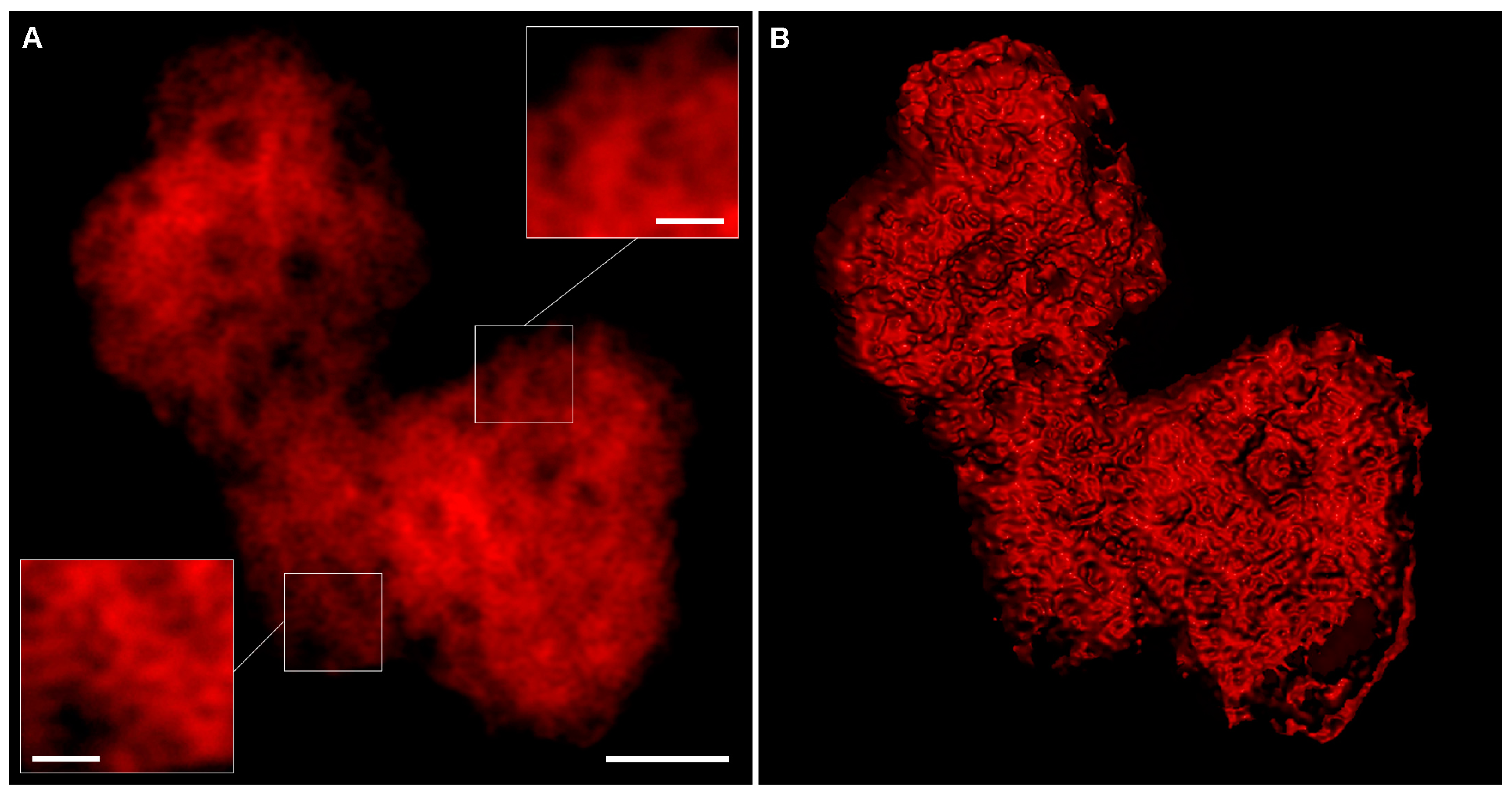
Abstract
:1. Introduction
2. Results
2.1. Visualization of PAMBs by AiryScan Confocal Microscopy
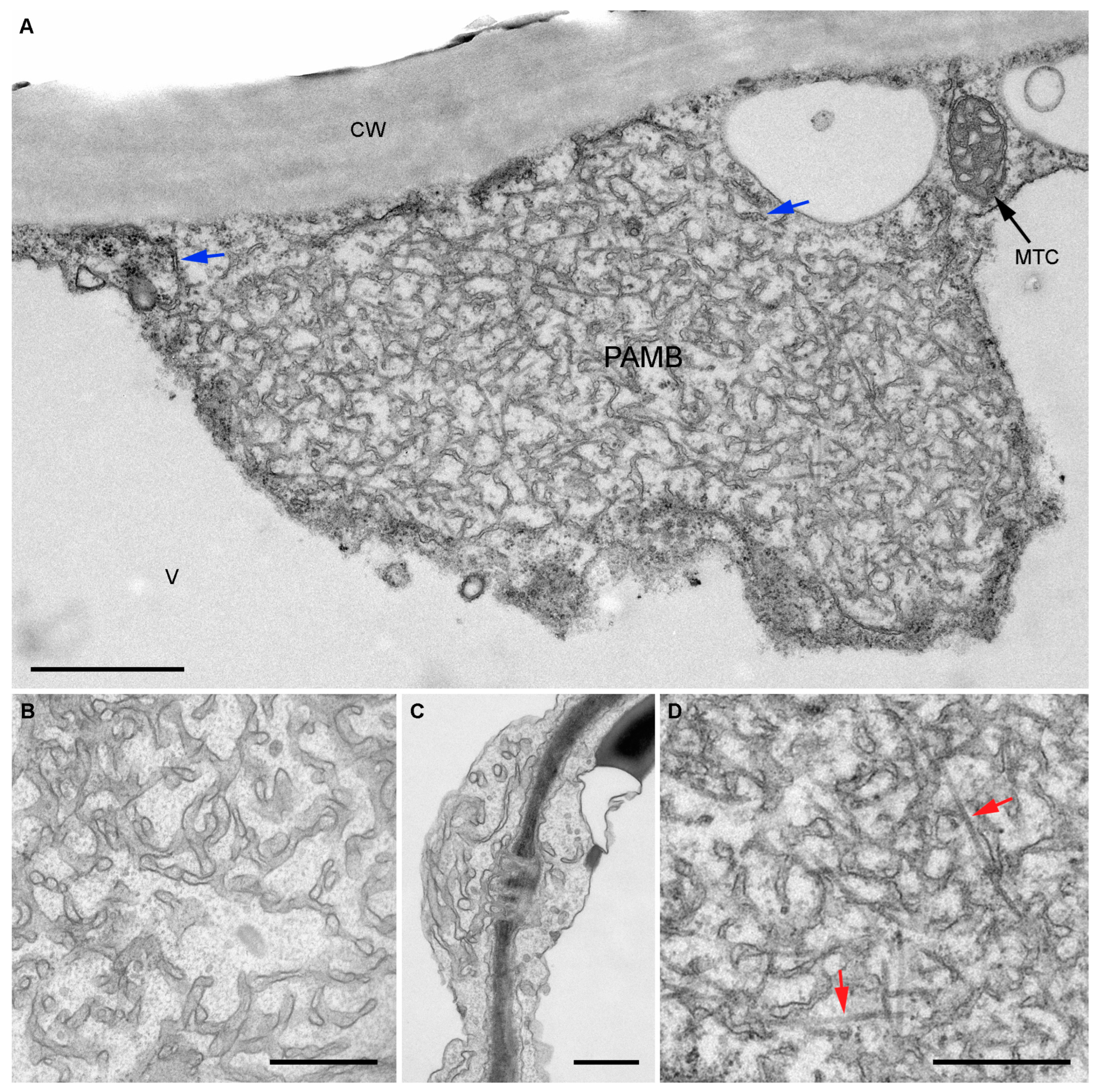
2.2. Analysis of the Structure of PAMBs by Transmission Electron Microscopy
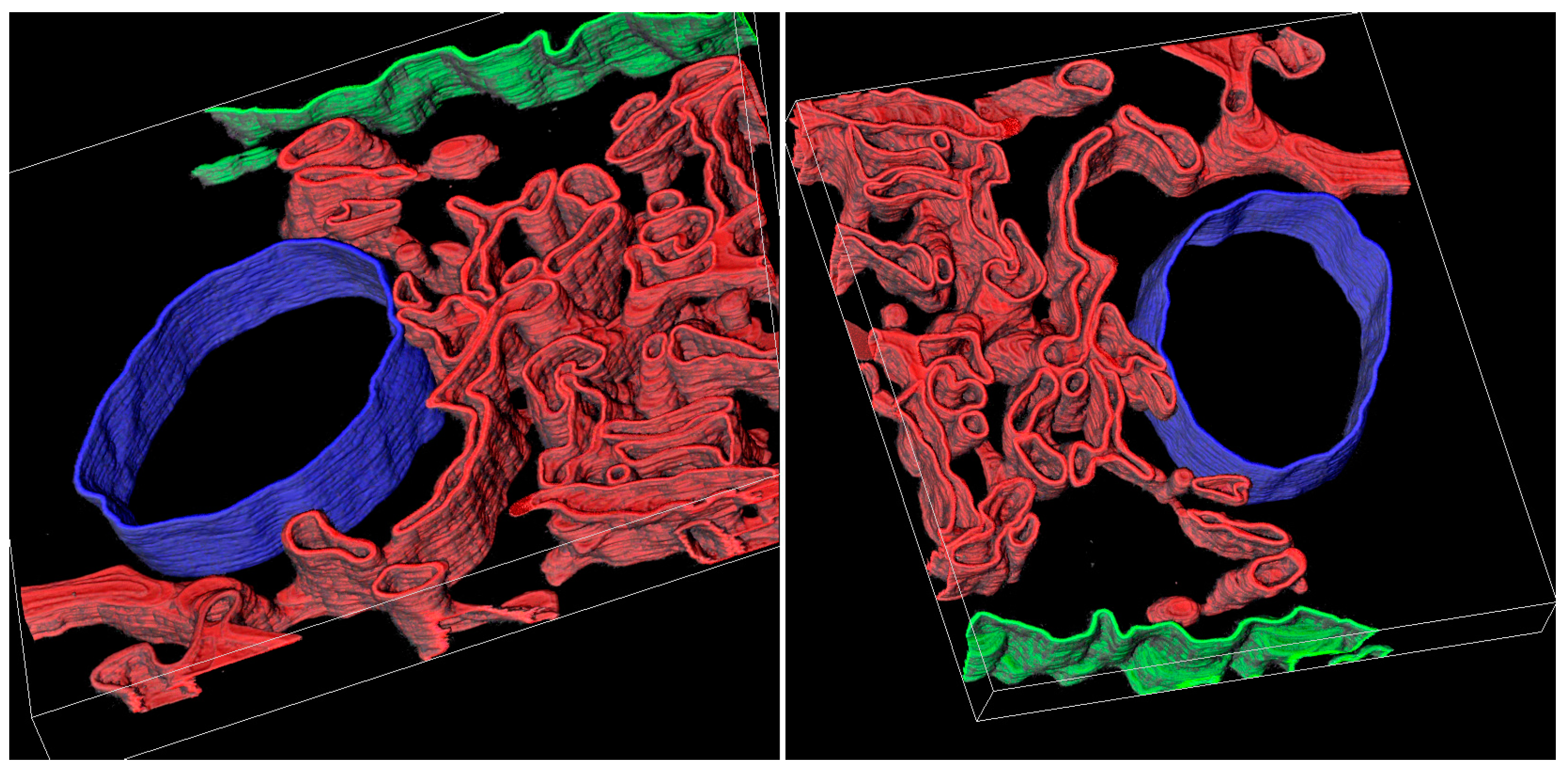
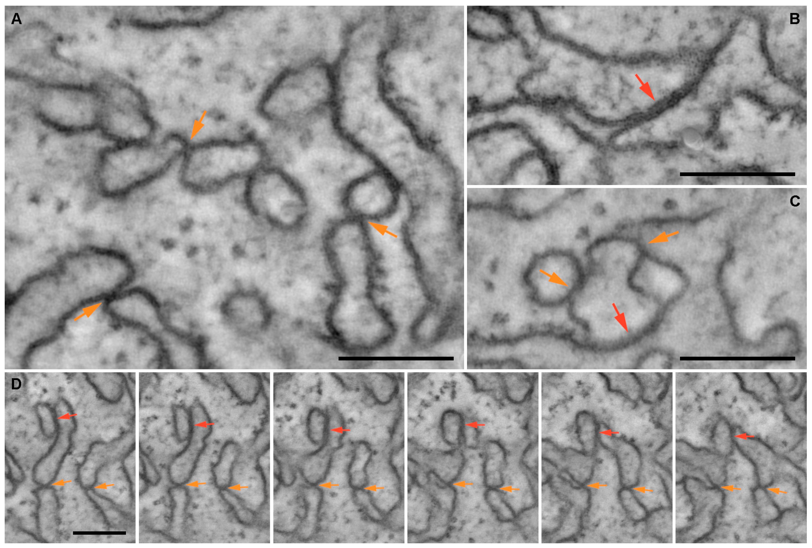
2.3. Analysis of the Structure of PAMBs by Electron Tomography
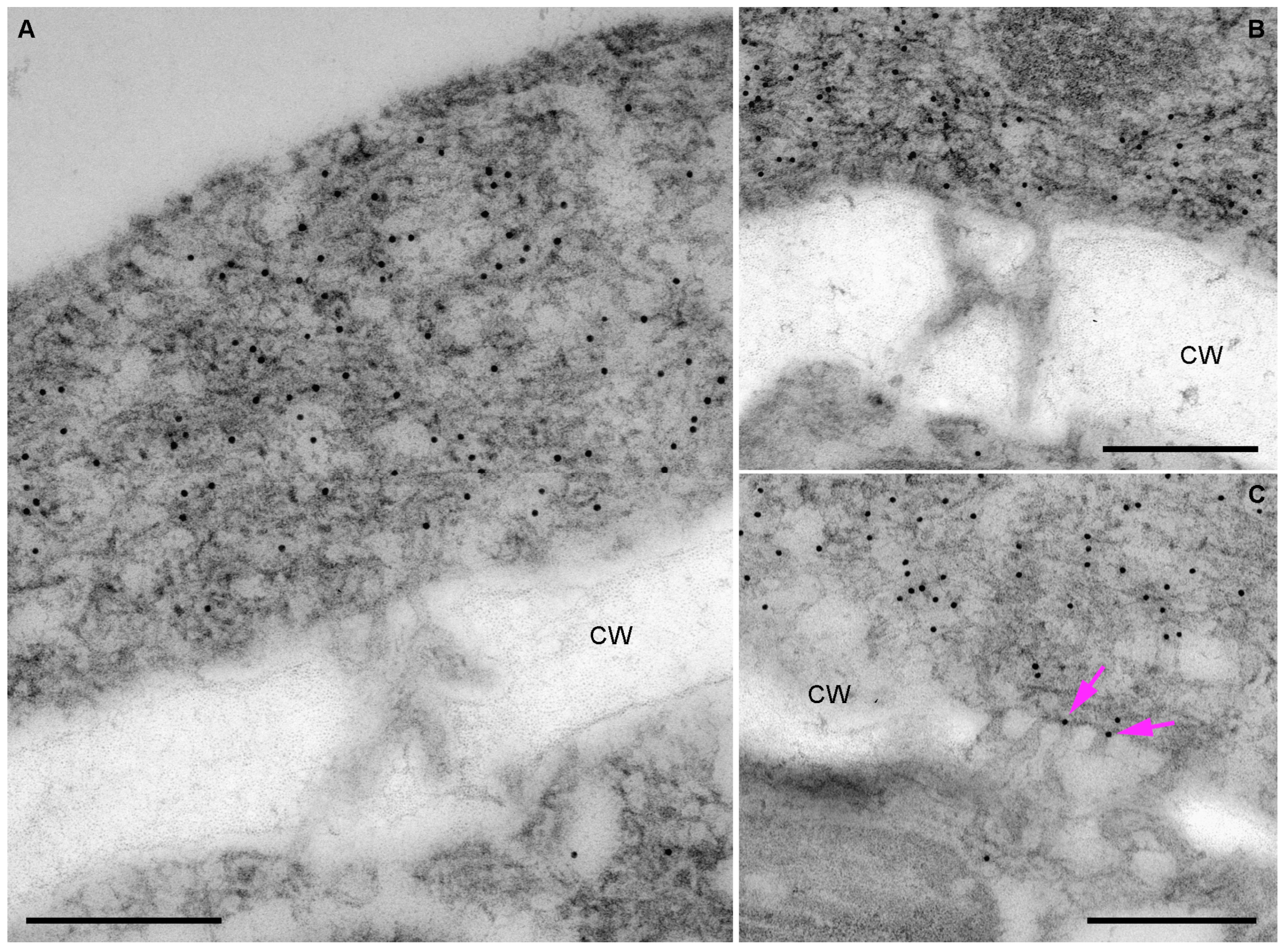
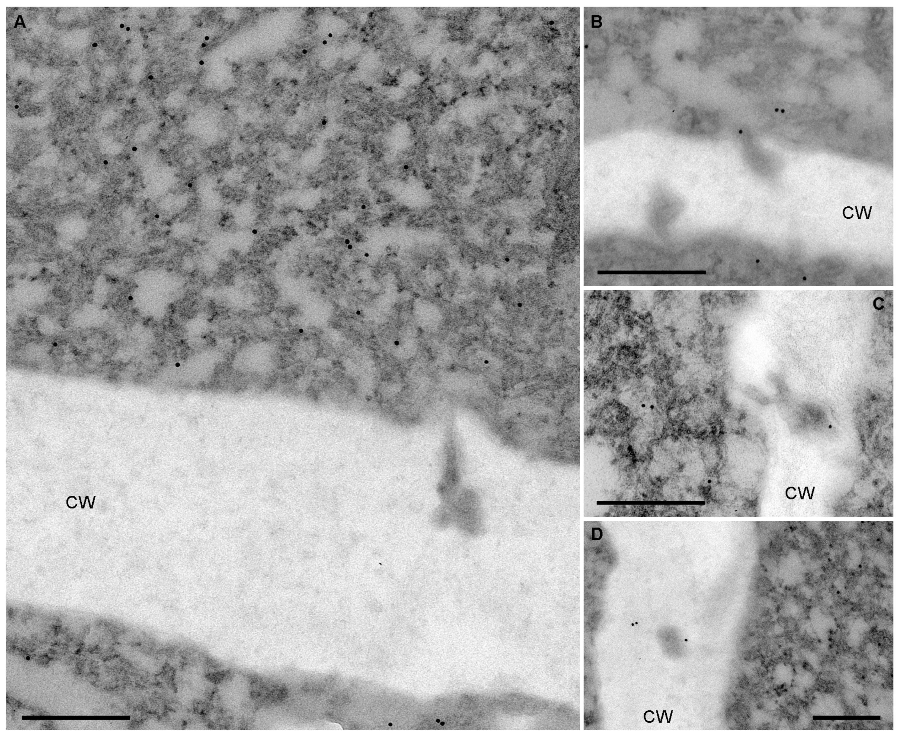
2.4. Analysis of BMB1 and BMB2 Localization in PAMBs and Plasmodesmata
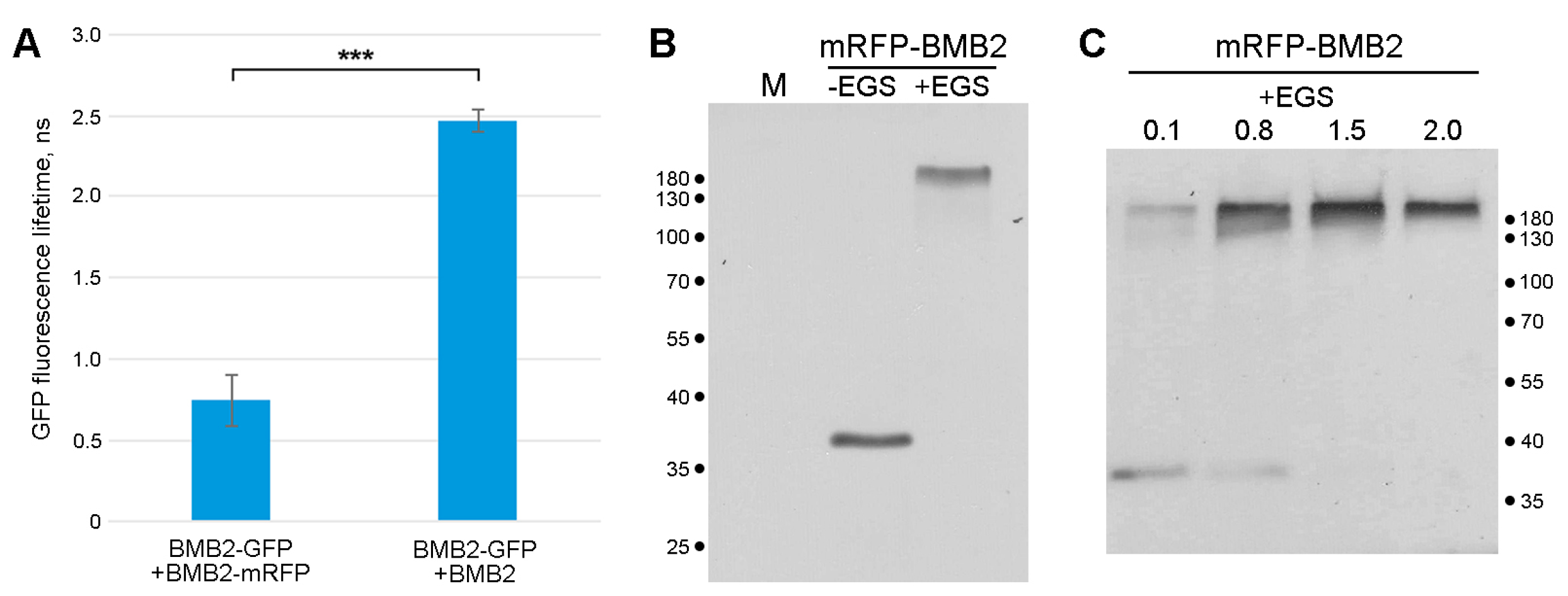
2.5. BMB2 Self-Interaction in PAMBs
3. Discussion
4. Materials and Methods
4.1. Agroinfiltration of Plants
4.2. AiryScan Confocal Microscopy
4.3. Sample Collection and Preparation for EM
4.4. EM Immunocytochemistry
4.5. Transmission Electron Microscopy and Tomography
4.6. EM Image Processing and Measurements
4.7. FRET-FLIM
4.8. Isolation of a Membrane Fraction and Chemical Cross-Linking Experiments
Supplementary Materials
Author Contributions
Funding
Data Availability Statement
Acknowledgments
Conflicts of Interest
References
- Lucas, W.J. Plant Viral Movement Proteins: Agents for Cell-to-Cell Trafficking of Viral Genomes. Virology 2006, 344, 169–184. [Google Scholar] [CrossRef]
- Heinlein, M. Plasmodesmata: Channels for Viruses on the Move. Methods Mol. Biol. 2015, 1217, 25–52. [Google Scholar] [CrossRef] [PubMed]
- Melcher, U. The “30K” Superfamily of Viral Movement Proteins. J. Gen. Virol. 2000, 81, 257–266. [Google Scholar] [CrossRef] [PubMed]
- Robles Luna, G.; Peña, E.J.; Borniego, M.B.; Heinlein, M.; García, M.L. Citrus Psorosis Virus Movement Protein Contains an Aspartic Protease Required for Autocleavage and the Formation of Tubule-Like Structures at Plasmodesmata. J. Virol. 2018, 92, e00355-18. [Google Scholar] [CrossRef] [PubMed]
- Kim, K.S.; Fulton, J.P. Tubules with Viruslike Particles in Leaf Cells Infected with Bean Pod Mottle Virus. Virology 1971, 43, 329–337. [Google Scholar] [CrossRef] [PubMed]
- Walkey, D.G.A.; Webb, M.J.W. Tubular Inclusion Bodies in Plants Infected with Viruses of the NEPO Type. J. Gen. Virol. 1970, 7, 159–166. [Google Scholar] [CrossRef]
- Kitajima, E.W.; Lauritis, J.A. Plant Virions in Plasmodesmata. Virology 1969, 37, 681–685. [Google Scholar] [CrossRef] [PubMed]
- Cheng, C.P.; Olszewski, N.E.; Lockhart, B.E.; Tzafrir, I. Tubules Containing Virions Are Present in Plant Tissues Infected with Commelina Yellow Mottle Badnavirus. J. Gen. Virol. 1998, 79, 925–929. [Google Scholar] [CrossRef]
- Kormelink, R.; Storms, M.; Van Lent, J.; Peters, D.; Goldbach, R. Expression and Subcellular Location of the NSM Protein of Tomato Spotted Wilt Virus (TSWV), a Putative Viral Movement Protein. Virology 1994, 200, 56–65. [Google Scholar] [CrossRef]
- van Lent, J.; Wellink, J.; Goldbach, R. Evidence for the Involvement of the 58K and 48K Proteins in the Intercellular Movement of Cowpea Mosaic Virus. J. Gen. Virol. 1990, 71, 219–223. [Google Scholar] [CrossRef]
- Heinlein, M. Plant Virus Replication and Movement. Virology 2015, 479–480, 657–671. [Google Scholar] [CrossRef]
- Taliansky, M.; Torrance, L.; Kalinina, N.O. Role of Plant Virus Movement Proteins. Methods Mol. Biol. 2008, 451, 33–54. [Google Scholar] [CrossRef]
- Brill, L.M.; Nunn, R.S.; Kahn, T.W.; Yeager, M.; Beachy, R.N. Recombinant Tobacco Mosaic Virus Movement Protein Is an RNA-Binding, α-Helical Membrane Protein. Proc. Natl. Acad. Sci. USA 2000, 97, 7112–7117. [Google Scholar] [CrossRef] [PubMed]
- Fujiki, M.; Kawakami, S.; Kim, R.W.; Beachy, R.N. Domains of Tobacco Mosaic Virus Movement Protein Essential for Its Membrane Association. J. Gen. Virol. 2006, 87, 2699–2707. [Google Scholar] [CrossRef]
- Reichel, C.; Beachy, R.N. Tobacco Mosaic Virus Infection Induces Severe Morphological Changes of the Endoplasmic Reticulum. Proc. Natl. Acad. Sci. USA 1998, 95, 11169–11174. [Google Scholar] [CrossRef] [PubMed]
- Heinlein, M.; Padgett, H.S.; Gens, J.S.; Pickard, B.G.; Casper, S.J.; Epel, B.L.; Beachy, R.N. Changing Patterns of Localization of the Tobacco Mosaic Virus Movement Protein and Replicase to the Endoplasmic Reticulum and Microtubules during Infection. Plant Cell 1998, 10, 1107–1120. [Google Scholar] [CrossRef]
- Epel, B.L. Plant Viruses Spread by Diffusion on ER-Associated Movement-Protein-Rafts through Plasmodesmata Gated by Viral Induced Host Beta-1,3-Glucanases. Semin. Cell Dev. Biol. 2009, 20, 1074–1081. [Google Scholar] [CrossRef] [PubMed]
- Uchiyama, A.; Shimada-Beltran, H.; Levy, A.; Zheng, J.Y.; Javia, P.A.; Lazarowitz, S.G. The Arabidopsis Synaptotagmin SYTA Regulates the Cell-to-Cell Movement of Diverse Plant Viruses. Front. Plant Sci. 2014, 5, 584. [Google Scholar] [CrossRef] [PubMed]
- Levy, A.; Zheng, J.Y.; Lazarowitz, S.G. Synaptotagmin SYTA Forms ER-Plasma Membrane Junctions That Are Recruited to Plasmodesmata for Plant Virus Movement. Curr. Biol. 2015, 25, 2018–2025. [Google Scholar] [CrossRef]
- Zavaliev, R.; Levy, A.; Gera, A.; Epel, B.L. Subcellular Dynamics and Role of Arabidopsis β-1,3-Glucanases in Cell-to-Cell Movement of Tobamoviruses. Mol. Plant. Microbe. Interact. 2013, 26, 1016–1030. [Google Scholar] [CrossRef]
- Ham, B.-K.; Wang, X.; Toscano-Morales, R.; Lin, J.; Lucas, W.J. Plasmodesmal Endoplasmic Reticulum Proteins Regulate Intercellular Trafficking of Cucumber Mosaic Virus in Arabidopsis. J. Exp. Bot. 2023, 74, 4401–4414. [Google Scholar] [CrossRef] [PubMed]
- Morozov, S.Y.; Solovyev, A.G. Triple Gene Block: Modular Design of a Multifunctional Machine for Plant Virus Movement. J. Gen. Virol. 2003, 84, 1351–1366. [Google Scholar] [CrossRef] [PubMed]
- Lazareva, E.A.; Lezzhov, A.A.; Komarova, T.V.; Morozov, S.Y.; Heinlein, M.; Solovyev, A.G. A Novel Block of Plant Virus Movement Genes. Mol. Plant Pathol. 2017, 18, 611–624. [Google Scholar] [CrossRef]
- Lazareva, E.A.; Lezzhov, A.A.; Chergintsev, D.A.; Golyshev, S.A.; Dolja, V.V.; Morozov, S.Y.; Heinlein, M.; Solovyev, A.G. Reticulon-like Properties of a Plant Virus-Encoded Movement Protein. New Phytol. 2021, 229, 1052–1066. [Google Scholar] [CrossRef]
- Schaad, M.C.; Jensen, P.E.; Carrington, J.C. Formation of Plant RNA Virus Replication Complexes on Membranes: Role of an Endoplasmic Reticulum-Targeted Viral Protein. EMBO J. 1997, 16, 4049–4059. [Google Scholar] [CrossRef] [PubMed]
- Grangeon, R.; Agbeci, M.; Chen, J.; Grondin, G.; Zheng, H.; Laliberté, J.-F. Impact on the Endoplasmic Reticulum and Golgi Apparatus of Turnip Mosaic Virus Infection. J. Virol. 2012, 86, 9255–9265. [Google Scholar] [CrossRef]
- Grangeon, R.; Jiang, J.; Laliberté, J.-F. Host Endomembrane Recruitment for Plant RNA Virus Replication. Curr. Opin. Virol. 2012, 2, 683–690. [Google Scholar] [CrossRef]
- Jiang, J.; Patarroyo, C.; Garcia Cabanillas, D.; Zheng, H.; Laliberté, J.-F. The Vesicle-Forming 6K2 Protein of Turnip Mosaic Virus Interacts with the COPII Coatomer Sec24a for Viral Systemic Infection. J. Virol. 2015, 89, 6695–6710. [Google Scholar] [CrossRef]
- Kaido, M.; Abe, K.; Mine, A.; Hyodo, K.; Taniguchi, T.; Taniguchi, H.; Mise, K.; Okuno, T. GAPDH--a Recruits a Plant Virus Movement Protein to Cortical Virus Replication Complexes to Facilitate Viral Cell-to-Cell Movement. PLoS Pathog. 2014, 10, e1004505. [Google Scholar] [CrossRef]
- Kaido, M.; Tsuno, Y.; Mise, K.; Okuno, T. Endoplasmic Reticulum Targeting of the Red Clover Necrotic Mosaic Virus Movement Protein Is Associated with the Replication of Viral RNA1 but Not That of RNA2. Virology 2009, 395, 232–242. [Google Scholar] [CrossRef]
- Kaido, M.; Funatsu, N.; Tsuno, Y.; Mise, K.; Okuno, T. Viral Cell-to-Cell Movement Requires Formation of Cortical Punctate Structures Containing Red Clover Necrotic Mosaic Virus Movement Protein. Virology 2011, 413, 205–215. [Google Scholar] [CrossRef] [PubMed]
- Tilsner, J.; Linnik, O.; Louveaux, M.; Roberts, I.M.; Chapman, S.N.; Oparka, K.J. Replication and Trafficking of a Plant Virus Are Coupled at the Entrances of Plasmodesmata. J. Cell Biol. 2013, 201, 981–995. [Google Scholar] [CrossRef] [PubMed]
- Linnik, O.; Liesche, J.; Tilsner, J.; Oparka, K.J. Unraveling the Structure of Viral Replication Complexes at Super-Resolution. Front. Plant Sci. 2013, 4, 6. [Google Scholar] [CrossRef] [PubMed]
- Tilsner, J.; Linnik, O.; Wright, K.M.; Bell, K.; Roberts, A.G.; Lacomme, C.; Santa Cruz, S.; Oparka, K.J. The TGB1 Movement Protein of Potato Virus X Reorganizes Actin and Endomembranes into the X-Body, a Viral Replication Factory. Plant Physiol. 2012, 158, 1359–1370. [Google Scholar] [CrossRef] [PubMed]
- Wu, X.; Liu, J.; Chai, M.; Wang, J.; Li, D.; Wang, A.; Cheng, X. The Potato Virus X TGBp2 Protein Plays Dual Functional Roles in Viral Replication and Movement. J. Virol. 2019, 93, e01635-18. [Google Scholar] [CrossRef] [PubMed]
- Solovyev, A.G.; Atabekova, A.K.; Lezzhov, A.A.; Solovieva, A.D.; Chergintsev, D.A.; Morozov, S.Y. Distinct Mechanisms of Endomembrane Reorganization Determine Dissimilar Transport Pathways in Plant RNA Viruses. Plants 2022, 11, 2403. [Google Scholar] [CrossRef]
- Melzer, M.J.; Sether, D.M.; Borth, W.B.; Hu, J.S. Characterization of a Virus Infecting Citrus Volkameriana with Citrus Leprosis-like Symptoms. Phytopathology 2012, 102, 122–127. [Google Scholar] [CrossRef]
- Lazareva, E.A.; Lezzhov, A.A.; Golyshev, S.A.; Morozov, S.Y.; Heinlein, M.; Solovyev, A.G. Similarities in Intracellular Transport of Plant Viral Movement Proteins BMB2 and TGB3. J. Gen. Virol. 2017, 98, 2379–2391. [Google Scholar] [CrossRef]
- Atabekova, A.K.; Lazareva, E.A.; Lezzhov, A.A.; Solovieva, A.D.; Golyshev, S.A.; Skulachev, B.I.; Solovyev, I.D.; Savitsky, A.P.; Heinlein, M.; Morozov, S.Y.; et al. Interaction between Movement Proteins of Hibiscus Green Spot Virus. Viruses 2022, 14, 2742. [Google Scholar] [CrossRef]
- Voeltz, G.K.; Prinz, W.A.; Shibata, Y.; Rist, J.M.; Rapoport, T.A. A Class of Membrane Proteins Shaping the Tubular Endoplasmic Reticulum. Cell 2006, 124, 573–586. [Google Scholar] [CrossRef]
- Tolley, N.; Sparkes, I.; Craddock, C.P.; Eastmond, P.J.; Runions, J.; Hawes, C.; Frigerio, L. Transmembrane Domain Length Is Responsible for the Ability of a Plant Reticulon to Shape Endoplasmic Reticulum Tubules in Vivo. Plant J. 2010, 64, 411–418. [Google Scholar] [CrossRef]
- Sparkes, I.; Tolley, N.; Aller, I.; Svozil, J.; Osterrieder, A.; Botchway, S.; Mueller, C.; Frigerio, L.; Hawes, C. Five Arabidopsis Reticulon Isoforms Share Endoplasmic Reticulum Location, Topology, and Membrane-Shaping Properties. Plant Cell 2010, 22, 1333–1343. [Google Scholar] [CrossRef]
- Shibata, Y.; Voss, C.; Rist, J.M.; Hu, J.; Rapoport, T.A.; Prinz, W.A.; Voeltz, G.K. The Reticulon and DP1/Yop1p Proteins Form Immobile Oligomers in the Tubular Endoplasmic Reticulum. J. Biol. Chem. 2008, 283, 18892–18904. [Google Scholar] [CrossRef] [PubMed]
- Pain, C.; Kriechbaumer, V.; Kittelmann, M.; Hawes, C.; Fricker, M. Quantitative Analysis of Plant ER Architecture and Dynamics. Nat. Commun. 2019, 10, 984. [Google Scholar] [CrossRef] [PubMed]
- Schoberer, J.; Botchway, S.W. Investigating Protein–Protein Interactions in the Plant Endomembrane System Using Multiphoton-Induced FRET-FLIM. Methods Mol. Biol. 2014, 1209, 81–95. [Google Scholar] [CrossRef] [PubMed]
- Lazareva, E.A.; Lezzhov, A.A.; Dolja, V.V.; Morozov, S.Y.; Heinlein, M.; Solovyev, A.G. Constriction of Endoplasmic Reticulum Tubules by the Viral Movement Protein BMB2 Is Associated with Local BMB2 Anchorage at Constriction Sites. Plant Signal. Behav. 2021, 16, 1856547. [Google Scholar] [CrossRef] [PubMed]
- Pérez-Sancho, J.; Tilsner, J.; Samuels, A.L.; Botella, M.A.; Bayer, E.M.; Rosado, A. Stitching Organelles: Organization and Function of Specialized Membrane Contact Sites in Plants. Trends Cell Biol. 2016, 26, 705–717. [Google Scholar] [CrossRef] [PubMed]
- Belov, G.A.; Sztul, E. Rewiring of Cellular Membrane Homeostasis by Picornaviruses. J. Virol. 2014, 88, 9478–9489. [Google Scholar] [CrossRef]
- He, W.; He, Y. Electron Tomography for Organelles, Cells, and Tissues. Methods Mol. Biol. 2014, 1117, 445–483. [Google Scholar] [CrossRef]
- Mastronarde, D.N. Automated Electron Microscope Tomography Using Robust Prediction of Specimen Movements. J. Struct. Biol. 2005, 152, 36–51. [Google Scholar] [CrossRef]
- Mastronarde, D.N. Dual-Axis Tomography: An Approach with Alignment Methods That Preserve Resolution. J. Struct. Biol. 1997, 120, 343–352. [Google Scholar] [CrossRef] [PubMed]
- Abas, L.; Luschnig, C. Maximum Yields of Microsomal-Type Membranes from Small Amounts of Plant Material without Requiring Ultracentrifugation. Anal. Biochem. 2010, 401, 217–227. [Google Scholar] [CrossRef] [PubMed]


Disclaimer/Publisher’s Note: The statements, opinions and data contained in all publications are solely those of the individual author(s) and contributor(s) and not of MDPI and/or the editor(s). MDPI and/or the editor(s) disclaim responsibility for any injury to people or property resulting from any ideas, methods, instructions or products referred to in the content. |
© 2023 by the authors. Licensee MDPI, Basel, Switzerland. This article is an open access article distributed under the terms and conditions of the Creative Commons Attribution (CC BY) license (https://creativecommons.org/licenses/by/4.0/).
Share and Cite
Atabekova, A.K.; Golyshev, S.A.; Lezzhov, A.A.; Skulachev, B.I.; Moiseenko, A.V.; Yastrebova, D.M.; Andrianova, N.V.; Solovyev, I.D.; Savitsky, A.P.; Morozov, S.Y.; et al. Fine Structure of Plasmodesmata-Associated Membrane Bodies Formed by Viral Movement Protein. Plants 2023, 12, 4100. https://doi.org/10.3390/plants12244100
Atabekova AK, Golyshev SA, Lezzhov AA, Skulachev BI, Moiseenko AV, Yastrebova DM, Andrianova NV, Solovyev ID, Savitsky AP, Morozov SY, et al. Fine Structure of Plasmodesmata-Associated Membrane Bodies Formed by Viral Movement Protein. Plants. 2023; 12(24):4100. https://doi.org/10.3390/plants12244100
Chicago/Turabian StyleAtabekova, Anastasia K., Sergei A. Golyshev, Alexander A. Lezzhov, Boris I. Skulachev, Andrey V. Moiseenko, Daria M. Yastrebova, Nadezda V. Andrianova, Ilya D. Solovyev, Alexander P. Savitsky, Sergey Y. Morozov, and et al. 2023. "Fine Structure of Plasmodesmata-Associated Membrane Bodies Formed by Viral Movement Protein" Plants 12, no. 24: 4100. https://doi.org/10.3390/plants12244100