Article

Unveiling the Chemical Composition and Biological Properties of Salvia cacaliifolia Benth. Essential Oil

1
Institute for Clinical and Biomedical Research, Health Sciences Campus, University of Coimbra, Azinhaga de S. Comba, 3000-548 Coimbra, Portugal
2
Faculty of Pharmacy, Health Sciences Campus, University of Coimbra, Azinhaga de S. Comba, 3000-548 Coimbra, Portugal
3
Laboratory of Plant Biology and Pharmaceutical Botany, Department of Life and Environmental Sciences, University of Cagliari, Viale Sant’Ignazio, 09123 Cagliari, Italy
4
Department of Chemical and Geological Sciences, University of Cagliari, Cittadella Universitaria, 09042 Monserrato, Italy
5
Chemical Process Engineering and Forest Products Research Centre, Department of Chemical Engineering, Faculty of Sciences and Technology, University of Coimbra, 3030-790 Coimbra, Portugal
6
Center for Neuroscience and Cell Biology, Faculty of Medicine, University of Coimbra, Rua Larga, 3004-504 Coimbra, Portugal
*
Author to whom correspondence should be addressed.
Plants 2023, 12(2), 359; https://doi.org/10.3390/plants12020359
Submission received: 28 November 2022 / Revised: 6 January 2023 / Accepted: 9 January 2023 / Published: 12 January 2023
(This article belongs to the Special Issue Phytochemistry of Aromatic and Medicinal Plants)

Abstract

:
Salvia is widely recognized for its therapeutic potential. However, the biological relevance of some species remains unknown, namely Salvia cacaliifolia Benth. Therefore, the aim of this study is to unveil the chemical composition and relevant properties to its essential oil (EO). The EO was characterized by GC and GC-MS and its antifungal effect was evaluated according to the CLSI guidelines on dermatophytes and yeasts. The anti-inflammatory potential was assessed on lipopolysaccharide-stimulated macrophages, by assessing the production of nitric oxide (NO) and the effect on the protein levels of two key pro-inflammatory enzymes, iNOS and COX-2 by western blot analysis. Wound healing capacity was determined using the scratch wound healing assay, and the anti-aging potential was assessed by evaluating the senescence marker β-galactosidase. The EO was mainly characterized by γ-curcumene, β-bisabolene, bicyclogermacrene and curzerenone. It is effective in inhibiting the growth of dermatophytes and C. neoformans. The EO significantly decreased iNOS and COX-2 protein levels and concomitantly reduced NO release. Additionally, it demonstrated anti-senescence potential and promoted wound healing. Overall, this study highlights relevant pharmacological properties of the EO of Salvia cacaliifolia, which should be further explored envisaging the development of sustainable, innovative, and environmentally friendly skin products.

1. Introduction

Fungal infections remain neglected by public health authorities, affecting over a billion people with 1.5 million deaths per year [1]. Immunocompromised individuals are more prone to fungal infections [1], being Candida and Cryptococcus two main etiological agents for invasive infections [1,2]. Dermatophytes, another group of pathogenic fungi that are highly relevant due to their role in the morbidity-associated with superficial mycoses [3], includes Trichophyton, Epidermophyton and Microsporum strains. Furthermore, they can cause invasive diseases in immunocompromised hosts [4].
Dermatophytes can activate the immune system due to the binding of fungal epitopes, such as mannan [5,6], to toll-like receptors (TLRs) thus activating the pro-inflammatory cascade associated with these infections. One of such cascades is the nuclear factor kappa B (NF-κB) pathway, which ultimately leads to an increase in the production of inflammatory cytokines and the protein levels of key pro-inflammatory enzymes, such as inducible nitric oxide synthase (iNOS) and cycloxygenase-2 (COX-2) [7]. The enzyme iNOS is responsible for the production of nitric oxide (NO), which is a well-known pro-inflammatory mediator overproduced in several chronic inflammatory-related diseases.
Furthermore, dermatophytes are associated with the formation of skin lesions [8]. Therefore, after the resolution of an infection, wound healing processes must occur in order to regenerate the damaged tissue. This process encompasses different phases, that must occur in the right order and at the correct times, otherwise the wound becomes chronic [9].
Due to relapses, resistances and antifungal side-effects, current antifungal treatments are many times inefficient with fungal infections remaining an unmet clinical need [10]. Strikingly, an increasing number of reports demonstrate that several fungal strains have already developed resistance to current antifungal agents [11,12], justifying the development of new antifungal approaches [13]. Furthermore, anti-inflammatory drugs are also reported to cause several adverse effects [14]. Having this in mind, therapeutic agents that could concomitantly treat fungal infections and decrease the associated inflammation would be of uttermost importance.
Aromatic plants and their metabolites have interesting organoleptic characteristics that also contribute to their high value in pharmaceutical, agronomic, food, sanitary, cosmetic, and perfume industries [15], and emerge as potential candidates as new anti-inflammatory/antifungal drugs.
Many aromatic plants from the Asteraceae, Apiaceae and Lamiaceae families are rich in essential oils that have demonstrated a wide array of biological effects, namely anti-inflammatory and antifungal properties [16,17,18,19,20,21,22]. The genus Salvia L., one of the most important genera of the Lamiaceae family, is cultivated to be used as food spices or flavoring agents in cosmetics and perfumery [23]. Several species are used in traditional medicine to treat microbial infections, malaria, inflammation, and to disinfect homes after sickness [23]. Salvia species have attracted researchers for their biological properties, showing strong antibacterial, antifungal, anticancer, and anti-inflammatory effects, as well as for improvement of cognitive performance [24,25,26,27,28,29,30]. Although this genus is largely studied, some species remain unknown regarding their chemical composition and their medicinal interest, such as Salvia cacaliifolia Benth. (syn Salvia atriplicifolia Fernald, Salvia hempsteadiana S.F.Blake and Salvia mendax Epling). S. cacaliifolia is an erect herbaceous perennial shrub reaching 70 cm in height, with triangular leaves and large racemes of deep blue, 2-lipped flowers 2 cm in length [31]. This species grows in high altitude regions from Mexico, Guatemala, and Honduras.
This works aims to chemically characterize the essential oil of S. cacaliifolia (cacalia sage) and to disclose its antifungal/anti-inflammatory potential. Furthermore, the wound healing capacity of the essential oil was also studied. To further promote the interest in this species, the anti-senescence potential was also examined.

2. Results

2.1. Chemical Composition of S. cacaliifolia Essential Oil

The essential oil was characterized by high amounts of hydrocarbons sesquiterpenes (70.10% ± 0.119) followed by oxygenated sesquiterpenes (11.71% ± 0.032), as shown in Table 1. Regarding individual compounds, the essential oil was rich in γ-curcumene (27.64% ± 0.195), β-bisabolene (18.01% ± 0.033), bicyclogermacrene (8.54% ± 0.074) and curzerenone (7.68% ± 0.055).

2.2. Antifungal Effect of Salvia cacaliifolia

The antifungal activity of the essential oil from S. cacaliifolia, particularly against dermatophytes and yeasts, was assessed. As shown in Table 2, the essential oil was effective against all tested dermatophyte strains, with T. mentagrophytes and T. rubrum being the most susceptible (MIC = 0.16 µL/mL). Regarding yeasts, C. neoformans was the most susceptible to essential oil (MIC = 0.64 µL/mL).

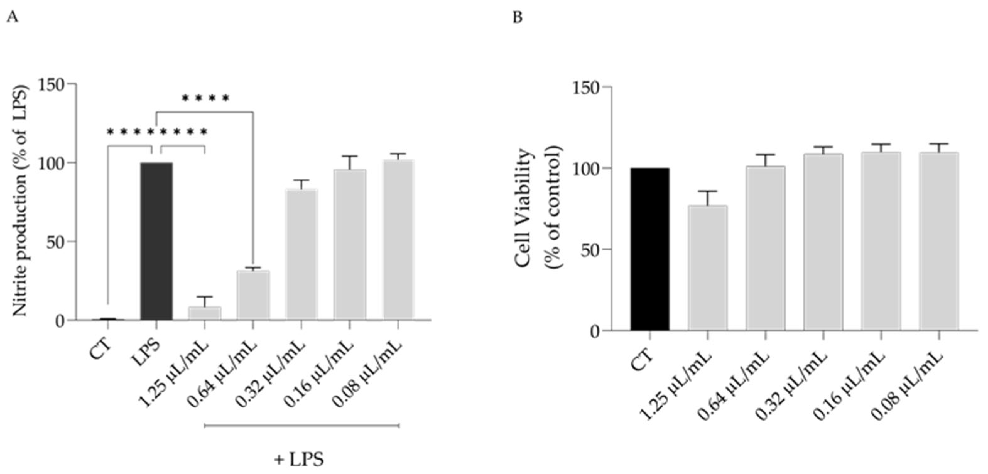
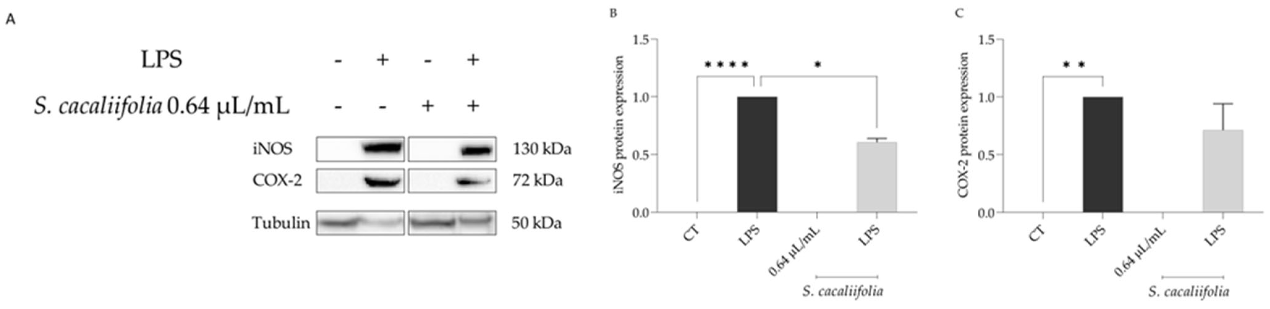
2.3. Anti-Inflammatory Potential of S. cacaliifolia

The anti-inflammatory potential of S. cacaliifolia essential oil was assessed using the lipopolysaccharide (LPS)-stimulated macrophages. As expected, the addition of LPS for 24 h lead to an increase in the secretion of nitric oxide (NO) to the culture medium ([NO] = 42.8 ± 7.4 µM). The addition of different concentrations of the essential oil led to a dose-dependent reduction in the nitrites detected (IC50 = 0.49 µL/mL). Both 1.25 and 0.64 µL/mL significantly decreased the detected nitrites (Figure 1A). Considering that at 1.25 µL/mL a significant decrease cell viability was observed (Figure 1B), the concentration of 0.64 µL/mL was used to disclose the underlying mechanism behind the anti-inflammatory potential of S. cacaliifolia essential oil.
Considering that the binding of LPS to the Toll-like receptor 4 (TLR4) leads to the nuclear translocation of NF-κB which in turn causes the expression of inducible nitric oxide synthase (Nos2) and cycloxygenease-2 (Cox2) [34], we then assessed whether the presence of the essential oil could modulate the levels of both proteins. As shown in Figure 2A–C, the presence of LPS alone caused a significant increase in the protein levels of iNOS and COX-2. In the presence of the essential oil, both proteins’ levels were decreased, particularly that of iNOS (Figure 2A,B), suggesting that the oil impaired NF-κB activation, and consequently the expression of Nos2 mRNA as well as its protein levels.

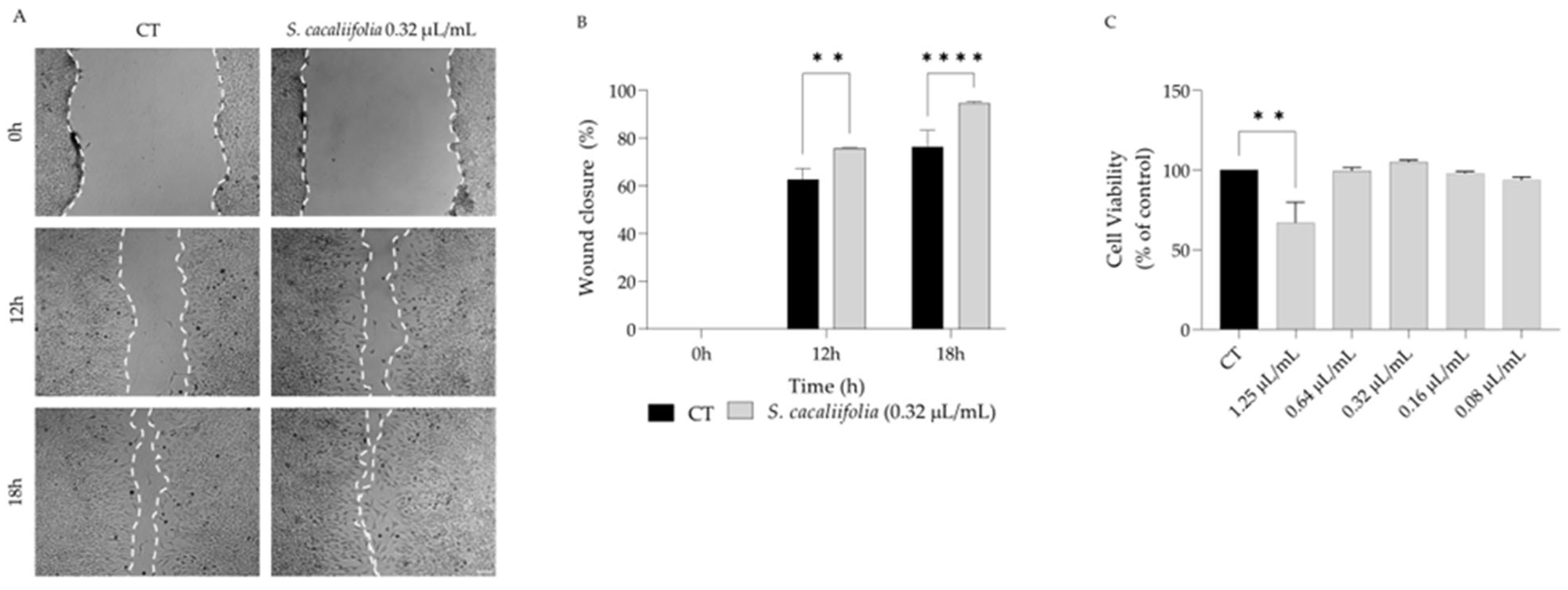
2.4. Wound Healing Properties of S. cacaliifolia Essential Oil

Considering that several plants from the genus Salvia have been reported as wound healing agents [27,35,36], we hypothesise that S. cacaliifolia could promote wound healing. For that we resorted to the scratch assay and wound closure was assessed in the presence and absence of the essential oil in fibroblasts. We report that the essential oil significantly promoted the migration of fibroblasts when compared to control cells (Figure 3A,B) at 0.32 µL/mL, a concentration devoid of toxicity towards the fibroblasts (Figure 3C).

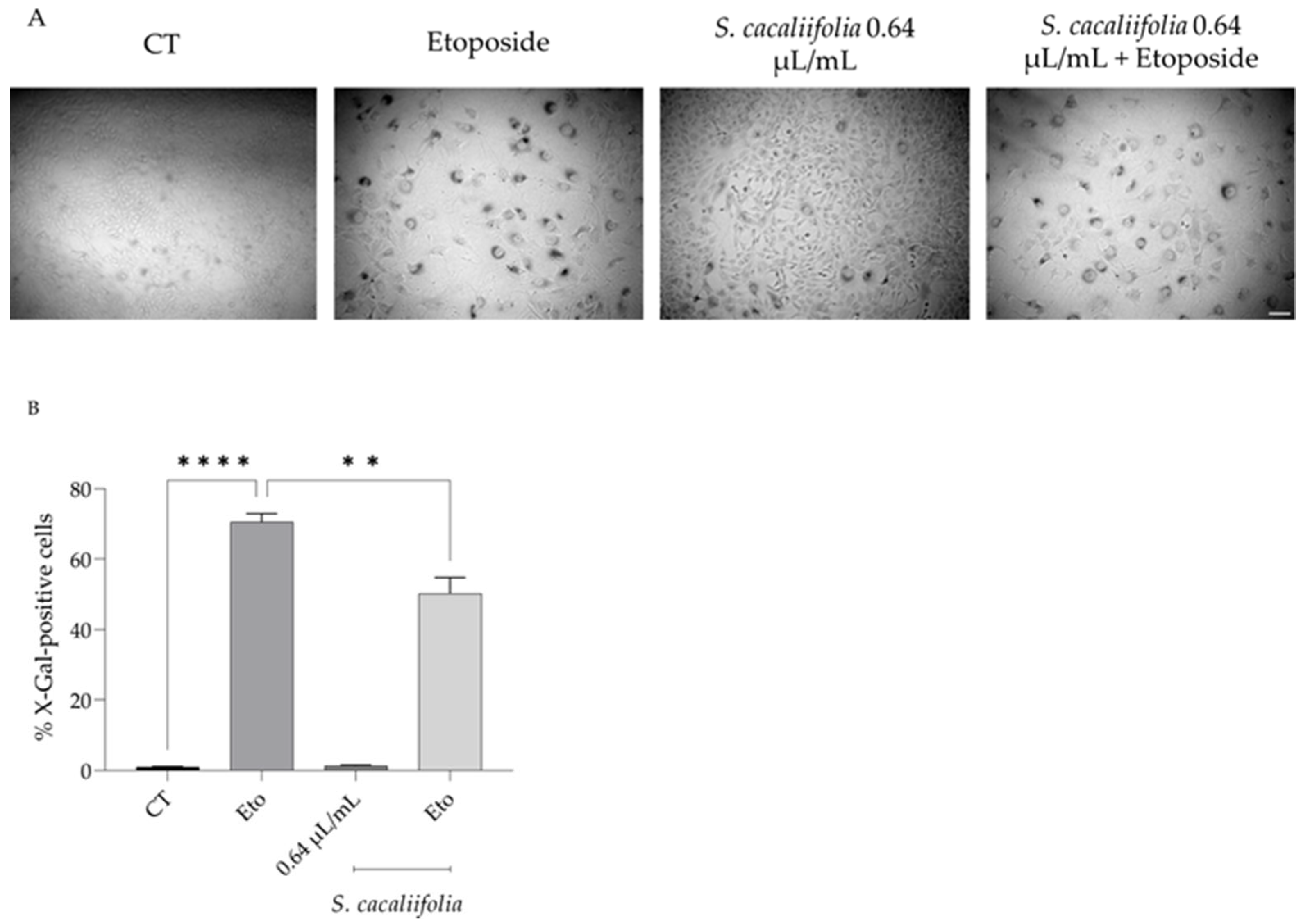
2.5. Anti-Senescence Potential of S. cacaliifolia Essential Oil

Since it is known that some Salvia species present anti-senescence properties [37,38], we assessed the anti-senescence potential of S. cacaliifolia essential oil using the Senescence-associated β-galactosidase (β-gal) assay with etoposide as senescence-inducing agent.
As shown in Figure 4, the addition of etoposide (12.5 µM) for 24 h followed by a recovery for 72 h in culture medium alone led to a significant increase in the β-gal-positive cells, showing that etoposide effectively induces cell senescence. Interestingly, when in the recovery phase S. cacaliifolia essential oil was added to the culture medium the percentage of β-gal-positive cells significantly decreased when compared to culture medium alone (Figure 4A,B), thus suggesting that S. cacaliifolia could also present anti-senescence effects.

3. Discussion

S. cacaliifolia is still largely unknown regarding the chemical composition of its essential oil as well as to any biologically relevant activities. To fill the gap in the knowledge, this work describes, for the first time, anti-inflammatory, anti-fungal, anti-senescence and wound healing properties for this species.
We reported that S. cacaliifolia essential oil is predominantly characterized by sesquiterpenes, particularly γ-curcumene, β-bisabolene, bicyclogermacrene and curzerenone. Similarly, the essential oils from S. scabra and S. aurea were also rich in sesquiterpenes (92.4 and 75%, respectively) [39], with β-caryophyllene, epi-α-cadinol, δ-cadinene being the major compounds in S. aurea while S. scabra was characterized by germacrene D, β-caryophyllene, germacrene B, and α-copaene. S. ceratophylla, S. verbenaca, S. stenophylla, S. runcinata and S. repens are other examples of Salvia essential oils predominantly rich in sesquiterpenes [40,41,42]. However, no essential oils from Salvia presents high amounts of γ-curcumene, β-bisabolene and curzerenone as the essential oil from S. cacaliifolia. Other Salvia species present a very distinct chemical composition, with predominance of monoterpenes, such as S. officinalis with high amounts of cis-thujone and camphor [43,44], or 1,8-cineole and camphor [45] and S. rosmarinus rich in 1,8-cineole, α-pinene, camphor [46].
Considering that some Salvia species have been reported as agents able to treat skin infections [36,47], we then aimed to assess if S. cacaliifolia oil could have potential in controlling fungal infection. The essential oil showed inhibitory effects against all dermatophyte strains and Cryptococcus neoformans, thus suggesting that it could be used to treat fungal infections, particularly dermatophytosis and cryptococcosis. The essential oil from other species such as S. officinalis have inhibitory effect against dermatophytes with MIC ranging from 0.63 to 2.5 µL/mL [43,45], thus showing a weaker activity than the one reported for S. cacaliifolia. Two essential oils from S. multicaulis presented a relevant anti-dermatophytic activity, particularly the chemotype rich in nerolidol [48]. Regarding the activity of the main compounds of S. cacaliifolia, no studies have been carried out. However, some studies have assessed the antifungal activity of essential oils rich in these compounds. Indeed, a study reported the activity of several essential oils from Helichrysum microphyllum subsp. tyrrhenicum and showed that those with highest content in γ-curcumene presented the best antifungal activity [49]. Other studies reported moderate antifungal activity for essential oils rich in β-bisabolene [50,51], bicyclogermacrene [51,52] and curzerenone [53]. Considering the weak antifungal of the previously reported essential oils, we hypothesize that the activity of S. cacaliifolia might be attributed to synergistic effect between all the compounds in the mixture. The mechanisms of action underlying the antifungal activity of S. cacaliifolia essential oil and isolated compounds are still unknown. However, considering their lipophilic nature we hypothesize that they might induce fungal growth inhibition by integrating into membrane structures which would increase permeability, promote the leakage of intracellular components and enzyme inactivation [15,54].
Considering that fungal infections lead to the activation of the immune system [55], the anti-inflammatory potential of S. cacaliifolia was also highlighted in the present study since a decrease in NO release was achieved when macrophages were pre-treated with the essential oil prior to LPS stimulation. In order to disclose the underlying mechanisms of the anti-inflammatory potential of S. cacaliifolia, the effect of the essential oil on the protein levels of iNOS and COX-2, two pro-inflammatory proteins associated with the TLR4/NF-κB signaling pathway, was assessed. A decrease in both protein levels was observed, thus suggesting that the decrease in NO release might be due to an inhibition of the NF-κB pathway. Indeed, one of the key molecular mechanisms that contributes to the perpetuation of chronic inflammation is the activation of NF-κB signaling cascade, which has emerged as the master regulator of inflammation and innate immune homeostasis. Activation of the transcriptional factor NF-kB causes induction of COX-2, iNOS and also aberrant expression of the inflammatory cytokines tumor necrosis factor (TNF)-α, IL-1β, IL-6, which can damage healthy neighboring cells and over a long period of time may lead to chronic illnesses. Further studies need to be conducted in order to further understand the full mechanism behind the anti-inflammatory potential of S. cacaliifolia essential oil, such as the nuclear translocation of p65. Regarding other Salvia species, it was shown that S. officinalis essential oil at 0.64 µL/mL was able to decrease NO production by 30–40% [45], whereas the essential oil from S. cacaliifolia at the same dose decreases NO production by 70%. In opposition, the essential oil from S. ceratophylla greatly inhibits NO production (IC50 = 90 µg/mL) [40]. The capacity of Salvia species to modulate NF-kB pathway is already reported. Indeed, S. officinalis was able to decrease the expression of NF-kB [44]. S. ceratophylla strongly inhibits NF-κB with an IC50 of 38 µg/mL [40]. It was shown that Helichrysum italicum essential oil rich in γ-curcumene, the main compound of S. cacaliifolia oil, was able to decrease NO production, which was dependent on the content in this compound, which was corroborated by fractioning of the essential oil where the curcumene-rich fraction induced the highest inhibition [56]. Similar effects were described for the γ-curcumene chemotype of H. italicum [57]. It was shown that essential oils with significant amounts of β-bisabolene [58] and bicyclogermacrene [59] exerted promising anti-inflammatory effects. Regarding curzerenone, no studies have been conducted assessing its anti-inflammatory potential. However, it was reported that two essential oils rich in curzerenone have relevant antioxidant properties [60,61], which could also contribute to the anti-inflammatory activity of S. cacaliifolia by allowing NO scavenging. Having this in mind, it is possible that γ-curcumene, the main compound of the essential oil, might be the major player responsible for S. cacaliifolia anti-inflammatory activity, however the contribution of the remaining major compounds cannot be discarded.
Considering that dermatophyte infection can lead to the formation of skin lesions [8], we assessed whether S. cacaliifolia could promote wound healing. Our results showed that this essential oil promotes the migration of fibroblasts, thus suggesting that it might play a role in the wound healing process. An essential oil from H. italicum rich in γ-curcumene promoted wound healing in diabetic rats [62], therefore we hypothesize that the wound healing capacity of S. cacaliifolia might be attributed to the high amount of this compound.
Considering that several Salvia species can act as anti-senescence agents [37], we aimed to disclose if S. cacaliifolia could also revert senescence. Indeed, a reduction in the activity of senescence-induced pH-dependent β-galactosidase was observed. This activity might be attributed mainly to the presence of β-bisabolene since an essential oil rich in this compound also presented a strong anti-senescence effect on fibroblasts [63].

4. Conclusions

This work highlights the chemical composition of a Salvia species, that remains largely unknown, and demonstrates that this plant possesses several biologically relevant activities. The essential oil from S. cacaliifolia presents a distinct chemical composition, being predominantly rich in γ-curcumene, β-bisabolene, bicyclogermacrene and curzerenone. Furthermore, this is the first report showing the antifungal, anti-inflammatory, anti-senescence and healing properties of the S. cacaliifolia essential oil, suggesting its potential use as an ingredient for the development of cosmetic products for skin inflammageing prevention, as well as for the treatment of dermatophytosis and associated inflammation.

5. Materials and Methods

5.1. Plant Material

Salvia cacaliifolia seedlings have been grown from seed in “Planta Medica” greenhouse in the Laboratory of Plant Biology and Pharmaceutical Botany of the University of Cagliari (UNICA). Cacalia sage seeds, from Mexican origin, have been purchased from an Italian specialist store (http://www.leessenzedilea.com/). After 5 weeks, seedlings were transplanted to “Planta Medica” greenhouse in accordance with the eco-physiological needs of species. After 2 years of growth, the plants were harvested (blooming period) and dried in a laboratory for two days. Voucher specimen (6/23.9/V1) was deposited in the Herbarium Karalitanum (CAG), University of Cagliari, Viale S. Ignazio, 13 Cagliari, Italy.

5.2. Essential Oil Analysis

Isolation of essential oils by hydrodistillation were performed in a Clevenger-type apparatus for 3 h [64].
Analyses of the oils were carried out by both gas chromatography (GC) and gas chromatography/mass spectrometry (GC/MS). GC analyses were performed using a gas chromatograph (Agilent 7890A, Palo Alto, CA, USA), equipped with a 30 m × 0.25 mm i.d. with 0.25 µm stationary film thickness HP-5 capillary column (Agilent J&W, Santa Clara, CA, USA). The following temperature program was used: from 60 °C to 246 °C at a rate of 3 °C min−1 and then held at 246 °C for 20 min (total analysis time 82 min). Other operating conditions were the following: carrier gas helium (purity ≥ 99.9999%—Air Liquide, Milan, Italy); flow rate, 1.0 mL.min−1; injector temperature, 250 °C; detector temperature, 300 °C. Injection of 1 μL of diluted sample (1:100 in n-hexane, w/w) was performed with 1:20 split ratio, using an autosampler (Agilent, Model 7683B, Santa Clara, CA, USA).
GC-MS analyses were carried out using a gas chromatograph (Agilent 6890N, Santa Clara, CA, USA) equipped with a 30 m × 0.25 mm i.d. with 0.25 µm stationary film thickness HP-5ms capillary column (Agilent J&W, Santa Clara, CA, USA) coupled with a mass selective detector having an electron ionization device, EI, and a quadrupole analyzer (Agilent 5973, Santa Clara, CA, USA). The temperature program and the chromatographic operating conditions (except detector) were the same used for GC-FID. The MS conditions were as follows: MS transfer line temperature 240 °C; EI ion source temperature, 200 °C with ionization energy of 70 eV; quadrupole temperature 150 °C; scan rate, 3.2 scan.s−1 at m/z scan range, (30 to 480). To handle and process chromatograms and mass spectra was used the software MSD ChemStation (Agilent, rev. E.01.00.237, Santa Clara, CA, USA). Compounds were identified by comparison of their mass spectra with those of NIST02 library data of the GC/MS system and Adams libraries [32,33]. The results were further confirmed by comparison with the compounds elution order with their retention indices on semi-polar phases reported in the literature [32]. Retention indices of the components were determined relative to the retention times of a series of n-alkanes (two standard mix C8–C20 and C21–C40) with linear interpolation [65]. Percentage of individual components was calculated based on GC peak areas without FID response factor correction. The results are shown as the % of individual peaks ± standard deviation of two independent chromatographic runs.

5.3. Antifungal Activity

5.3.1. Fungal Strains

The antifungal activity of the essential oil of S. cacaliifolia was evaluated against filamentous fungi and yeasts. Three dermatophyte clinical strains isolated from nails and skin (Epidermophyton floccosum FF9, Trichophyton mentagrophytes FF7 and Microsporum canis FF1), and four dermatophyte type strains from the Colección Espanõla de Cultivos Tipo (T. mentagrophytes var. interdigitale CECT 2958, T. rubrum CECT 2794, T. verrucosum CECT 2992, and M. gypseum CECT 2908), one Cryptococcus neoformans type strain (C. neoformans YPO186), two clinical Candida strain isolated from recurrent cases of vulvovaginal (C. krusei LF33, C. guillermondii MAT23) and three Candida type strains (C. albicans ATCC 10231, C. tropicalis YPO128 and C. parapsilopsis ATCC 90018). All strains were stored in Sabouraud dextrose broth with 20% glycerol at −80 °C and subcultured in Sabouraud dextrose agar (SDA) or Potato dextrose agar (PDA) before each test, to ensure optimal growth conditions and purity.

5.3.2. Macrodilution Broth Assay

A macrodilution broth method was used to determine the minimal inhibitory concentrations (MIC) and the minimum lethal concentration (MLC) of the oil according to the Clinical and Laboratory Standards Institute (CLSI) reference protocol M38-A2 [66] or M27-A3 [67] for filamentous fungi and yeasts, respectively. The MIC was the lowest concentration in which no growth was observed in the inoculated test tubes, whereas the MLC was the lowest concentration where no growth was observed after inoculation in SDA of all the negative tubes. A negative (non-inoculated medium) and a positive (inoculated medium with 1% DMSO) controls were also included.

5.4. Anti-Inflammatory Activity

5.4.1. Cell Culture

RAW 264.7, a mouse leukemic macrophage cell line obtained from the American Type Culture Collection (ATCC TIB-71), was cultured as previously reported by our group [22].

5.4.2. Nitric Oxide Production

NO production was measured by quantifying the accumulation of nitrites in culture supernatants, using the Griess reagent [68]. Cells (0.6 × 10⁶ cells/well) were cultured in 48-well culture plates. After an overnight stabilization, macrophages were pre-treated for 1 h with the essential oil (0.08–1.25 μL/mL) diluted in DMSO and then activated with 50 ng/mL of LPS during 24 h. Positive (LPS-stimulated macrophages) and negative controls (untreated macrophages) were performed. After this incubation period, equal volumes of culture supernatants and Griess reagent [1:1 of 0.1% (w/v) N-(1-naphthyl) ethylenediaminedihydrochloride and 1% (w/v) sulphanilamide containing 5% (w/v) H3PO4] were mixed and incubated for 30 min, in the dark. The absorbance at 550 nm was registered in an automated plate reader (Agilent, Santa Clara, CA, USA) and nitrite concentration was determined from a sodium nitrite standard curve. DMSO at the maximum concentration used (0.4%) was already demonstrated by our group to be devoid of anti-inflammatory and cytotoxicity effects (data not shown).

5.4.3. Expression of Pro-Inflammatory Proteins, iNOS and COX-2

RAW 264.7 (1.2 × 10⁶ cells/well) were cultured in 6-well plates and allowed to stabilize for 12 h. Next, cells were incubated with the essential oil (0.64 µL/mL) for 1 h followed by LPS (50 ng/mL) activation during 24 h. A negative control (untreated cells) and a positive control (LPS only treated cells) was considered. Cell lysates were prepared as previously described by Zuzarte et al. [22].
Western blot analysis was carried out to measure the protein levels of inducible nitric oxide synthase (iNOS) and cyclooxygenase-2 (COX-2). The proteins were separated by electrophoresis on SDS-polyacrylamide 10% (v/v), at 130 V for 1.5 h, and transferred to polyvinylidene fluoride membranes (previously activated with methanol) at 400 mA for 3 h. After blocking non-specific IgGs with 5% (w/v) milk in TBS-T for 1 h at room temperature, membranes were incubated with specific antibodies against iNOS (1:500; R & D Systems) or COX-2 (1:5000; Abcam, Cambridge, UK) overnight, at 4 °C. Next, membranes were washed for 30 min with TBS-T (10 min, 3 times) and incubated at room temperature, for 1 h, with secondary antibodies (1:40,000; Santa Cruz Biotechnology, Dallas, TX, USA) conjugated with horseradish peroxidase. Immunocomplexes were detected using a chemiluminescence scanner (Image Quant LAS 500, GE, Boston, MA, USA). Membranes were probed with an anti-tubulin antibody (1:20,000; Sigma) to guarantee that an equivalent amount of protein was loaded. Protein quantification was performed using ImageLab version 6.1.0 (Bio-Rad Laboratories Inc., Hercules, CA, USA).

5.5. Cell Migration

5.5.1. Cell Culture

NIH 3T3, a mouse embryonic fibroblast cell line (ATCC CRL-1658), was cultured as previously described in our group [69].

5.5.2. Cell Migration Assay

Cell migration was carried out using the scratch wound assay as reported by Martinotti and colleagues [70] with slight modifications. Briefly, NIH 3T3 fibroblasts were seeded at 2.5 × 10⁵ cells/mL in 12-well plates. After 24 h of growth, a scratch was carried out in the cell monolayer using a 20–200 µL pipette tip. Detached cells were removed by washing cells with sterile PBS 1x. DMEM with 2% serum was added to all plates, in the presence or absence of the essential oil. Using phase-contract microscope, images were acquired 0, 12 and 18 h post-scratch, and the wound area was measured using ImageJ/Fiji software. The results presented were obtained using the following equation
w o u n d   c l o s u r e   ( % ) = A t = 0 h A t = x h A t = 0 h × 100
where At=0h is the area of the wound 0 h after the scratch and At=xh is the area at the different time post-scratch (0 h, 12 h and 18 h).

5.6. Cell Viability

The effect of different concentrations of the essential oil on the viability of both macrophages and fibroblasts was carried out using the resazurin reduction assay. Briefly, macrophages (0.6 × 10⁶ cells/mL), or fibroblasts (1.25 × 10⁵ cells/mL) were seeded in 48-well plates. After an overnight stabilization, different concentrations of the essential oil (0.08–1.25 μL/mL) diluted in DMSO were added for 24 h. At the end of the experiment, the medium was removed and fresh medium containing resazurin (1:10) was added for 1 h or 4 h, for macrophages and fibroblasts, respectively. The absorbance at 570 nm with a reference filter 620 nm was registered in an automated plate reader (Agilent, Santa Clara, CA, USA). Cell viability was determined using the following equation:
C e l l   v i a b i l i t y   ( % ) = A b s E x p A b s C T × 100
where AbsExp is the absorbance (difference between 570 and 620 nm) in the different experimental conditions and AbsCT is the absorbance in control cells (no essential oil).

5.7. Etoposide-Induced Senescence

Senescence was assessed using a commercially available beta-galactosidase staining kit according to the manufacturer’s protocol (Cell Signaling Technology). Briefly, 2.5 × 10⁴ fibroblasts were seeded in 12-well plates and allow for overnight adherence. Next, senescence was induced by incubating cells with 12.5 μM of etoposide for 24 h. Etoposide was removed, and cells were washed with PBS 1x. Next, the cells were allowed to recover for 72 h in DMEM in the absence or in the presence of S. cacaliifolia essential oil, and changes in morphology were assessed daily. After 72 h, cells were fixed for 15 min using 1x fixative solution (provided in the commercial kit), followed by PBS washes, and incubated overnight with beta-galactosidase staining solution in a dry incubator at 37 °C without CO2 supply. Different fields were viewed under microscope for blue color development and were photographed for image analysis (8 images per condition). A distinct blue color staining was indicative of beta-galactosidase activity. Quantitative analysis was carried out using ImageJ, and the percentage of senescent cells to total cells was calculated.

5.8. Statistical Analysis

The results are presented as mean values ± SEM (standard error of the mean) from at least three independent experiments performed in duplicate. Statistical significance for anti-inflammatory, cell viability and senescence assays were determined using one-way analysis of variance (ANOVA) followed by Dunnett’s post-hoc test using GraphPad Prism version 9.3.0 (GraphPad Software, San Diego, CA, USA). For cell migration assays statistical significance was determined by two-way ANOVA followed by Sydák’s multiple comparison test. A p value < 0.05 was considered to represent significant differences.

Author Contributions

Conceptualization, L.S. and A.M.; validation, A.S., M.J.G., M.T.C. and S.P.; formal analysis, J.M.A.-S., M.J.G., A.P. and D.F.; investigation, J.M.A.-S., A.M. and A.P.; resources, A.M., E.C., M.T.C. and L.S.; data curation, A.P.; writing—original draft preparation, J.M.A.-S., A.P. and A.M.; writing—review and editing, M.T.C., L.S. and A.M.; visualization, J.M.A.-S.; supervision, L.S. and A.M.; project administration, L.S.; funding acquisition, L.S. and M.T.C. All authors have read and agreed to the published version of the manuscript.

Funding

This work was financed by COMPETE 2020—Operational Programme for Competitiveness and Internationalisation and Portuguese national funds via FCT—Fundação para a Ciência e a Tecnologia, under projects UIDB/04539/2020, UIDP/04539/2020 and LA/P/0058/2020.

Data Availability Statement

Data will be available upon request.

Conflicts of Interest

The authors declare no conflict of interest.

References

  1. Bongomin, F.; Gago, S.; Oladele, R.; Denning, D. Global and Multi-National Prevalence of Fungal Diseases—Estimate Precision. J. Fungi 2017, 3, 57. [Google Scholar] [CrossRef] [PubMed]
  2. Campoy, S.; Adrio, J.L. Antifungals. Biochem. Pharm. 2017, 133, 86–96. [Google Scholar] [CrossRef] [PubMed]
  3. Gupta, A.K.; Cooper, E.A. Update in Antifungal Therapy of Dermatophytosis. Mycopathologia 2008, 166, 353–367. [Google Scholar] [CrossRef]
  4. Matiz, C.; Friedlander, S.F. Subcutaneous Tissue Infections and Abscesses. In Principles and Practice of Pediatric Infectious Diseases; Elsevier: Amsterdam, The Netherlands, 2012; pp. 454–462.e3. [Google Scholar]
  5. de Oliveira, C.B.; Vasconcellos, C.; Sakai-Valente, N.Y.; Sotto, M.N.; Luiz, F.G.; Belda Júnior, W.; de Sousa, M.d.G.T.; Benard, G.; Criado, P.R. Toll-like Receptors (TLR) 2 and 4 Expression of Keratinocytes from Patients with Localized and Disseminated Dermatophytosis. Rev. Inst. Med. Trop. Sao Paulo 2015, 57, 57–61. [Google Scholar] [CrossRef] [PubMed] [Green Version]
  6. Celestrino, G.A.; Reis, A.P.C.; Criado, P.R.; Benard, G.; Sousa, M.G.T. Trichophyton Rubrum Elicits Phagocytic and Pro-Inflammatory Responses in Human Monocytes Through Toll-Like Receptor 2. Front. Microbiol. 2019, 10, 2589. [Google Scholar] [CrossRef] [Green Version]
  7. Sun, S.-C. The Non-Canonical NF-B Pathway in Immunity and Inflammation. Nat. Rev. Immunol. 2017, 17, 545–558. [Google Scholar] [CrossRef] [PubMed]
  8. Sharma, A.; Gupta, S. Protective Manifestation of Herbonanoceuticals as Antifungals: A Possible Drug Can-didate for Dermatophytic Infection. Health Sci. Rep. 2022, 5. [Google Scholar] [CrossRef]
  9. Guo, S.; DiPietro, L.A. Factors Affecting Wound Healing. J. Dent. Res. 2010, 89, 219–229. [Google Scholar] [CrossRef]
  10. Zuzarte, M.; Gonçalves, M.J.; Cavaleiro, C.; Canhoto, J.; Vale-Silva, L.; Silva, M.J.; Pinto, E.; Salgueiro, L. Chemical Composition and Antifungal Activity of the Essential Oils of Lavandula Viridis LHér. J. Med. Microbiol. 2011, 60, 612–618. [Google Scholar] [CrossRef] [Green Version]
  11. Martinez-Rossi, N.M.; Bitencourt, T.A.; Peres, N.T.A.; Lang, E.A.S.; Gomes, E.V.; Quaresemin, N.R.; Martins, M.P.; Lopes, L.; Rossi, A. Dermatophyte Resistance to Antifungal Drugs: Mechanisms and Prospectus. Front. Microbiol. 2018, 9, 1108. [Google Scholar] [CrossRef]
  12. Mourad, A.; Perfect, J.R. The War on Cryptococcosis: A Review of the Antifungal Arsenal. Mem. Inst. Oswaldo. Cruz. 2018, 113, e170391. [Google Scholar] [CrossRef] [PubMed]
  13. McCarthy, M.W.; Kontoyiannis, D.P.; Cornely, O.A.; Perfect, J.R.; Walsh, T.J. Novel Agents and Drug Targets to Meet the Challenges of Resistant Fungi. J. Infect. Dis. 2017, 216, S474–S483. [Google Scholar] [CrossRef] [PubMed] [Green Version]
  14. Vonkeman, H.E.; van de Laar, M.A.F.J. Nonsteroidal Anti-Inflammatory Drugs: Adverse Effects and Their Prevention. Semin. Arthritis. Rheum. 2010, 39, 294–312. [Google Scholar] [CrossRef] [PubMed]
  15. Bakkali, F.; Averbeck, S.; Averbeck, D.; Idaomar, M. Biological Effects of Essential Oils A Review. Food Chem. Toxicol. 2008, 46, 446–475. [Google Scholar] [CrossRef] [PubMed]
  16. Christaki, E.; Bonos, E.; Giannenas, I.; Florou-Paneri, P. Aromatic Plants as a Source of Bioactive Compounds. Agriculture 2012, 2, 228–243. [Google Scholar] [CrossRef] [Green Version]
  17. Edris, A.E. Pharmaceutical and Therapeutic Potentials of Essential Oils and Their Individual Volatile Con-stituents: A Review. Phytother. Res. 2007, 21, 308–323. [Google Scholar] [CrossRef] [PubMed]
  18. Pinto, E.; Vale-Silva, L.; Cavaleiro, C.; Salgueiro, L. Antifungal Activity of the Clove Essential Oil from Syzygium Aromaticum on Candida, Aspergillus and Dermatophyte Species. J. Med. Microbiol. 2009, 58, 1454–1462. [Google Scholar] [CrossRef]
  19. Pinto, E.; Hrimpeng, K.; Lopes, G.; Vaz, S.; Gonçalves, M.J.; Cavaleiro, C.; Salgueiro, L. Antifungal Activity of Ferulago Capillaris Essential Oil against Candida, Cryptococcus, Aspergillus and Dermatophyte Species. Eur. J. Clin. Microbiol. Infect. Dis. 2013, 32, 1311–1320. [Google Scholar] [CrossRef]
  20. Pinto, E.; Pina-Vaz, C.; Salgueiro, L.; Gonçalves, M.J.; Costa-de-Oliveira, S.; Cavaleiro, C.; Palmeira, A.; Ro-drigues, A.; Martinez-de-Oliveira, J. Antifungal Activity of the Essential Oil of Thymus Pulegioides on Can-dida, Aspergillus and Dermatophyte Species. J. Med. Microbiol. 2006, 55, 1367–1373. [Google Scholar] [CrossRef] [Green Version]
  21. Valente, J.; Zuzarte, M.; Gonçalves, M.J.; Lopes, M.C.; Cavaleiro, C.; Salgueiro, L.; Cruz, M.T. Antifungal, An-tioxidant and Anti-Inflammatory Activities of Oenanthe Crocata L. Essential Oil. Food Cheml. Toxicol. 2013, 62, 349–354. [Google Scholar] [CrossRef] [PubMed]
  22. Zuzarte, M.; Alves-Silva, J.M.; Alves, M.; Cavaleiro, C.; Salgueiro, L.; Cruz, M.T. New Insights on the An-ti-Inflammatory Potential and Safety Profile of Thymus Carnosus and Thymus Camphoratus Essential Oils and Their Main Compounds. J. Ethnopharmacol. 2018, 225, 10–17. [Google Scholar] [CrossRef]
  23. Walker, J.B.; Sytsma, K.J.; Treutlein, J.; Wink, M. Salvia (Lamiaceae) Is Not Monophyletic: Implications for the Systematics, Radiation, and Ecological Specializations of Salvia and Tribe Mentheae. Am. J. Bot. 2004, 91, 1115–1125. [Google Scholar] [CrossRef] [PubMed]
  24. Su, C.-Y.; Ming, Q.-L.; Rahman, K.; Han, T.; Qin, L.-P. Salvia Miltiorrhiza: Traditional Medicinal Uses, Chemistry, and Pharmacology. Chin. J. Nat. Med. 2015, 13, 163–182. [Google Scholar] [CrossRef] [PubMed]
  25. Ghorbani, A.; Esmaeilizadeh, M. Pharmacological Properties of Salvia Officinalis and Its Components. J. Tradit. Complement. Med. 2017, 7, 433–440. [Google Scholar] [CrossRef]
  26. Afonso, A.F.; Alves-Silva, J.M.; Pereira, O.R.; Cardoso, S.M. Beneficial Effects of Salvia Plants: Correlation with Bioactive Components. In Recent Progress in Medicinal Plants Volume 44: Phytotherapeutics III; Govil, J.N., Pathak, M., Eds.; Studium Press: New Delhi, India, 2016; pp. 161–198. [Google Scholar]
  27. Salimikia, I.; Aryanpour, M.; Abdollahi, M.; Abdolghaffari, A.; Samadi, N.; Monsef-Esfahani, H. Phytochem-ical and Wound Healing Effects of Methanolic Extract of Salvia Multicaulis Vahl. in Rat. Planta Med. 2016, 81, S1–S381. [Google Scholar] [CrossRef] [Green Version]
  28. Gali-Muhtasib, H.; Hilan, C.; Khater, C. Traditional Uses of Salvia Libanotica (East Mediterranean Sage) and the Effects of Its Essential Oils. J. Ethnopharmacol. 2000, 71, 513–520. [Google Scholar] [CrossRef] [PubMed]
  29. Hamidpour, M.; Hamidpour, R.; Hamidpour, S.; Shahlari, M. Chemistry, Pharmacology, and Medicinal Property of Sage (Salvia) to Prevent and Cure Illnesses Such as Obesity, Diabetes, Depression, Dementia, Lupus, Autism, Heart Disease, and Cancer. J. Tradit. Complement. Med. 2014, 4, 82–88. [Google Scholar] [CrossRef] [Green Version]
  30. Askari, S.F.; Avan, R.; Tayarani-Najaran, Z.; Sahebkar, A.; Eghbali, S. Iranian Salvia Species: A Phytochemi-cal and Pharmacological Update. Phytochemistry 2021, 183, 112619. [Google Scholar] [CrossRef]
  31. Davidse, G.; Sousa Sánchez, M.; Knapp, S.D.; Chian Cabrera, F. Rubiaceae a Verbenaceae. 4(2): I–Xvi, 1–533. In Flora Mesoamericana; Davidse, G., Sousa Sánchez, M., Knapp, S.D., Chian Cabrera, F., Eds.; Missouri Bota-nical Garden: St. Louis, MO, USA, 2012; pp. 402–403. [Google Scholar]
  32. Adams, R.P. Identification of Essential Oil Components by Gas Chromatography/Quadrupole Mass Spectroscopy, 4th ed.; Allured Publishing Corporation: Carol Stream, IL, USA, 2007. [Google Scholar]
  33. NIST/EPA/NIH Mass Spectral Library 2005. Available online: https://chemdata.nist.gov/ (accessed on 4 December 2022).
  34. Guijarro-Muñoz, I.; Compte, M.; Álvarez-Cienfuegos, A.; Álvarez-Vallina, L.; Sanz, L. Lipopolysaccharide Activates Toll-like Receptor 4 (TLR4)-Mediated NF-ΚB Signaling Pathway and Proinflammatory Response in Human Pericytes. J. Biol. Chem. 2014, 289, 2457–2468. [Google Scholar] [CrossRef] [Green Version]
  35. Scrima, M.; Melito, C.; Merola, F.; Iorio, A.; Vito, N.; Giori, A.M.; Ferravante, A. Evaluation of Wound Healing Activity of Salvia Haenkei Hydroalcoholic Aerial Part Extract on in Vitro and in Vivo Experimental Models. Clin. Cosmet. Investig. Derm. 2020, 13, 627–637. [Google Scholar] [CrossRef] [PubMed]
  36. Farahpour, M.R.; Pirkhezr, E.; Ashrafian, A.; Sonboli, A. Accelerated Healing by Topical Administration of Salvia Officinalis Essential Oil on Pseudomonas Aeruginosa and Staphylococcus Aureus Infected Wound Model. Biomed. Pharmacother. 2020, 128, 110120. [Google Scholar] [CrossRef]
  37. Matic, I.; Revandkar, A.; Chen, J.; Bisio, A.; Dall’Acqua, S.; Cocetta, V.; Brun, P.; Mancino, G.; Milanese, M.; Mattei, M.; et al. Identification of Salvia Haenkei as Gerosuppressant Agent by Using an Integrated Senes-cence-Screening Assay. Aging 2016, 8, 3223–3240. [Google Scholar] [CrossRef] [Green Version]
  38. Park, C.H.; Shin, S.H.; Lee, E.K.; Kim, D.H.; Kim, M.-J.; Roh, S.-S.; Yokozawa, T.; Chung, H.Y. Magnesium Lithospermate B from Salvia Miltiorrhiza BUNGE Ameliorates Aging-Induced Renal Inflammation and Senescence via NADPH Oxidase-Mediated Reactive Oxygen Generation. Phytother. Res. 2017, 31, 721–728. [Google Scholar] [CrossRef]
  39. Najar, B.; Mecacci, G.; Nardi, V.; Cervelli, C.; Nardoni, S.; Mancianti, F.; Ebani, V.V.; Giannecchini, S.; Pistelli, L. Volatiles and Antifungal-Antibacterial-Antiviral Activity of South African Salvia Spp. Essential Oils Cul-tivated in Uniform Conditions. Molecules 2021, 26, 2826. [Google Scholar] [CrossRef] [PubMed]
  40. Abu-Darwish, M.S.; Cabral, C.; Ali, Z.; Wang, M.; Khan, S.I.; Jacob, M.R.; Jain, S.K.; Tekwani, B.L.; Zulfiqar, F.; Khan, I.A.; et al. Salvia Ceratophylla L. from South of Jordan: New Insights on Chemical Composition and Biological Activities. Nat. Prod. Bioprospect. 2020, 10, 307–316. [Google Scholar] [CrossRef] [PubMed]
  41. Taarit, M.B.; Msaada, K.; Hosni, K.; Chahed, T.; Marzouk, B. Essential Oil Composition of Salvia Verbenaca Growing Wild in Tunisia. J. Food Biochem. 2010, 34, 142–151. [Google Scholar] [CrossRef]
  42. Viljoen, A.M.; Gono-Bwalya, A.; Kamatou, G.P.P.; Başer, K.H.C.; Demirci, B. The Essential Oil Composition and Chemotaxonomy of Salvia Stenophylla and Its Allies S. Repens and S. Runcinata. J. Essent. Oil Res. 2006, 18, 37–45. [Google Scholar] [CrossRef]
  43. Pinto, E.; Salgueiro, L.R.; Cavaleiro, C.; Palmeira, A.; Gonçalves, M.J. In Vitro Susceptibility of Some Species of Yeasts and Filamentous Fungi to Essential Oils of Salvia Officinalis. Ind. Crop. Prod. 2007, 26, 135–141. [Google Scholar] [CrossRef]
  44. Tosun, A.; Khan, S.; Kim, Y.; Calín-Sánchez, A.; Hysenaj, X.; Carbonell-Barrachina, A. Essential Oil Composi-tion and Anti-Inflammatory Activity of Salvia Officinalis L (Lamiaceae) in Murin Macrophages. Trop. J. Pharm. Res. 2014, 13, 937. [Google Scholar] [CrossRef] [Green Version]
  45. Abu-Darwish, M.S.; Cabral, C.; Ferreira, I.V.; Gonçalves, M.J.; Cavaleiro, C.; Cruz, M.T.; Al-bdour, T.H.; Sal-gueiro, L. Essential Oil of Common Sage (Salvia Officinalis L.) from Jordan: Assessment of Safety in Mamma-lian Cells and Its Antifungal and Anti-Inflammatory Potential. Biomed. Res. Int. 2013, 2013, 1–9. [Google Scholar] [CrossRef]
  46. Leporini, M.; Bonesi, M.; Loizzo, M.R.; Passalacqua, N.G.; Tundis, R. The Essential Oil of Salvia Rosmarinus Spenn. from Italy as a Source of Health-Promoting Compounds: Chemical Profile and Antioxidant and Cho-linesterase Inhibitory Activity. Plants 2020, 9, 798. [Google Scholar] [CrossRef]
  47. Choi, J.K.; Oh, H.-M.; Lee, S.; Kwon, T.K.; Shin, T.-Y.; Rho, M.-C.; Kim, S.-H. Salvia Plebeia Suppresses Atopic Dermatitis-Like Skin Lesions. Am. J. Chin. Med. 2014, 42, 967–985. [Google Scholar] [CrossRef]
  48. Fahed, L.; Stien, D.; Ouaini, N.; Eparvier, V.; el Beyrouthy, M. Chemical Diversity and Antimicrobial Activity of Salvia Multicaulis Vahl Essential Oils. Chem. Biodivers. 2016, 13, 591–595. [Google Scholar] [CrossRef] [Green Version]
  49. Juliano, C.; Marchetti, M.; Campagna, P.; Usai, M. Antimicrobial Activity and Chemical Composition of Essential Oil from Helichrysum Microphyllum Cambess. Subsp. Tyrrhenicum Bacch., Brullo & Giusso Collected in South-West Sardinia. Saudi. J. Biol. Sci. 2019, 26, 897–905. [Google Scholar] [CrossRef] [PubMed]
  50. He, X.; Zhang, L.; Chen, J.; Sui, J.; Yi, G.; Wu, J.; Ma, Y. Correlation between Chemical Composition and Anti-fungal Activity of Clausena Lansium Essential Oil against Candida spp. Molecules 2019, 24, 1394. [Google Scholar] [CrossRef] [Green Version]
  51. Ruiz-Vásquez, L.; Ruiz Mesia, L.; Caballero Ceferino, H.D.; Ruiz Mesia, W.; Andrés, M.F.; Díaz, C.E.; Gonza-lez-Coloma, A. Antifungal and Herbicidal Potential of Piper Essential Oils from the Peruvian Amazonia. Plants 2022, 11, 1793. [Google Scholar] [CrossRef] [PubMed]
  52. Fontenelle, R.O.S.; Morais, S.M.; Brito, E.H.S.; Brilhante, R.S.N.; Cordeiro, R.A.; Nascimento, N.R.F.; Kerntopf, M.R.; Sidrim, J.J.C.; Rocha, M.F.G. Antifungal Activity of Essential Oils of Croton Species from the Brazilian Caatinga Biome. J. Appl. Microbiol. 2008, 104, 1383–1390. [Google Scholar] [CrossRef] [PubMed]
  53. Mathela, C.; Joshi, S. Antioxidant and Antibacterial Activities of the Leaf Essential Oil and Its Constituents Furanodienone and Curzerenone from Lindera Pulcherrima (Nees.) Benth. Ex Hook. f. Pharmacogno. Res. 2012, 4, 80. [Google Scholar] [CrossRef] [Green Version]
  54. Serra, E.; Hidalgo-Bastida, L.; Verran, J.; Williams, D.; Malic, S. Antifungal Activity of Commercial Essential Oils and Biocides against Candida Albicans. Pathogens 2018, 7, 15. [Google Scholar] [CrossRef] [Green Version]
  55. Burstein, V.L.; Beccacece, I.; Guasconi, L.; Mena, C.J.; Cervi, L.; Chiapello, L.S. Skin Immunity to Dermato-phytes: From Experimental Infection Models to Human Disease. Front. Immunol. 2020, 11, 605644. [Google Scholar] [CrossRef]
  56. Genčić, M.S.; Aksić, J.M.; Živković Stošić, M.Z.; Randjelović, P.J.; Stojanović, N.M.; Stojanović-Radić, Z.Z.; Radulović, N.S. Linking the Antimicrobial and Anti-Inflammatory Effects of Immortelle Essential Oil with Its Chemical Composition—The Interplay between the Major and Minor Constituents. Food Chem. Toxicol. 2021, 158, 112666. [Google Scholar] [CrossRef] [PubMed]
  57. Aćimović, M.; Ljujić, J.; Vulić, J.; Zheljazkov, V.D.; Pezo, L.; Varga, A.; Tumbas Šaponjac, V. Helichrysum Ital-icum (Roth) G. Don Essential Oil from Serbia: Chemical Composition, Classification and Biological Activi-ty—May It Be a Suitable New Crop for Serbia? Agronomy 2021, 11, 1282. [Google Scholar] [CrossRef]
  58. Amorim, J.L.; Simas, D.L.R.; Pinheiro, M.M.G.; Moreno, D.S.A.; Alviano, C.S.; da Silva, A.J.R.; Dias Fernandes, P. Anti-Inflammatory Properties and Chemical Characterization of the Essential Oils of Four Citrus Species. PLoS ONE 2016, 11, e0153643. [Google Scholar] [CrossRef] [Green Version]
  59. Ascari, J.; de Oliveira, M.S.; Nunes, D.S.; Granato, D.; Scharf, D.R.; Simionatto, E.; Otuki, M.; Soley, B.; Heiden, G. Chemical Composition, Antioxidant and Anti-Inflammatory Activities of the Essential Oils from Male and Female Specimens of Baccharis Punctulata (Asteraceae). J. Ethnopharmacol. 2019, 234, 1–7. [Google Scholar] [CrossRef] [Green Version]
  60. Singh, P.; Singh, S.; Kapoor, I.P.S.; Singh, G.; Isidorov, V.; Szczepaniak, L. Chemical Composition and Anti-oxidant Activities of Essential Oil and Oleoresins from Curcuma Zedoaria Rhizomes, Part-74. Food Biosci. 2013, 3, 42–48. [Google Scholar] [CrossRef]
  61. Jena, S.; Ray, A.; Banerjee, A.; Sahoo, A.; Nasim, N.; Sahoo, S.; Kar, B.; Patnaik, J.; Panda, P.C.; Nayak, S. Chemical Composition and Antioxidant Activity of Essential Oil from Leaves and Rhizomes of Curcuma Angustifolia Roxb. Nat. Prod. Res. 2017, 31, 2188–2191. [Google Scholar] [CrossRef]
  62. Andjić, M.; Božin, B.; Draginić, N.; Kočović, A.; Jeremić, J.N.; Tomović, M.; Milojević Šamanović, A.; Kladar, N.; Čapo, I.; Jakovljević, V.; et al. Formulation and Evaluation of Helichrysum Italicum Essential Oil-Based Topical Formulations for Wound Healing in Diabetic Rats. Pharmaceuticals 2021, 14, 813. [Google Scholar] [CrossRef]
  63. Ahlina, F.N.; Nugraheni, N.; Salsabila, I.A.; Haryanti, S.; Da’i, M.; Meiyanto, E. Revealing the Reversal Effect of Galangal (Alpinia Galanga L.) Extract Against Oxidative Stress in Metastatic Breast Cancer Cells and Normal Fibroblast Cells Intended as a Co- Chemotherapeutic and Anti-Ageing Agent. Asian Pac. J. Cancer Prev. 2020, 21, 107–117. [Google Scholar] [CrossRef] [Green Version]
  64. Council of Europe. European Pharmacopoeia, 7th ed.; Directorate for the Quality of Medicines & HealthCare of the Council of Europe: Strasbourg, France, 2010; ISBN 978-92-871-6700-2. [Google Scholar]
  65. van den Dool, H.; Kratz, P.D. A Generalization of the Retention Index System Including Linear Tempera-ture Programmed Gas—Liquid Partition Chromatography. J. Chromatogr. A 1963, 11, 463–471. [Google Scholar] [CrossRef]
  66. Clinical and Laboratory Standards Institute. Reference Method for Broth Dilution Antifungal Susceptibility Testing of Filamentous Fungi; Approved Standard M38-A2, 2nd ed.; Clinical and Laboratory Standards Institute: Wayne, PA, USA, 2008; ISBN 1-56238-668-9. [Google Scholar]
  67. Clinical and Laboratory Standards Institute. Reference Method for Broth Dilution Antifungal Susceptibility Testing of Yeasts; Approved Standard M27-A3, 3rd ed.; Clinical and Laboratory Standards Institute: Wanye, PA, USA, 2008; ISBN 1-56238-666-2. [Google Scholar]
  68. Green, L.C.; Wagner, D.A.; Glogowski, J.; Skipper, P.L.; Wishnok, J.S.; Tannenbaum, S.R. Analysis of Nitrate, Nitrite, and [15N]Nitrate in Biological Fluids. Anal. Biochem. 1982, 126, 131–138. [Google Scholar] [CrossRef]
  69. Piras, A.; Maccioni, A.; Falconieri, D.; Porcedda, S.; Gonçalves, M.J.; Alves-Silva, J.M.; Silva, A.; Cruz, M.T.; Salgueiro, L.; Maxia, A. Chemical Composition and Biological Activity of Essential Oil of Teucrium Scordium L. Subsp. Scordioides (Schreb.) Arcang. (Lamiaceae) from Sardinia Island (Italy). Nat. Prod. Res. 2021, 36, 5828–5835. [Google Scholar] [CrossRef] [PubMed]
  70. Martinotti, S.; Ranzato, E. Scratch Wound Healing Assay. In Epidermal Cells: Methods in Molecular Biology; Turksen, K., Ed.; Humana: New York, NY, USA, 2019; Volume 2109, pp. 225–229. [Google Scholar]
Figure 1. Effect of S. cacaliifolia essential oil on nitrite production (A) on LPS-stimulated macrophages after 1 h of pre-treatment with the essential oil followed by 24 h stimulation with LPS and on (B) cell viability after 24 h of cells treatment with the essential oil. The results show the mean ± SEM. **** p < 0.0001 when compared to control or LPS after one-way ANOVA followed by Dunnett’s multiple comparison test.
Plants 12 00359 g001
Figure 2. Effect of S. cacaliifolia essential oil on iNOS and COX-2 protein levels. (A) Representative blots of iNOS and COX-2 protein levels; + or − represents, respectively, the presence or absence of the condition in the table line. (B) Semi-quantitative analysis of iNOS protein levels. (C) Semi-quantitative analysis of COX-2 protein analysis. Protein levels were normalized to tubulin and to LPS. The results show the mean ± SEM. * p < 0.05, ** p < 0.01, **** p < 0.0001 when compared to control (CT) or LPS after one-way ANOVA followed by Dunnett’s multiple comparison test.
Plants 12 00359 g002
Figure 3. Effect of S. cacaliifolia on wound healing scratch assay. (A) Representative phase-contrast images of NIH 3T3 fibroblasts in the presence or absence of S. cacaliifolia essential oil (0.32 µL/mL) at 0 h, 12 h and 18 h post-scratch. (B) Quantification of the percentage of closed “wound”. (C) Effect of S. cacaliifolia essential oil on NIH 3T3 fibroblast viability. The results show the mean ± SEM. ** p < 0.01, **** p < 0.0001 when compared to control (CT) after two-way ANOVA followed by Sydák’s multiple comparison test or one-way ANOVA followed by Dunnett’s multiple comparison test.
Plants 12 00359 g003
Figure 4. Effect of S. cacaliifolia essential oil on etoposide-induced cell senescence. (A) Representative phase-contrast images of NIH 3T3 fibroblasts treated with etoposide for 24 h followed by a recovery period of 72 h in the presence or absence of S. cacaliifolia essential oil. (B) Percentage of β-gal-positive cells. The results show the mean ± SEM. ** p < 0.01, **** p < 0.0001 when compared to control (CT) or etoposide (Eto) after one-way ANOVA followed by Dunnett’s multiple comparison test.
Plants 12 00359 g004
Table 1. Chemical composition of S. cacaliifolia essential oil.
RICompoundS. cacaliifolia
(% Peak Area, Mean ± SD)
1039(Z)-β-Ocimene0.24 ± 0.004
1050(E)-β-Ocimene1.02 ± 0.019
1375α-Copaene0.34 ± 0.004
1383β-Bourbonene0.22 ± 0.026
1390β-Elemene0.40 ± 0.002
1400iso-Italicene0.54 ± 0.005
1409α-Cedrene0.49 ± 0.021
1417β-Cedrene1.41 ± 0.008
1457(E)-β-Farnesene0.39 ± 0.009
1460allo-Aromadendrene3.02 ± 0.018
1462cis-Cadina-1(6),4-diene0.84 ± 0.005
1480γ-Curcumene27.64 ± 0.195
1483Germacrene D4.47 ± 0.043
1495Bicyclogermacrene8.54 ± 0.074
1510β-Bisabolene18.01 ± 0.033
1512β-Curcumene1.46 ± 0.060
1522δ-Cadinene0.68 ± 0.003
1537α-Copaen-11-ol0.68 ± 0.005
1555Germacrene B0.98 ± 0.014
1575α-Cedrene epoxide1.40 ± 0.018
1603Curzerenone7.68 ± 0.055
1652Atractylone0.68 ± 0.012
1683α-Bisabolol0.93 ± 0.016
1693Germacrone1.02 ± 0.014
Total identified83.07 ± 0.112
Hydrocarbon monoterpenes1.26 ± 0.024
Hydrocarbon sesquiterpenes70.10 ± 0.119
Oxygenated sesquiterpenes11.71 ± 0.032
RI—Retention index determined on a HP-5ms fused silica column relative to a series of n-alkanes. The results shown as mean ± SD of two independent analysis. Identification was achieved by comparing mass spectra (MS) to those found in NIST02 and Adams libraries [32,33] and retention indices (RI).
Table 2. Antifungal activity of S. cacaliifolia essential oil against dermatophytes and yeasts.
StrainsS. cacaliifolia Essential Oil
MIC aMLC a
Trichophyton mentagrophytes FF70.160.16
T. rubrum CECT 27940.160.64
T. mentagrophytes var. interdigitale CECT 29580.320.32
T. verrucosum CECT 29921.252.5
Microsporum canis FF10.320.32
M. gypseum CECT 29080.321.25
Epidermophyton floccosum FF90.320.32
Cryptococcus neoformans YPO1860.641.25
Candida albicans ATCC 10231>2.5>2.5
C. tropicalis YPO128>2.5>2.5
C. krusei LF33>2.5>2.5
C. guillermondii MAT232.5>2.5
C. parapsilosis ATCC 90018>2.5>2.5
a MIC and MLC were determined by a macrodilution method and expressed in μL/mL (V/V). The results were obtained from 3 independent experiments performed in duplicate.
Disclaimer/Publisher’s Note: The statements, opinions and data contained in all publications are solely those of the individual author(s) and contributor(s) and not of MDPI and/or the editor(s). MDPI and/or the editor(s) disclaim responsibility for any injury to people or property resulting from any ideas, methods, instructions or products referred to in the content.

