Article

Yellow Leaf Disease Resistance and Melanaphis sacchari Preference in Commercial Sugarcane Cultivars

by
Luiz Eduardo Tilhaqui Bertasello
1,
Marcel Fernando da Silva
2,
Luciana Rossini Pinto
1,2,
Paula Macedo Nóbile
2,
Michele Carmo-Sousa
3,
Ivan Antônio dos Anjos
2,
Dilermando Perecin
1,
João Roberto Spotti Lopes
3 and
Marcos Cesar Gonçalves
1,4,*
1
School of Agricultural and Veterinary Sciences-FCAV, São Paulo State University-UNESP, Jaboticabal 17884-900, Brazil
2
Sugarcane Research Centre, Instituto Agronômico de Campinas-IAC, Ribeirão Preto 14001-970, Brazil
3
Department of Entomology and Acarology, Escola Superior de Agricultura Luiz de Queiroz (ESALQ), University of São Paulo, Piracicaba 13418-900, Brazil
4
Crop Protection Research Centre, Instituto Biológico-IB, São Paulo 04014-002, Brazil
*
Author to whom correspondence should be addressed.
Plants 2023, 12(17), 3079; https://doi.org/10.3390/plants12173079
Submission received: 26 June 2023 / Revised: 18 August 2023 / Accepted: 21 August 2023 / Published: 28 August 2023

Abstract

:
Sugarcane yellow leaf disease (YLD) caused by sugarcane yellow leaf virus (ScYLV) is a major threat for the sugarcane industry worldwide, and the aphid Melanaphis sacchari is its main vector. Breeding programs in Brazil have provided cultivars with intermediate resistance to ScYLV, whereas the incidence of ScYLV has been underestimated partly due to the complexity of YLD symptom expression and identification. Here, we evaluated YLD symptoms in a field assay using eight sugarcane genotypes comprising six well-established commercial high-sucrose cultivars, one biomass yield cultivar, and a susceptible reference under greenhouse conditions, along with estimation of virus titer through RT-qPCR from leaf samples. Additionally, a free-choice bioassay was used to determine the number of aphids feeding on the SCYLV-infected cultivars. Most of the cultivars showed some degree of resistance to YLD, while also revealing positive RT-qPCR results for ScYLV and virus titers with non-significant correlation with YLD severity. The cultivars IACSP01-5503 and IACBIO-266 were similar in terms of aphid preference and ScYLV resistance traits, whereas the least preferred cultivar by M. sacchari, IACSP96-7569, showed intermediate symptoms but similar virus titer to the susceptible reference, SP71-6163. We conclude that current genetic resistance incorporated into sugarcane commercial cultivars does not effectively prevent the spread of ScYLV by its main aphid vector.

1. Introduction

Sugarcane (Saccharum spp.) is a major cash crop cultivated in tropical and subtropical regions for sugar and energy production [1,2]. Aside from ethanol production from sucrose and starch, this crop has potential for biofuel and bioelectricity production from lignocellulosic biomass and industrial residues [1,3,4]. Several diseases hamper sugarcane production [2,5], with yellow leaf disease (YLD), caused by sugarcane yellow leaf virus (ScYLV), being one of the most important and widespread viral diseases among sugarcane growing countries [6,7,8]. The disease is responsible for yield losses ranging from 10%–30% [9,10] to up to 60% [11], and has been controlled mainly by the use of resistant cultivars and certified virus-free seed cane [6,12].
ScYLV is a member of the genus Polerovirus, family Solemoviridae, and is primarily transmitted through infected vegetative cuttings [13]. Similar to other members of the genus Polerovirus, ScYLV is phloem-limited, with secondary transmission taking place through extensive feeding by aphids in a persistent circulative manner [14]. Among aphid species that colonize sugarcane, Melanaphis sacchari (Hemiptera: Aphididae) has been reported as the most prominent vector of ScYLV [15,16,17,18]. The most predominant YLD symptom is the yellowing of the midrib on the abaxial surface of the leaf midrib that can spread laterally to the leaf lamina, which afterwards becomes dry and bleached, with tissue necrosis eventually taking place, starting from the leaf tip [6,8]. Significant sucrose accumulation in leaves of cultivars with yellowing symptoms has been reported [11,19,20]. In some cultivars, YLD symptoms may also include reddish coloration of the midrib on the adaxial surface and shortening of terminal internodes [6,7,8,21]. The midrib yellowing usually appears between 6 and 8 months of crop growth, being usually favored by abiotic stresses, such as drought and cold [22,23], whereas it is not a specific response to ScYLV, but also associated with other biotic and abiotic factors [24]. Additionally, the asymptomatic phase of YLD is a major epidemiological factor contributing to the primary spread of ScYLV [21,24].
The complexity of YLD symptom expression challenges the phenotyping for the disease resistance, therefore supportive tools for ScYLV detection have been developed. These include methods that rely on ScYLV-specific polyclonal antiserum, viz., tissue blot immuno-assay (TBIA) and double antibody sandwich (DAS)-ELISA [25,26]; and on ScYLV-specific primers, viz., conventional reverse transcription–polymerase chain reaction (RT-PCR) [27], real-time quantitative PCR (RT-qPCR) [28,29], and methods based on isothermal amplification and gel-free detection of amplification products, which may consist on molecular beacons and a sequence detection system, i.e., the AmpliDet RNA system [30], or visualization of solution color change within microcentrifuge tubes, i.e., RT-loop-mediated isothermal amplification (LAMP) [31]. The serological assays, which have been widely used for ScYLV diagnosis [20,32,33,34,35], have demonstrated lower sensitivity in comparison with molecular diagnostic tools [28,30,36,37,38]. Among the latter, RT-qPCR allows precise detection and estimations of ScYLV titer via either absolute or relative quantifications, enabling the phenotyping of sugarcane clones for YLD in conditions of absence of disease symptoms [38,39].
Several studies have reported that virus-mediated changes in host plants influence vector behavior in ways that enhance virus transmission, which include alterations in host plant defenses, nutritional properties, and sensory cues presented to the vector insects [40,41]. Additionally, viral infections in many cases are also beneficial for vector colonization, which include increased vector performance and possible synergism in compromising host plant perception and defenses, thus indicating mutualistic relationships [41,42,43,44,45,46,47]. Host plant resistance to insect vectors, on the other hand, can lead to the decrease in viral disease incidence, especially for propagative-persistent and circulative-persistent virus transmission modes [48,49]. The general expression of plant resistance against insects, including vectors of viral diseases, relies on resistance mechanisms such as antixenosis, that is the non-preference of the insect due to repellence or deterrence phenomena, affecting insect settling and feeding behaviors; antibiosis, consisting of adverse effects of host plants on the biology of insects; and tolerance, which allows plants to maintain growth and production after insect feeding [50,51,52,53,54]. In sugarcane, sources of resistance against M. sacchari involving antibiosis and antixenosis have been identified by biological assays, while electrical penetration graph (EPG) recordings demonstrated the unsuccessful feeding of M. sacchari on phloem vessels in aphid-resistant cultivars [51,55,56,57].
Conventional sugarcane breeding in Brazil has provided cultivars with tolerance or intermediate resistance to ScYLV [6]; however, since most sugarcane cultivars do not express visual symptoms [6,21,38], little is known about the impact of YLD in Brazilian sugarcane areas. On the other hand, the phenotyping based on virus titration and further knowledge about plant-virus-vector interactions have been stated as promising strategies for the selection of genotypes with durable resistance to ScYLV; however, there is scant information about the virus titers in the sugarcane genotypes and their resistance to aphid infestations [38,51]. In this regard, the present study aimed to evaluate YLD symptoms, ScYLV titration, and the preference of aphid vector M. sacchari of eight sugarcane genotypes, comprising well-established commercial high-sucrose cultivars and a biomass yield cultivar.

2. Results

2.1. YLD Phenotyping

Variance analysis showed significant variation among genotypes in response to YLD (p < 0.05; Supplementary Table S1). According to the LSD test, SP71-6163 was the most susceptible over all the MAP evaluations. At 8 and 9 MAP, the genotypes IACSP95-5000 and IACSP96-7569 were the most susceptible to YLD after SP71-6163, with IACSP96-7569 not differing from IACSP95-5094, IACSP01-5503, and IACSP01-3127 at 8 MAP; and from IACSP95-5094 at 9 MAP. At 10 and 11 MAP, IACSP95-5000 was the second most susceptible to YLD, followed by IACSP95-5094, IACCTC05-2562, and IACSP96-7569 at 10 MAP; and by IACSP96-7569 at 11 MAP (Table 1). The most resistant genotype was IACBIO-266, with absence of YLD symptoms in all evaluations. The remaining genotypes showed significantly higher YLD symptom means in comparison to IACBIO-266, at 6 MAP (IACSP01-5503), at 9 MAP (IACCTC05-2562), and at 11 MAP (IACSP95-5094, IACCTC05-2562, IACSP01-5503, and IACSP01-3127).

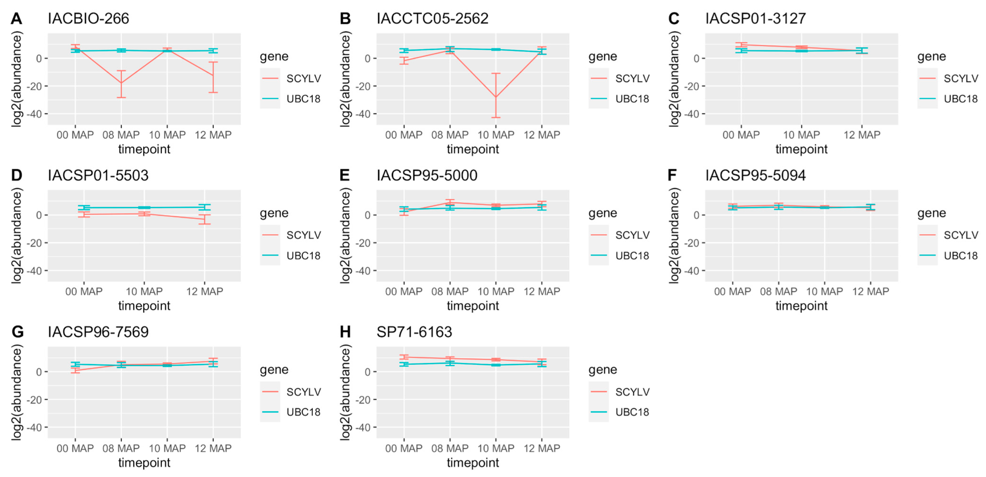
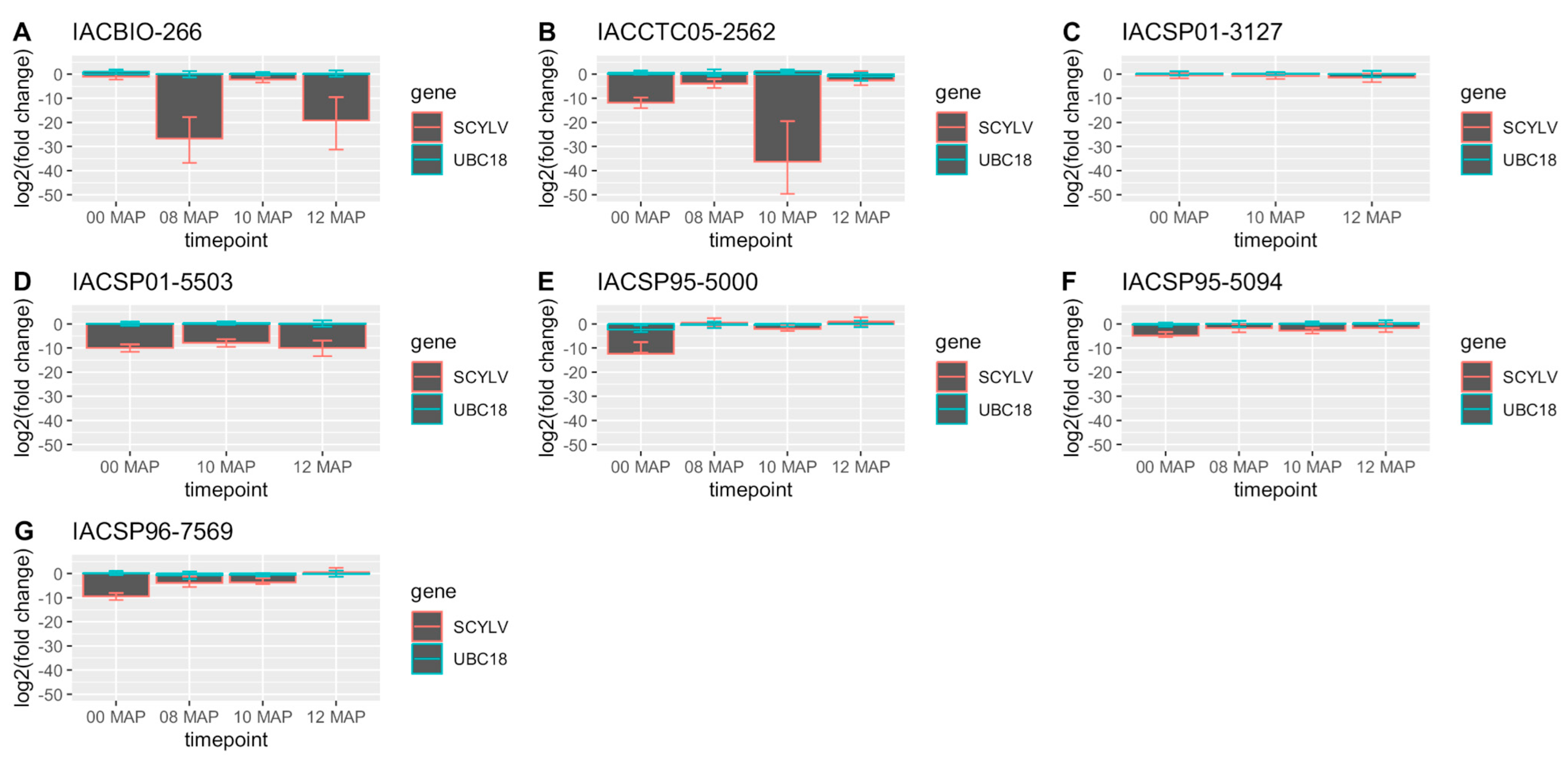
2.2. RT-qPCR and Virus Titer

The raw Ct values of the six candidate reference genes and the target gene, i.e., 181 bp fragment of ScYLV capsid protein, along with their mean Ct values, coefficient of variation (CV%) values, individual and mean amplification efficiency, and correlation coefficients (R2) are listed in the Supplementary Tables S2 and S3. The mean amplification efficiency among these genes ranged from 1.85 ± 0.09 to 2.13 ± 0.14, which is close to the optimal range of 90–110% [58]. The mean Ct values of the target gene per genotype were 27.23 ± 1.35, 28.47 ± 1.75, 30.22 ± 0.6, 30.84 ± 3.69, 32.33 ± 2.49, 34.23 ± 6.16, 34.38 ± 4.11, and 36.49 ± 0.85 for SP71-6163, IACSP01-3127, IACSP95-5094, IACSP95-5000, IACSP96-7569, IACBIO-266, IACCTC05-2562, and IACSP01-5503, respectively (Supplementary Table S3). Melting curve analysis showed unique peaks of fluorescence for all primer pairs, indicating RT-qPCR amplification of single fragments (Supplementary Figures S1 and S2). Positive RT-qPCR results for ScYLV were observed for all varieties in at least three sampling times and one technical replicate (Supplementary Table S4; Supplementary Figure S2). The stability ranking of the six candidate reference genes according to the NormFinder algorithm was the following: UBC18 > TUB > GAPDH > TPI > UK > SAND (Supplementary Table S5). Thus, UBC18 was employed as the reference gene for the virus titer assessment and considered as a prior in the Bayesian MCMC model for the transcript abundance analysis.
Virus titer variations significant at the 95% credible interval among sampling times were observed for all genotypes, except for IACSP01-5503 and SP71-6163 (Figure 1). These variations comprise the increase in virus titer between 0 and 8 MAP, decrease between 8 and 10 MAP, and increase between 10 and 12 MAP for IACCTC05-2562 and the reverse trend, i.e., the decrease between 0 and 8 MAP, increase between 8 and 10 MAP, and decrease between 10 and 12 MAP, for IACBIO-266. Increases in virus titer between 0 and 8 MAP were also observed for IACSP95-5000 and IACSP96-7569, with the latter also showing an increase in virus titer between 8 and 12 MAP, while decreases between 0 and 12 MAP were observed for IACSP01-3127 and IACSP95-5094, and also between 8 and 12 MAP for the latter. The genotypes with significantly higher virus titer across sampling times were SP71-6163, IACSP01-3127, and IACBIO-266 at 0 MAP; SP71-6163, IACSP95-5000, and IACSP95-5094 at 8 MAP; SP71-6163, IACSP01-3127, and IACSP95-5000 at 10 MAP; and IACSP95-5000, IACSP96-7569, SP71-6163, IACCTC05-2562, IACSP01-3127, and IACSP95-5094 at 12 MAP. It is noteworthy that the susceptible reference genotype, i.e., SP71-6163, was amongst those with higher virus titer across all sampling timepoints. The virus titer fold change in relation to SP71-6163 revealed either significantly lower SCYLV titers or non-significant values in the remaining cultivars across sampling timepoints, according to the 95% credible intervals (Figure 2). The average values of the significant virus titer fold decrease ranged from 4.48 × 10⁻² to 2.7 × 10⁻⁴ for IACSP95-5094, IACSP96-7569, IACSP01-5503, IACSP95-5000, and IACCTC05-2562 at 0 MAP; from 2.91 × 10⁻¹ to 9.11 × 10⁻⁹ for IACSP95-5094, IACSP96-7569, IACCTC05-2562, and IACBIO-266 at 8 MAP; from 3.15 × 10⁻¹ to 1.21 × 10⁻¹¹ for IACSP95-5000, IACBIO-266, IACSP95-5094, IACSP96-7569, IACSP01-5503, and IACCTC05-2562 at 10 MAP; and from 9.93 × 10⁻⁴ to 1.7 × 10⁻⁶ for IACSP01-5503 and IACBIO-266, respectively. The non-significant fold changes were observed for IACSP01-3127 and IACBIO-266 at 0 MAP; IACSP95-5000 at 8 MAP; IACSP01-3127 at 10 MAP; and IACSP95-5000, IACSP96-7569, IACSP01-3127, IACCTC05-2562, and IACSP95-5094 at 12 MAP.
Virus titer was not correlated with YLD symptom expression among sugarcane cultivars (r2 = 0.3254328; p = 0.1394). An evident contribution to the non-significative correlation between these traits was the significant virus titer fluctuation in the resistant cultivar, IACBIO-266. Other contributions for the poor correlation consisted of the mismatch between YLD severity and virus titer fluctuations between 8 and 10 MAP and between 10 and 12 MAP for IACCTC05-2562, and by the non-significant increases in virus titers among cultivars with increasing YLD severity across sampling timepoints, namely, IACSP01-3127, IACSP01-5503, and IACSP95-5094. On the other hand, the increasing YLD severity between 0 and 8 MAP observed for IACSP95-5000, IACSP96-7569, and IACCTC05-2562 was coherent with their increasing ScYLV titers.

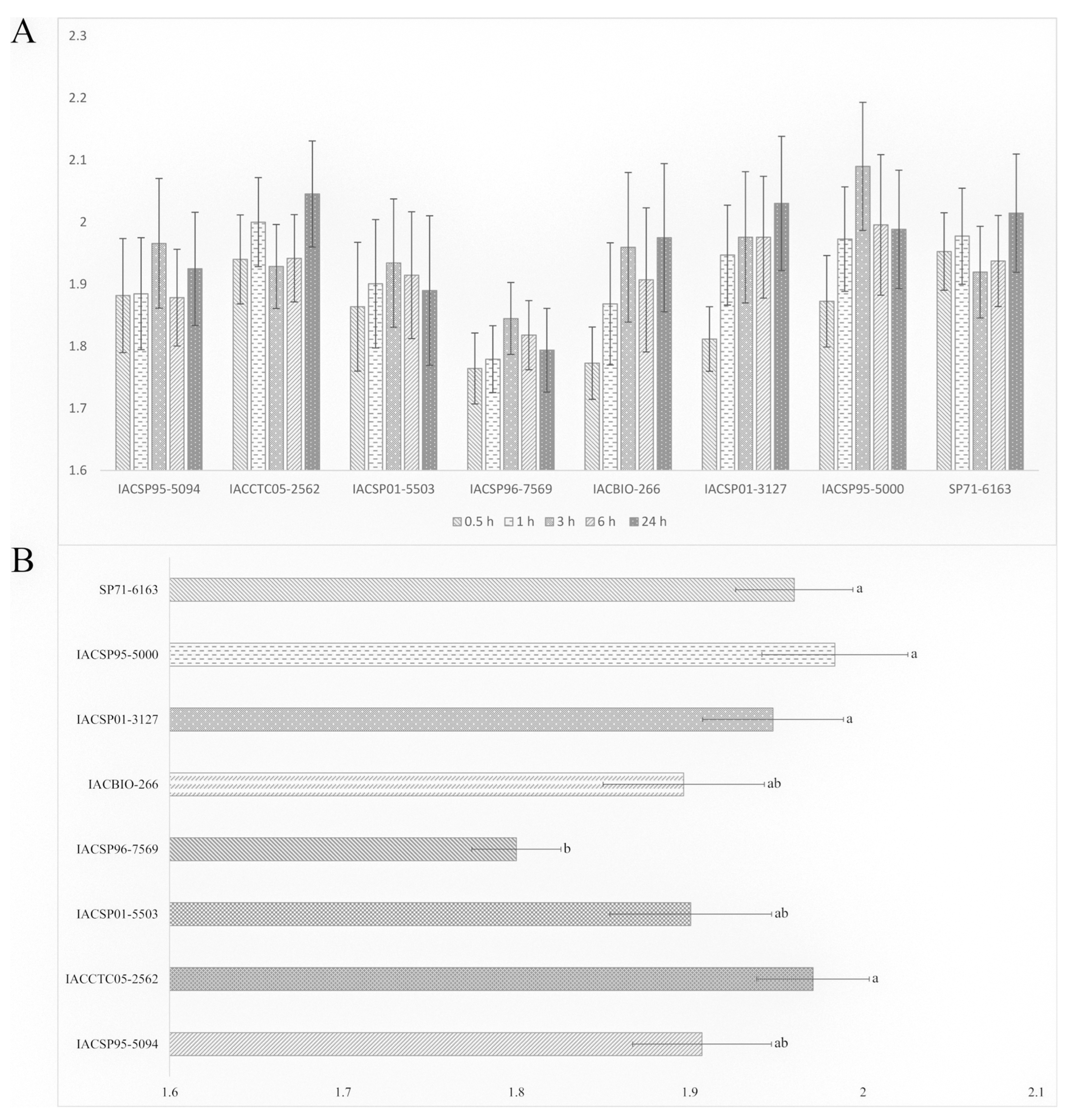
2.3. Aphid Preference

The one-way ANOVA for each timepoint of the free-choice bioassay revealed non-significant differences among cultivars for number of M. sacchari (p > 0.05) (Supplementary Table S6). The mean number of aphids across timepoints indicated increases from 0.5 to 3 h for the cultivars IACSP01-5503, IACSP96-7569, IACBIO-266, IACSP01-3127, and IACSP95-5000; decreases from 3 to 24 h for IACSP01-5503, IACSP96-7569, and IACSP95-5000; and increases from 3 to 24 h for IACSP01-3127. Another noteworthy change was the increase in the mean number of aphids from 6 to 24 h observed for IACCTC05-2562 (Figure 3A). Nonetheless, the two-way ANOVA with all 640 observations of the free-choice bioassay showed non-significant effects for time after release and its interaction with cultivar, while a significant F value (p < 0.05) was observed for the variable cultivar (Table 2). The least M. sacchari preferred cultivar according to the LSD test (p < 0.05) was IACSP96-7569, followed by IACBIO-266, IACSP01-5503, and IACSP95-5094. The latter three genotypes, on the other hand, did not differentiate from the most preferred ones, namely, SP71-6163, IACSP95-5000, IACSP01-3127, and IACCTC05-2562 (Figure 3B).

3. Discussion

ScYLV appeared in the mid 1990’s and became endemic in the main sugarcane-growing areas of Brazil [59], impelling the breeding for YLD resistance for over the last two decades by Brazilian sugarcane breeding programs. Such efforts have relied on phenotypical clone selection and resulted in tolerance or intermediate resistance of most of the sugarcane genotypes cultivated in the country [6]. On the other hand, the incidence of ScYLV has been underestimated, with the impacts on yield being neglected due to the near to absence of YLD symptoms in most cultivars [6]. The majority of the sugarcane cultivars assayed in the present study showed variable degrees of resistance to YLD while revealing positive RT-qPCR results for ScYLV presence, indicating that they may represent inoculum sources for the secondary spread of the virus in the field by aphid vectors. Although Ct values for IACBIO-266 were also recorded by Burbano et al. [38], ScYLV was considered non-detectable in this cultivar due to melting temperatures below 81.5 °C in two out of three blocks from a field experiment under a randomized complete block design. Here, melting temperatures above this threshold were observed for IACBIO-266 in all cDNA samples, each representing pools of RNA from three biological replicates, i.e., blocks, and thus were considered positive for ScYLV.
The virus titer assessment of the cultivars assayed here revealed significant fold decreases ranging from 3.15 × 10⁻¹ to 1.21 × 10⁻¹¹ in relation to the susceptible reference genotype, while ScYLV titers also varied among sampling timepoints for most of the cultivars and showed non-significant correlation with YLD severity. Previous studies have investigated the correlation between ScYLV titer and YLD symptom expression in sugarcane genotypes. The presence of ScYLV, revealed by TBIA, and the development of YLD were reported as correlated in a non-strict manner by Lehrer and Komor [20] based on the higher YLD incidence and severity within ScYLV-infected cultivars. According to the authors, the inconsistencies were due to the differential responses of some cultivars to ScYLV infection and virus titer fluctuations with the plant age, above and below the threshold detection of TBIA. Such titer fluctuations were confirmed by RT-qPCR, in a range of 103–104-fold, with the causes remaining unknown [60]. RT-qPCR assays have also revealed that ScYLV titers in resistant sugarcane cultivars were at least 106-fold lower than in susceptible ones [60], while symptomatic sugarcane plants showed 103–104 and virus titers up to 291-fold higher than the asymptomatic ones, with evidence pointing to a ScYLV titer threshold to cause YLD symptoms [60,61]. Accordingly, such virus titer thresholds appear to vary among sugarcane genotypes and may also vary with environmental conditions [21,38,60]. In assays for resistance screening to ScYLV on sugarcane panels comprising basic germplasm, elite clones, and commercial cultivars, no quantitative correlation between YLD severity and ScYLV titer was observed by Pimenta et al. [62], while Burbano et al. [38] reported high coefficient of correlation values for the moderately susceptible and susceptible classes of genotypes and very low and non-significant values for the moderately resistant ones.
The absence of YLD symptoms in all of the IACBIO-266 phenotyping assays, in combination with the fluctuating ScYLV titers, indicates that this cultivar is able to restrict the expression of YLD symptoms even at high virus titers, what was also observed for more susceptible cultivars at 0 and 10 MAP. The resistance of this genotype found in this study may be attributed to the genetic background inherited from the female parent SES069, a S. spontaneum accession. The contribution of S. spontaneum to YLD resistance has been previoulsy highlighted in studies screening sugarcane panels for resistance to ScYLV [38,63]. Conversely, aside from the contribution to the primary spread of ScYLV, during the asymptomatic phase of YLD yield losses of 20–30% have been reported [22]. Thus, the presence of ScYLV over the plant development of some genotypes requires attention, and stresses the importance of combining YLD phenotyping with ScYLV titer assessment to improve and speed up the selection of resistant genotypes [21,38].
The results obtained in the free-choice bioassay revealed IACSP96-7569 as the least preferred cultivar, followed by IACSP95-5094, IACSP01-5503, and IACBIO266, which supports the expression of antixenosis among the cultivars evaluated here. Additionally, the apparent movement of aphids among cultivars throughout the time intervals of 0.5, 1, 3, 6, and 24 h after release further indicates that host factors may have affected the settling behavior of the non-viruliferous aphids. Such outcome differs from the reported by Akbar et al. [64], in which antixenosis assays revealed no preference of M. sacchari and Sipha flava among sugarcane cultivars, with aphids finding their hosts within an hour followed by no apparent movement among cultivars for the 24 h duration of the assay. Conversely, significant differences on aphid settling on potted sugarcane plantlets were observed by Fartek et al. [51] when comparing the cultivars R 365 and MQ 76/53 under no-choice conditions at 8, 24, 48, and 72 h after aphid deposit, indicating the expression of antixenosis in the former cultivar from 8 to 24 h, followed by a similar decrease in aphid populations between cultivars from 24 to 72 h. Previous studies assessing the resistance of sugarcane to aphids have also revealed significant variance among cultivars in terms of aphid incidence [65,66] as well as life history parameters and feeding behavior [51,56,57,64,67]. In the cultivar IACSP96-7569, M. sacchari has also showed the worst biological performance, indicating that antibiosis is also expressed here [57]. Regarding the cultivars which showed intermediary antixenosis in this study, i.e., IACSP95-5094, IACSP01-5503, and IACBIO266, data of resistance to non-viruliferous M. sacchari in terms of reproductive parameters were correspondingly observed for the first two cultivars, whereas the latter was amongst the most favorable to M. sacchari reproduction [57], exhibiting diversity in terms of vector resistance mechanisms among these cultivars.
The host-vector interaction is another determining factor in viral epidemiology, with the effects of vector resistance on disease spread being strongly dependent on the virus transmission modes [48]. For instance, increase in the spread of non-persistently transmitted viruses has been reported in aphid-resistant cowpea plants [68], while reduction in the spread of persistently and semipersistently transmitted viruses associated with host plant resistance to whitefly has been reported in tomato [49,69]. Regarding sugarcane YLD, modest and positive correlations between aphid and ScYLV incidence were reported by Fartek et al. [66], indicating some degree of independence between these traits, possibly due to genotypes possessing virus resistance genes while showing susceptibility to M. sacchari. Viswanathan et al. [70] investigated the population dynamics of M. sacchari in field assays comprising hybrid varieties, inter-specific hybrids, and sugarcane varieties under typical tropical conditions in India, and reported higher aphid populations in plants expressing YLD comparatively to the symptomless ones from the same genotype. Additionally, the authors also observed comparatively higher number of aphids on the YLD-susceptible varieties, whereas instances of genotypes with high YLD severity and low aphid populations indicated that a limited population or even a single viruliferous aphid may spread the disease. Here, the cultivars IACSP01-5503 and IACBIO266 have shown agreements between aphid antixenosis and virus resistance traits. More specifically, the cultivar IACSP01-5503 showed intermediary resistance to YLD, relatively lower ScYLV titer over all evaluations, and lower M. sacchari preference. Similarly, IACBIO266 showed high resistance to YLD whilst relatively less preferred by the aphid vector. On the other hand, the putative resistance mechanisms carried by IACSP96-7569 involving antibiosis and antixenosis seemed to not prevent the secondary infection by ScYLV, which may have occurred either under our experimental conditions or previously in the field, followed by spread via virus-infected seed cane.
The present results indicate that current genetic resistance incorporated into sugarcane commercial cultivars may restrict YLD incidence in the field, but not necessarily reduce ScYLV titer. Further studies are necessary to determine the extent of varietal degeneration caused by YLD, unveil which genes are behind the genetic resistance of sugarcane against aphid vectors, and establish the relationship of this trait with the spread of ScYLV.

4. Material and Methods

4.1. Plant Material

YLD was evaluated in eight sugarcane cultivars, namely IACSP95-5094, IACCTC05-2562, IACSP01-5503, IACSP96-7569, IACSP01-3127, IACSP95-5000, SP71-6163, and IACBIO-266, maintained at the IAC Sugarcane Breeding Station, Ribeirão Preto, São Paulo, Brazil (latitude 21°10′39″ S and longitude 47°48′37″ W). The first six consist of high-sucrose cultivars currently cultivated in Brazil, while SP71-6163 was replaced from commercial sugarcane areas due to its high susceptibility to YLD, and therefore adopted as the susceptible reference in this study. IACBIO-266, in turn, is an energy cane cultivar aiming for biofuel production. Buds were obtained from stalks collected from sugarcane stools and planted in plastic trays containing commercial substrate composed by sphagnum turf, expanded vermiculite, dolomitic limestone, and gypsum. The trays were placed in a photoperiod chamber at 28 °C and photoperiod of 12 h for sprouting/germination. The obtained seedlings were individually transferred to stiff plastic tubes (volume of 50 cm3) and maintained on stands in an aphid-proof greenhouse during ninety days until being taken to the field.

4.2. Aphid Rearing

A colony of M. sacchari was established from a clone provided by Centre of Molecular Biology and Genetic Engineering, Campinas, São Paulo, Brazil (latitude 22°49′13″ S and longitude 47°3′34″ W), which colonies originated from aphid samplings previously performed at the IAC Sugarcane Breeding Station. Viruliferous aphid colonies were reared on detached leaves of RT-PCR tested ScYLV-infected plants of the cultivar IACSP95-5000 [57]. The youngest fully emerged leaves of these plants were cut, and the fragments were kept in test tubes containing 1% agar solution within a growth chamber (12 L:12 D, 29 ± 1 °C). Non-viruliferous aphid colonies, on the other hand, were reared on fragments of the youngest fully emerged leaves of healthy plants of IACISP95-5000 obtained from meristem tip culture, maintained under aphid-proof greenhouse conditions, and indexed by RT-PCR. In both conditions the sugarcane leaves were changed every 6 ± 1 day and newly born individuals were sampled and reared on new detached leaves. The presence and absence of the virus in the aphid colonies were confirmed by RT-PCR analysis.

4.3. YLD Phenotyping

Viruliferous M. sacchari colonies were released biweekly in the aphid-proof greenhouse during ninety days to promote uniform ScYLV infection. After the last release of viruliferous aphid colonies, sugarcane plants were taken to the field (November 2019) in a complete randomized block design with six replicates. Each plot consisted of four lines spaced 1.5 m between rows and 0.5 m between plants. Six additional biweekly releases of viruliferous aphids were performed in the field, after planting. YLD symptoms were recorded in six monthly evaluations in the two inner lines of each genotype plot, while the outer lines served as side borders, starting six months after planting (MAP; May 2020) up to eleven MAP (October 2020). A diagrammatic symptoms scale was used to evaluate YLD severity: (1) green leaf without symptoms; (2) slight yellowing of the midrib and leaf blade; (3) intense yellowing of the midrib and partial yellowing of the leaf blade; (4) intense yellowing of the midrib and leaf blade (Supplementary Figure S3). The symptom records were performed by four different evaluators to reduce the experimental error. Data were ln (x + 5) transformed and the normality was tested with Shapiro–Wilk W test. The transformed data were submitted to variance analysis using the general linear model (Proc GLM) and mixed model (Proc MIXED) procedures of SAS package version 9.3 [71]. Means were subsequently compared using the least significant difference test (LSD test) at p < 0.05.

4.4. RNA Extraction and RT-qPCR

ScYLV diagnosis and estimation of virus titer were performed via RT-qPCR from field samples collected at 0, 8, 10, and 11 MAP. For each cultivar x MAP, total RNA was extracted from three biological replicates, corresponding to the first leaf with visible dewlap from top to bottom (+1 leaf), and stored at −80 °C. Total RNA was extracted using 1000 µL of Trizol (Invitrogen, Carlsbad, CA, USA) per 100 mg of leaf tissue, according to the manufacturer’s instructions. RNA concentration was evaluated using a NanoDrop (Termofisher Scientific, Wilmington DE, USA). RNA integrity was checked by electrophoresis in a 1% agarose gel stained with ethidium bromide in TAE buffer. The RNA from the three biological replicates were pooled together for each cultivar x MAP for cDNA synthesis (n = 32 RNA pools). From these pools, 1 µg of RNA was treated with RQ1 RNase-Free DNase to remove genomic DNA, following manufacturer’s instructions (Promega, Fitchburg WI, USA). The reverse transcription was performed using the GoScript Reverse Transcription System (Promega), in a final volume of 10 µL containing 500 ng of DNAse treated RNA, 2 µL of GoScript Reaction Buffer + Random Primers, and 1 µL of GoScript Enzime Mix. The RT-qPCR reactions were performed on a IQTM5 Real-Time PCR Detection Systems BIO-RAD. The reaction mixture consisted of 3 μL of (1:10) diluted cDNA, 5 μL of Syber qPCR, and 2 μM of each forward and reverse primers in a total volume of 10 μL. The thermal cycling conditions consisted of denaturation at 95 °C for 2 min, followed by 40 cycles at 95 °C for 15 s, 60 °C for 1 min, 95 °C for 1 min, and 55 °C for 1 min. The target amplicon was 181 bp long, obtained with forward (5′-GGACCGAACCTATCTCAGTAC-3′) and reverse (5′-TAGTAATCTTGGAGCCTGTTGTTG-3′) primers annealing to the ScYLV capsid protein [18]. Six potential reference genes previously reported in sugarcane, i.e., triosephosphate isomerase (TPI), Uridylate kinase (UK), SAND protein family (SAND), Ubiquitin-conjugating enzyme 18 gene (UBC18), glyceraldehyde-3-phosphate dehydrogenase (GAPDH), and TUBULIN (TUB) (Supplementary Table S7), were tested by RT-qPCR in another set of four cDNA pools, each representing a sampling time and comprising the eight genotypes, using three technical replicates plus three non-template controls (NTCs), yielding a total of 90 RT-qPCR reactions. The RT-qPCR efficiency of each primer set and the corresponding product threshold cycle (Ct) were obtained via LinReg PCR analysis [72]. The Ct and reaction efficiency data from the potential reference genes were used as input in NormFinder software [73] for stability evaluation. The target gene and the most stable gene, according to NormFinder, were amplified in the 32 cDNA pools using three technical replicates, yielding a total of 192 RT-qPCR reactions plus three NTCs per gene per RT-qPCR plate. The amplicon specificity was assessed for all primer sets via dissociation curve profiles (melting curves). Regarding the target gene, cDNA samples with melting temperatures of 82.5 ± 1.0 °C were considered positive for ScYLV [74]. RT-qPCR data were analyzed using the Poisson-lognormal generalized mixed model and the Markov Chain Monte Carlo (MCMC) sampling scheme from MCMC.qpcr package [75] using genotype and time point as fixed factors, with the best reference gene added as prior to the model function, allowing 1.2-fold changes. Posterior mean estimates of virus titer were plotted as log2 mRNA abundance with error representing 95% credible intervals, where differences among cDNA samples were considered significant when credible intervals did not overlap. Pearson correlation test between virus titer and YLD symptoms was performed using the cor.test function of the R program [76].

4.5. Aphid Settling Behavior

The settling behavior of non-viruliferous M. sacchari was investigated in a free-choice bioassay aiming to determine the aphid preference among eight ScYLV-infected sugarcane cultivars. The experiments were conducted in glasshouse facilities at Department of Entomology and Acarology, ESALQ-USP, and cubic arenas (30 × 30 × 30 cm) (Supplementary Figure S4) were used to assess the preference of aphids (a similar experiment was described by Ramos et al. [77]). The cage structure was made from acrylic to allow natural light to penetrate the cage. The plants of each treatment had their leaves exposed through one of the side openings, one leaf from each plant per opening, with the treatments distributed equidistantly and interspersed. A circular acrylic arena coated with black adhesive tape was used to release the apterous aphids, and a squared black platform was fixed on the roof of the cage to support a leaf of each sugarcane cultivar (without detaching from the plant), interspersed at random and without touching (Supplementary Figure S4).
Fifty apterous adult non-viruliferous M. sacchari were released in the circular arena after a fasting period of one hour. The number of aphids remaining on each sugarcane cultivar was counted at short (0.5, 1, 3, 6 h) and long term (24 h) intervals after release, and the assay replicated sixteen times. Each arena being a repetition, in each repetition the plants and aphids were replaced. Data were ln (x + 5) transformed and mean comparisons were performed by ANOVA with significance level at p < 0.05 using the SAS package version 9.3 [71].

Supplementary Materials

The following supporting information can be downloaded at https://www.mdpi.com/article/10.3390/plants12173079/s1, Table S1 Variance analysis (ANOVA) for yellow leaf severity of eight sugarcane cultivars across six monthly evaluations in a field assay; Table S2: Cycle threshold (Ct) of the reference gene candidates with respective mean Ct, standard deviation, individual and mean efficiencies (eff.), and coefficient of determination (R2) estimated by RT-qPCR for eight sugarcane cultivars planted in a field trial; Table S3: Cycle threshold (Ct) of the viral target and reference gene (UBC18) with respective mean Ct, standard deviation (SD), individual and mean efficiency, and coefficient of determination (R2) estimated by RT-qPCR for eight sugarcane cultivars planted in a field trial; Table S4: Melting temperature and detection of 181 bp RT-qPCR products of the SCYLV capsid protein for eight sugarcane cultivars in each sampling time point, namely 0, 8, 10, and 12 months after planting (MAP); Table S5: Analyses of candidate reference genes by the NormFinder algorithm; Table S6: One-way variance analysis (ANOVA) for the number apterous adult non-viruliferous Melanaphis sacchari for the timepoints of 0.5, 1, 3, 6, and 24 h after release, in a free-choice bioassay with eight sugarcane cultivars; Table S7: Primer pairs sequences and amplicon size (A) and melting temperatures (Tm), estimated by RT-qPCR, of six genes selected for stability assessment across four sampling timepoints of sugarcane under sugarcane yellow leaf virus (SCYLV) infection, namely, triosephosphate isomerase (TPI), Uridylate kinase (UK), SAND protein family (SAND), Ubiquitin-conjugating enzyme 18 gene (UBC18), glyceraldehyde-3-phosphate dehydrogenase (GAPDH), and TUBULIN (TUB); Figures S1: Dissociation curve of six genes selected for stability assessment across four cDNA pools, each representing the sampling times of 0, 8, 10, and 12 months after planting (MAP) and comprising eight sugarcane cultivars under sugarcane yellow leaf virus (SCYLV) infection. The candidate reference genes were triosephosphate isomerase (TPI), Uridylate kinase (UK), SAND protein family (SAND), Ubiquitin-conjugating enzyme 18 gene (UBC18), glyceraldehyde-3-phosphate; Figure S2: Dissociation curve of the target gene, i.e., 181 bp fragment of ScYLV capsid protein, across cDNA pools from eight sugarcane cultivars under sugarcane yellow leaf virus (SCYLV) infection and four sampling timepoints, namely 0, 8, 10, and 12 months after planting (MAP); Figure S3: Diagrammatic scale for SCYLV symptoms intensity. Grade 1: green leaf without symptoms; Grade 2: slight yellowing of the midrib and leaf blade; Grade 3: intense yellowing of the midrib and partial yellowing of the leaf blade; Grade 4: intense yellowing of the midrib and leaf blade; Figure S4: Free-choice bioassay aiming to evaluate the settling behavior of Melanaphis sacchari among eight sugarcane cultivars. (A) Acrylic cages (30 cm × 30 cm × 30 cm), each comprising individual leaves of plantlets from eight sugarcane cultivars. Plantlets were maintained in stiff plastic tubes and randomly distributed. (B) Individual leaves of eight sugarcane cultivars placed in a squared black platform within an acrylic cage. The squared black platform supported the circular acrylic arena where fifty apterous adult non-viruliferous were released. (C) Side view of an acrylic cage with side openings (4 × 20 cm) sealed with “voile” fabric for ventilation (a) and openings through which individual leaves were inserted (b). On top of the acrylic cage (c) there are openings (4 × 20 cm) also sealed with “voile” fabric; for a top view of a closed acrylic cage, see Ramos et al. [64]. (D) Circular arena (d) supported by a squared black platform (e), which was fixed on the roof of the acrylic cage. (E) Internal side view showing individual leaves inserted through side openings, e.g., (f) and (g).

Author Contributions

Conceived and designed the experiments, L.E.T.B., M.C.G., L.R.P. and J.R.S.L.; performed the experiments, L.E.T.B., M.F.d.S., M.C.-S. and I.A.d.A.; analyzed the data, M.F.d.S., D.P., P.M.N., L.E.T.B., M.C.-S. and M.C.G.; wrote and edited the paper M.F.d.S., L.E.T.B. and M.C.G. All authors have read and agreed to the published version of the manuscript.

Funding

Financial support was received from Coordenação de Aperfeiçoamento de Pessoal de Nível Superior (CAPES)—under financing code 001. The first author was a recipient of a PhD scholarship from CAPES Brazil, and the eighth author received a research fellowship from Conselho Nacional de Desenvolvimento Científico e Tecnológico (CNPq)/Brazil (Proc. 314181/2020-2).

Data Availability Statement

The data presented in this study are available in the Supplementary Material.

Conflicts of Interest

The authors declare no competing interests. The funders had no role in the design of the study; in the collection, analyses, or interpretation of data; in the writing of the manuscript, or in the decision to publish the results.

References

  1. Waclawovsky, A.J.; Sato, P.M.; Lembke, C.G.; Moore, P.H.; Souza, G.M. Sugarcane for bioenergy production: An assessment of yield and regulation of sucrose content. Plant Biotechnol. J. 2010, 8, 263–276. [Google Scholar] [CrossRef] [PubMed]
  2. Yadav, S.; Jackson, P.; Wei, X.; Ross, E.M.; Aitken, K.; Deomano, E.; Atkin, F.; Hayes, B.J.; Voss-Fels, K.P. Accelerating Genetic Gain in Sugarcane Breeding Using Genomic Selection. Agronomy 2020, 10, 585. [Google Scholar] [CrossRef]
  3. Carpio, L.G.; Souza, F.S. Optimal allocation of sugarcane bagasse for producing bioelectricity and second generation ethanol in Brazil: Scenarios of cost reductions. Renew. Energy 2017, 111, 771–780. [Google Scholar] [CrossRef]
  4. Reis, C.E.R.; Hu, B. Vinasse from sugarcane Ethanol production: Better treatment or better utilization? Front. Energy Res. 2017, 5, 7. [Google Scholar]
  5. Cheavegatti-Gianotto, A.; Abreu, H.M.C.; Arruda, P.; Filho, J.C.B.; Burnquist, W.L.; Creste, S.; Ciero, L.; Ferro, J.A.; Figueira, A.V.O.; Filgueiras, T.S.; et al. Sugarcane (Saccharum x officinarum): A reference study for the regulation of genetically modified cultivars in Brazil. Trop. Plant Biol. 2011, 4, 62–89. [Google Scholar] [CrossRef]
  6. Gonçalves, M.C.; Pinto, L.R.; Souza, S.C.; Landell, M.G.A. Virus Diseases of Sugarcane. A Constant Challenge to Sugarcane Breeding in Brazil. Funct. Plant Sci. Biotechnol. 2012, 6, 108–116. [Google Scholar]
  7. ElSayed, A.I.; Komor, E.; Boulila, M.; Viswanathan, R.; Odero, D.C. Biology and management of sugarcane yellow leaf vírus: An historical overview. Arch. Virol. 2015, 160, 2921–2934. [Google Scholar] [CrossRef]
  8. Holkar, S.K.; Balasubramaniam, P.; Kumar, A.; Kadirvel, N.; Shingote, P.R.; Chhabra, M.L.; Kumar, S.; Kumar, P.; Viswanathan, R.; Jain, R.K.; et al. Present status and future management strategies for sugarcane yellow leaf virus: A major constraint to the global sugarcane production. Plant Pathol. J. 2020, 36, 536–557. [Google Scholar] [CrossRef]
  9. Grisham, M.P.; Eggleston, G.; Hoy, J.W.; Viator, R.P. The effect of Sugarcane yellow leaf virus infection on yield of sugarcane in Louisiana. Sugar Cane Int. 2009, 27, 3–91. [Google Scholar]
  10. Lehrer, A.T.; Wu, K.K.; Komor, E. Impact of sugarcane yellow leaf virus (ScYLV) on growth and sugar yield of sugarcane. J. Gen. Plant Pathol. 2009, 75, 288–296. [Google Scholar] [CrossRef]
  11. Izaguirre-Mayoral, M.L.; Carballo, O.; Alceste, C.; Romano, M.; Nass, H.A. Physiological performance of asymptomatic and yellow leaf syndrome-affected sugarcanes in Venezuela. J. Phytopathol. 2002, 150, 13–19. [Google Scholar] [CrossRef]
  12. Boukari, W.; Kaye, C.; Wei, C.; Hincapie, M.; LaBorde, C.; Irey, M.; Rott, P. Field infection of virus-free sugarcane by Sugarcane yellow leaf virus and effect of yellow leaf on sugarcane grown on organic and on mineral soils in Florida. Plant Dis. 2019, 103, 2367–2373. [Google Scholar] [CrossRef] [PubMed]
  13. Viswanathan, R.; Balamuralikrishnan, M.; Karuppaiah, R. Yellow leaf disease of sugarcane: Occurrence and impact of infected sets on disease severity and yield. Proc. Sugar Cancer Technol. Assoc. India 2006, 67, 74–89. [Google Scholar]
  14. Gray, S.; Cilia, M.; Ghanim, M. Circulative, “nonpropagative” virus transmission: An orchestra of virus, insect, and plant-derived instruments. Adv. Virus Res. 2014, 89, 141–199. [Google Scholar] [PubMed]
  15. Rott, P.; Mirkov, T.E.; Schenck, S.; Girard, J.C. Recent advances in research on sugarcane yellow leaf vírus, the causal agents of sugarcane yellow leaf. Sugar Cane Int. 2008, 26, 18–22. [Google Scholar]
  16. Ahmad, A.Y.; Costet, L.; Daugrois, J.H.; Nibouche, S.; Letourmy, P.; Girard, J.C.; Rott, P. Variation in infection capacity and in virulence exists between genotypes of Sugarcane yellow leaf virus. Plant Dis. 2007, 91, 253–259. [Google Scholar] [CrossRef]
  17. Lehrer, A.T.; Schenck, S.; Yan, S.-L.; Komor, E. Movement of aphid-transmitted Sugarcane yellow leaf vírus (ScYLV) within and between sugarcane plants. Plant Pathol. 2007, 56, 711–717. [Google Scholar] [CrossRef]
  18. Chinnaraja, C.; Viswanathan, R. Quantification of sugarcane yellow leaf virus in sugarcane following transmission through aphid vector Melanaphis sacchari. Virus Dis. 2015, 26, 237–242. [Google Scholar] [CrossRef]
  19. Gonçalves, M.C.; Vega, J.; Oliveira, J.G.; Gomes, M.M.A. Sugarcane yellow leaf virus infection leads to alterations in photosynthetic efficiency and carbohydrate accumulation in sugarcane leaves. Fitop. Brasil. 2005, 30, 10–16. [Google Scholar] [CrossRef]
  20. Lehrer, A.T.; Komor, E. Symptom expression of yellow leaf disease in sugarcane cultivars with different degrees of infection by Sugarcane yellow leaf vírus. Plant Pathol. 2008, 57, 178–189. [Google Scholar] [CrossRef]
  21. Viswanathan, R. Impact of yellow leaf disease in sugarcane and its successful disease management to sustain crop production. Indian Phytopathol. 2021, 74, 573–586. [Google Scholar] [CrossRef]
  22. Rassaby, L.; Girard, J.C.; Letourmy, P.; Chaume, J.; Irey, M.S.; Lockhart, B.E.; Kodja, H.; Rott, P. Impact of sugarcane yellow leaf virus on sugarcane yield and juice quality in Reunion Island. Eur. J. Plant Pathol. 2003, 109, 459–466. [Google Scholar] [CrossRef]
  23. Bertani, R.P.; Perera, M.F.; Arias, M.E.; Luque, C.; Funes, C.; González, V.; Cuenya, M.I.; Ploper, L.D.; Welin, B.; Castagnaro, A.P. A study of the sugarcane yellow leaf disease in Argentina. Plant Dis. J. 2014, 98, 1036–1042. [Google Scholar] [CrossRef] [PubMed]
  24. Viswanathan, R.; Karuppaiah, R.; Malathi, P.; Ganesh Kumar, V.; Chinnaraja, C. Diagnosis of sugarcane yellow leaf virus in asymptomatic sugarcane by RT-PCR. Sugar Tech. 2009, 11, 368–372. [Google Scholar] [CrossRef]
  25. Comstock, J.C.; Irey, M.S.; Lockhart, B.E.L.; Wang, Z.K. Incidence of yellow leaf syndrome in CP cultivars based on polymerase chain reaction and serological techniques. Sugar Cane 1998, 4, 21–24. [Google Scholar]
  26. Gao, S.J.; Lin, Y.H.; Pan, Y.B.; Damaj, M.B.; Wang, Q.N.; Mirkov, T.E.; Chen, R.K. Molecular characterization and phylogenetic analysis of Sugarcane yellow leaf virus isolates from China. Virus Genes 2012, 45, 340–349. [Google Scholar] [CrossRef]
  27. Grisham, M.P.; Comstock, J.C.; Flynn, J.L. Comparing the natural spread of Sugarcane yellow leaf virus in Florida to its natural spread in Louisiana. J. Am. Soc. Sugar Cane Technol. 2010, 30, 151. [Google Scholar]
  28. Zhu, Y.J.; Lim, S.T.; Schenck, S.; Arcinas, A.; Komor, E. RT-PCR and quantitative real-time RT-PCR detection of Sugarcane yellow leaf virus (SCYLV) in symptomatic and asymptomatic plants of Hawaiian sugarcane cultivars and the correlation of SCYLV titre to yield. Eur. J. Plant Pathol. 2010, 127, 263–273. [Google Scholar] [CrossRef]
  29. ElSayed, A.I.; Boulila, M.; Rott, P. Molecular evolutionary history of Sugarcane yellow leaf vírus based on sequence analysis of RNA-dependent RNA polymerase and putative aphid transmission factor-coding genes. J. Mol. Evol. 2014, 78, 349–365. [Google Scholar] [CrossRef]
  30. Gonçalves, M.C.; Klerks, M.M.; Verbeek, M.; Veja, J.; Van den Heuvel, J.F.J.M. The use of molecular beacons combined with NASBA for the sensitive detection of sugarcane yellow leaf virus. Eur. J. Plant Pathol. 2002, 108, 401–407. [Google Scholar] [CrossRef]
  31. Amata, R.L.; Fernandez, E.; Filloux, D.; Martin, D.P.; Rott, P.; Roumagnac, P. Prevalence of Sugarcane yellow leaf virus in sugarcane producing regions in Kenya revealed by reverse transcription loop-mediated isothermal amplification method. Plant Dis. 2015, 100, 260–268. [Google Scholar] [CrossRef] [PubMed]
  32. Scagliusi, S.M.; Lockhart, B.E.L. Transmission, characterization, and serology of a luteovirus associated with yellow leaf syndrome of sugarcane. Phytopathology 2000, 90, 120–124. [Google Scholar] [CrossRef]
  33. Chatenet, M.; Delage, C.; Ripolles, M.; Irey, M.S.; Lockhart, B.E.L.; Rott, P. Detection of Sugarcane yellow leaf vírus in quarantine and production of virus-free sugarcane by apical meristem culture. Plant Dis. 2001, 85, 1177–1180. [Google Scholar] [CrossRef] [PubMed]
  34. Viswanathan, R.; Balamuralikrishnan, M. Detection of sugarcane yellow leaf virus, the causal agent of yellow leaf syndrome in sugarcane by DAS-ELISA Arch. Phytopathol. Plant Protect 2004, 37, 169–176. [Google Scholar] [CrossRef]
  35. Schenck, S.; Hu, J.S.; Lockhart, B.E. Use of a tissue blot immunoassay to determine the distribution of sugarcane yellow leaf virus in Hawaii. Sugar Cane 1997, 4, 5–8. [Google Scholar]
  36. Parakh, D.R.; Shamloul, A.M.; Hadidi, A.; Waterworth, H.E.; Scott, S.W.; Howell, H.E.; Mink, G.I. Detection of Prune dwarf ilarvirus from infected stone fruits using reverse transcription polymerase chain reaction. Acta Hortic. 1995, 386, 421–430. [Google Scholar] [CrossRef]
  37. Nassuth, A.; Pollari, E.; Helmeczy, K.; Stewart, S.; Ko-falvi, A. Improved RNA extraction and one-tube RT-PCR assay for simultaneous detection of control plantRNA plus several viruses in plant extracts. J. Virol. Methods 2000, 90, 37–49. [Google Scholar] [CrossRef]
  38. Burbano, R.C.; Gonçalves, M.C.; Nobile, P.M.; dos Anjos, I.A.; da Silva, M.F.; Perecin, D.; Anjos, L.O.S.; Fernandes, V.B.V.R.; Pinto, L.R. Screening of Saccharum spp. genotypes for sugarcane yellow leaf virus resistance by combining symptom phenotyping and highly precise virus titration. Crop Prot. 2021, 144, 105577. [Google Scholar] [CrossRef]
  39. Chinnaraja, C.; Viswanathan, R.; Sathyabhama, M.; Parameswari, B.; Bagyalakshmi, K.; Malathi, P.; Neelamathi, D. Quantification of sugarcane yellow leaf virus in in vitro plantlets and asymptomatic plants of sugarcane by RT-qPCR. Curr. Sci. 2014, 106, 729–734. [Google Scholar]
  40. Carr, J.P.; Murphy, A.M.; Tungadi, T.; Yoon, J.Y. Plant defense signals: Players and pawns in plant-virus-vector interactions. Plant Sci. 2019, 279, 87–95. [Google Scholar] [CrossRef]
  41. Ray, S.; Casteel, C.L. Effector-mediated plant–virus–vector interactions. The Plant Cell 2022, 34, 1514–1531. [Google Scholar] [CrossRef] [PubMed]
  42. Su, Q.; Mescher, M.C.; Wang, S.; Chen, G.; Xie, W.; Wu, Q.; Wang, W.; Zhang, Y. Tomato yellow leaf curl virus differentially influences plant defense responses to a vector and a non-vector herbivore. Plant Cell Environ. 2016, 39, 597–607. [Google Scholar] [CrossRef] [PubMed]
  43. Ziebell, H.; Murphy, A.M.; Groen, S.C.; Tungadi, T.; Westwood, J.H.; Lewsey, M.G.; Moulin, M.; Kleczkowski, A.; Smith, A.G.; Stevens, M.; et al. Cucumber mosaic virus and its 2b RNA silencing suppressor modify plant aphid interactions in Tobacco. Sci. Rep. 2011, 1, 187. [Google Scholar] [CrossRef] [PubMed]
  44. Li, R.; Weldegergis, B.T.; Li, J.; Jung, C.; Qu, J.; Sun, Y.; Qian, H.; Tee, C.; Van Loon, J.J.; Dicke, M.; et al. Virulence factors of geminivirus interact with MYC2 to subvert plant resistance and promote vector performance. Plant Cell 2014, 26, 4991–5008. [Google Scholar] [CrossRef]
  45. Casteel, C.L.; Falk, B.W. Plant virus-vector interactions: More than just for virus transmission. In Current Research Topics in Plant Virology; Wang, A., Zhou, X., Eds.; Springer International Publishing: Cham, Switzerland, 2016; pp. 217–240. [Google Scholar]
  46. Mauck, K.E.; Chesnais, Q.; Shapiro, L.R. Evolutionary determinants of host and vector manipulation by plant viruses. In Virology and Virus Ecology; Malmstrom, C., Ed.; Environmental Academic Press: Cambridge, MA, USA, 2018; pp. 189–250. [Google Scholar]
  47. Patton, M.F.; Bak, A.; Sayre, J.M.; Heck, M.L.; Casteel, C.L. A polerovirus, Potato leafroll virus, alters plant–vector interactions using three viral proteins. Plant Cell Environ. 2020, 43, 387–399. [Google Scholar] [CrossRef]
  48. Madden, L.V.; Jeger, M.J.; van den Bosch, F. A theoretical assessment of the effects of vector-virus transmission mechanism on plant virus disease epidemics. Phytopathology 2000, 90, 576–594. [Google Scholar] [CrossRef]
  49. Fortes, I.M.; Muñoz, R.F.; Moriones, E. Host plant resistance to Bemisia tabaci to control damage caused in tomato plants by the emerging crinivirus tomato chlorosis vírus. Front. Plant. Sci. 2020, 11, 585510. [Google Scholar] [CrossRef]
  50. Painter, R.H. Insect Resistance in Crop Plants; The Macmillan Company: New York, NY, USA, 1951; Volume 72, p. 481. [Google Scholar]
  51. Fartek, B.; Nibouche, S.; Turpin, P.; Costet, B.R. Resistance to Melanaphis sacchari in the sugarcane cultivar R 365. Entomol. Exp. Appl. 2012, 144, 270–278. [Google Scholar] [CrossRef]
  52. Van Emden, H.F.; Harrington, R. Aphids as Crop Pests; Cabi: Wallingford, UK, 2007. [Google Scholar]
  53. Williams, W.; Kennedy, G.G.; Yamamoto, R.T.; Thacker, J.; Bordner, J. 2-Tridecanone: A naturally occurring insecticide from the wild tomato Lycopersicon hirsutum f. glabratum. Science 1980, 207, 888–889. [Google Scholar] [CrossRef] [PubMed]
  54. Smith, C.M. Plant Resistance to Arthropods: Molecular and Conventional Approaches; Springer Science & Business Media: Berlin, Germany, 2005. [Google Scholar]
  55. Fartek, B.; Nibouche, S.; Costet, L.; Reynaud, B. Characterization of Sugarcane Resistance to Melanaphis sacchari Using Biological Tests and Electrical Penetration Graph (DC-EPG); Symposium on Plant Interactions with Aphids: Wageningen, The Netherlands, 2008; p. 1. [Google Scholar]
  56. Akbar, W.; Showler, A.T.; Reagan, T.E.; Davis, J.A.; Beuzelin, J.M. Feeding by sugarcane aphid, Melanaphis sacchari, on sugarcane cultivars with differential susceptibility and potential mechanism of resistance. Entomol. Exp. Appl. 2014, 150, 32–44. [Google Scholar] [CrossRef]
  57. Bertasello, L.E.T.; Sousa, M.C.; Maluta, N.K.P.; Pinto, L.R.; Lopes, J.R.S.; Gonçalves, M.C. Effect of sugarcane cultivars infected with sugarcane yellow leaf vírus (ScYLV) on feeding behavior and biological performance of Melanaphis sacchari (Hemiptera: Aphididae). Plants 2021, 10, 2122. [Google Scholar] [CrossRef]
  58. Ramakers, C.; Ruijter, J.M.; Deprez, R.H.L.; Moorman, A.F.M. Assumption-free analysis of quantitative real-time polymerase chain reaction (PCR) data. Neurosci. Lett. 2003, 339, 62–66. [Google Scholar] [CrossRef]
  59. Gonçalves, M.C. Doenças causadas por vírus. In Cana-de-Açúcar; Dinardo-Miranda, L.L., Vasconcelos, A.C.M., Landell, M.G.A., Eds.; Instituto Agronômico de Campinas: São Paulo, Brasil, 2008; Volume 1, pp. 150–165. [Google Scholar]
  60. Zhu, Y.J.; McCafferty, H.; Osterman, G.; Lim, S.; Agbayani, R.; Lehrer, A.; Schenck, S.; Komor, E. Genetic transformation with untranslatable coat protein gene of sugarcane yellow leaf virus reduces virus titers in sugarcane. Transgenic Res. 2011, 20, 503–512. [Google Scholar] [CrossRef]
  61. Bagyalakshmi, K.; Viswanathan, R.; Ravichandran, V. Impact of the viruses associated with mosaic and yellow leaf disease on varietal degeneration in sugarcane. Phytoparasitica 2019, 47, 591–604. [Google Scholar] [CrossRef]
  62. Pimenta, R.J.G.; Aono, A.H.; Burbano, R.C.V.; Coutinho, A.E.; Silva, C.C.; Dos Anjos, I.A.; Perecin, D.; Landell, M.G.A.; Gonçalves, M.C.; Pinto, L.R.; et al. Genome-wide approaches for the identification of markers and genes associated with sugarcane yellow leaf vírus resistance. Sci. Rep. 2021, 11, 15730. [Google Scholar] [CrossRef]
  63. Yang, X.; Sood, S.; Luo, Z.; Todd, J.; Wang, L. Genome-wide association studies identified resistance loci to orange rust and yellow leaf virus diseases in sugarcane (Saccharum spp.). Phytopathology 2019, 109, 623–631. [Google Scholar] [CrossRef] [PubMed]
  64. Akbar, W.; Showier, A.T.; White, W.H.; Reagan, T.E. Categorizing sugarcane cultivars for resistance to the sugarcane aphid and yellow sugarcane aphid (Hemiptera: Aphididae). J. Econ. Entomol. 2010, 103, 1431–1437. [Google Scholar] [CrossRef]
  65. Akbar, W.; Showler, A.T.; Beuzelin, J.M.; Reagan, T.E.; Gravois, K.A. Evaluation of Aphid Resistance Among Sugarcane Cultivars in Louisiana. Ann. Entomol. Soc. Am. 2011, 104, 699–704. [Google Scholar] [CrossRef]
  66. Fartek, B.; Nibouche, S.; Atiama-Nurbel, T.; Reynaud Costet, B.L. Genotypic variability of sugarcane resistance to the aphid Melanaphis sacchari, vector of the Sugarcane yellow leaf virus. Plant Breed. 2014, 133, 771–776. [Google Scholar] [CrossRef]
  67. White, W.H. Yellow sugarcane aphid (Homoptera: Aphididae) resistance mechanisms in selected cultivars. J. Econ. Entomol. 1990, 83, 2111–2114. [Google Scholar] [CrossRef]
  68. Atiri, G.I.; Ekpot, E.J.A.; Thottappilly, G. The effect of aphid-resistance in cowpea on infestation and development of Aphis cracivora and the transmission of cowpea aphid-borne mosaic virus. Ann. Appl. Biol. 1984, 104, 339–346. [Google Scholar] [CrossRef]
  69. Rodríguez-López, M.J.; Garzo, E.; Bonani, J.P.; Fereres, A.; Fernández-Muñoz, R.; Moriones, E. Whitefly resistance traits derived from the wild tomato Solanum pimpinellifolium affect the preference and feeding behavior of Bemisia tabaci and reduce the spread of Tomato yellow leaf curl virus. Phytopathology 2011, 101, 1191–1201. [Google Scholar] [CrossRef]
  70. Viswanathan, R.; Ramasubramanian, T.; Chinnaraja, C.; Selvakumar, R.; Lakshmi Pathy, T.; Manivannan, K.; Nithyanantham, R. Population dynamics of Melanaphis sacchari (Zehntner), the aphid vector of sugarcane yellow leaf virus under tropical conditions in India. Trop Plant Pathol. 2022, 47, 260–277. [Google Scholar] [CrossRef]
  71. SAS Institute. SAS/STAT User’s Guide, Version 9.3; SAS: Cary, NC, USA, 2013. [Google Scholar]
  72. Ruijter, J.M.; Ramakers, C.; Hoogaars, W.M.; Karlen, Y.; Bakker, O.; van den Hoff, M.J.; Moorman, A.F. Amplification efficiency: Linking baseline and bias in the analysis of quantitative PCR data. Nucleic Acids Res. 2009, 37, e45. [Google Scholar] [CrossRef]
  73. Andersen, C.L.; Jensen, J.L.; Ørntoft, T.F. Normalization of real-time quantitative reverse transcription-PCR data: A model-based variance estimation approach to identify genes suited for normalization, applied to bladder and colon cancer data sets. Cancer Res. 2004, 64, 5245–5250. [Google Scholar] [CrossRef]
  74. Ririe, K.M.; Rasmussen, R.P.; Wittwer, C.T. Product differentiation by analysis of DNA melting curves during the polymerase chain reaction. Anal. Biochem. 1997, 245, 154–160. [Google Scholar] [CrossRef]
  75. Matz, M.V.; Wright, R.M.; Scott, J.G. No control genes requires: Bayesian analysis of qRT-PCR data. PLoS ONE 2013, 8, e71448. [Google Scholar] [CrossRef]
  76. R Core Team. R: A Language and Environment for Statistical Computing; R Foundation for Statistical Computing: Vienna, Austria, 2016; Available online: https://www.R-project.org (accessed on 6 February 2023).
  77. Ramos, A.; Esteves, M.B.; Cortés, M.T.B.; Lopes, J.R.S. Maize Bushy Stunt Phytoplasma Favors Its Spread by Changing Host Preference of the Insect Vector. Insects 2020, 11, 600. [Google Scholar] [CrossRef]
Figure 1. Sugarcane yellow leaf virus (SCYLV) titer (log2 abundance) in eight sugarcane genotypes across four sampling times. Data were modeled using the Bayesian MCMC sampling scheme of the ‘MCMC.qpcr’ package, considering UBC18 as a prior. Error bars represent 95% credible intervals surrounding the posterior mean estimate, with differences among samples significant at no overlapping intervals.
Plants 12 03079 g001
Figure 2. Sugarcane yellow leaf virus (SCYLV) log2 fold changes in seven sugarcane cultivars relative to the susceptible reference cultivar, SP71-6163. Data were modeled using the Bayesian MCMC sampling scheme of the ‘MCMC.qpcr’ package, considering UBC18 as a prior. Error bars represent 95% credible intervals surrounding the posterior mean estimate, with differences with SP71-6163 significant at no overlapping with zero.
Plants 12 03079 g002
Figure 3. (A) Ln (x + 5) transformed mean number of apterous adult non-viruliferous Melanaphis sacchari per sugarcane cultivar at short (0.5, 1, 3, 6 h) and long-term (24 h) intervals after the release of fifty individuals in a free-choice bioassay. The differences among cultivars are not significantly different according to the LSD test after one-way ANOVA for each timepoint (p > 0.05). Error bars represent the standard error (n = 16). (B) Ln (x + 5) transformed mean number of adult non-viruliferous M. sacchari per sugarcane cultivar, each consisting of the mean of the five timepoints. Means with the same letter are not significantly different according to the LSD test after two-way ANOVA with all observations (p < 0.05). Error bars represent the standard error (n = 80).
Plants 12 03079 g003
Table 1. Yellow leaf disease (YLD) mean severity compared by LSD test (p = 0.05) after ln(x + 5) transformation.
GenotypesSymptom Evaluations
6 MAP7 MAP8 MAP9 MAP10 MAP11 MAPMeanClass
IACSP95-50941.07 c1.11 b1.28 cd1.78 cd1.84 c1.79 d1.48 dMR
IACCTC05-25621.00 c1.00 b1.03 d1.43 de1.94 c1.78 d1.36 dMR
IACSP01-55031.21 b1.17 b1.17 cd1.08 ef1.25 d1.35 e1.20 eMR
IACSP96-75691.00 c1.15 b1.53 bc2.00 bc1.93 c2.1 c1.62 cMR
IACBIO-2661.00 c1.00 b1.00 d1.00 f1.00 d1.00 f1.00 fR
IACSP01-31271.00 c1.00 b1.10 cd1.13 ef1.18 d1.38 e1.13 eMR
IACSP95-50001.00 c1.00 b1.78 b2.26 b2.93 b3.12 b2.01 bMS
SP71-61631.98 a2.46 a3.29 a3.98 a4.00 a4.00 a3.29 aS
Means with the same letter are not significantly different.
Table 2. Two-way variance analysis (ANOVA) for the number apterous adult non-viruliferous Melanaphis sacchari across eight sugarcane cultivars in a free-choice bioassay.
SourceDFSum of SquaresMean SquareF ValuePr > F
Model393.648442570.093549810.740.8748
Error60075.590457790.12598410
SourceDFType III SSMean SquareF ValuePr > F
Cultivar71.965704270.280814902.230.0305
Time after release40.817479840.204369961.620.1670
Cultivar x time280.865258460.030902090.251.0000
CV (%) 18.47719
Disclaimer/Publisher’s Note: The statements, opinions and data contained in all publications are solely those of the individual author(s) and contributor(s) and not of MDPI and/or the editor(s). MDPI and/or the editor(s) disclaim responsibility for any injury to people or property resulting from any ideas, methods, instructions or products referred to in the content.

