Article

Phytochemical Characterization of Saudi Mint and Its Mediating Effect on the Production of Silver Nanoparticles and Its Antimicrobial and Antioxidant Activities

1
Department of Medical Laboratory Science, College of Applied Medical Sciences, University of Ha’il, Ha’il 55476, Saudi Arabia
2
Department of Biological Sciences, University of Ha’il, Hail 81451, Saudi Arabia
3
Department of Biology, College of Science, University of Jeddah, Jeddah 21589, Saudi Arabia
4
Department of Clinical Laboratory Sciences, College of Applied Medical Sciences, Taif University, Taif 21944, Saudi Arabia
5
Medical Laboratory, East Jeddah General Hospital, Jeddah 22253, Saudi Arabia
*
Author to whom correspondence should be addressed.
Plants 2023, 12(11), 2177; https://doi.org/10.3390/plants12112177
Submission received: 6 May 2023 / Revised: 23 May 2023 / Accepted: 29 May 2023 / Published: 30 May 2023

Abstract

:
The green synthesis of nanoparticles (NPs) is attracting enormous attention as a new area of study that encompasses the development and discovery of new agents for their utilization in different fields, such as pharmaceuticals and food. Nowadays, the use of plants, particularly medicinal plants, for the creation of NPs has emerged as a safe, ecofriendly, rapid, and simple approach. Therefore, the present study aimed to use the Saudi mint plant as a medicinal plant for the synthesis of silver nanoparticles (AgNPs) and to evaluate the antimicrobial and antioxidant activities of AgNPs compared to mint extract (ME). A phenolic and flavonoid analysis that was conducted by using HPLC indicated the presence of numerous compounds in the ME. Through an HPLC analysis, chlorogenic acid at a concentration of 7144.66 µg/mL was the main detected component in the ME, while catechin, gallic acid, naringenin, ellagic acid, rutin, daidzein, cinnamic acid, and hesperetin were identified in varying concentrations. AgNPs were synthesized by using ME and were confirmed via UV–visible spectroscopy at 412 nm of the maximum absorption. The mean diameter of the synthesized AgNPs was measured by TEM to be 17.77 nm. Spectra obtained by using energy-dispersive X-ray spectroscopy indicated that silver was the main element formation in the created AgNPs. The presence of various functional groups, analyzed by using Fourier transform infrared spectroscopy (FTIR), indicated that the mint extract was responsible for reducing Ag+ to Ag0. The spherical structure of the synthesized AgNPs was confirmed by X-ray diffraction (XRD). Furthermore, the ME showed reduced antimicrobial activity (a zone of inhibition of 30, 24, 27, 29, and 22 mm) compared with the synthesized AgNPs (a zone of inhibition of 33, 25, 30, 32, 32, and 27 mm) against B. subtilis, E. faecalis, E. coli, P. vulgaris, and C. albicans, respectively. The minimum inhibitory concentration of the AgNPs was lower than that of the ME for all of the tested micro-organisms, except for P. vulgaris. The MBC/MIC index suggested that the AgNPs revealed a higher bactericidal effect compared to the ME. The synthesized AgNPs exhibited antioxidant activity with a reduced IC50 (IC50 of 8.73 µg/mL) compared to that of the ME (IC50 of 13.42 µg/mL). These findings demonstrate that ME could be applied as a mediator for AgNPs synthesis and natural antimicrobial and antioxidant agents.

Graphical Abstract

1. Introduction

Since ancient times, the Mentha species has been recognized as possessing several aromatic and pharmaceutical purposes, due to their potential antifungal, antioxidant, and inhibitory action regarding mycotoxin production in addition to their potential use as natural food preservatives [1]. A diversity of secondary metabolites was associated with the genus of Mentha, as it was applied in traditional medicines, household remedies for digestive disorders and food additives, and was utilized to treat inflammation of the mouth; minimize bacterial infection; and also as a tonic, mild laxative, culinary herb, and astringent [2]. These biochemical features are principally caused by the presence of various aromatic and phenolic chemicals in the various sections of the Mentha spp. The chemical composition of the Mentha species extracts may depend on the specific growing season, species, and climatic conditions [3,4]. For the present investigation, two biological potentials, including the antimicrobial and antioxidant activities of mint plant extract and AgNPs, were studied because using the mediated plant for AgNPs synthesis is more efficient in the presence of safe compounds, which play a vital role as antioxidants. This study also focused on antimicrobial activities because AgNPs can be used to fight numerous micro-organisms and therefore may be used as a topical ointment to repress human pathogenic micro-organisms.
Several cultivated Mentha species were studied in Saudi Arabia, illustrating their potential use as a natural antioxidant and antifungal treatment [5,6,7]. Leaves of cultivated M. longifolia, collected from the Al Baha region, were investigated and chemically analyzed to detect the active constituents that combat pathogenic micro-organisms [8]. M. longifolia, M. microphylla, and M. australis were planted in Al Madinah Al Munawwarah, Saudi Arabia. Their analysis showed they were able to provide different amounts of essential oils [9]. Aldogman et. al. [5] recently reported that M. suaveolens L. in Saudi Arabia could be used as a natural flavoring, antioxidant, and antifungal agent. They reported that the extract of M. suaveolens L. displayed the highest antifungal activity (53%) and more than 90% antioxidant activity [5]. The antioxidant activity of two Saudi Mentha species was evaluated by Ahmed et. al. [6], who found that Mentha longifolia exhibited a stronger antioxidant than M. pulegium, which was attributed to its high flavonoid content. In addition, Anwar et. al. [7] reported that several biological activities, such as serving as an antioxidant and antimicrobial, were due to the presence of certain phenolic compounds. In another report, M. suaveolens L. was used as a strong inhibitor of fungal growth as well as mycotoxins production, and also reflected antioxidant activities [1]. Candida albicans and Aspergillus niger were inhibited by the volatile oils of M. suaveolens [10]. Several biological activities were associated with M. suaveolens volatile oils, particularly antioxidant activity [11]. Farnad et. al. [12] reported that the most widely detected phenolic acids were caffeic acid, followed by cinnamic acid, protocatechuic acid, gentisic acid, vanillic acid, and hydroxybenzoic acid. In addition, the antioxidant activity of three species, including M. pulegium, M. spicata, and M. suaveolens, was recently documented due to the existence of phenolic compounds [13].
Several new discoveries in the fields of physics, engineering, biology, and agriculture have been documented and developed in the last decade due to nanotechnology [14]. Various methods, including chemical, physical, physicochemical, and biological approaches, were applied for the purpose of nanoparticles (NPs) synthesis, but the biological method, using bacteria, fungi, algae, and plants, has been characterized as the main safe, ecofriendly, and cheap process for NPs creation [15,16,17,18]. During the last decade, many scientists have investigated the use of various plant extracts in the fabrication of silver nanoparticles (AgNPs). However, due to their availability and widespread use, common medicinal and edible plants have frequently been the subject of important research studies [19]. As several previous studies mention, plants are rich in various secondary metabolites [20,21,22,23] that play an essential role in the reduction and capping process throughout the creation of NPs [19]. One of the main constituents (menthol) of Mentha piperita is responsible for the creation of AgNPs [24]. The creation of AgNPs by Mentha arvensis [25] and Mentha pulegium [26] were reported. Several biological utilities of AgNPs, particularly green ones, such as being antimicrobial, antioxidant, anticancer, antimalarial, and antidiabetic, were documented [27]. In addition, AgNPs showed an inhibition of phytopathogenic fungi, such as Fusarium solani and Alternaria alternata [26]. The inhibitory action of AgNPs was also reported against multidrug-resistant bacteria, such as Methicillin-resistant S. aureus, Salmonella enterica, E. coli, and P. aeruginosa [27,28,29]. In other studies, the antimicrobial activity against bacteria and fungi as well as mycotoxins was enhanced when the plant extract incorporated AgNPs [30,31]. Several mechanisms of the antimicrobial activities of AgNPs were explained through the interactions with DNA and cytoplasmic contents of microbial cells, leading to the blocking of cell division [25]. The ecofriendly, cost-effective creation of AgNPs is being requested by both the public and environmentalists for pharmacological and ecological applications. The current study suggests that the creation of AgNPs via the leaf extract of M. longifolia can help to overcome the pathogenic microbes. Additionally, studying the antioxidant activity of synthesized AgNPs can facilitate the comparison of the antioxidant activity between the mint extract and the created AgNPs. An HPLC analysis of the mint extract was also conducted in addition to exploring its antimicrobial and antioxidant properties.

2. Results and Discussion

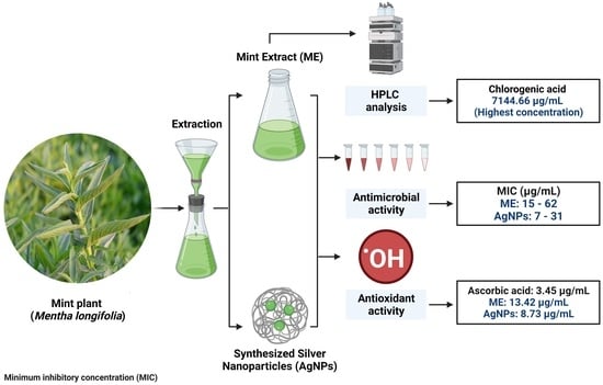
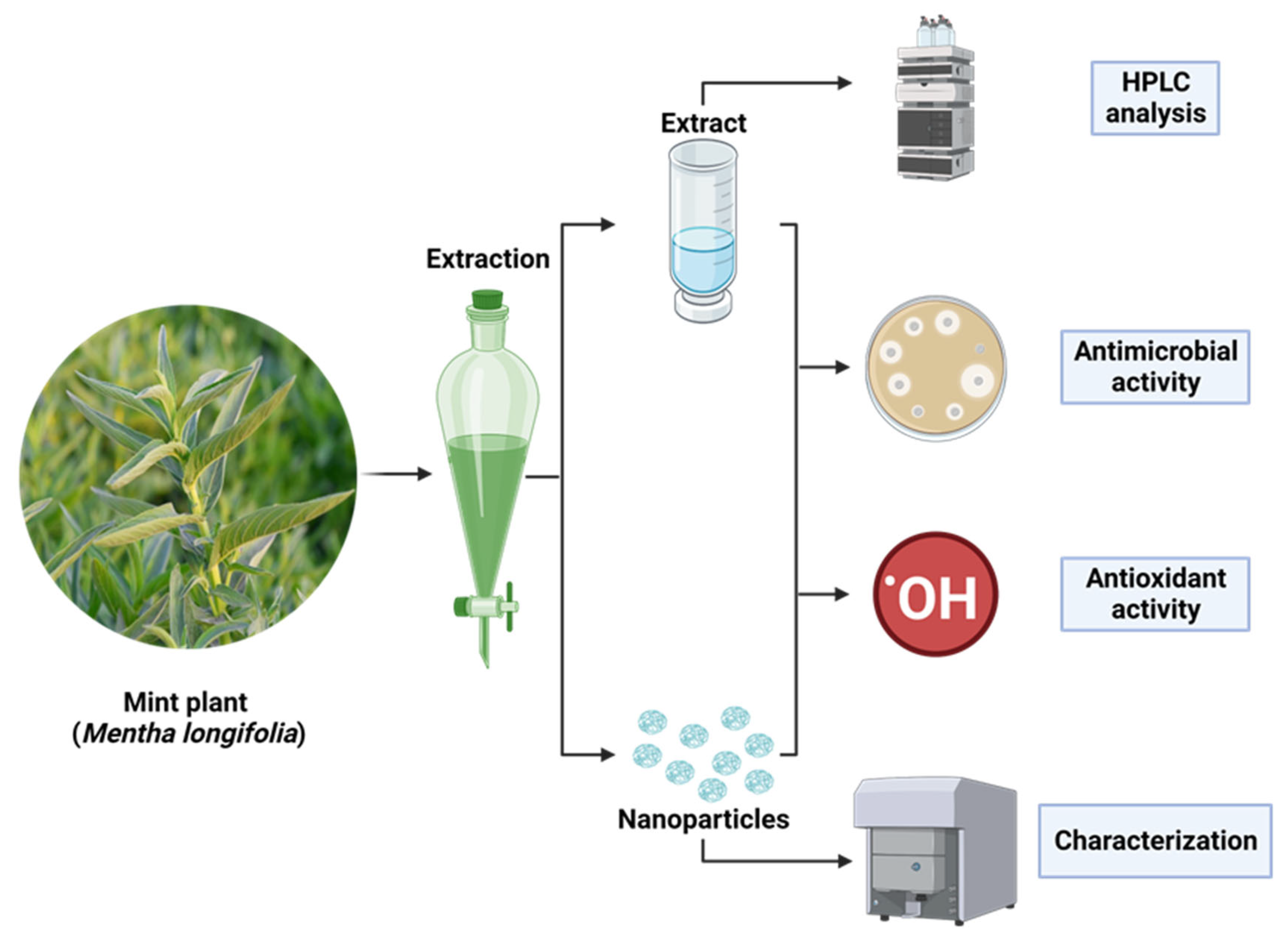
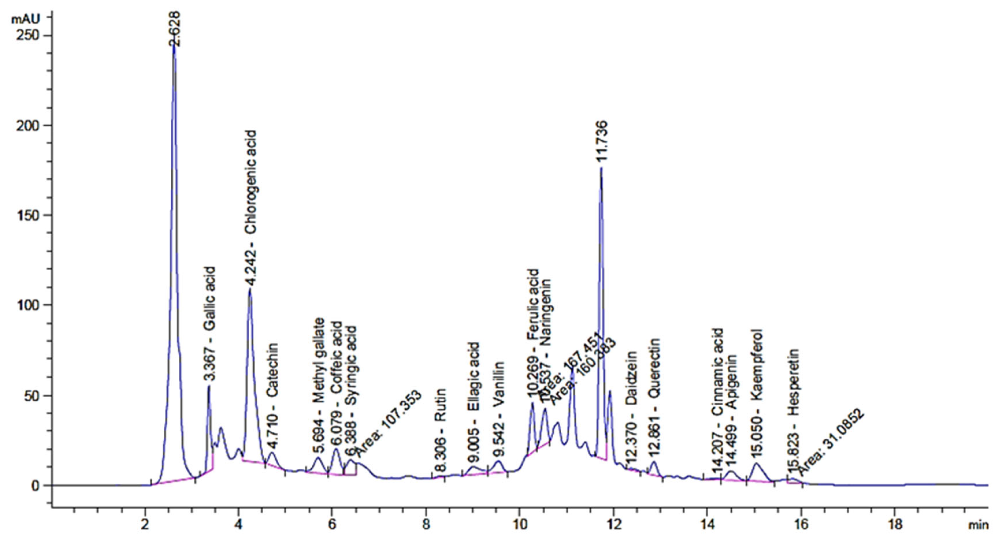
2.1. Phenolic and Flavonoid Characterizations of Mint Extract (ME)

Saudi mint was selected for the current study due to its traditional and medicinal applications. The antimicrobial and antioxidant activities of the extract as well as the created AgNPs were evaluated. The experiments on mint and mint-mediated AgNPs synthesis were described (Figure 1). The phenolic and flavonoid constituents of Saudi mint were analyzed via HPLC. The analysis showed the existence of various constituents with a dissimilar retention time, area, area%, and levels of concentration (Table 1 and Figure 2). In addition, the whole chemical construction of the detected compounds was documented (Figure S1). It was clear that the chlorogenic acid was found in a very high concentration (7144.66 µg/mL) compared to the other identified compounds. Catechin, gallic acid, and naringenin were detected in a narrow concentration range of 966.93, 980.13, and 958.50 µg/mL, respectively, followed by ellagic acid (841.23 µg/mL). Hesperetin (83.77 µg/mL), rutin (47.82 µg/mL), daidzein (43.25 µg/mL), and cinnamic acid (10.40 µg/mL) were detected at low concentrations. Chlorogenic acid, according to the study conducted by Yan et. al., showed an effective improvement in tolerance to glucose as well as insulin resistance and could decrease the abundance of microbial infections [32]. Choi et. al. reported that rutin could minimize the severity of inflammation and blood coagulation [33]. Several studies focused on the pharmacological applications of quercetin, as it could be used to prevent cancer proliferation, protect against Alzheimer’s Disease, and inhibit diabetes development, as well as possessing some antioxidant properties [34,35]. In another previous study, both catechin and gallic acid exhibited retardation potential against Helicobacter pylori, but a higher inhibitory effect was attributed to gallic acid compared to catechin [36]. Moreover, Bai et. al. documented that gallic acid had an anti-inflammatory action [37]. Ellagic acid also displayed several pharmaceutical activities, including antioxidant, antiproliferative, anti-inflammatory, antimutagenic, antiallergic, antiatherosclerotic, antiallergic, neuroprotective, and nephroprotective properties [38]. In the current study, the detected flavonoids (kaempferol and quercetin) were reported to acquire antifungal and antibacterial properties, and the results were consistent with Jan et. al. [39].

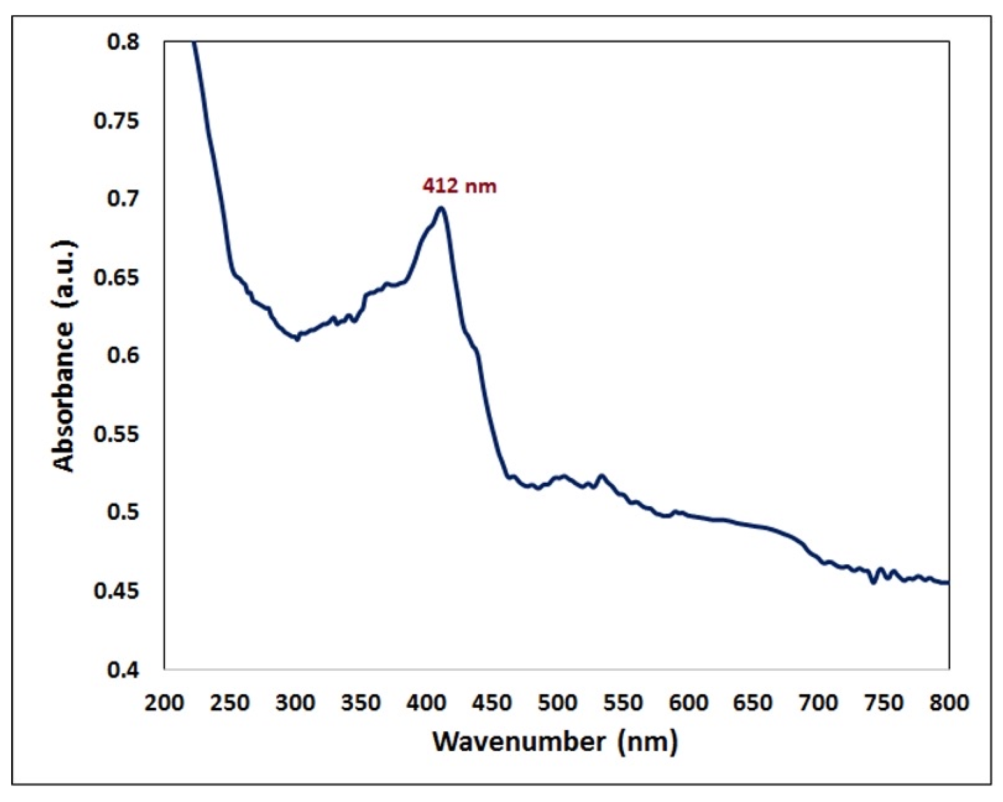
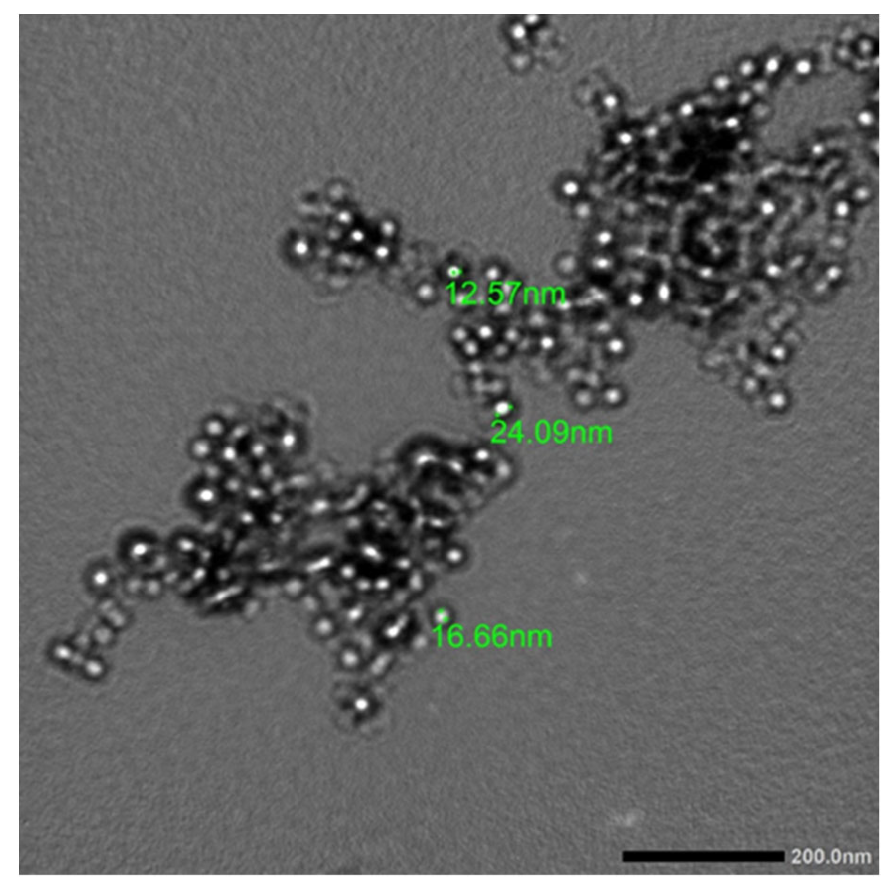
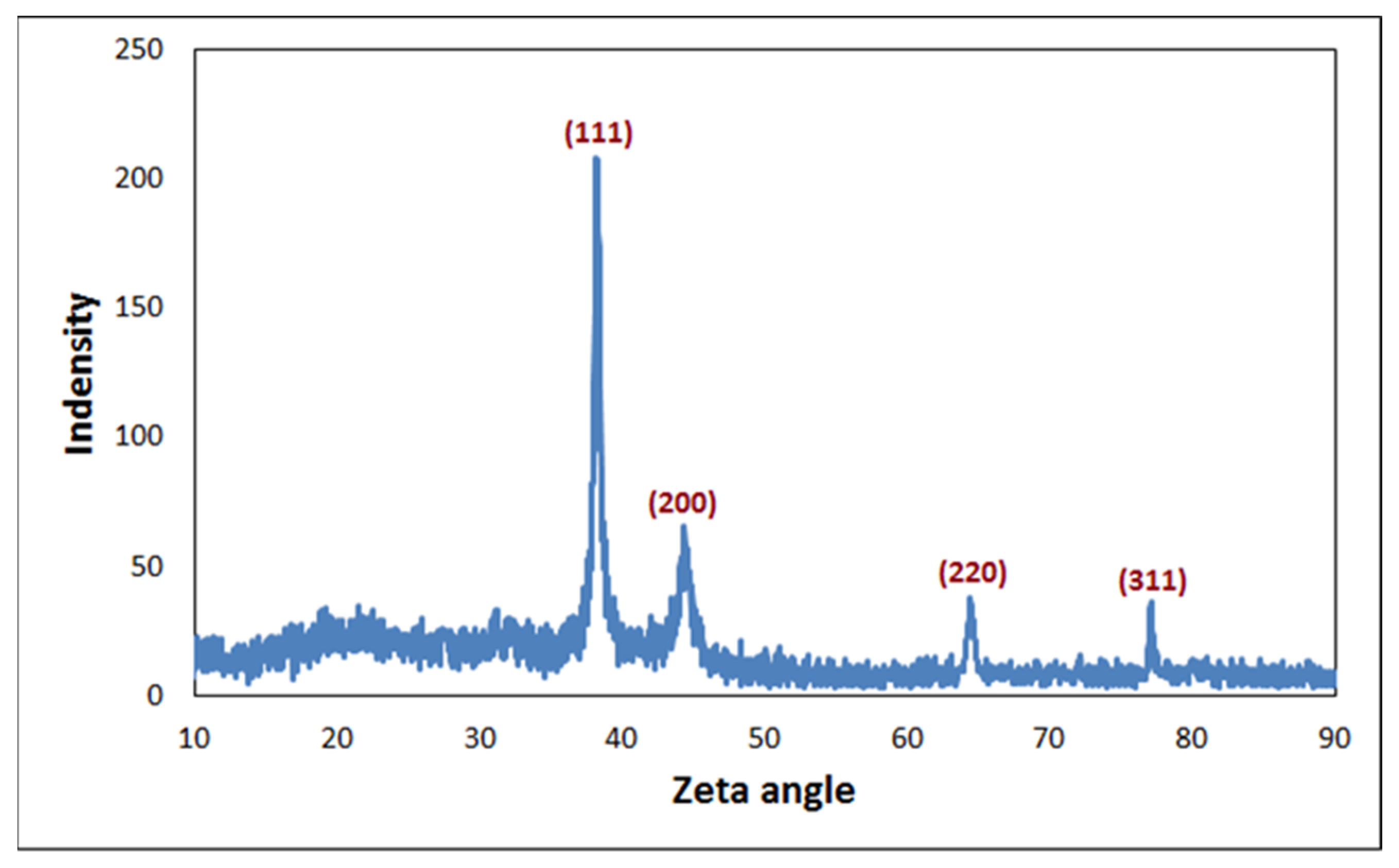
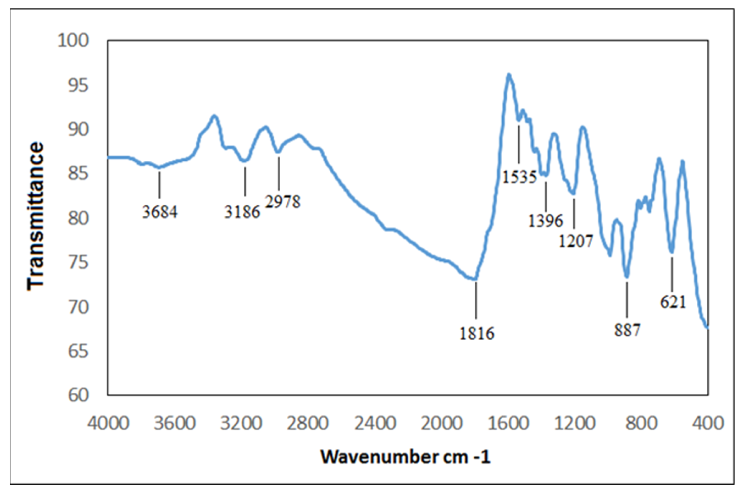
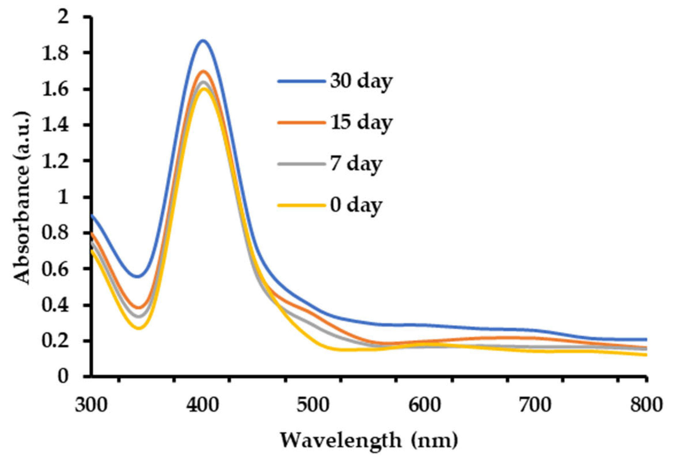
2.2. Characterization of Phyto-Created AgNPs by Mint Plant (ME)

In this study, the production of AgNPs in the mixture with a surface plasmon resonance (SRP) spectrum of 412 nm of maximum absorption was validated by using a UV–visible optical microscope (Figure 3). The obtained results were analogous to those published in the literature, in which biological extract was employed to produce AgNPs, and the UV–visible absorbance spectra indicated that the SPR band for Ag particles was in the 400–450 nm range [40,41,42]. The creation of AgNPs was reported when using several Mentha spp., such as Mentha arvensis and Mentha pulegium [25,26]. The synthesized AgNPs were characterized via transmission electron microscopy (TEM) in order to detect the morphology and diameter of the NPs (Figure 4). The synthesized AgNPs tended to be spherical with a mean diameter of 17.77 nm. The findings of the current study were consistent with Rizwana and Alwhibi [26], where the size of the created AgNPs, which were created by using M. pulegium extract, ranged between 4 and 60 nm. Moreover, the results were consistent with Bashir et. al. [40], who found that the size of the AgNPs created by using M. asiatica was 29 nm.
The quantitative elemental compositions of phyto-created AgNPs were analyzed and compared by using a scanning electron microscopy–energy dispersive X-ray (SEM-EDX) analysis. The existence of spherical AgNPs was found, and an elemental analysis confirmed the fabrication of AgNPs at a rate of 76.67% (Figure 5). The EDX spectra also clearly showed the presence of AgNPs with a strong peak at 3 KeV, which indicated that silver was the major element formation. Moreover, the distinct peak signals of oxygen and carbon were recorded to be 13.20% and 10.13%, respectively, which was related to the presence of metabolites, such as carbohydrates, proteins, and others [43,44].
An XRD pattern of the AgNPs that demonstrated the spherical structure of the formed AgNPs was observed (Figure 6). The typical diffraction peaks, at 2θ = 38.2°, 44.3°, 64.4°, and 77.1°, were ascribed to (111), (200), (220), and (311), respectively, which reflected the planes of the silver cubic face of the cubic-centered (FCC) crystalline form (JCPDS No 87-0720). The results showed that the AgNPs biosynthesized by reducing Ag+ ions from the biological extracts had a crystalline nature, and the sharp peak at 2θ of ~38.2° indicated the position (111) of Ag that corresponded to the face cubic center [43,45]. The results indicated that the biosynthesized AgNPs were composed of crystalline silver nano with a high purity.
The functional groups found in the AgNPs were characterized by using Fourier-transform infrared (FTIR) analysis [40,46]. The FTIR of the biogenic AgNPs displayed varying peaks at 3684, 3186, and 2978 cm−1 (OH, NH stretching, and aliphatic primary amines and OH broad stretching, respectively) (Figure 7). Other peaks appeared at 1816 cm−1 and 1535 cm−1 (amide-I band) and 1396 and 1207 cm−1 (CN stretching of the amines). Interestingly, the FTIR peaks at 887 and 621 cm−1 corresponded to the formation of Ag. Moreover, the stability of the AgNPs under different storage conditions at 30 °C was documented through UV/Vis spectra (Figure 8). The surface plasmon resonance (SPR) bands were symmetrical, where the peaks’ position remained stable over time for up to 30 days, which indicated that the AgNPs were stable.

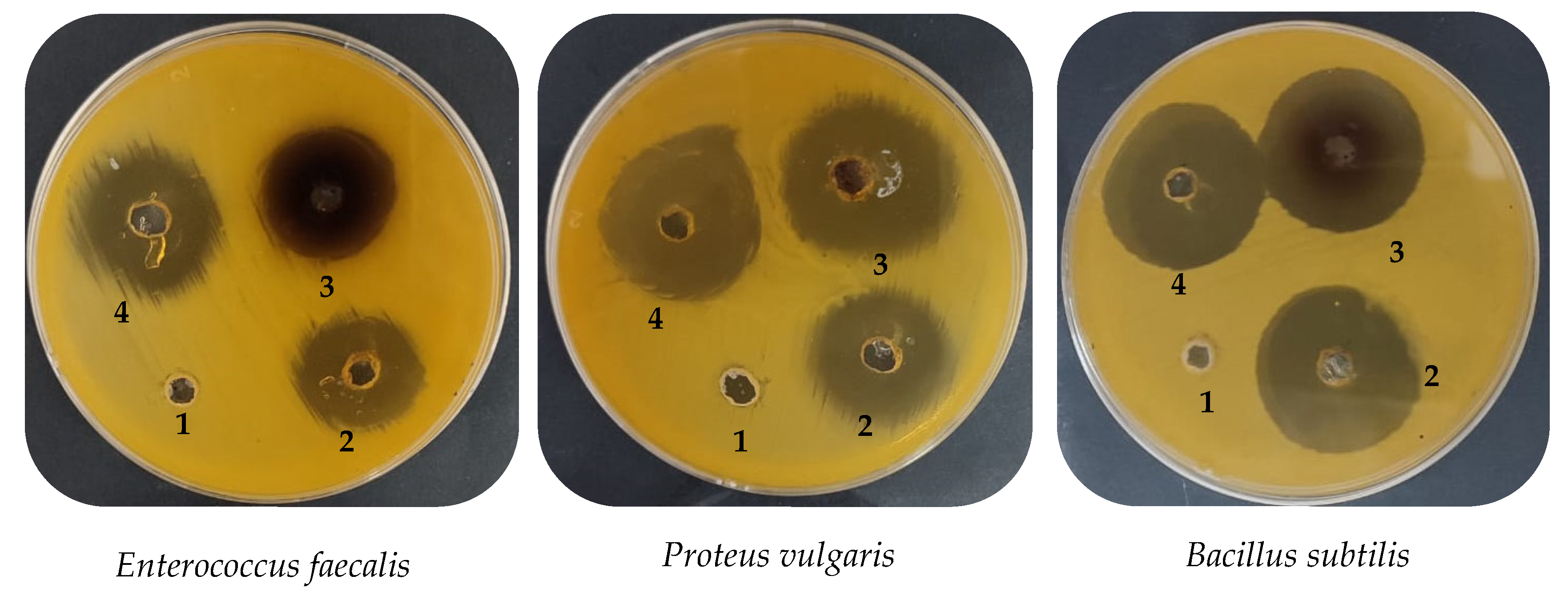
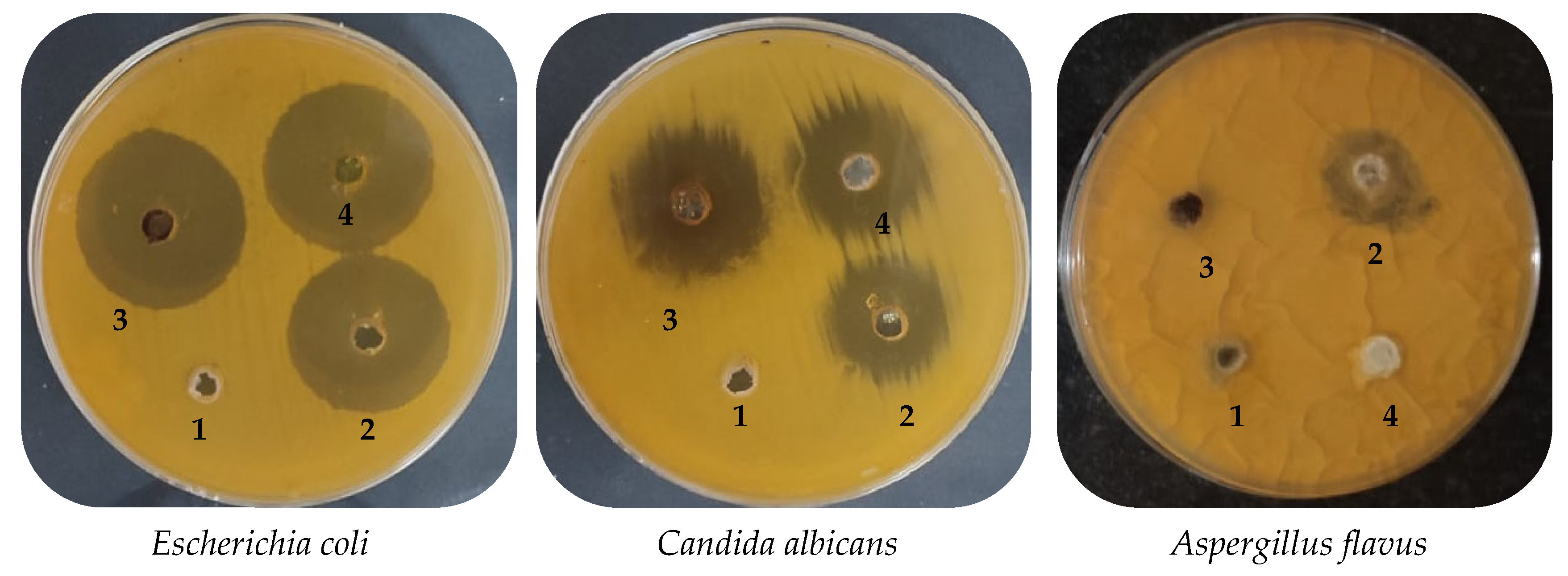
2.3. Antimicrobial Activity of Mint Extract and Phyto-Created AgNPs

The mint extract (ME) exhibited antimicrobial activity against all the tested microbes, except for A. flavus, with various inhibition zones of 30.67, 24.33, 27.83, 29.67, and 22.83 mm (Table 2 and Figure 9); various MICs of 15.70, 15.77, 7.87, 7.90, and 62.67 µg/mL; and various MBCs of 31.83, 62.33, 15.50, 15.67, and 124.33 µg/mL, compared with the inhibition zones of positive control gentamycin/ketoconazole against B. subtilis, E. faecalis, E. coli, P. vulgaris, and C. albicans, respectively. Various bacterial species, including Pseudomonas aeruginosa, Escherichia coli, Staphylococcus aureus, Shigella flexneri, and Klebsiella pneumoniae were inhibited by using an ethanolic extract of M. arvensis, and S. aureus was the most susceptible (a 21 mm inhibition zone diameter) of all of the tested bacteria in a similar way to standard antibiotic tetracycline [47]. Moldovan et. al. documented the antimicrobial activity of five Mentha spp., such as M. suaveolens var. rispate, M. rotundifolia, M. spicata subsp. rispate, M. piperita var. officinalis F. pallescens, and F. rubescens, with inhibition zones ranging from 8 to 26 mm against S. aureus, Listeria monocytogenes, Salmonella typhimurium, E. coli, and C. albicans [48].
The phyto-mediated AgNPs that were created by using mint extract (ME) exhibited greater antimicrobial activity compared to the ME. Moreover, the AgNPs showed a promising inhibition zone, compared to standard antibiotics tested against bacteria and C. albicans, with the exception of E. coli (Table 2 and Figure 9). These results are promising, in comparison with Aziz and Jassim [49], where the diameters of the inhibition zones of the AgNPs synthesized with mint against B. subtilis and E. coli were 25 mm and 20 mm, respectively. In another previous study, the ability of AgNPs synthesized by M. longifolia to inhibit the phytopathogens bacteria was recorded against Pectobacterium carotovorum, Xanthomonas vesicatoria, Xanthomonas oryzae, and Ralstonia solanacearum, and potential inhibitory activity was detected between 2–12 μg ml−m [50].
The minimum inhibitory concentration (MIC) of AgNPs was reported to be lower in the case of E. faecalis (15.53 µg/mL), E. coli (7.73 µg/mL), and C. albicans (31.33 µg/mL), but higher (7.83 and 15.57 µg/mL) in the case of B. Subtilis (7.83 µg/mL) and P. vulgaris (15.57 µg/mL) compared to the ME (Table 2). In addition, the antimicrobial property of the synthesized AgNPs was evaluated against common oral microbes, including Streptococcus mutans, Staphylococcus aureus, Candida albicans, and Enterococcus faecalis [51]. AgNPs, synthesized by using the leaves of Saudi Mentha pulegium, displayed antibacterial activity against Listeria monocytogenes, Salmonella typhimurium, Staphylococcus aureus, Pseudomonas aeruginosa, Escherichia coli, and Bacillus cereus, with the diameters of the inhibition zones ranging from 17 to 26 mm. The most effective antimicrobial activity was reported against E. coli, while less effective antimicrobial activity was observed against L. monocytogenes [52]. AgNPs were found to be more effective than commercial antibiotics in the current study, in line with Naghmouchi et. al. [52]. The cidal effect of the extract or AgNPs was determined by calculating the MBC/MIC Index, where the cidal properties increased as the index of the MBC/MIC decreased. Values less than four indicated that the present ME and AgNPs exerted a cidal effect. The mechanisms of the antimicrobial activity of phyto-mediated AgNPs were studied in several reports, where the denaturation of bacterial protein [53], disorders of the bacterial respiratory chain [19], and lipopolysaccharides changes [54] were reported.

2.4. Antioxidant Activity of Mint Extract and Phyto-Created AgNPs

The rise in DPPH scavenging (%) indicated the efficient antioxidant activity of the ME and AgNPs. There was a relationship between antioxidant activity and the concentration of either the ME or AgNPs (Table 3). A promising IC50 was recorded when using ME (13.42 µg/mL); however, the AgNPs were more effective with a lower IC50 (8.73 µg/mL). In the present study, the antioxidant activity of the ME and AgNPs was compared to that of ascorbic acid, which differed slightly, particularly at high concentrations of 500 and 1000 µg/mL. It was common to record a low value for IC50 (8.73 µg/mL) when using ascorbic acid, but the IC50 values of the ME and AgNPs were surprisingly low. The most frequently detected flavonoid in the ME was reported to execute several biofunctional antioxidant activities. The findings were consistent with previous studies based in Saudi Arabia, where M. longifolia and M. pulegium displayed efficient antioxidant activity but with different levels of activity [6]. Additionally, the ethanolic extract of fresh or dry M. suaveolens exhibited a strong antioxidant potential [5]. Mint extract (ME) appeared to represent a promising antioxidant, due to its enriched content of rutin (252.16 µg/g) [54] and naringenin (53.22 µg/g) [55] in a whole plant. Therefore, the AgNPs that were synthesized by using ME could be applied to the treatment of several diseases that are affected by oxidative stress. The findings of the current study are in line with a recent study conducted by Bashir et. al., where the green fabricated AgNPs, that were created by utilizing leaf extract of M. asiatica, displayed a significant antioxidant potential [40]. Due to the several side effects caused by a synthetic antioxidant, researchers may consider using a natural antioxidant as a safe alternative [56].

3. Materials and Methods

3.1. Chemicals Used in the Study

All of the materials used in the current study were obtained from Sigma Aldrich, (Taufkirchen, Germany), in an analytical grade form.

3.2. Collection and Extraction of the Mint Plant

Fresh mint plants (Mentha longifolia) were collected from local markets in the cities of Medina and Hail, Saudi Arabia. Areal parts, including the leaves and stems, were washed in distilled water, and then dried at 30 °C until a constant weight was obtained. The dried collected plant (100 g) was ground down and immersed in 300 mL of methanol under a shaking condition by using a magnetic stirrer for 24 h. The mixture was centrifuged for 10 min at 5000 rpm. The obtained supernatant was concentrated by using a rotary evaporator before being completely concentrated and redissolved in DEMSO, and it was stored at 4 °C for further study.

3.3. Detection of the Phenolic and Flavonoid Constituents of Mint Extract via HPLC

A total of 5 μL of mint extract as well as the AgNPs were added to the HPLC device (Agilent 1260 series, Agilent Technologies, Santa Clara, CA, USA) once the extraction process had been completed. The Eclipse C18 column employed had the following specifications: 4.6 mm and 250 mm, i.e., 5 μm and 40 °C. During the mobile phase, two buffers were used: buffer A (military Q water and 0.05% trifluoroacetic acid) and buffer B (acetonitrile and 0.05% trifluoroacetic acid). A flow rate of 0.9 mL/min was used to apply the buffers. The serial dilutions for buffer A were 82, 80, 60, 60, 82, 82, and 82% for 0, 0–5, 5–8, 12–15, 15–16, and 20–20 min, respectively. A flow rate of 0.9 mL/min was used for the buffers. The mobile phase took 20 min to run, with the following serial dilutions of buffer A: 82, 80, 60, 60, 82, 82, and 82% for 0, 0–5, 5–8, 12–15, 15–16, and 20–20 min, respectively. The presence of phenolic compounds and flavonoids was determined by using an ultraviolet (UV) detector with a wavelength of 280 nm. Based on the injected standard compounds, the amount of each was recorded [16,57].

3.4. Synthesis of Silver Nanoparticles (AgNPs)

The aqueous solution of 1 mM of AgNO3 was prepared by using distilled water. A total of 10 mL of mint extract was added to 90 mL of the aqueous solution of AgNO3. The reaction mixture was kept at 70 °C for 40 min, and the color change was subsequently observed. A color change from yellow to brown was used to detect the creation of AgNPs via Ag ion reduction.

3.5. Characterization of Synthesized AgNPs

The reaction mixture containing the AgNPs was characterized by using ultraviolet–visible spectroscopy (Perkin Elmer, MA, USA) to measure the absorption spectrum in a range from 200 up to 800 nm. The morphology and size of the created AgNPs were characterized by using a transmission electron microscope (TEM) (JEOL-JEM-Plus-1400, Tokyo, Japan). Fourier-transform infrared (FTIR) spectroscopy was applied to characterize the functional groups that are responsible for encapsulating and reducing the Ag ions to AgNPs by utilizing a pellet of potassium bromide on a Thermo Fisher Spectrometer (Thermo Scientific Model-Nicolet 6700, Hillsboro, OR, USA). The infrared spectrum was measured at a range of wavelength of 400–4000 cm−1. The elemental contents of the synthesized AgNPs were examined with an energy dispersive X-ray detector (EDX-FESEM-model no-JSM-7610F, Akishima, Japan) at an accelerating 30 kilo voltage. An X-ray diffraction (XRD) (Model diffractometer, Shimadzu 7000, Tokyo, Japan) analysis was conducted to determine the crystal structure of the created AgNPs.

3.6. Antimicrobial Activity of Mint Extract and AgNPs Using Agar Diffusion Method

All of the tested micro-organisms were provided by the American Type Culture Collection (Manassas, VA, USA). Mint extract and the AgNPs’ ability to inhibit micro-organisms (Bacillus Subtilis (ATCC 6633), Enterococcus faecalis (ATCC 10541), Escherichia coli (ATCC 8739), P. vulgaris, Candida albicans (ATCC 10221), and Aspergillus flavus) was evaluated in accordance with the Clinical and Laboratory Standards Institute (CLSI) guidelines. An agar diffusion technique was used to test the antibacterial activity. Mueller–Hinton agar (MHA) plates were used to maintain the tested bacteria and C. albicans, while a Dox medium was used for the tested fungus. A seedling of 2 × 10⁸ CFU/mL of tested bacteria and 2 × 10⁶ of the tested fungus was applied, and the MHA and Dox agar media were inoculated. A 6 mm well was then made in the inoculated media, and 100 µL of the mint extract and AgNPs was separately added to the wells. The cultured plates underwent a 24-h incubation period at 37 °C for the bacteria and C. albicans, while the tested fungi were incubated at 30 °C for 3 days. The inhibition zone that appeared following the incubation period was measured. The solvent employed for the extraction was applied as a negative control, while the standard antibiotic/antifungal treatments (Gentamycin, 0.1 mg/mL and ketoconazole, 0.1 mg/mL) were applied to inhibit the bacteria and fungi, respectively; additionally, silver nitrate (200 µg/mL) was utilized as a positive control [58,59].

3.7. The Minimal Inhibitory Concentration (MIC) Experiment

To determine the MIC of the mint extract and AgNPs against the tested micro-organisms, a Mueller–Hinton broth was used as the microdilution broth [60]. Various serial dilutions containing concentrations of mint extract and AgNPs ranging from 0.98 to 1000 μg/mL were prepared. We used 96-well polystyrene microtitrate plates, with 200 μL of each appropriate dilution of mint extract and AgNPs being dispensed per well. A fresh-tested micro-organism culture inoculum was created in sterile NaCl (0.85%) to match the required 1.0 McFarland turbidity. A final concentration of 2 × 10⁶ CFU/mL was obtained by inoculating each well with a total of 2 mL of the tested micro-organisms, and the plates were incubated at 35 °C for three days. The minimal inhibitory concentration was visually assessed to determine the lowest concentration required to inhibit the tested micro-organisms. Each microplate contained an inoculum of the tested micro-organisms without either mint extract or AgNPs as a positive control, and the extract and AgNPs without the tested micro-organisms as a negative control [61].

3.8. The Minimal Bactericidal Concentration (MBC) Experiment

A total of 100 mL of the tested micro-organisms that showed no growth onto the sterile Mueller–Hinton agar plates were subcultured from each well [62]. The cultivated plates were incubated at 35 °C for three days. The MBCs were detected as the lowest concentration of mint extract and AgNPs that visually showed no growth for the tested micro-organisms. To determine whether the mint extract and AgNPs possessed bactericidal or bacteriostatic properties, the ratios of the MBC/MIC were calculated. The extract was considered to be an efficient bactericidal agent if the MBC/MIC ratio was less than four times the MIC [63].

3.9. The Antioxidant Activity of the Mint Extract and AgNPs

Mint extract’s capacity to scavenge free radicals was assessed by using a DPPH (1,1-diphenyl-2-picrylhydrazyl) assay. A 0.1 mM DPPH solution in ethanol was created. In total, 3 mL of mint extract and AgNPs at different concentrations (ranging from 1.95 to 1000 µg/mL) were mixed with 1 mL of a 0.1 mM DPPH solution in ethanol. Only ethanol-soluble extracts were used, and various concentrations of those extracts were created by diluting the original extract. The mixture was shaken vigorously and then left to stand at 25 °C for 30 min. In the following step, a UV-VIS Milton Roy spectrophotometer was used to measure the absorbance at 517 nm. Ascorbic acid was used as a standard antioxidant agent. The concentration of mint extract or AgNPs necessary to inhibit 50% of the DPPH free radical (IC50) was calculated by utilizing a Log dose of the inhibition curve, as described by Al-Rajhi et. al. [23]. The percentage of the DPPH scavenging influence was recorded via the following formula:
DPPH   scavenging   % = A 0 A 1 A 0   × 100
where the control reaction absorbance is A0, and the mint or AgNPs’ reaction absorbance is A1.

3.10. Statistical Analysis

The biological activities of the mint extract and AgNPs were studied in triplicate, and the results were assessed as a mean value of the standard deviation (±SD).

4. Conclusions

In the current investigation, Saudi mint extract appeared to represent an ecofriendly source for creating AgNPs. In addition, several characterizations, including UV–visible spectroscopy, TEM, FTIR, and XRD confirmed the complete synthesis of AgNPs. The mint extract displayed numerous phenolic and flavonoid compounds, which were identified by HPLC, such as catechin, gallic acid, ellagic acid, naringenin, ferulic acid, kaempferol, and Quercetin. The Saudi mint extract was also shown to be enriched with chlorogenic acid. The findings of the present study indicate that mint extract and the synthesized AgNPs possess significant antimicrobial potential to combat several bacteria, including Bacillus Subtilis (ATCC 6633), Enterococcus faecalis (ATCC 10541), Escherichia coli (ATCC 8739), P. vulgaris, and unicellular fungus (C. albicans). Additionally, promising antioxidant activities were attributed to mint extract and AgNPs with an IC50 of 13.42 µg/mL and 8.73 µg, respectively, compared to the IC50 value of ascorbic acid (3.45 µg/mL). However, the AgNPs displayed the highest level of antimicrobial and antioxidant activities, which supported the use of nanotechnology to permit and enhance the application of plant-derived products during treatment.

Supplementary Materials

The following supporting information can be downloaded at: https://www.mdpi.com/article/10.3390/plants12112177/s1, Figure S1: Chemical constructions of the identified phenolic and flavonoid in mint extract.

Author Contributions

Conceptualization, H.Q.; formal analysis—antibacterial activity, A.S.B.; formal analysis—antifungal activity, N.A.; formal analysis—antioxidant activity, N.K.B. and B.A.; data curation, S.H.Q., H.M.A. and A.A.B.; investigation, H.Q., A.S.B., N.K.B., B.A. and N.A.; supervision, H.Q.; project administration, H.Q. and A.S.B.; funding acquisition, H.Q.; visualization and validation, H.Q., A.S.B., N.K.B., B.A., N.A., S.H.Q., H.M.A. and A.A.B.; writing—original draft preparation, H.Q.; writing—review and editing, H.Q., A.S.B., N.K.B., B.A., N.A., S.H.Q., H.M.A. and A.A.B. All authors have read and agreed to the published version of the manuscript.

Funding

This research was funded by the Scientific Research Deanship at the University of Ha’il, Saudi Arabia; project number BA-22 035.

Data Availability Statement

All data are available within the manuscript.

Acknowledgments

All authors thank the Scientific Research Deanship at the University of Ha’il, Saudi Arabia, for supporting this project (project number BA-22 035). Figure 1 and the graphical abstract were created with BioRender.com.

Conflicts of Interest

The authors declare no conflict of interest.

References

  1. Shahbazi, Y. Application of carboxymethyl cellulose and chitosan coatings containing Mentha spicata essential oil in fresh strawberries. Int. J. Biol. Macromol. 2018, 112, 264–272. [Google Scholar] [CrossRef] [PubMed]
  2. Sarikurkcu, C.; Eryigit, F.; Cengiz, M.; Tepe, B.; Cakir, A.; Mete, E. Screening of the Antioxidant Activity of the Essential Oil and Methanol Extract of Mentha pulegium L. from Turkey. Spectrosc. Lett. 2012, 45, 352–358. [Google Scholar] [CrossRef]
  3. Božović, M.; Pirolli, A.; Ragno, R. Mentha suaveolens Ehrh. (Lamiaceae) Essential Oil and Its Main Constituent Piperitenone Oxide: Biological Activities and Chemistry. Molecules 2015, 20, 8605–8633. [Google Scholar] [CrossRef] [PubMed]
  4. Dwivedy, A.K.; Prakash, B.; Chanotiya, C.S.; Bisht, D.; Dubey, N.K. Chemically characterized Mentha cardiaca L. essential oil as plant based preservative in view of efficacy against biodeteriorating fungi of dry fruits, aflatoxin secretion, lipid peroxidation and safety profile assessment. Food Chem. Toxicol. 2017, 106, 175–184. [Google Scholar] [CrossRef] [PubMed]
  5. Aldogman, B.; Bilel, H.; Moustafa, S.M.N.; Elmassary, K.F.; Ali, H.M.; Alotaibi, F.Q.; Hamza, M.; Abdelgawad, M.A.; El-Ghorab, A.H. Investigation of Chemical Compositions and Biological Activities of Mentha suaveolens L. from Saudi Arabia. Molecules 2022, 27, 2949. [Google Scholar] [CrossRef]
  6. Ahmed, A.M.; Ozbak, H.A.; Hemeg, H.A. Effect of essential oil of traditional two Saudi mint types and its possible role in cardiovascular and throat health. Int. J. Clin. Exp. Med. 2015, 8, 8060–8068. [Google Scholar]
  7. Anwar, F.; Abbas, A.; Mehmood, T.; Gilani, A.-H.; Rehman, N.-U. Mentha: A genus rich in vital nutra-pharmaceuticals—A review. Phytother. Res. 2019, 33, 2548–2570. [Google Scholar] [CrossRef]
  8. Burham, B.O.; Osman, O.A.; Nour, A.A.M. Chemical Composition and Antibacterial Activity of Essential oil of Mentha longifolia Leaf from Albaha Area Southern Saudi Arabia. Asian J. Biol. Life Sci. 2019, 8, 48–52. [Google Scholar] [CrossRef]
  9. Ibrahim, S.R.M.; Abdallah, H.M.; Mohamed, G.A.; Farag, M.A.; Alshali, K.Z.; Alsherif, E.A.; Ross, S.A. Volatile oil profile of some lamiaceous plants growing in Saudi Arabia and their biological activities. Z. Nat. C 2017, 72, 35–41. [Google Scholar] [CrossRef]
  10. El-Sayeda, A.E.-K.; Hesham, I.E.-A.; Zeinab, A.K.; Shahira, M.E.; Mohamed, A.S.; Amany, A.S. Chemical and biological study of Mentha suaveolens Ehrh. Cultivated in Egypt. J. Med. Plant Res. 2014, 8, 747–755. [Google Scholar] [CrossRef]
  11. El-Kashoury, E.-S.A.; El-Askary, H.I.; Kandil, Z.; Salem, M.A.; Sleem, A.A. Chemical composition and biological activities of the essential oil of Mentha suaveolens Ehrh. Z. Nat. C J. Biosci. 2012, 67, 571–579. [Google Scholar] [CrossRef]
  12. Farnad, N.; Heidari, R.; Aslanipour, B. Phenolic composition and comparison of antioxidant activity of alcoholic extracts of Peppermint (Mentha piperita). J. Food Meas. Charact. 2014, 8, 113–121. [Google Scholar] [CrossRef]
  13. Zekri, N.; Elazzouzi, H.; Ailli, A.; Gouruch, A.A.; Radi, F.Z.; El Belghiti, M.A.; Zair, T.; Nieto, G.; Centeno, J.A.; Lorenzo, J.M. Physicochemical Characterization and Antioxidant Properties of Essential Oils of M. pulegium (L.), M. suaveolens (Ehrh.) and M. spicata (L.) from Moroccan Middle-Atlas. Foods 2023, 12, 760. [Google Scholar] [CrossRef]
  14. Bayda, S.; Adeel, M.; Tuccinardi, T.; Cordani, M.; Rizzolio, F. The History of Nanoscience and Nanotechnology: From Chemical-Physical Applications to Nanomedicine. Molecules 2020, 25, 112. [Google Scholar] [CrossRef]
  15. Abdelghany, T.M. Stachybotrys chartarum: A Novel Biological Agent for The Extracellular Synthesis of Silver Nanoparticles and Their Antimicrobial Activity. Indones. J. Biotechnol. 2015, 18, 75. [Google Scholar] [CrossRef]
  16. Yahya, R.; Al-Rajhi, A.M.H.; Alzaid, S.Z.; Al Abboud, M.A.; Almuhayawi, M.S.; Al Jaouni, S.K.; Selim, S.; Ismail, K.S.; Abdelghany, T.M. Molecular Docking and Efficacy of Aloe vera Gel Based on Chitosan Nanoparticles against Helicobacter pylori and Its Antioxidant and Anti-Inflammatory Activities. Polymers 2022, 14, 2994. [Google Scholar] [CrossRef]
  17. Al-Rajhi, A.M.H.; Yahya, R.; Bakri, M.M.; Yahya, R.; Abdelghany, T.M. In situ green synthesis of Cu-doped ZnO based polymers nanocomposite with studying antimicrobial, antioxidant and anti-inflammatory activities. Appl. Biol. Chem. 2022, 65, 35. [Google Scholar] [CrossRef]
  18. Qanash, H.; Bazaid, A.S.; Aldarhami, A.; Alharbi, B.; Almashjary, M.N.; Hazzazi, M.S.; Felemban, H.R.; Abdelghany, T.M. Phytochemical Characterization and Efficacy of Artemisia judaica Extract Loaded Chitosan Nanoparticles as Inhibitors of Cancer Proliferation and Microbial Growth. Polymers 2023, 15, 391. [Google Scholar] [CrossRef]
  19. Abdelghany, T.M.; Al-Rajhi, A.M.H.; Al Abboud, M.A.; Alawlaqi, M.M.; Ganash Magdah, A.; Helmy, E.A.M.; Mabrouk, A.S. Recent Advances in Green Synthesis of Silver Nanoparticles and Their Applications: About Future Directions. A Review. BioNanoScience 2018, 8, 5–16. [Google Scholar] [CrossRef]
  20. Abdelghany, T.; Yahya, R.; Bakri, M.M.; Ganash, M.; Amin, B.H.; Qanash, H. Effect of Thevetia peruviana seeds extract for microbial pathogens and cancer control. Int. J. Pharmacol. 2021, 17, 643–655. [Google Scholar] [CrossRef]
  21. Al-Rajhi, A.M.H.; Qanash, H.; Almuhayawi, M.S.; Al Jaouni, S.K.; Bakri, M.M.; Ganash, M.; Salama, H.M.; Selim, S.; Abdelghany, T.M. Molecular Interaction Studies and Phytochemical Characterization of Mentha pulegium L. Constituents with Multiple Biological Utilities as Antioxidant, Antimicrobial, Anticancer and Anti-Hemolytic Agents. Molecules 2022, 27, 4824. [Google Scholar] [CrossRef] [PubMed]
  22. Al-Rajhi, A.M.H.; Yahya, R.; Abdelghany, T.M.; Fareid, M.A.; Mohamed, A.M.; Amin, B.H.; Masrahi, A.S. Anticancer, Anticoagulant, Antioxidant and Antimicrobial Activities of Thevetia peruviana Latex with Molecular Docking of Antimicrobial and Anticancer Activities. Molecules 2022, 27, 3165. [Google Scholar] [CrossRef] [PubMed]
  23. Al-Rajhi, A.M.H.; Qanash, H.; Bazaid, A.S.; Binsaleh, N.K.; Abdelghany, T.M. Pharmacological Evaluation of Acacia nilotica Flower Extract against Helicobacter pylori and Human Hepatocellular Carcinoma In Vitro and In Silico. J. Funct. Biomater. 2023, 14, 237. [Google Scholar] [CrossRef] [PubMed]
  24. Kamatou, G.P.; Vermaak, I.; Viljoen, A.M.; Lawrence, B.M. Menthol: A simple monoterpene with remarkable biological properties. Phytochemistry 2013, 96, 15–25. [Google Scholar] [CrossRef]
  25. Sivakumar, T.R.T.; Thangapandian, V.; Shankar, T. Silver Nanoparticles Synthesis of Mentha arvensis Extracts and Evaluation of Antioxidant Properties. J. Biosci. Bioeng. 2015, 1, 22–28. [Google Scholar]
  26. Rizwana, H.; Alwhibi, M.S. Biosynthesis of silver nanoparticles using leaves of Mentha pulegium, their characterization, and antifungal properties. Green Process. Synth. 2021, 10, 824–834. [Google Scholar] [CrossRef]
  27. Chung, I.-M.; Park, I.; Seung-Hyun, K.; Thiruvengadam, M.; Rajakumar, G. Plant-Mediated Synthesis of Silver Nanoparticles: Their Characteristic Properties and Therapeutic Applications. Nanoscale Res. Lett. 2016, 11, 40. [Google Scholar] [CrossRef]
  28. Ali, K.; Ahmed, B.; Dwivedi, S.; Saquib, Q.; Al-Khedhairy, A.A.; Musarrat, J. Microwave Accelerated Green Synthesis of Stable Silver Nanoparticles with Eucalyptus globulus Leaf Extract and Their Antibacterial and Antibiofilm Activity on Clinical Isolates. PLoS ONE 2015, 10, e0131178. [Google Scholar] [CrossRef]
  29. Das, B.; Dash, S.K.; Mandal, D.; Ghosh, T.; Chattopadhyay, S.; Tripathy, S.; Das, S.; Dey, S.K.; Das, D.; Roy, S. Green synthesized silver nanoparticles destroy multidrug resistant bacteria via reactive oxygen species mediated membrane damage. Arab. J. Chem. 2017, 10, 862–876. [Google Scholar] [CrossRef]
  30. Ganash, M.; Ghany, T.M.A.; Omar, A.M. Morphological and Biomolecules Dynamics of Phytopathogenic Fungi under Stress of Silver Nanoparticles. BioNanoScience 2018, 8, 566–573. [Google Scholar] [CrossRef]
  31. Bakri, M.M.; El-Naggar, M.A.; Helmy, E.A.; Ashoor, M.S.; Ghany, T.M.A. Efficacy of Juniperus procera Constituents with Silver Nanoparticles Against Aspergillus fumigatus and Fusarium chlamydosporum. BioNanoScience 2020, 10, 62–72. [Google Scholar] [CrossRef]
  32. Yan, Y.; Li, Q.; Shen, L.; Guo, K.; Zhou, X. Chlorogenic acid improves glucose tolerance, lipid metabolism, inflammation and microbiota composition in diabetic db/db mice. Front. Endocrinol. 2022, 13, 1042044. [Google Scholar] [CrossRef]
  33. Choi, S.S.; Park, H.R.; Lee, K.A. A Comparative Study of Rutin and Rutin Glycoside: Antioxidant Activity, Anti-Inflammatory Effect, Effect on Platelet Aggregation and Blood Coagulation. Antioxidants 2021, 10, 1696. [Google Scholar] [CrossRef]
  34. Khan, H.; Ullah, H.; Aschner, M.; Cheang, W.S.; Akkol, E.K. Neuroprotective Effects of Quercetin in Alzheimer’s Disease. Biomolecules 2019, 10, 59. [Google Scholar] [CrossRef]
  35. Bhatiya, M.; Pathak, S.; Jothimani, G.; Duttaroy, A.K.; Banerjee, A. A Comprehensive Study on the Anti-cancer Effects of Quercetin and Its Epigenetic Modifications in Arresting Progression of Colon Cancer Cell Proliferation. Arch. Immunol. Ther. Exp. 2023, 71, 6. [Google Scholar] [CrossRef]
  36. Díaz-Gómez, R.; López-Solís, R.; Obreque-Slier, E.; Toledo-Araya, H. Comparative antibacterial effect of gallic acid and catechin against Helicobacter pylori. LWT—Food Sci. Technol. 2013, 54, 331–335. [Google Scholar] [CrossRef]
  37. Bai, J.; Zhang, Y.; Tang, C.; Hou, Y.; Ai, X.; Chen, X.; Zhang, Y.; Wang, X.; Meng, X. Gallic acid: Pharmacological activities and molecular mechanisms involved in inflammation-related diseases. Biomed. Pharm. 2021, 133, 110985. [Google Scholar] [CrossRef]
  38. Sharifi-Rad, J.; Quispe, C.; Castillo, C.M.S.; Caroca, R.; Lazo-Vélez, M.A.; Antonyak, H.; Polishchuk, A.; Lysiuk, R.; Oliinyk, P.; De Masi, L.; et al. Ellagic Acid: A Review on Its Natural Sources, Chemical Stability, and Therapeutic Potential. Oxidative Med. Cell. Longev. 2022, 2022, 3848084. [Google Scholar] [CrossRef]
  39. Jan, R.; Khan, M.; Asaf, S.; Lubna; Asif, S.; Kim, K.M. Bioactivity and Therapeutic Potential of Kaempferol and Quercetin: New Insights for Plant and Human Health. Plants 2022, 11, 2623. [Google Scholar] [CrossRef] [PubMed]
  40. Bashir, I.; Khan, M.A.; Dilshad, E.; Siyo, B.; Hussain, E.; Ali, I. Antioxidant and anticancer silver nanoparticles of Mentha asiatica aerial part extract: A novel study. Inorg. Nano-Met. Chem. 2021, 1–7. [Google Scholar] [CrossRef]
  41. Al-Rajhi, A.M.H.; Salem, S.S.; Alharbi, A.A.; Abdelghany, T.M. Ecofriendly synthesis of silver nanoparticles using Kei-apple (Dovyalis caffra) fruit and their efficacy against cancer cells and clinical pathogenic microorganisms. Arab. J. Chem. 2022, 15, 103927. [Google Scholar] [CrossRef]
  42. Salem, S.S. Baker’s Yeast-Mediated Silver Nanoparticles: Characterisation and Antimicrobial Biogenic Tool for Suppressing Pathogenic Microbes. BioNanoScience 2022, 12, 1220–1229. [Google Scholar] [CrossRef]
  43. Aref, M.S.; Salem, S.S. Bio-callus synthesis of silver nanoparticles, characterization, and antibacterial activities via Cinnamomum camphora callus culture. Biocatal. Agric. Biotechnol. 2020, 27, 101689. [Google Scholar] [CrossRef]
  44. Soliman, M.K.Y.; Abu-Elghait, M.; Salem, S.S.; Azab, M.S. Multifunctional properties of silver and gold nanoparticles synthesis by Fusarium pseudonygamai. Biomass Convers. Biorefin. 2022, 1–18. [Google Scholar] [CrossRef]
  45. Salem, S.S.; Ali, O.M.; Reyad, A.M.; Abd-Elsalam, K.A.; Hashem, A.H. Pseudomonas indica-Mediated Silver Nanoparticles: Antifungal and Antioxidant Biogenic Tool for Suppressing Mucormycosis Fungi. J. Fungi 2022, 8, 126. [Google Scholar] [CrossRef]
  46. Alsharif, S.M.; Salem, S.S.; Abdel-Rahman, M.A.; Fouda, A.; Eid, A.M.; El-Din Hassan, S.; Awad, M.A.; Mohamed, A.A. Multifunctional properties of spherical silver nanoparticles fabricated by different microbial taxa. Heliyon 2020, 6, e03943. [Google Scholar] [CrossRef]
  47. Do Nascimento, E.M.M.; Rodrigues, F.F.G.; Campos, A.R.; Da Costa, J.G.M. Phytochemical prospection, toxicity and antimicrobial activity of Mentha arvensis (labiatae) from Northeast of Brazil. J. Young Pharm. 2009, 1, 210. [Google Scholar] [CrossRef]
  48. Moldovan, R.I.; Oprean, R.; Benedec, D.; Hanganu, D.; Duma, M.; Oniga, I.; Vlase, L. LC-MS analysis, antioxidant and antimicrobial activities for five species of Mentha cultivated in Romania. Dig. J. Nanomater. Biostruct. 2014, 9, 559–566. [Google Scholar]
  49. Aziz, W.J.; Jassim, H.A. Green Chemistry for the Preparation of Silver Nanoparticles Using Mint Leaf Leaves Extracts and Evaluation of their Antimicrobial Potential. World News Nat. Sci. 2018, 18, 163–170. [Google Scholar]
  50. Javed, B.; Nadhman, A.; Rehman, Z. Optimization, Characterization and Antimicrobial Activity of Silver Nanoparticles against Plant Bacterial Pathogens Phyto-synthesized by Mentha longifolia. Mater. Res. Express 2020, 7, 085406. [Google Scholar] [CrossRef]
  51. Harsha, L.; Jain, R.K.; Prasad, A.S. Synthesis, characterization, and antimicrobial activity of silver nanoparticles derived from Mentha X piperita + Ocimum tenuiflorum: An in vitro study. J. Adv. Pharm. Technol. Res. 2022, 13, S272–S276. [Google Scholar] [PubMed]
  52. Naghmouchi, S.; Al-Zaban, M.I.; Al-Zaben, M.; Alharbi, N.; Bahatheq, A. Generation and Characterization of Silver Nanoparticles in Mentha pulegium Extract and Evaluation of Biological Activities of the Prepared Extract. J. Nanomater. 2022, 2022, 5410274. [Google Scholar] [CrossRef]
  53. Patil, R.S.; Kokate, M.R.; Kolekar, S.S. Bioinspired synthesis of highly stabilized silver nanoparticles using Ocimum tenuiflorum leaf extract and their antibacterial activity. Spectrochim. Acta Part A Mol. Biomol. Spectrosc. 2012, 91, 234–238. [Google Scholar] [CrossRef]
  54. Lu, J.; Wang, Y.; Jin, M.; Yuan, Z.; Bond, P.; Guo, J. Both silver ions and silver nanoparticles facilitate the horizontal transfer of plasmid-mediated antibiotic resistance genes. Water Res. 2020, 169, 115229. [Google Scholar] [CrossRef]
  55. Mundlia, J.; Ahuja, M.; Kumar, P.; Pillay, V. Improved antioxidant, antimicrobial and anticancer activity of naringenin on conjugation with pectin. 3 Biotech 2019, 9, 312. [Google Scholar] [CrossRef]
  56. Xu, D.P.; Li, Y.; Meng, X.; Zhou, T.; Zhou, Y.; Zheng, J.; Zhang, J.J.; Li, H.B. Natural Antioxidants in Foods and Medicinal Plants: Extraction, Assessment and Resources. Int. J. Mol. Sci. 2017, 18, 96. [Google Scholar] [CrossRef]
  57. Qanash, H.; Yahya, R.; Bakri, M.M.; Bazaid, A.S.; Qanash, S.; Shater, A.F.; Abdelghant, T.M. Anticancer, antioxidant, antiviral and antimicrobial activities of Kei Apple (Dovyalis caffra) fruit. Sci. Rep. 2022, 12, 5914. [Google Scholar] [CrossRef]
  58. Al-Rajhi, A.M.H.; Ghany, T.M.A. Nanoemulsions of some edible oils and their antimicrobial, antioxidant, and anti-hemolytic activities. BioResources 2023, 18, 1465–1481. [Google Scholar] [CrossRef]
  59. Qanash, H.; Bazaid, A.S.; Alharazi, T.; Barnawi, H.; Alotaibi, K.; Shater, A.-R.M.; Abdelghany, T.M. Bioenvironmental applications of myco-created bioactive zinc oxide nanoparticle-doped selenium oxide nanoparticles. Biomass Convers. Biorefin. 2023, 1–12. [Google Scholar] [CrossRef]
  60. Khalid, A.; Ahmad, P.; Khandaker, M.U.; Modafer, Y.; Almukhlifi, H.A.; Bazaid, A.S.; Aldarhami, A.; Alanazi, A.M.; Jefri, O.A.; Uddin, M.; et al. Biologically Reduced Zinc Oxide Nanosheets Using Phyllanthus emblica Plant Extract for Antibacterial and Dye Degradation Studies. J. Chem. 2023, 2023, 3971686. [Google Scholar] [CrossRef]
  61. Parvekar, P.; Palaskar, J.; Metgud, S.; Maria, R.; Dutta, S. The minimum inhibitory concentration (MIC) and minimum bactericidal concentration (MBC) of silver nanoparticles against Staphylococcus aureus. Biomater. Investig. Dent. 2020, 7, 105–109. [Google Scholar] [CrossRef] [PubMed]
  62. Aldarhami, A.; Bazaid, A.S.; Alhamed, A.S.; Alghaith, A.F.; Ahamad, S.R.; Alassmrry, Y.A.; Alharazi, T.; Snoussi, M.; Qanash, H.; Alamri, A.; et al. Antimicrobial Potential of Pithecellobium dulce Seed Extract against Pathogenic Bacteria: In Silico and In Vitro Evaluation. BioMed Res. Int. 2023, 2023, 2848198. [Google Scholar] [CrossRef] [PubMed]
  63. Rajakumar, S.; Revanth, M.; Kasi, A.; Sujitha, P. Evaluating the antibacterial efficacy and minimal bactericidal concentration (MBC) of three different herbal extracts on recalcitrant endodontic pathogens—An in vitro study. J. Int. Oral Health 2022, 14, 266–272. [Google Scholar] [CrossRef]
Figure 1. Image illustrating the mint plant (Mentha longifolia) and mint mediate AgNPs synthesis, which was subjected to effective extraction methods and several appropriate assays to identify the phytochemical analysis of the mint extract and the antimicrobial and antioxidant activities of the extracted product compared to the synthesized AgNPs.
Plants 12 02177 g001
Figure 2. High-performance liquid chromatography (HPLC) of the detected phenolic and flavonoid constituents in mint extract.
Plants 12 02177 g002
Figure 3. UV–visible spectra of the phyto-created AgNPs.
Plants 12 02177 g003
Figure 4. Transmission electron microscopy (TEM) of phyto-created AgNPs.
Plants 12 02177 g004
Figure 5. SEM-EDX analysis for phyto-created Ag-NPs.
Plants 12 02177 g005
Figure 6. X-ray diffraction (XRD) analysis for phyto-created AgNPs.
Plants 12 02177 g006
Figure 7. Fourier-transform infrared spectrum of phyto-created AgNPs.
Plants 12 02177 g007
Figure 8. The UV/Vis spectra of AgNPs at different days at 30 °C.
Plants 12 02177 g008
Figure 9. Antimicrobial activity of mint extract and created AgNPs; negative control (1); positive control (2); AgNPs (3); and mint extract (4).
Plants 12 02177 g009aPlants 12 02177 g009b
Table 1. Identified contents of phenolic and flavonoid in mint extract.
CompoundRetention TimeAreaArea (%)Concentration (µg/mL)
Gallic acid3.367221.003.60966.93
Chlorogenic acid4.2421031.1516.817144.66
Catechin4.71078.591.28980.13
Methyl gallate5.694114.421.87316.86
Caffeic acid6.079160.352.61631.52
Syringic acid6.388107.351.75379.83
Rutin8.3068.030.1347.82
Ellagic acid9.00581.011.32841.23
Vanillin9.54285.371.39189.64
Ferulic acid10.269167.452.73574.81
Naringenin10.537160.382.62958.50
Daidzein12.37013.930.2343.25
Quercetin12.86164.901.06428.51
Cinnamic acid14.20711.15150.1810.40
Apigenin14.49974.851.22283.88
Kaempferol15.050156.562.55570.72
Hesperetin15.82331.090.5183.77
Table 2. The inhibitory activity, minimum inhibitory concentration (MIC), and minimal bactericidal concentration (MBC) of mint extract (ME) and synthesized AgNPs against the tested microbes.
Tested OrganismMean Inhibition Zones (mm)MIC (µg/mL)MBC (µg/mL)MBC/MIC
Index
Mean Inhibition Zones (mm) of AgNO3
AgNPsMEControl *AgNPsMEAgNPsMEAgNPsME200 µg/mL
Bacillus subtilis33.67 ± 1.1530.67 ± 0.5830.17 ± 1.267.83 ± 0.0615.70 ± 0.1731.17 ± 0.1431.83 ± 1.013.982.0324.22 ± 1.15
Enterococcus faecalis25.33 ± 1.5324.33 ± 0.5823.17 ± 0.2915.53 ± 0.1215.77 ± 0.1531.37 ± 0.2062.33 ± 0.292.023.9514.33 ± 1.53
Escherichia coli30.00 ± 1.7327.83 ± 0.2930.33 ± 0.297.73 ± 0.127.87 ± 0.067.90 ± 0.1715.50 ± 0.171.021.9721. 33 ± 1.53
Proteus vulgaris32.33 ± 1.5329.67 ± 0.5826.00 ± 0.5015.57 ± 0.067.90 ± 0.1015.70 ± 0.1715.67 ± 0.121.011.9820.33 ± 1.15
Candida albicans27.50 ± 0.8722.83 ± 0.2922.17 ± 0.2931.33 ± 0.1462.67 ± 0.2963.0 ± 0.87124.33 ± 1.150.021.9816.00 ± 1.00
Aspergillus flavusNANA17.33 ± 0.58------NA
* Gentamycin and ketoconazole were applied as positive controls for bacteria and fungi, respectively.
Table 3. DPPH scavenging (%) of mint extract (ME) and ascorbic acid.
Concentration (µg/mL)Ascorbic AcidMEAgNPs
DPPH Scavenging (%)SDDPPH Scavenging (%)SDDPPH Scavenging (%)SD
100095.50.00292.10.00293.70.003
50092.90.00485.60.00288.60.002
25091.10.00579.30.00283.00.003
12584.90.00672.60.00476.30.001
62.5076.40.00665.70.00469.90.003
31.2569.60.00459.50.00163.20.005
15.6362.60.00552.10.00756.10.006
7.8154.70.00345.50.00348.50.004
3.9044.30.00235.80.00641.60.005
1.9540.20.00730.50.00435.00.011
00.00.0040.00.0160.00.016
IC503.45 µg/mL13.42 µg/mL8.73 µg/mL
Disclaimer/Publisher’s Note: The statements, opinions and data contained in all publications are solely those of the individual author(s) and contributor(s) and not of MDPI and/or the editor(s). MDPI and/or the editor(s) disclaim responsibility for any injury to people or property resulting from any ideas, methods, instructions or products referred to in the content.

