Article

Biocontrol Potential of an Endophytic Pseudomonas poae Strain against the Grapevine Trunk Disease Pathogen Neofusicoccum luteum and Its Mechanism of Action

by
Jennifer Millera Niem
1,2,3,4,*,
Regina Billones-Baaijens
1,
Benjamin J. Stodart
1,2,
Pierluigi Reveglia
1,2,5 and
Sandra Savocchia
1,2
1
Gulbali Institute, Charles Sturt University, Locked Bag 588, Wagga Wagga, NSW 2678, Australia
2
Faculty of Science and Health, School of Agricultural, Environmental and Veterinary Sciences, Charles Sturt University, Locked Bag 588, Wagga Wagga, NSW 2678, Australia
3
UPLB Museum of Natural History, University of the Philippines Los Baños, College, Los Baños 4031, Laguna, Philippines
4
Institute of Weed Science, Entomology, and Plant Pathology, College of Agriculture and Food Science, University of the Philippines Los Baños, College, Los Baños 4031, Laguna, Philippines
5
Institute for Sustainable Agriculture, CSIC, 14004 Córdoba, Spain
*
Author to whom correspondence should be addressed.
Plants 2023, 12(11), 2132; https://doi.org/10.3390/plants12112132
Submission received: 31 March 2023 / Revised: 11 May 2023 / Accepted: 21 May 2023 / Published: 28 May 2023
(This article belongs to the Special Issue Integrated Disease Management in Fruit Crops)

Abstract

:
Grapevine trunk diseases (GTDs) impact the sustainability of vineyards worldwide and management options are currently limited. Biological control agents (BCAs) may offer a viable alternative for disease control. With an aim to develop an effective biocontrol strategy against the GTD pathogen Neofusicoccum luteum, this study investigated the following: (1) the efficacy of the strains in suppressing the BD pathogen N. luteum in detached canes and potted vines; (2) the ability of a strain of Pseudomonas poae (BCA17) to colonize and persist within grapevine tissues; and (3) the mode of action of BCA17 to antagonize N. luteum. Co-inoculations of the antagonistic bacterial strains with N. luteum revealed that one strain of P. poae (BCA17) suppressed infection by 100% and 80% in detached canes and potted vines, respectively. Stem inoculations of a laboratory-generated rifampicin-resistant strain of BCA17 in potted vines (cv. Shiraz) indicated the bacterial strain could colonize and persist in the grapevine tissues, potentially providing some protection against GTDs for up to 6 months. The bioactive diffusible compounds secreted by BCA17 significantly reduced the spore germination and fungal biomass of N. luteum and the other representative GTD pathogens. Complementary analysis via MALDI-TOF revealed the presence of an unknown cyclic lipopeptide in the bioactive diffusible compounds, which was absent in a non-antagonistic strain of P. poae (JMN13), suggesting this novel lipopeptide may be responsible for the biocontrol activity of the BCA17. Our study provided evidence that P. poae BCA17 is a potential BCA to combat N. luteum, with a potential novel mode of action.

1. Introduction

The biocontrol of plant pathogens has been intensified these recent years, particularly the use of beneficial microorganisms, among which are those described as endophytes [1,2,3,4]. The environmental pollution caused by the excessive use and misuse of agrochemicals and the hazard that these chemicals pose to human health and the ecosystem has led to strict regulations on synthetic pesticide use and the banning of the most hazardous from the market. This has paved the way for the discovery of alternative methods for controlling plant diseases. Among those alternatives is biological control. Biological control is the “decrease of inoculum or the disease-producing activity of a pathogen accomplished through one or more organisms” [5].
Due to the limited number of management options available for the control of grapevine trunk diseases (GTDs), biocontrol agents (BCAs) have been explored as a viable alternative. Studies have been conducted on the use of bacteria for the control of grapevine trunk diseases (GTDs) [6,7,8,9,10,11,12]. Bacillus subtilis, for instance, has shown antagonistic activity against GTD pathogens Eutypa lata [6,7,8] and Neofusicoccum parvum [9,10]. A study conducted by Trotel-Aziz et al. in 2019 [9] reported that B. subtilis strain PTA-271 protected grapevine cuttings against N. parvum by inducing the immune responses of the host plant and by detoxifying the fungal toxins terremutin and R-mellein. Erwinia herbicola [8] demonstrated biocontrol activity against E. lata in the laboratory although it has subsequently had little success in the field. Actinobacteria were also shown to be promising in controlling GTD pathogens in grafted nursery plants [11]. An endophytic strain, Streptomyces sp. VV/E1, and two rhizosphere Streptomyces isolates, Streptomyces sp. sp. VV/R1 and Streptomyces sp. VV/R4, also significantly reduced young grapevine decline infection by the fungal pathogens Dactylonectria sp., Ilyonectria sp., Phaeomoniella chlamydospora, and Phaeoacremonium minimum [9]. Another strain, Streptomyces sp. E1 + R4, suppressed black-foot disease of grapevine caused by Dactylonectria torresensis, D. macrodidyma, Ilyonectria liriodendri, and D. alcacerensis [12]. The use of Pseudomonas spp. as BCAs has been widely studied within various pathosystems, including potato scab [13], late blight [14], Rhizoctonia root rot on bean [15], damping-off and root rot in tomato [16], anthracnose in beans [17], post-harvest diseases of stone fruit [18], and take-all disease of wheat [19,20,21]. In grapevines, Pseudomonas spp. were shown to suppress Botrytis cinerea [22,23], root-rot pathogens [24], and Rhizobium vitis, which causes crown gall [25]. Pseudomonas spp. Were found to be present in the grapevine endosphere [26,27,28]. The presence of Pseudomonas spp. in the inner grapevine tissues may be an indication of their ability to colonize and survive in planta.
Recently, the antagonistic activity of 10 endophytic strains of Pseudomonas from grapevines was demonstrated against the pathogens associated with Botryosphaeria, Eutypa dieback, and Esca/Petri disease [29]. The in vitro inhibition of these pathogens may imply that these antagonistic strains are potential BCAs. However, the demonstrated inhibition of a pathogen in vitro does not always translate to effective biocontrol in vivo. A successful BCA must also be able to survive in the host plant and efficiently colonize the plant tissues for a sufficient period to provide protection [13,30]. Hence, in planta assays to investigate the efficacy of antagonistic strains of Pseudomonas against GTD pathogens in grapevines and their colonization potential of the plant is essential.
Plant protection against pathogen infection using Pseudomonas spp. is mediated by a variety of secondary metabolites, among which are antibiotics such as 2,4-diacetylphloroglucinol (DAPG) [21,31,32,33] and phenazine and its derivatives [13,34,35,36]. These antibiotics play a role in the induction of systemic resistance, biofilm formation, and membrane permeability. Beneficial fluorescent Pseudomonas spp. also enhance plant growth and exert antagonistic activity against plant pathogens by producing siderophores [37,38,39], which cause a reduction in iron availability for other microorganisms, including plant pathogens, resulting in less efficient iron-sequestering systems [40,41]. Cyclic lipopeptides (CLPs) are also produced by many strains of Pseudomonas spp. [42,43,44,45]. CLPs can insert into membranes and perturb their function, which results in broad antibacterial and antifungal activities [46]. Some CLPs also act as a biosurfactant, providing the organism improved surface colonization and utilization of surface-bound substrates [47]. The production of volatile organic compounds [33,48,49,50], niche exclusion [51,52], production of lytic enzymes, [53] and induction of systemic resistance [54,55,56,57,58,59,60] have also been implicated in the plant-protecting capacity for different strains of Pseudomonas. Thus, the mechanism of action involved in the control of GTD pathogens by these Pseudomonas strains requires further investigation. Understanding their mode of action may help determine their capacity to efficiently suppress disease in natural environments.
This study aims to determine the potential of an antagonistic Pseudomonas poae strain (BCA17) to control the GTD pathogen N. luteum. The objectives are as follows: (1) to investigate the ability of Pseudomonas spp. to suppress N. luteum in detached canes and potted grapevines; (2) to demonstrate the endophytic colonization of grapevine tissues by a rifampicin-resistant BCA17 strain in pre-planted cuttings and potted vines; and (3) to investigate a potential direct mechanism of BCA17 in the control of GTD pathogens and disease.

2. Results

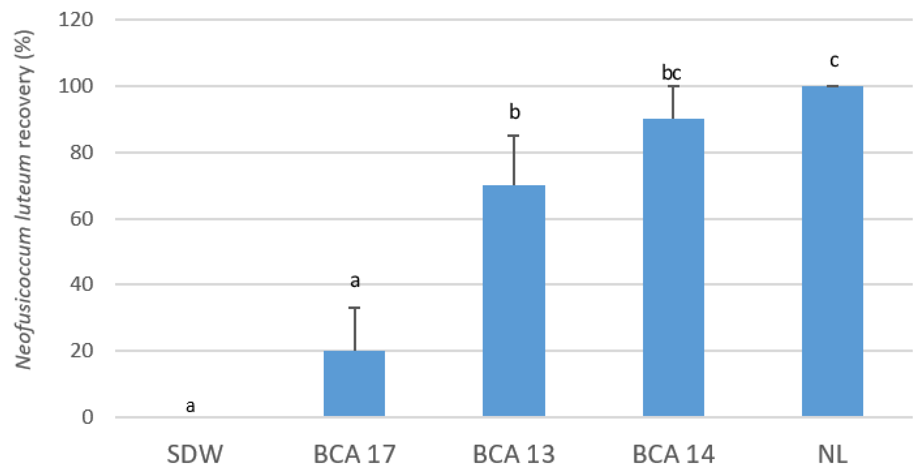
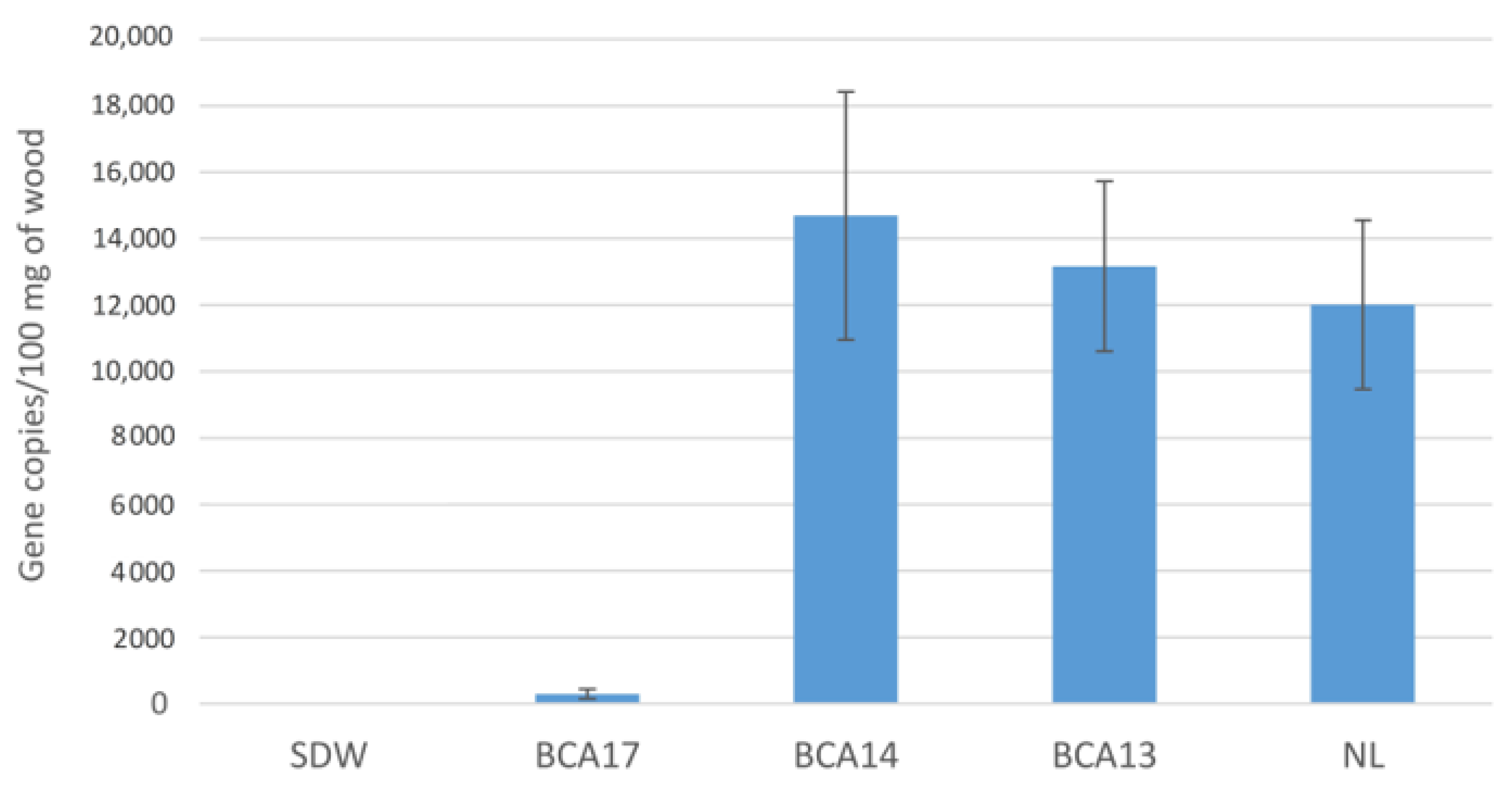
2.1. Suppression of Neofusicoccum luteum Infection in Planta by Pseudomonas Strains

Of the 10 antagonistic strains of Pseudomonas inoculated on detached canes, six significantly reduced the recovery of Neofusicoccum luteum (p ≤ 0.005), while only BCA17 resulted in no pathogen recovery 4 weeks post-inoculation of conidia (Figure 1). Pathogen recovery in canes treated with BCA12, BCA15, and BCA20 (56–78%) was not significantly different from that in the positive control canes (100%) inoculated with N. luteum only.
At 3 months post-inoculation in potted grapevines, BCA17 resulted in the lowest percentage recovery of N. luteum at 20% (p ≤ 0.001); BCA13 and BCA14 resulted in 70% and 90% recovery; and N. luteum was not recovered from the negative control vines, which were uninoculated but treated with sterile distilled water (SDW) (Figure 2).

Assessment of N. luteum Infections by qPCR

A quantitative PCR assay of tissue samples from potted vines treated with strains of Pseudomonas prior to inoculation with N. luteum confirmed the previous results. The amount of Botryosphaeriaceae DNA detected in vines treated with BCA17 was significantly lower, with 300 copies of DNA detected compared to 12,000 for infected vines not treated with BCA (N. luteum only), a 40-fold reduction (Figure 3). Grapevine plants treated with BCA13 and BCA14 did not significantly reduce N. luteum with an average of 3000–14,000 copies of Botryosphaeriaceae DNA detected. Pathogen DNA was not detected in any of the uninoculated vines, indicating that there was no background infection in the plant material due to the Botryosphaeriaceae species.

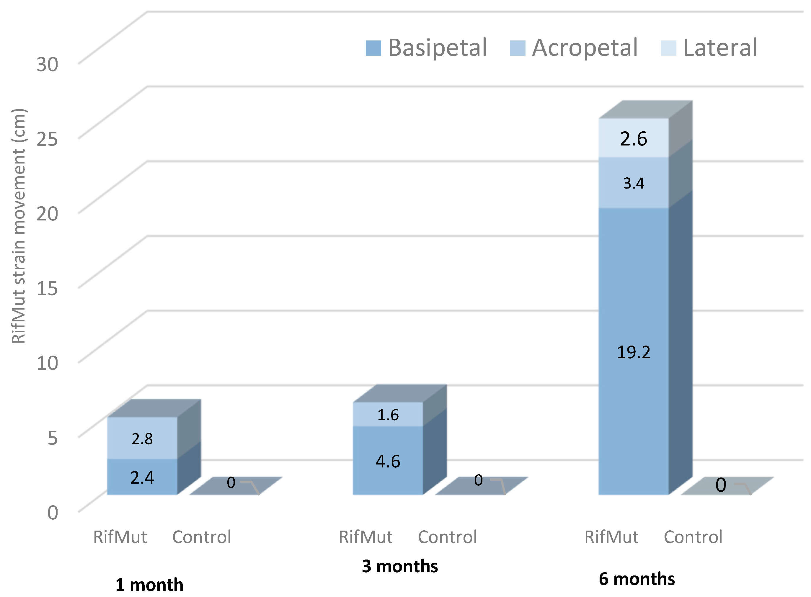
2.2. Pseudomonas Colonization and Establishment in Grapevine Tissues

2.2.1. Pre-Planting Application of Pseudomonas RifMut Strain to Canes

The analysis of canes treated for 24 h with the rifampicin-resistant (RifMut) strain of Pseudomonas poae BCA17 prior to rooting demonstrated that Pseudomonas spp. can be reisolated from the five randomly selected canes, revealing that the bacterial strain was translocated into the grapevine canes (Figure 4). The RifMut strain was reisolated in all five wood pieces obtained from the base and middle sections (approximately 13–15 cm distance) of the canes, while only two wood pieces of the top end of the canes were positive to RifMut (Figure 4). Bacteria were not re-isolated from canes dipped in Ringer’s solution only.
Subsequent re-isolations post-rooting showed the RifMut strain still persisted in the grapevine tissues at 8 weeks post-treatment. The RifMut strain was present in all the 10 rooted vines from the base and middle sections, while the RifMut strain was only re-isolated from three vines of the top sections at this time-point of analysis (Figure 4). Bacteria were not recovered from the control canes in Ringer’s solution at post-rooting.

2.2.2. Post-Planting Application of Pseudomonas

The assessment of potted grapevine plants inoculated with RifMut shows that at one month after inoculation RifMut was reisolated up to 2.4 and 2.8 cm below and above the inoculation point, respectively, from all sub-sampled vines (Figure 5). Such acropetal and basipetal movements of the RifMut strain were also observed at 3 months after inoculations. At 6 months, the entire plant was colonized by RifMut for the five sampled vines. The bacterial strain was reisolated from the apical tip of the living tissue down to the base of the trunk below the ground. Subsequent re-isolation on lateral shoots also indicates lateral movement of RifMut up to a distance of 3 cm in the shoot sections. Bacteria were not recovered from control plants inoculated with SDW only.

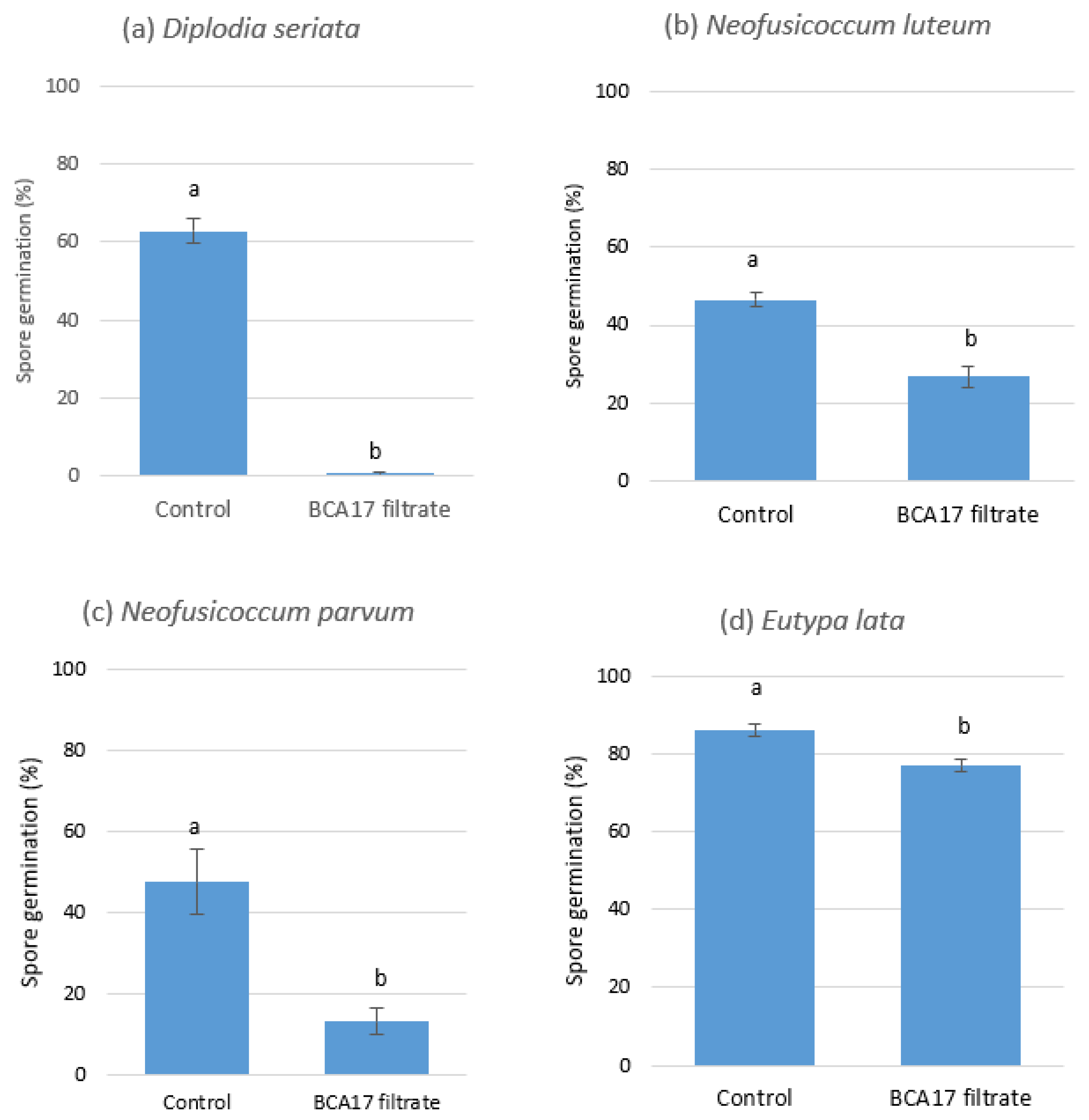
2.3. Bioactivity of P. poae BCA17 Culture Filtrate on Mycelial Growth and Spore Germination of GTD Pathogens

2.3.1. Effect of BCA17 Culture Filtrate on Mycelial Growth

The cell-free culture filtrates derived from BCA17 were inhibitory to the mycelial growth of Diplodia seriata DAR79990, N. luteum DAR81016, N. parvum DAR80004, and Eutypa lata WB052, regardless of whether these were obtained from cultures co-inoculated with the pathogen or not (Figure 6a–d). The mycelial weight of D. seriata DAR79990 grown in NB only was significantly higher (76.26 mg; p ≤ 0.01) when compared to mycelia grown in the culture filtrate of BCA17, where mycelial biomasses of 5.23 mg (filtrate obtained from BCA17 + pathogen co-culture) and 1.87 mg (filtrate obtained from BCA17 only culture) were obtained (Figure 6a). A similar pattern was observed with N. luteum DAR81016 with 181.7 mg of mycelial biomass (Figure 6b) and N. parvum DAR80004 with 190.63 mg in filtrates (Figure 6c) containing NB only. N. luteum DAR81016 and N. parvum DAR80004 did not grow in the culture filtrate of BCA17 (Figure 6b,c). E. lata WB052 was typically slow growing and the mycelial weight in NB was 15 mg, which was significantly different from the biomass of the pathogen that was grown in the culture filtrate of BCA17 (p ≤ 0.01) where no growth of the pathogen was observed after 7 days (Figure 6d). No significant differences in fungal biomass (p ≥ 0.05) were observed between culture filtrates derived from broth cultures with or without the pathogen (Figure 6a–d).

2.3.2. Effect of BCA17 Culture Filtrate on Spore Germination

The spore germination of D. seriata DAR79990, N. luteum DAR81016, N. parvum DAR80004, and E. lata WB052 was inhibited by the culture filtrates of BCA17 at varying degrees 24 h after incubation. The BCA17 culture filtrate reduced D. seriata DAR79990 spore germination to 1%, which was significantly lower (p ≤ 0.0001) than the untreated control at 60% germination (Figure 7a). The BCA17 culture filtrate also reduced N. luteum DAR81016 (Figure 7b) and N. parvum DAR80004 (Figure 7c) spore germination with 27 and 13% germination, respectively, which was significantly lower than the untreated control at 48 and 47% spore germination, respectively. E. lata WB052 was the least sensitive to the culture filtrate resulting in 86% germination in the control treatment and 77% in the BCA17 culture filtrate (Figure 7d).

2.4. Identification of Secondary Metabolite Biosynthetic Gene Clusters

The secondary metabolites and their corresponding biosynthetic gene clusters are summarized in Supplementary Table S1. The genome features of the antagonistic P. poae strains BCA13, BCA14, and BCA17 and the non-antagonistic P. poae strain JMN1 were very similar. Genome analysis using antiSMASH 4.0 [61] identified the consistent presence of gene clusters coding for the synthesis of the siderophore pyoverdine and APEs (aryl polyenes) Vf and Ec, which comprised the largest biosynthetic gene cluster (BGC). The four BCA strains of P. poae also displayed a notable array of BGC associated with the production of various cyclic lipopeptides (CLPs), including rhizoxin, viscosin, orfamide, putisolvin, poaeamide, bananamide, entolysin, anikasin, white-line-inducing principle (WLIP), cichopeptin, tolaasin, sessilin, xantholysin, and arthrofactin (Supplementary Table S1). Some of these CLPs have previously been described to have an antifungal property.

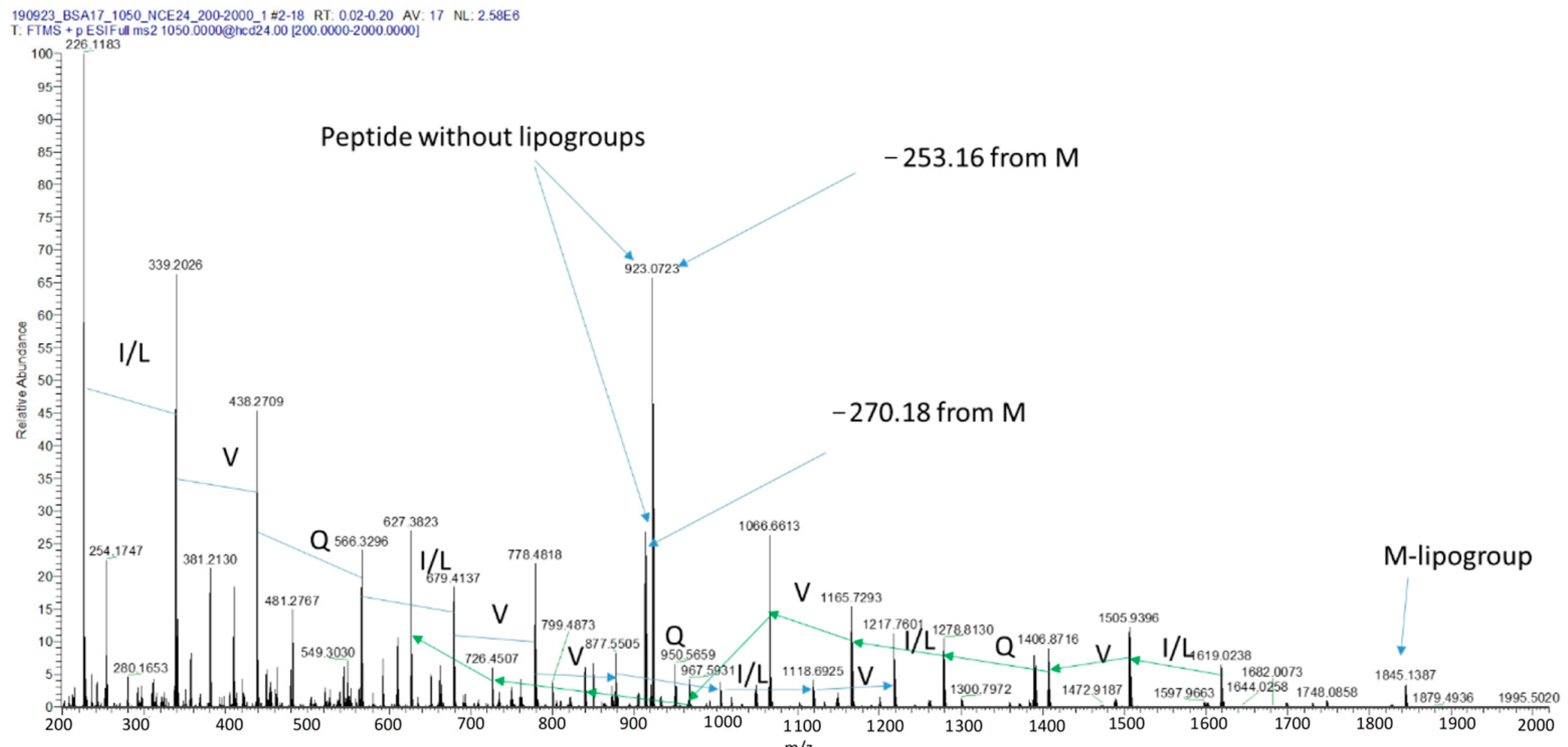
2.5. Identification of the Bioactive Lipopeptides Produced by BCA17

Due to the abundance of CLP-associated genes within the strains of P. poae, a complementary analysis was undertaken to verify which lipopeptides are specifically produced by BCA17, and therefore could be involved in the biocontrol activity of the antagonistic P. poae BCA17. Utilizing MALDI-TOF MS, a possible lipopeptide with a [M + H]+ mass to charge ratio at 2098.2913 m/z was detected in P. poae BCA17 (Figure 8) but was absent in P. poae JMN1 (Figure 9). A comprehensive assessment of literature articles indicated that the [M+H]+ ion at 2098.2913 m/z is likely a new lipopeptide; however, the sequence of the compound could not be confirmed with MS/MS analysis due to a lack of specific MS data within online databases.
From de novo sequencing, the partial protein sequence was confirmed to be leu-val-gln-leu-val-val-gln-leu-val (LVQLVVQLV) (C48H87N11O12) (Figure 10), with a monoisotopic mass of 1009.654. This finding does not directly match with the literature [62,63,64,65,66], but a ring opening reaction with ammonia confirmed that the unknown lipopeptide is cyclic in nature. Therefore, and in accordance with the literature, this new lipopeptide is likely related to or belongs to the group of tolaasin. The partial identified sequence shares 8 out of 9 amino acids with the L4VSLVVQLV12 sequence of tolaasin with only 1difference (a Glu instead of the Ser present in tolaasin) [66,67].

3. Materials and Methods

3.1. Plant Materials

Vitis vinifera (cv. Shiraz) was used for all in planta experiments. One-year-old dormant canes with no apparent GTD symptoms were collected from grapevines in the Hilltops and Riverina regions, New South Wales (NSW), Australia, in the winters of 2017 and 2019, respectively. All canes were stored at 4 °C for 4–6 weeks until rooting or used in the detached cane assays. For rooting, canes were callused in moist perlite in heat mats maintained at ~30 °C under ambient, winter conditions until roots developed. Rootlings with well-developed roots were transferred to 10 L pots filled with commercial garden soil (compost, 60%; washed sand, 20%; screened loam, 20%). All vines were maintained in the glasshouse (17–27 °C).

3.2. Bacterial Strains, Culture Conditions, Inoculum Preparation

The 10 strains of Pseudomonas (nine from the Pseudomonas poae group and one closely related to P. moraviensis) that inhibited the mycelial growth of GTD pathogens in dual culture tests [29] were selected for in planta assays. Bacterial cells were obtained from single colonies of each antagonistic strain and stored in 20% glycerol at −80 °C.
To prepare the bacterial inoculum, a loopful of the bacterial suspension in glycerol was streaked onto nutrient agar (NA, Oxoid Ltd., Hampshire, England) and incubated at 25 °C for 2 days. A loopful of the subsequent bacterial growth was then transferred to nutrient broth (NB, Oxoid Ltd.) and incubated for 24 h in an OM15 orbital shaking incubator (RATEK Instruments Pty. Ltd., Victoria, Australia) at 25 °C, 150 rpm. The optical density of the bacterial broth culture was measured at 600 nm (UNICAM 8625 UV/VIS spectrometer, UNICAM Limited, Cambridge, UK) and the bacterial density was adjusted to 1010 CFU, which was further confirmed by dilution plating.

3.3. Fungal Isolates

Neofusicoccum luteum was used as a model pathogen for all the challenge inoculation experiments. Grapevine isolates of N. luteum (DAR 80983, 81013, 81016) were used for inoculating both detached canes and potted vines. All isolates were previously identified by sequence analyses of the ITS, EF1-α, and β-tubulin genes [61]. The three isolates were cultured on potato dextrose agar (PDA, Difco Laboratories, Detroit, MI, USA) amended with chloramphenicol (PDAC, 100 mg/L of agar) and incubated at 25 °C overnight in the dark before being transferred under UV light (Phillips 20 w UVB) and exposed to a 12 h light–dark regime at 18–22 °C for 4–5 weeks until fruiting bodies developed. To harvest the conidia, fruiting bodies were scraped and ground with a sterile mortar and pestle before sieving using two layers of sterile Miracloth (Merk Millipore). The conidial suspensions from each of the three isolates were combined and standardized to 1 × 10⁴ conidia/mL and 20 µL (200 conidia) was used for the subsequent inoculations. The viability of the spores was determined by serial dilution plating at three replicate plates.
In studying the antagonistic mechanisms of P. poae in vitro, four GTD fungal pathogens were used: Diplodia seriata DAR79990, N. parvum DAR80004, N. luteum DAR81016, and Eutypa lata WB052 were used. The fungal pathogens were originally isolated from grapevines exhibiting symptoms of GTD in Australian vineyards and were previously identified by a partial sequencing of the ribosomal RNA gene [68,69,70].

3.4. Challenge Inoculation

3.4.1. Detached Canes

The 10 antagonistic strains of Pseudomonas (29) were initially screened for their ability to suppress GTD infection using detached canes. Dormant one-year-old Shiraz canes were surface-sterilized with a 0.5% sodium hypochlorite solution for 1 min and rinsed twice with tap water before being cut into single nodes (~10 cm). The fresh wound at the apical end of each cane was inoculated with 20 µL of bacterial suspension (1010 CFU) for each of the 10 antagonistic strains [29]. One hour after inoculation, the wounds were subsequently inoculated with the conidia (200 conidia) of N. luteum. Canes inoculated with conidia only served as positive controls, while the negative control canes were inoculated with sterile distilled water (SDW). Canes were placed in 5 mL plastic containers containing tap water and incubated for 4 weeks at 25 °C under a 12:12 dark–light regime. For each treatment, three replicate canes were used.
After 4 weeks, all canes were collected, peeled and, surface sterilized in 2.5% sodium hypochlorite and rinsed twice with SDW. The top 2 cm of the cane where the inoculated point was located was excised, cut in half, and transferred onto PDAC. The plates were incubated at 25 °C in darkness and visually assessed for a yellow pigment in the media typical of N. luteum for 7–14 days. Recovery of N. luteum was evaluated in the following manner: 0-N, luteum was not recovered in both cane sections; 1-N, luteum was recovered in only 1 section; 2-N, luteum was recovered in both cane sections.

3.4.2. Potted Grapevines

The three strains identified as P. poae (BCA13, BCA14, and BCA17), which resulted in the lowest pathogen recovery in the detached cane assay, were further used for challenge inoculation using glasshouse potted vines. Apparently healthy 1-yr-old rootlings (cv. Shiraz, ~40 cm) were grown in 10 L pots filled with commercial garden soil (compost, 60%; washed sand, 20%; screened loam, 20%) and maintained in the glasshouse (17–27 °C). The plants were grown and allowed to establish in the potted soil for 3 months prior to inoculation.
In the summer of 2018, inoculations were performed on fresh wounds that were created by cutting the top node of each vine and 20 µL of the BCA suspension (1010 CFU) was pipetted onto the wounds as described in detached cane assays. After 1 h, mixed conidial suspensions of three N. luteum isolates (DAR 80983, 81013, 81016) were prepared as described previously and 20 µL of the suspension (200 conidia) was applied onto the same inoculation point. Positive control vines were inoculated with conidia of N. luteum only, while the negative control vines were inoculated with SDW. Ten replicate vines per treatment were used.
At 3 months after inoculation, the top end for each vine (~2 cm) where the inoculation point was located was collected. Each section was peeled, and surface sterilized as previously described, then cut longitudinally into quarters. Two of the quarters were transferred onto PDAC and incubated at 25 °C in darkness and assessed for growth of N. luteum for 5–7 days as described in Section 3.4.1. The remaining two quarters were placed in sterile 2 mL tubes and stored at −80 °C. The frozen wood samples were subsequently lyophilized and used for DNA extractions and quantitative PCR (qPCR) analysis.
The efficacy of BCA13, BCA14, and BCA17 to suppress N. luteum colonization in potted vines was further assessed by qPCR. DNA was extracted from lyophilized wood samples (100 mg) following the methods of Pouzoulet et al. [71] as modified by Niem et al. [29]. All DNA samples were quantified using a Quantus™ Fluorometer (Promega, Madison, WI, USA) prior to qPCR. The qPCR assay was performed in a RotorGene 6000 system (Corbett Life Science, Qiagen, Germantown, MD, USA) using the Botryosphaeriaceae multi-species primers Bot-BtF1 (5′-GTATGGCAATCTTCTGAACG-3′) and Bot-BtR1 (5′-CAGTTGTTACCGGCRCCAGA-3′) and the hydrolysis probe Taq-Bot probe (5′-/56- FAM/TCGAGCCCG/ZEN/GCACCATGGAT/3IBkFQ/-3′) [70]. The PCR amplification was carried out in a 20 µL reaction containing 10 µL of 2× GoTaq® Master Mix (Promega, Madison, WI, USA), 500 nm each of the primers Bot-BtF1 and Bot-BtR1, 250 nm of Taq-Bot probe, 5 µL of template DNA, and nuclease-free water (Qiagen, Germantown, MD, USA) with four technical replicates per sample. Standard Bot-Btub gBlock (500 pg) and a non-template control were included in the assay as positive and negative controls, respectively. The qPCR analysis was performed following the conditions optimized by Billones-Baaijens et al. [70] with the following thermal cycling conditions: 2 min at 98 °C, followed by 40 cycles of 98 °C for 10 s, 60 °C for 30 s, and fluorescence detection at 60 °C for 30 s.
The amount of pathogen DNA amplified was interpolated from a previously developed standard curve [70]. The imported standard curve was calibrated using the standard Bot-Btub gBlock (500 pg) (Integrated DNA Technologies, Inc., Coralville, IA, USA). The number of Botryosphaeriaceae β-tubulin gene copies in each inoculated wood sample was calculated using the following formula:
N = g(d × c)/t × c
where
  • N = the calculated number of β-tubulin gene copies in 100 mg of wood sample
  • g = the mean number of gene copies detected by qPCR
  • d = the total gDNA extracted from 100 mg of wood (100 µL)
  • c = DNA concentration (µL)
  • t = the amount of DNA template (5 µL) in one reaction
The amount of pathogen detected in wood sections inoculated with BCA13, BCA14, BCA17, pathogen only (positive control), and SDW (negative control) were compared.

3.5. Pseudomonas Colonization and Establishment in Grapevine Tissues

3.5.1. Production of Rifampicin-Resistant Strain

The endophytic colonization of grapevine tissues by Pseudomonas was studied using a laboratory-induced rifampicin-resistant (RifMut) strain from BCA17 (P. poae). The RifMut strain was developed following the methods of Adorada et al. [72] and West et al. [27]. A single colony of BCA17 was streaked onto NA amended with 1 ppm of rifampicin (Sigma Chemical Co., St. Louis, MO, USA) and resulting colonies subsequently exposed to increasing concentrations of rifampicin (5, 10, 50, and 100 pm). The final resistant strain, now referred to as RifMut, was re-inoculated onto NA with 100 ppm rifampicin on five subsequent occasions to ensure the stability of the resistance.

3.5.2. Pre-Planting Application of RifMut Strain

The potential for transmission of strains of Pseudomonas in nursery propagation materials was tested by treating grapevine canes with RifMut prior to rooting. To acclimatize them at room temperature, dormant canes (cv. Shiraz) were removed from the 4 °C cold room 24 h prior to RifMut inoculation. All canes were surface sterilized using the methods described earlier.
To prepare the bacterial suspension, a loopful of 2-day-old RifMut culture grown on NA was suspended in 500 mL of ¼ strength Ringer’s solution (Sigma Aldrich, St. Louis, MO, USA) and incubated in an orbital shaker as described previously. The bacterial density was determined after 24 h prior to cane treatment.
For inoculations, canes were trimmed (5 mm length) at the apical and basal ends to create fresh wounds and then placed in a 500 mL beaker containing 250 mL of RifMut suspension with the basal ends of the canes (~4 cm length) immersed in the bacterial suspension. Control canes were placed in a 500 mL beaker containing 250 mL of ¼ Ringer’s solution only. Fifteen replicate canes per treatment were used. All treatments were incubated at 25 °C under a 12:12 dark:light regime. A 500 mL beaker containing 250 mL of ¼ Ringer’s solution without canes was also incubated to account for water loss due to evaporation. After 24 h, the canes were removed from the beakers and the amount of inoculum absorbed by the canes was calculated, after accounting for loss to evaporation.
After inoculation, five canes per treatment were randomly selected and used for re-isolation to determine if the RifMut strain was inside the canes. Tissue samples (~1 cm length) were cut from the base, middle, and top sections of each cane and surface sterilized as described previously. Each section was cut longitudinally and plated onto NA amended with 100 ppm of Rifampicin and observed for bacterial growth following incubation at 25 °C in the dark for 5–7 days. The 10 remaining canes per treatment were rooted in plastic containers containing perlite. After eight weeks, the plants were uprooted, washed, and had their roots removed. The re-isolation of the RifMut was further carried out and observed for bacterial growth as described previously. The recovery of the bacteria was evaluated in the following manner: 0—bacteria not recovered in both cane sections; 1—bacteria were recovered in only 1 section; 2—bacteria were recovered in both cane sections.”

3.5.3. Post-Planting Application of RifMut Strain

The ability of Pseudomonas to colonize and establish in potted grapevines was further investigated using RifMut. Dormant cuttings (cv. Shiraz) were rooted and planted in pots in the spring of 2017. For inoculation, fresh wounds were created by drilling through the second internode of the grapevine plant and a 20 µL suspension of RifMut (estimated to be 1010 CFU) was pipetted onto the fresh wounds and wrapped with Parafilm®M (Merck, Sydney, NSW, Australia). The potted vines with five replicates per sampling period were maintained at ambient temperature (17–27 °C) inside the glasshouse for up to 6 months. The negative control was inoculated with sterile distilled water. The translocation of the RifMut strain within the grapevine tissue was assessed at 1-, 3-, and 6-months post-inoculation.
For each assessment, five replicate vines were uprooted, washed, and had their roots removed. Each vine was cut into 1 cm sections from the apical tip down to the crown and processed as described earlier. At 6 months, further isolations were conducted from the lateral shoots above the inoculation point to determine the lateral movement of the RifMut strain. All wood sections were plated onto NA amended with 100 ppm of rifampicin and observed for bacterial growth as previously described. The recovery of the bacteria was evaluated as described in Section 3.5.2.

3.6. Elucidation of Antagonistic Mechanisms of P. poae BCA17 against GTD Pathogens

3.6.1. Bioactivity of P. poae-Derived Culture Filtrates on Mycelial Growth and Spore Germination of GTD Pathogens

To prepare the cell-free culture filtrates, P. poae BCA17 was streaked on NA then incubated for 24 h, and a loopful from a single colony was transferred into 10 mL of NB with and without the mycelial plug (4 mm) from either D. seriata DAR79990, N. parvum DAR80004, N. luteum DAR81016, or E. lata WB052. All cultures were incubated for 24 h in an orbital shaking incubator at 25 °C, 150 rpm. The resulting bacterial suspensions (10 mL) were passed twice through a 0.22 µm Minisart syringe filter (Sartorius Stedim Biotech, Goettingen, Germany) to remove bacterial cells, creating a cultural filtrate. The filtrate was plated onto NA to ensure that it was free from bacterial cells.

Effect of Culture Filtrates on Mycelial Growth

To investigate the effects of culture filtrates on mycelial growth, the culture filtrates (10 mL) derived from P. poae BCA17 with and without co-incubation with a GTD pathogen were transferred into 55 mm Petri plates. A mycelial plug (4 mm) obtained from the margin of 5-day old cultures D. seriata DAR79990, N. parvum DAR80004, N. luteum DAR81016, and E. lata WB052 was added to individual Petri plates containing the culture filtrates and to plates containing only NB, which acted as negative controls. Three replicate plates were used per treatment. All plates were incubated at 25 °C in the dark. After 7 days, the mycelium was harvested and placed on a sterile filter paper (Whatman™, GE Healthcare Life Sciences, Hartlepool, Cleveland, UK). The agar plug was removed, and the mycelia was allowed to air dry for 30 min before being weighed.

Effect of Culture Filtrates on Spore Germination

Conidia were produced by transferring mycelial plugs of D. seriata DAR79990, N. parvum DAR80004, and N. luteum DAR81016 onto prune extract agar [73] and incubated following the methods previously described for N. luteum. Conidial suspensions for each isolate were prepared, as previously described. An ascospore suspension of E. lata isolated from infected grapevine wood was obtained from the South Australian Research and Development Institute, Australia (SARDI). All spore densities were adjusted to 3 × 10⁵ spores/mL. Aliquots (200 µL) of the spore suspensions from each of the pathogens were transferred into a sterile 1.5 mL tube and an equal volume of the BCA17 culture filtrate without co-inoculation with fungal pathogen was added. For the control treatment, an equal volume of NB was added to the spore suspension instead of the culture filtrate. The assay was performed with three replicate tubes per treatment. All tubes were incubated for 24 h at 25 °C. To assess germination, a loopful of the spore suspension was transferred onto a microscope slide. The first 100 spores in the field of view (40×) were counted at 2 counts per isolate and the average number of spores that germinated (with a germ tube half the length of the spore) were recorded.

3.6.2. Identification of Secondary Metabolite Biosynthetic Gene Clusters

The genomes of four strains of P. poae (BCA13, BCA14, BCA17, and JMN1) were sequenced, as described by Niem et al. [74]. Strains BCA13, BCA14, and BCA17 were antagonistic towards GTD pathogens, while P. poae JMN1 did not exhibit any antagonistic activity [29]. Draft genomes were assembled and annotated as described by Niem et al. [74]. Genome contigs were submitted to the antiSMASH 4.0 web-server [61] to identify gene clusters that may play a role in the control of plant pathogens.

3.6.3. Extraction and Identification of Bioactive Lipopeptides

The detection of antifungal lipopeptides was performed using Matrix-Assisted Laser Desorption/Ionization Time-Of-Flight mass spectrometry (MALDI-TOF MS) analysis. A crude extraction of lipopeptide was achieved following the methods of Sajitha et al. [75]. Two strains, P. poae BCA17 (antagonistic) and P. poae JMN1 (non-antagonistic), were analyzed for the presence of bioactive lipopeptides. A bacterial suspension (10 µL) of each strain was streaked on one side of a PDA plate, while a mycelial plug of the GTD pathogen D. seriata was placed on the opposite side. After 72 h of incubation at 25 °C, one loopful of the bacterial cells from the interface of the dual culture was suspended in 500 µL of acetonitrile with trifluoroacetic acid (0.1%) for 2 min. The mixture was vortexed to obtain a homogenized suspension. Bacterial cells were separated from the supernatant by centrifugation at 5000 rpm for 10 min. The cell-free supernatants were transferred to new microcentrifuge tubes and were sent to the Australian Proteome Analysis Facility (APAF) for MALDI-TOF-MS analysis.
The samples were diluted 1:10 with 50% acetonitrile 0.1% formic acid and directly infused into a QExactive Plus mass spectrometer. MS1 scan indicated the detection of intact lipopeptides, and MS/MS and subsequent de novo sequencing was performed on the ion at 1049.6654 m/z to assess its sequence.

3.7. Data Analysis

All data were analyzed using GenStat (18th edition) (VSN International Ltd., Hemel Hempstead, Hertfordshire, UK) and tested for homogeneity using Levene’s test at p ≤ 0.05. The effects of treatments for all the above-mentioned experiments were individually analyzed by one-way ANOVA. Pairwise comparison of treatment means was performed using least significant difference (LSD) at p ≤ 0.05. The standard error of the mean was also obtained to estimate the standard deviation of the sampling distribution of the mean.
For the culture filtrate assays, the pathogen growth rates differed, thus the effects of BCA17 culture filtrate on mycelial biomass for each pathogen were determined separately using one-way ANOVA, with significance at p ≤ 0.05. Pairwise comparison of means was performed using LSD (p ≤ 0.05 significance level). Differences on the effect of BCA17 culture filtrate on spore germination for each species were determined using t-tests at p ≤ 0.05 level.

4. Discussion

The main aim of this study was to investigate the antagonistic ability of strains belonging to Pseudomonas poae and P. moraviensis against the GTD pathogen Neofusicoccum luteum in planta and some of the direct mechanisms of action employed by P. poae in the control of GTDs. This study also investigated the colonization capacity and persistence of these bacterial strains when artificially inoculated in living hosts. The results demonstrated that particular strains of P. poae could prevent/suppress N. luteum infection in planta, indicating that they have the potential to be used as BCAs against Botryosphaeria dieback (BD). Further, a diffusible compound, possibly a cyclic lipopeptide, has been identified to exert an antagonistic activity against GTD pathogens in vitro. To our knowledge, this study is the first attempt to investigate Pseudomonas spp. endophytic to grapevine for their potential to control the GTD pathogen N. luteum.
In planta experiments affirmed the efficacy of an antagonistic P. poae strain as a BCA. The recovery of the pathogen N. luteum was nil to low in the presence of BCA17 in both detached cane and potted vine inoculations. This was further confirmed by a challenge inoculation on potted vines, which resulted in an 80% reduction in pathogen recovery when canes were treated with BCA17. The qPCR analysis further corroborated these results with a significant decrease in the detectable amount of pathogen DNA (40-fold reduction) when the vines were treated with BCA17. A study in New Zealand reported endophytic Pseudomonas obtained from stem tissues of Leptospermum scoparium (mānuka) demonstrated strong inhibitory activity against N. luteum in a dual culture assay [76]. This strain of Pseudomonas also reduced the lesion lengths and colonization of the host by N. luteum by up to 40% [77]. P. poae strain BCA17 from the inner grapevine tissues from this study was shown to have similar antagonistic characteristics to the mānuka strain reported by Wicaksono et al. [77]. The challenge inoculations showed that BCA17 was able to suppress N. luteum in planta using detached canes and potted vines and had a demonstrable efficacy as a wound protectant on potted vines. However, this needs to be placed in the context of being applied towards a single GTD pathogen, namely N. luteum. Additional experiments are now required to test this BCA efficiency in vineyards and determine its stability and persistence under field conditions and then its efficacy against a wider range of GTD pathogens.
Pseudomonas poae has been isolated in a few other hosts, with some strains also exhibiting antagonistic activity against certain plant pathogens. P. poae strain DSM 14936 was first reported and described by Behrendt et al. [78] and was isolated from the phyllosphere of grasses (Poa spp.). Another strain isolated from the internal root tissue of sugar beet, P. poae RE*1-1-14, was found to inhibit the sugar beet root pathogens Phoma betae, Rhizoctonia solani, and Sclerotium rolfsii [79,80]. Field trials using this strain of antagonistic bacteria demonstrated control of late root rot caused by R. solani [80].
In this study, P. poae strain BCA17 was determined to be an efficient colonizer, capable of multiplying and establishing itself within the grapevine. The RifMut, a derivative resistant to the rifampicin generated from BCA17, was able to endophytically colonize grapevine tissues regardless of whether it was inoculated basally as a pre-planting treatment on cuttings or apically applied on established glasshouse potted vines. The upward movement of bacteria in grapevines has previously been documented for the pathogenic Agrobacterium tumefaciens, which has been reported to move up to 30 cm within 24 h when freshly cut basal ends of the shoots are dipped in a bacterial suspension [81]. In grapevine, rapid bacterial colonization via the primary xylem is possible due to the presence of numerous long vessels (up to 1 m), which facilitate ease in bacterial movement within the stem [82]. Thorne et al. [82] further postulated that there are open, continuous vessels from the stem to the leaf lamina of grapevine. As BCA17 has shown promise as a BCA for N. luteum in grapevine, there may be potential for this to be used in nurseries where the grapevine cuttings can be pre-treated with the bacterial strain. These findings may be important, especially with the emerging issue of propagation materials being a source of inoculum of GTDs in new vineyards [83,84].
The inoculation of three-month-old potted vines also resulted in the successful colonization and persistence of the bacterial BCA17. Upward and downward movement of the RifMut strain was noted at 1 month post-inoculation and complete colonization 6 months post-inoculation, with the RifMut strain being recovered from the tip of the living tissue down to the basal end of the trunk, below the soil line, and in the lateral shoots. In 1-yr-old and 3-yr-old potted vines, West et al. [27] demonstrated movement of the bacteria Bacillus cereus from inoculated leaves down to the vine shoots. However, after 4 weeks B. cereus was found only at a 6.5 cm distance from the inoculation point. Wicaksono et al. [77] found a strain of Pseudomonas from mānuka was able to colonize the wound site of a 1-yr-old Sauvignon Blanc vine for 6 months. However, the distance travelled by this endophytic Pseudomonas within the vine was not reported [77]. In this study, BCA17 was able to persist within the grapevine for longer than 4 weeks and its ability to colonize the grapevine tissue 6 months after introduction implies that it may be a promising BCA. Host colonization by the BCA can be considered another asset for the suppression of plant diseases. To assess the long-term establishment and persistence of BCA17 in the grapevine, further studies are required using mature vines growing in vineyard conditions.
This study also attempted to identify potential compounds that can explain the antagonistic activity of BCA17. The production of bioactive diffusible compounds by BCA17 was evident by the reduction in biomass produced by Diplodia seriata, N. luteum, N. parvum, and Eutypa lata when incubated in the cell-free culture filtrate of BCA17. The culture filtrate had a similar inhibitory effect on the spore germination of the above-mentioned GTD pathogens, with a 10–99% reduction in spore germination.
Several antagonistic Pseudomonas with a biocontrol ability against a wide range of fungal pathogens have been reported to produce various antifungal compounds [13,20,21,32,33,52,53,85,86,87], including hydrolytic enzymes, such as proteases, cellulases, chitinase, and ß-glucosidase; antibiotics, such as DAPG, pyrrolnitrin, pyoluteorin, phenazines, and siderophores; and several CLPs [88,89]. Cell-free extracts of Pseudomonas poae RE*1-1-14 and purified poaeamide were reported to cause the lysis and immobilization of Phytophthora capsici and P. infestans zoospores but a cell-free culture supernatant of the poaeamide-deficient mutants did not [73].
The draft genomes of P. poae strains BCA13, BCA14, BCA17, and JMN1 revealed that all strains possess the same set of genes [67] encoding for pyoverdines (also known as pseudobactins) and for APEs (aryl polyenes), APE Vf, and APE Ec, which comprised the largest biosynthetic gene cluster in the four strains examined here. Pyoverdines are important for the acquisition of Fe3+ ions (competition for iron) from the environment and may serve as intracellular signaling compounds controlling gene expression [90]. In general, APEs play a role in protecting bacterial cells from exogenous oxidative stress by reducing the concentration of free radicals that would otherwise cause damage to other cellular lipids, proteins, or nucleic acids [91]. Focusing on the secondary metabolite (SM)-encoding genes present in the genome of strains BCA13, BCA14, BCA17, and JMN1 revealed the presence of biosynthetic genes (BGs) that could be involved in the production of antifungal compounds. Biosynthetic gene clusters (BGCs) for the synthesis of different CLPs were abundant in the genomes of the four strains. CLPs are produced by non-ribosomal peptide synthetases (NRPSs) of bacteria, mostly Pseudomonas, Bacillus, and Streptomyces spp. [88], and are described to perturb membrane function, leading to broad antibacterial and antifungal activities [72]. Among the many CLP-SMs produced by P. poae strains BCA13, BCA14, BCA17, and JMN are viscosin [92,93,94,95], orfamide [96], sessilin [96,97], bananamide [98], white-line-inducing principle (WLIP) [99], anikasin [100], and poaemide [73], which have been suggested to play a role in the biological control of plant pathogens [92]. Poaemide produced by Pseudomonas poae RE*1-1-14 has been reported to be the determinant factor in the biocontrol ability of the bacteria against sugar beet pathogens [73]. Like other CLPs, this lipopeptide was also essential for the surface motility and swarming ability of P. poae RE*1-1-14 and enhanced the root colonization of sugar beet seedlings [73].
While all the SM-BGC coding for the reported antifungal CLP biosynthetic paths are present in both the antagonistic and non-antagonistic P. poae strains, it remains unknown if these genes are expressed similarly by the four strains. As part of the continuing effort to understand the biocontrol ability of BCA17, an attempt to identify the CLP profile of the antagonistic and non-antagonistic Pseudomonas strains was undertaken using MALDI-TOF analysis. An unknown cyclic lipopeptide only produced by the antagonistic strains of P. poae peaked at 2098.2913 m/z with the molecular formula C48H87N11O12 and the amino acid sequence Leu-Val-Gln-Leu-Val-Val-Gln-Leu-Val (LVQLVVQLV). The CLP poaemide from P. poae RE*1-1-14 appears to be distinct from the lipopeptide produced by the four strains used in this study, peaking at m/z 1275.7805 in the high-resolution electrospray ionization mass spectrometry (ESI-MS), with a molecular formula C61H108N10O17 and the peptide sequence Xle-Glu-Thr-Xle-Xle-Ser-Xle-Xle-Ser-Xle (Xle = Leu or Ile) [73].
To date, the role of the potentially novel CLP in the biocontrol ability of BCA17 requires further confirmation by in vitro testing with purified compounds and in planta assays to assess the effect on pathogen infection and disease development. This can only be elucidated after the careful identification and purification of the compounds. However, this may prove to be challenging as a BCA can employ more than one mechanism to suppress a pathogen. Furthermore, the relative importance of a particular mechanism may be affected by many other factors, including nutrient availability, stress factors, or signal molecules of a microbial or plant origin, which may affect the expression of biocontrol traits. Gene knockout studies targeting the biosynthetic genes may be a more effective way to confirm the importance of these compounds in the biocontrol ability of P. poae BCA17. The full potential of any BCA can only be achieved if there is a better understanding of the mechanisms that govern their biocontrol ability. Such knowledge can be useful in providing optimum conditions that will favor production of these antagonistic compounds.

Supplementary Materials

The following supporting information can be downloaded at: https://www.mdpi.com/article/10.3390/plants12112132/s1, Table S1: Secondary metabolites of P. poae BCA strains 13, 14, 17, and JMN1 and their corresponding biosynthetic gene clusters.

Author Contributions

Conceptualization of project proposal, J.M.N., R.B.-B., B.J.S. and S.S.; design of methodology, J.M.N., R.B.-B., B.J.S., P.R. and S.S.; conduct of experiment, J.M.N.; writing of original draft, J.M.N.; review and editing, J.M.N., R.B.-B., B.J.S., P.R. and S.S. All authors have read and agreed to the published version of the manuscript.

Funding

This research project was supported by a Charles Sturt University Australian Government Research Training Program (AGRTP) scholarship and a Wine Australia PhD Scholarship (PPA001773).

Data Availability Statement

The data presented in this study are contained within the article and Supplementary Material.

Conflicts of Interest

The authors declare no conflict of interest.

References

  1. Ryan, R.P.; Germaine, K.; Franks, A.; Ryan, D.J.; Dowling, D.N. Bacterial endophytes: Recent developments and applications. FEMS Microbiol. Lett. 2008, 278, 1–9. [Google Scholar] [CrossRef]
  2. Gómez-Lama Cabanás, C.; Schiliró, E.; Valverde-Corredor, A.; Mercado-Blanco, J. The biocontrol endophytic bacterium Pseudomonas fluorescens PICF7 induces systemic defense responses in aerial tissues upon colonization of olive roots. Front. Microbiol. 2014, 5, 1–14. [Google Scholar] [CrossRef]
  3. Rybakova, D.; Cernava, T.; Köberl, M.; Liebminger, S.; Etemadi, M.; Berg, G. Endophytes-assisted biocontrol: Novel insights in ecology and the mode of action of Paenibacillus. Plant Soil 2015, 405, 125–140. [Google Scholar] [CrossRef]
  4. Hong, C.E.; Park, J.M. Endophytic bacteria as biocontrol agents against plant pathogens: Current state-of-the-art. Plant Biotechnol. Rep. 2016, 10, 353–357. [Google Scholar] [CrossRef]
  5. Baker, K.F. Evolving concepts of biological control of plant pathogens. Annu. Rev. Phytopathol. 1987, 25, 67–85. [Google Scholar] [CrossRef]
  6. Kotze, C.; Van Niekerk, J.; Mostert, L.; Halleen, F.; Fourie, P. Evaluation of biocontrol agents for grapevine pruning wound protection against trunk pathogen infection. Phytopathol. Mediterr. 2011, 50, S247–S263. [Google Scholar]
  7. Ferreira, J.H.S.; Matthee, F.N.; Thomas, A.C. Biological control of Eutypa lata on grapevine by an antagonistic strain of Bacillus subtilis. Phytopathology 1991, 81, 283–287. [Google Scholar] [CrossRef]
  8. Schmidt, C.S.; Lorenz, D.; Wolf, G.A. Biological control of the grapevine dieback fungus Eutypa lata I: Screening of bacterial antagonists. J. Phytopathol. 2001, 149, 427–435. [Google Scholar] [CrossRef]
  9. Trotel-Aziz, P.; Abou-Mansour, E.; Courteaux, B.; Rabenoelina, F.; Clément, C.; Fontaine, F.; Aziz, A. Bacillus subtilis PTA-271 counteracts Botryosphaeria dieback in grapevine, triggering immune responses and detoxification of fungal phytotoxins. Front. Plant Sci. 2019, 10, 25. [Google Scholar] [CrossRef]
  10. Leal, C.; Richet, N.; Guise, J.-F.; Gramaje, D.; Armengol, J.; Fontaine, F.; Trotel-Aziz, P. Cultivar contributes to the beneficial effects of Bacillus subtilis PTA-271 and Trichoderma atroviride SC1 to protect grapevine against Neofusicoccum parvum. Front. Microbiol. 2021, 12, 726132. [Google Scholar] [CrossRef]
  11. Álvarez-Pérez, J.M.; González-García, S.; Cobos, R.; Olego, M.Á.; Ibañez, A.; Díez-Galán, A.; Garzón-Jimeno, E.; Coque, J.J.R. Use of endophytic and rhizosphere actinobacteria from grapevine plants to reduce nursery fungal graft infections that lead to young grapevine decline. Appl. Environ. Microbiol. 2017, 83, e01564-17. [Google Scholar] [CrossRef]
  12. Martínez-Diz, M.D.P.; Díaz-Losada, E.; Andrés-Sodupe, M.; Bujanda, R.; Maldonado-González, M.M.; Ojeda, S.; Yacoub, A.; Rey, P.; Gramaje, D. Field evaluation of biocontrol agents against black-foot and Petri-diseases of grapevine. Pest Manag. Sci. 2021, 77, 697–708. [Google Scholar] [CrossRef] [PubMed]
  13. Arseneault, T.; Goyer, C.; Filion, M. Biocontrol of potato common scab is associated with high Pseudomonas fluorescens LBUM223 populations and phenazine-1-carboxylic acid biosynthetic transcript accumulation in the potato geocaulosphere. Phytopathology 2016, 106, 963–970. [Google Scholar] [CrossRef]
  14. Hultberg, M.; Bengtsson, T.; Liljeroth, E. Late blight on potato is suppressed by the biosurfactant-producing strain Pseudomonas koreensis 2.74 and its biosurfactant. BioControl 2010, 55, 543–550. [Google Scholar] [CrossRef]
  15. D’aes, J.; Hua, G.K.H.; De Maeyer, K.; Pannecoucque, J.; Forrez, I.; Ongena, M.; Dietrich, L.E.P.; Thomashow, L.S.; Mavrodi, D.V.; Höfte, M. Biological control of Rhizoctonia root rot on bean by phenazine- and cyclic lipopeptide-producing Pseudomonas CMR12a. Phytopathology 2011, 101, 996–1004. [Google Scholar] [CrossRef]
  16. Solanki, M.K.; Singh, R.K.; Srivastava, S.; Kumar, S.; Kashyap, P.L.; Srivastava, A.K.; Arora, D.K. Isolation and characterization of siderophore producing antagonistic rhizobacteria against Rhizoctonia solani. J. Basic Microbiol. 2014, 54, 585–597. [Google Scholar] [CrossRef]
  17. Bardas, G.A.; Lagopodi, A.L.; Kadoglidou, K.; Tzavella-Klonari, K. Biological control of three Colletotrichum lindemuthianum races using Pseudomonas chlororaphis PCL1391 and Pseudomonas fluorescens WCS365. Biol. Control 2009, 49, 139–145. [Google Scholar] [CrossRef]
  18. Aiello, D.; Restuccia, C.; Stefani, E.; Vitale, A.; Cirvilleri, G. Postharvest biocontrol ability of Pseudomonas synxantha against Monilinia fructicola and Monilinia fructigena on stone fruit. Biocatal. Agric. Biotechnol. 2019, 13, 46–52. [Google Scholar] [CrossRef]
  19. Weller, D.M.; Cook, R.J. Suppression of take-all of wheat by seed treatments with fluorescent pseudomonads. Phytopathology 1983, 73, 463–469. [Google Scholar] [CrossRef]
  20. Thomashow, L.S.; Weller, D.M. Role of a phenazine antibiotic from Pseudomonas fluorescens in biological control of Gaeumannomyces graminis var. tritici. J. Bacteriol. 1988, 170, 3499–3508. [Google Scholar] [CrossRef]
  21. Keel, C.; Schnider, U.; Maurhofer, M.; Voisard, C.; Laville, J.; Burger, U.; Wirthner, P.; Haas, D.; Défago, G. Suppression of root diseases by Pseudomonas fluorescens CHA0: Importance of the bacterial secondary metabolite 2,4-diacetylphloroglucinol. Mol. Plant-Microbe Interact. 1992, 5, 4–13. [Google Scholar] [CrossRef]
  22. Ait Barka, E.; Gognies, S.; Nowak, J.; Audran, J.C.; Belarbi, A. Inhibitory effect of endophyte bacteria on Botrytis cinerea and its influence to promote grapevine growth. Biol. Control 2002, 24, 135–142. [Google Scholar] [CrossRef]
  23. Trotel-Aziz, P.; Couderchet, M.; Biagianti, S.; Aziz, A. Characterization of new biocontrol agents Acinetobacter, Bacillus, Pantoea and Pseudomonas spp, 2008. mediating grapevine resistance against Botrytis cinerea. Environ. Exp. Bot. 2008, 64, 21–32. [Google Scholar] [CrossRef]
  24. Ziedan, E.H.E.; El-Mohamedy, R.S.R. Application of Pseudomonas fluorescens for controlling root-rot disease of grapevine. Res. J. Agric. Biol. Sci. 2008, 4, 346–353. [Google Scholar]
  25. Khmel, I.A.; Sorokina, T.A.; Lemanova, N.B.; Lipasova, V.A.; Metlitski, O.Z.; Burdeinaya, T.V.; Chernin, L.S. Biological control of crown gall in grapevine and raspberry by two Pseudomonas spp. with a wide spectrum of antagonistic activity. Biocontrol Sci. Technol. 1998, 8, 45–57. [Google Scholar] [CrossRef]
  26. Bell, C.R.; Dickie, G.A.; Harvey, W.L.G.; Chan, J.W.Y.F. Endophytic bacteria in grapevine. Can. J. Microbiol. 1995, 41, 46–53. [Google Scholar] [CrossRef]
  27. West, E.R.; Cother, E.J.; Steel, C.C.; Ash, G.J. The characterization and diversity of bacterial endophytes of grapevine. Can. J. Microbiol. 2010, 56, 209–216. [Google Scholar] [CrossRef]
  28. Deyett, E.; Roper, M.C.; Ruegger, P.; Yang, J.; Borneman, J.; Rolshausen, P.E. Microbial landscape of the grapevine endosphere in the context of Pierce’s disease. Phytobiomes J. 2017, 1, 138–149. [Google Scholar] [CrossRef]
  29. Niem, J.M.; Billones-Baaijens, R.; Stodart, B.; Savocchia, S. Diversity profiling of grapevine microbial endosphere and antagonistic potential of endophytic Pseudomonas against Grapevine trunk diseases. Front. Microbiol. 2020, 11, 477. [Google Scholar] [CrossRef] [PubMed]
  30. Weller, D.M. Biological control of soilborne plant pathogens in the rhizosphere with bacteria. Annu. Rev. Phytopathol. 1988, 26, 379–407. [Google Scholar] [CrossRef]
  31. Keel, C.; Wirthner, P.H.; Oberhänsli, T.H.; Voisard, C.; Burger, P.U.; Haas, D.; Défago, G. Pseudomonads as antagonists of plant pathogens in the rhizosphere–role of the antibiotic 2,4 diacetylphloroglucinol in the suppression of black root-rot of tobacco. Symbiosis 1990, 9, 327–341. [Google Scholar]
  32. Raaijmakers, J.; Weller, D.M. Natural plant protection by 2, 4 diacetylphloroglucinol-producing Pseudomonas spp. in take-all decline soils. Mol. Plant-Microbe Interact. 1998, 11, 144–152. [Google Scholar] [CrossRef]
  33. Ramette, A.; Moënne-Loccoz, Y.; Défago, G. Prevalence of fluorescent pseudomonads producing antifungal phloroglucinols and/or hydrogen cyanide in soils naturally suppressive or conducive to tobacco black root rot. FEMS Microbiol. Ecol. 2003, 44, 35–43. [Google Scholar] [CrossRef]
  34. Bull, C.T.; Weller, D.M.; Thomashow, L.S. Relationship between root colonization and suppression of Gaeumannomyces graminis var. tritici by Pseudomonas fluorescence strain 2-79. Phytopathology 1991, 81, 954–959. [Google Scholar] [CrossRef]
  35. Mazzola, M.; Cook, R.J.; Thomashow, L.S.; Weller, D.M.; Pierson, L.S. Contribution of phenazine antibiotic biosynthesis to the ecological competence of fluorescent pseudomonads in soil habitats. Appl. Environ. Microbiol. 1992, 58, 2616–2624. [Google Scholar] [PubMed]
  36. Raaijmakers, J.M.; Vlami, M.; de Souza, J.T. Antibiotic production by bacterial biocontrol agents. Antonie Leeuwenhoek. 2002, 81, 537–547. [Google Scholar] [CrossRef]
  37. Scher, F.M.; Baker, R. Effect of Pseudomonas putida and a synthetic iron chelator on induction of soil suppressiveness to Fusarium wilt pathogens. Phytopathology 1982, 72, 1567–1573. [Google Scholar] [CrossRef]
  38. Ahl, P.; Voisard, C.; Défago, G. Iron bound–siderophores, cyanic acid, and antibiotics involved in suppression of Thielaviopsis basicola by a Pseudomonas fluorescens strains. J. Phytopathol. 1986, 116, 121–134. [Google Scholar] [CrossRef]
  39. Becker, O.; Cook, R.J. Role of siderophores in suppression of Pythium species and production of increased growth response of wheat by fluorescent pseudomonads. Phytopathology 1988, 78, 778–782. [Google Scholar] [CrossRef]
  40. Kloepper, J.W.; Leong, J.; Teintze, M.; Schroth, M.N. Pseudomonas siderophores: A mechanism explaining disease suppressive soils. Curr. Microbiol. 1980, 4, 317–320. [Google Scholar] [CrossRef]
  41. Weller, D.M. Pseudomonas biocontrol agents of soilborne pathogens: Looking back over 30 years. Phytopathology 2007, 97, 250–256. [Google Scholar] [CrossRef] [PubMed]
  42. Nielsen, T.H.; Sørensen, J. Production of cyclic lipopeptides by Pseudomonas fluorescence strains in bulk soil and in the sugar beet rhizosphere. Appl. Environ. Microbiol. 2003, 69, 861–868. [Google Scholar] [CrossRef] [PubMed]
  43. Martinez, E.P.; Osorio, J.A. Preliminary studies for the production of an active biosurfactant against Phytophthora infestans (Mont.) de Bary. Rev. Corpoica Cienc. Y Technol. Agropecu. 2007, 8, 5–16. [Google Scholar] [CrossRef]
  44. Tran, H.; Ficke, A.; Asiimwe, T.; Höfte, M.; Raaijmakers, J.M. Role of the cyclic lipopeptide massetolide A in biological control of Phytophthora infestans and in colonization of tomato plants by Pseudomonas fluorescens. New Phytol. 2007, 175, 731–742. [Google Scholar] [CrossRef]
  45. Hultberg, M.; Alsberg, T.; Khalil, S.; Alsanius, B. Suppression of disease in tomato infected by Pythium ultimum with a biosurfactant produced by Pseudomonas koreensis. BioControl 2010, 55, 435–444. [Google Scholar] [CrossRef]
  46. Stanghellini, M.E.; Miller, R.M. Biosurfactants: Their identity and potential efficacy in the biological control of zoosporic plant pathogens. Plant Dis. 1997, 81, 4–12. [Google Scholar] [CrossRef]
  47. Nielsen, T.H.; Christophersen, C.; Anthoni, U.; Sørensen, J. Viscosinamide, a new cyclic depsipeptide with surfactant and antifungal properties produced by Pseudomonas fluorescens DR54. J. Appl. Microbiol. 1999, 86, 80–90. [Google Scholar] [CrossRef]
  48. Voisard, C.; Keel, C.; Haas, D.; Défago, G. Cyanide production by Pseudomonas fluorescens helps suppress black root rot of tobacco under gnotobiotic conditions. EMBO J. 1989, 8, 351–358. [Google Scholar] [CrossRef] [PubMed]
  49. Lo Cantore, P.; Giorgio, A.; Lacobellis, N.S. Bioactivity of volatile organic compounds produced by Pseudomonas tolaasii. Front. Microbiol. 2015, 6, 1082. [Google Scholar] [CrossRef]
  50. Rojas-Solís, D.; Zetter-Salmón, E.; Contreras-Pérez, M.; Rocha-Granados, M.D.C.; MacíasRodríguez, L.; Santoyo, G. Pseudomonas stutzeri E25 and Stenotrophomonas maltophilia CR71 endophytes produce antifungal volatile organic compounds and exhibit additive plant growth-promoting effects. Biocatal. Agric. Biotechnol. 2018, 13, 46–52. [Google Scholar] [CrossRef]
  51. Suslow, T.V.; Schroth, M.N. Rhizobacteria of sugar beets: Effects of seed application and root colonization on yield. Phytopathology 1982, 72, 199–206. [Google Scholar] [CrossRef]
  52. Santoyo, G.; Orozco-Mosqueda, M.; Govindappa, M. Mechanisms of biocontrol and plant growth-promoting activity in soil bacterial species of Bacillus and Pseudomonas: A review. Biocontrol Sci. Technol. 2012, 22, 855–872. [Google Scholar] [CrossRef]
  53. Lorito, M.; Peterbauer, C.; Hayes, C.K.; Harman, G.E. Synergistic interaction between fungal cell-wall degrading enzymes and different antifungal compounds enhances inhibition of spore germination. Microbiology 1994, 140, 623–629. [Google Scholar] [CrossRef]
  54. Smith, J.A.; Hammerschmidt, R.; Fulbright, D.W. Rapid induction of systemic resistance in cucumber by Pseudomonas syringae pv syringae. Physiol. Mol. Plant Pathol. 1991, 38, 223–235. [Google Scholar] [CrossRef]
  55. van Peer, R.; Niemann, G.J.; Schippers, B. Induced resistance and phytoalexin accumulation in biological control of Fusarium wilt of carnation by Pseudomonas spp. strain WCS417r. Phytopathology 1991, 81, 728–734. [Google Scholar] [CrossRef]
  56. Benhamou, N.; Bélanger, R.R.; Paulitz, T.C. Induction of differential host responses by Pseudomonas fluorescens in Ri-T DNA transformed pea roots upon challenge with Fusarium oxysporum f.sp. pisi and Pythium ultimum. Phytopathology 1996, 86, 1174–1185. [Google Scholar] [CrossRef]
  57. M’Piga, P.; Bélanger, R.R.; Paulitz, T.C.; Benhamou, N. Increased resistance to Fusarium oxysporum f. sp. radicis-lycopersici in tomato plants treated with the endophytic bacterium Pseudomonas fluorescens strain 63-28. Physiol. Mol. Plant Pathol. 1997, 50, 301–320. [Google Scholar] [CrossRef]
  58. Audenaert, K.; Pattery, T.; Cornelis, P.; Höfte, M. Induction of systemic resistance to Botrytis cinerea in tomato by Pseudomonas aeruginosa 7NSK2: Role of salicylic acid, pyochelin, and pyocyanin. Mol. Plant-Microbe Interact. 2002, 15, 1147–1156. [Google Scholar] [CrossRef]
  59. Iavicoli, A.; Boutet, E.; Buchala, A. Métraux, J.-P. Induced systemic resistance in Arabidopsis thaliana in response to root inoculation with Pseudomonas fluorescens CHA0. Mol. Plant Microbe Interact. 2003, 16, 851–858. [Google Scholar] [CrossRef]
  60. de Vleesschauwer, D.; Djavaheri, M.; Bakker, P.A.H.M.; Höfte, M. Pseudomonas fluorescens WCS374r-induced systemic resistance in rice against Magnaporthe oryzae is based on pseudobactin-mediated priming for a salicylic acid- repressible multifaceted defense response. Plant Physiol. 2008, 148, 1996–2012. [Google Scholar] [CrossRef]
  61. Blin, K.; Wolf, T.; Chevrette, M.G.; Lu, X.; Schwalen, C.J.; Kautsar, S.A.; Suarez Duran, H.G.; De Los Santos, E.L.C.; Kim, H.U.; Nave, M.; et al. AntiSMASH 4.0—Improvements in chemistry prediction and gene cluster boundary identification. Nucleic Acids Res. 2017, 45, W36–W41. [Google Scholar] [CrossRef]
  62. Nielsen, T.H.; Sørensen, D.; Tobiasen, C.; Andersen, J.B.; Christeophersen, C.; Givskov, M.; Sørensen, J. Antibiotic and biosurfactant properties of cyclic lipopeptides produced by fluorescent Pseudomonas spp. from the sugar beet rhizosphere. Appl. Environ. Microbiol. 2002, 68, 3416–3423. [Google Scholar] [CrossRef]
  63. de Souza, J.T.; de Boer, M.; de Waard, P.; van Beek, T.A.; Raaijmakers, J.M. Biochemical, genetic, and zoosporicidal properties of cyclic lipopeptide surfactants produced by Pseudomonas fluorescens. Appl. Environ. Microbiol. 2003, 69, 7161–7172. [Google Scholar] [CrossRef] [PubMed]
  64. Raaijmakers, J.M.; de Bruijn, I.; de Kock, M.J.D. Cyclic lipopeptide production by plant-associated Pseudomonas spp.: Diversity, activity, biosynthesis, and regulation. Mol. Plant-Microbe Interact. 2006, 19, 699–710. [Google Scholar] [CrossRef] [PubMed]
  65. Zachow, C.; Jahanshah, G.; de Bruijn, I.; Song, C.; Ianni, F.; Pataj, Z.; Gerhardt, H.; Pianet, I.; Lämmerhofer, M.; Berg, G. The novel lipopeptide poaeamide of the endophyte Pseudomonas poae RE*1–1–14 is involved in pathogen suppression and root colonization. Mol. Plant-Microbe Interact. 2015, 28, 800–810. [Google Scholar] [CrossRef]
  66. Geudens, N.; Martins, J.C. Cyclic lipodepsipeptides from Pseudomonas spp.–biological swiss-army knives. Front. Microbiol. 2018, 9, 1867. [Google Scholar] [CrossRef] [PubMed]
  67. Bassarello, C.; Lazzaroni, S.; Bifulco, G.; Lo Cantore, P.; Iacobellis, N.S.; Riccio, R.; Gomez-Paloma, L.; Evidente, A. Tolaasins A-E, five new lipodepsipeptides produced by Pseudomonas tolaasii. J. Nat. Prod. 2004, 67, 811–816. [Google Scholar] [CrossRef]
  68. Wunderlich, N.; Ash, G.J.; Steel, C.C.; Raman, H.; Savocchia, S. Association of Botryosphaeriaceae grapevine trunk disease fungi with the reproductive structures of Vitis vinifera. Vitis 2011, 50, 89–96. [Google Scholar]
  69. Qiu, Y.; Steel, C.C.; Ash, G.J.; Savocchia, S. Survey of Botryosphaeriaceae associated with grapevine decline in the Hunter Valley and Mudgee grape growing regions of New South Wales. Australas. Plant Pathol. 2011, 40, 1–11. [Google Scholar] [CrossRef]
  70. Billones-Baaijens, R.; Úrbez-Torres, J.R.; Liu, M.; Ayres, M.; Sosnowski, M.; Savocchia, S. Molecular methods to detect and quantify Botryosphaeriaceae inocula associated with grapevine dieback in Australia. Plant Dis. 2018, 102, 1489–1499. [Google Scholar] [CrossRef]
  71. Pouzoulet, J.; Mailhac, N.; Couderc, C.; Besson, X.; Daydé, J.; Lummerzheim, M.; Jacques, A. A method to detect and quantify Phaeomoniella chlamydospora and Phaeoacremonium aleophilum DNA in grapevine-wood samples. Appl. Microbiol. Biotechnol. 2013, 97, 10163–10175. [Google Scholar] [CrossRef]
  72. Adorada, D.L.; Stodart, B.J.; Pangga, I.B.; Ash, G.J. Implications of bacterial contaminated seed lots and endophytic colonization by Pseudomonas fuscovaginae on rice establishment. Plant Pathol. 2015, 64, 43–50. [Google Scholar] [CrossRef]
  73. Talboys, P.W. A culture-medium aiding the identification of Verticillium albo-atrum and V. dahliae. Plant Pathol. 1960, 9, 57–58. [Google Scholar] [CrossRef]
  74. Niem, J.; Billones-Baaijens, R.; Savocchia, S.; Stodart, B. Draft genome sequences of endophytic Pseudomonas spp. isolated from grapevine tissue and antagonistic to grapevine trunk disease pathogens. Microbiol. Resour. Announc. 2019, 8, e00345-19. [Google Scholar] [CrossRef] [PubMed]
  75. Sajitha, K.L.; Dev, S.A.; Florence, E.J.M. Identification and characterization of lipopeptides from Bacillus subtilis B1 against sapstain fungus of rubberwood through MALDI-TOF-MS and RT-PCR. Curr. Microbiol. 2016, 73, 46–53. [Google Scholar] [CrossRef]
  76. Behrendt, U.; Ulrich, A.; Schumann, P. Fluorescent pseudomonads associated with the phyllosphere of grasses; Pseudomonas trivialis sp. nov., Pseudomonas poae sp. nov. and Pseudomonas congelans sp. nov. Int. J. Syst. Evol. Microbiol. 2003, 53, 1461–1469. [Google Scholar] [CrossRef] [PubMed]
  77. Zachow, C.; Fatehi, J.; Cardinale, M.; Tilcher, R.; Berg, G. Strain-specific colonization pattern of Rhizoctonia antagonists in the root system of sugar beet. FEMS Microbiol. Ecol. 2010, 74, 124–135. [Google Scholar] [CrossRef]
  78. Müller, H.; Zachow, C.; Alavi, M.; Tilcher, R.; Krempl, P.M.; Thallinger, G.G.; Berg, G. Complete genome sequence of the sugar beet endophyte Pseudomonas poae RE*1-1-14, a disease-suppressive bacterium. Genome Announc. 2013, 1, e00020-13. [Google Scholar] [CrossRef]
  79. Wicaksono, W.A.; Jones, E.E.; Monk, J.; Ridgway, H.J. The bacterial signature of Leptospermum scoparium (mānuka) reveals core and accessory communities with bioactive properties. PLoS ONE 2016, 11, e0163717. [Google Scholar] [CrossRef]
  80. Wicaksono, W.A.; Jones, E.E.; Monk, J.; Ridgway, H.J. Using bacterial endophytes from a New Zealand native medicinal plant for control of grapevine trunk diseases. Biol. Control 2017, 114, 65–72. [Google Scholar] [CrossRef]
  81. Tarbah, F.; Goodman, R.N. Systemic spread of Agrobacterium tumefaciens biovar 3 in the vascular system of grapes. Phytopathology 1987, 77, 915–920. [Google Scholar] [CrossRef]
  82. Thorne, E.T.; Young, B.M.; Young, G.M.; Stevenson, J.F.; Labavitch, J.M.; Matthews, M.A.; Rost, T.L. The structure of xylem vessels in grapevines (Vitaceae) and a possible passive mechanism for the systemic spread of bacterial disease. Am. J. Bot. 2006, 93, 497–504. [Google Scholar] [CrossRef]
  83. Billones-Baaijens, R.; Ridgway, H.J.; Jones, E.E.; Cruickshank, R.H.; Jaspers, M.V. Prevalence and distribution of Botryosphaeriaceae species in New Zealand grapevine nurseries. Eur. J. Plant Pathol. 2012, 135, 175–185. [Google Scholar] [CrossRef]
  84. Leal, C.; Gramaje, D.; Fontaine, F.; Richet, N.; Trotel-Aziz, P.; Armengol, J. Evaluation of Bacillus subtilis PTA-271 and Trichoderma atroviride SC1 to control Botryosphaeria dieback and black-foot pathogens in grapevine propagation material. Pest Manag. Sci. 2023, 79, 1674–1683. [Google Scholar] [CrossRef] [PubMed]
  85. Howell, C.R.; Stipanovic, R.D. Suppression of Pythium ultimum-induced damping-off of cotton seedlings by Pseudomonas fluorescens and its antibiotic, pyoluteorin. Phytopathology 1980, 70, 712–715. [Google Scholar] [CrossRef]
  86. Loper, J.E.; Hassan, K.A.; Mavrodi, D.V.; Davis, E.W.I.I.; Lim, C.K.; Shaffer, B.T.; Elbourne, L.D.H.; Stockwell, V.O.; Hartney, S.L.; Breakwell, K.; et al. Comparative genomics of plant-associated Pseudomonas spp.: Insights into diversity and inheritance of traits involved in multitrophic interactions. PLoS Genet. 2012, 8, e1002784. [Google Scholar] [CrossRef]
  87. Arseneault, T.; Goyer, C.; Filion, M. Phenazine production by Pseudomonas sp. LBUM223 contributes to the biological control of potato common scab. Phytopathology 2013, 103, 995–1000. [Google Scholar] [CrossRef]
  88. Raaijmakers, J.M.; De Bruijn, I.; Nybro., O.; Ongena, M. Natural functions of lipopeptides from Bacillus and Pseudomonas: More than surfactants and antibiotics. FEMS Microbiol. Rev. 2010, 34, 1037–1062. [Google Scholar] [CrossRef]
  89. Calderón, C.E.; Ramos, C.; de Vicente, A.; Cazorla, F.M. Comparative genomic analysis of Pseudomonas chlororaphis PCL1606 reveals new insight into antifungal compounds involved in biocontrol. Mol. Plant-Microbe Interact. 2018, 28, 249–260. [Google Scholar] [CrossRef]
  90. Gross, H.; Loper, J.E. Genomics of secondary metabolite production by Pseudomonas spp. Nat. Prod. Rep. 2009, 26, 1408–1446. [Google Scholar] [CrossRef]
  91. Cimermancic, P.; Medema, M.H.; Claesen, J.; Kurita, K.; Wieland Brown, L.C.; Mavrommatis, K.; Pati, A.; Godfrey, P.A.; Koehrsen, M.; Clardy, J.; et al. Insights into secondary metabolism from a global analysis of prokaryotic biosynthetic gene clusters. Cell 2014, 158, 412–421. [Google Scholar] [CrossRef] [PubMed]
  92. De Bruijn, I.; de Kock, M.J.D.; Yang, M.; de Waard, P.; van Beek, T.A.; Raaijmakers, J.M. Genome-based discovery, structure prediction and functional analysis of cyclic lipopeptide antibiotics in Pseudomonas species. Mol. Microbiol. 2007, 63, 417–428. [Google Scholar] [CrossRef]
  93. Alsohim, A.S.; Taylor, T.B.; Barrett, G.A.; Gallie, J.; Zhang, X.; Altamirano-Junqueira, A.E.; Johnson, L.J.; Rainey, P.B.; Jackson, R.W. The biosurfactant viscosin produced by Pseudomonas fluorescens SBW25 aids spreading motility and plant growth promotion. Environ. Microbiol. 2014, 16, 2267–2281. [Google Scholar] [CrossRef]
  94. Yang, M.; Wen, S.; Mavrodi, M.V.; Mavrodi, O.V.; von Wettstein, D.; Thomashow, L.S.; Guo, J. Biological control of wheat root diseases by the CLP-producing strain Pseudomonas fluorescens HC1-07. Phytopathology 2014, 104, 248–256. [Google Scholar] [CrossRef] [PubMed]
  95. Kakembo, D.; Lee, Y.H. Analysis of traits for biocontrol performance of Pseudomonas parafulva JBCS1880 against bacterial pustule in soybean plants. Biol. Control 2019, 134, 72–81. [Google Scholar] [CrossRef]
  96. Olorunleke, F.E.; Hua, G.K.H.; Kieu, N.P.; Ma, Z.; Höfte, M. Interplay between orfamides, sessilins and phenazines in the control of Rhizoctonia diseases by Pseudomonas sp. CMR12a. Environ. Microbiol. Rep. 2015, 7, 774–781. [Google Scholar] [CrossRef]
  97. Olorunleke, F.E.; Kieu, N.P.; De Waele, E.; Timmerman, M.; Ongena, M.; Höfte, M. Coregulation of the cyclic lipopeptides orfamide and sessilin in the biocontrol strain Pseudomonas sp. CMR12a. Microbiol. Open 2017, 6, e499. [Google Scholar] [CrossRef]
  98. Omoboye, O.O.; Geudens, N.; Duban, M.; Chevalier, M.; Flahaut, C.; Martins, J.C.; Leclère, V.; Oni, F.E.; Höfte, M. Pseudomonas sp. COW3 produces new bananamide-type cyclic lipopeptides with antimicrobial activity against Pythium myriotylum and Pyricularia oryzae. Molecules 2019, 24, 4170. [Google Scholar] [CrossRef]
  99. Rokni-Zadeh, H.; Li, W.; Sanchez-Rodriguez, A.; Sinnaeve, D.; Rozenski, J.; Martins, J.C.; De Mot, R. Genetic and functional characterization of cyclic lipopeptide white-line-inducing principle (WLIP) production by rice rhizosphere isolate Pseudomonas putida RW10S2. Appl. Environ. Microbiol. 2012, 78, 4826–4834. [Google Scholar] [CrossRef]
  100. Götze, S.; Herbst-Irmer, R.; Klapper, M.; Görls, H.; Schneider, K.R.A.; Barnett, R.; Burks, T.; Neu, U.; Stallforth, P. Structure, biosynthesis, and biological activity of the cyclic lipopeptide anikasin. ACS Chem. Biol. 2017, 12, 2498–2502. [Google Scholar] [CrossRef]
Figure 1. Recovery of the pathogen Neofusicoccum luteum (NL) at 4 weeks post-inoculation, from detached canes treated with different strains of Pseudomonas. Bars with different letters indicate significant differences in pathogen recovery with the strains of Pseudomonas at p ≤ 0.05 LSD. Error bars represent standard error of the means. Sterile distilled water (SDW) served as the negative control. BCA strains used for challenge inoculation are BCAs 11, 12, 13, 14, 15, 16, 17, 18, 19, and 20.
Plants 12 02132 g001
Figure 2. Recovery of the pathogen Neofusicoccum luteum (NL) at 3 months post-inoculation, from glasshouse potted vines treated with Pseudomonas poae strains BCA13, BCA14, and BCA17 prior to pathogen inoculation. Bars with different letters indicate significant differences in pathogen recovery upon challenge inoculation with the selected BCA strains at p ≤ 0.05 LSD. Error bars represent standard error of the means. Sterile distilled water (SDW) served as the negative control. BCA strains used for challenge inoculation in potted vines are BCAs 13, 14, and 17.
Plants 12 02132 g002
Figure 3. Number of Botryosphaeriaceae-associated β-tubulin gene copies detected by quantitative PCR analysis of DNA extracted from potted vines treated with BCA strains prior to inoculation with Neofusicoccum luteum (NL). Each treatment is represented by 100 mg of dried wood from 10 replicate vines, with 4 technical replicates per DNA sample. Bars with different letters indicate significant differences in the number of Botryosphaeriaceae β-tubulin gene copies detected by qPCR at p ≤ 0.05 LSD. Error bars represent standard error of the means. Sterile distilled water (SDW) served as the negative control.
Plants 12 02132 g003
Figure 4. Recovery of the rifampicin-resistant (RifMut) strain of Pseudomonas poae BCA17 from dormant canes dipped in bacterial suspensions for 24 h and incubated 1 week (pre-rooting) or rooted for 8 weeks (post-rooting).
Plants 12 02132 g004
Figure 5. Re-isolation of rifampicin-resistant mutant (RifMut) strain of Pseudomonas poae BCA17 from inoculated potted vines at three different sampling periods.
Plants 12 02132 g005
Figure 6. Effect of culture filtrates derived from Pseudomonas poae BCA17 on mycelial growth of (a) Diplodia seriata; (b) Neofusicoccum luteum; (c) N. parvum; and (d) Eutypa lata after 7 days in culture filtrate. The treatments were filtrates from the BCA17 strain nutrient broth culture with (BCA17 + pathogen) and without the pathogen (BCA only). Nutrient broth served as the control. Bars with different letters indicate significant differences in mycelial biomass at p ≤ 0.05 LSD.
Plants 12 02132 g006
Figure 7. Effect of culture filtrates derived from Pseudomonas poae BCA17 on spore germination of (a) Diplodia seriata; (b) Neofusicoccum luteum; (c) N. parvum; and (d) Eutypa lata after 24 h incubation of the spores in BCA17 filtrate. Nutrient broth served as the control. Bars with different letters for each species indicate significant differences in spore germination at p ≤ 0.002 using t-tests.
Plants 12 02132 g007
Figure 8. MALDI-TOF spectrum for the antagonistic secondary metabolite produced by Pseudomonas poae BCA17. Red box indicates the presence of a dominant lipopeptide at 2098.2913 m/z ([M + H]+ ion).
Plants 12 02132 g008
Figure 9. MALDI-TOF spectrum for the non-antagonistic secondary metabolite produced by Pseudomonas poae JMN1 indicating the absence of the lipopeptide at 2096.297 m/z ([M + H]+ ion).
Plants 12 02132 g009
Figure 10. Fragmentation pattern (MS/MS) of the new lipopeptide identified in P. poae BCA17 sample. Amino acid sequence information and key ions are labelled on the spectrum.
Plants 12 02132 g010
Disclaimer/Publisher’s Note: The statements, opinions and data contained in all publications are solely those of the individual author(s) and contributor(s) and not of MDPI and/or the editor(s). MDPI and/or the editor(s) disclaim responsibility for any injury to people or property resulting from any ideas, methods, instructions or products referred to in the content.

