


Comparative Metabolic Study of Tamarindus indica L.’s Various Organs Based on GC/MS Analysis, In Silico and In Vitro Anti-Inflammatory and Wound Healing Activities


Abstract
:1. Introduction
2. Results and Discussion
2.1. GC/MS Analysis of the N-Hexane Extract of Tamarindus indica L. Various Organs
2.2. Chemometric Analysis Based on GC/MS Analysis
2.3. Anti-Inflammatory Activity of Tamarindus indica L. Various Organs
2.4. Wound Healing Activity of Tamarindus indica L. Various Organs
2.4.1. Cytotoxicity Assay
2.4.2. Scratch Wound Assay
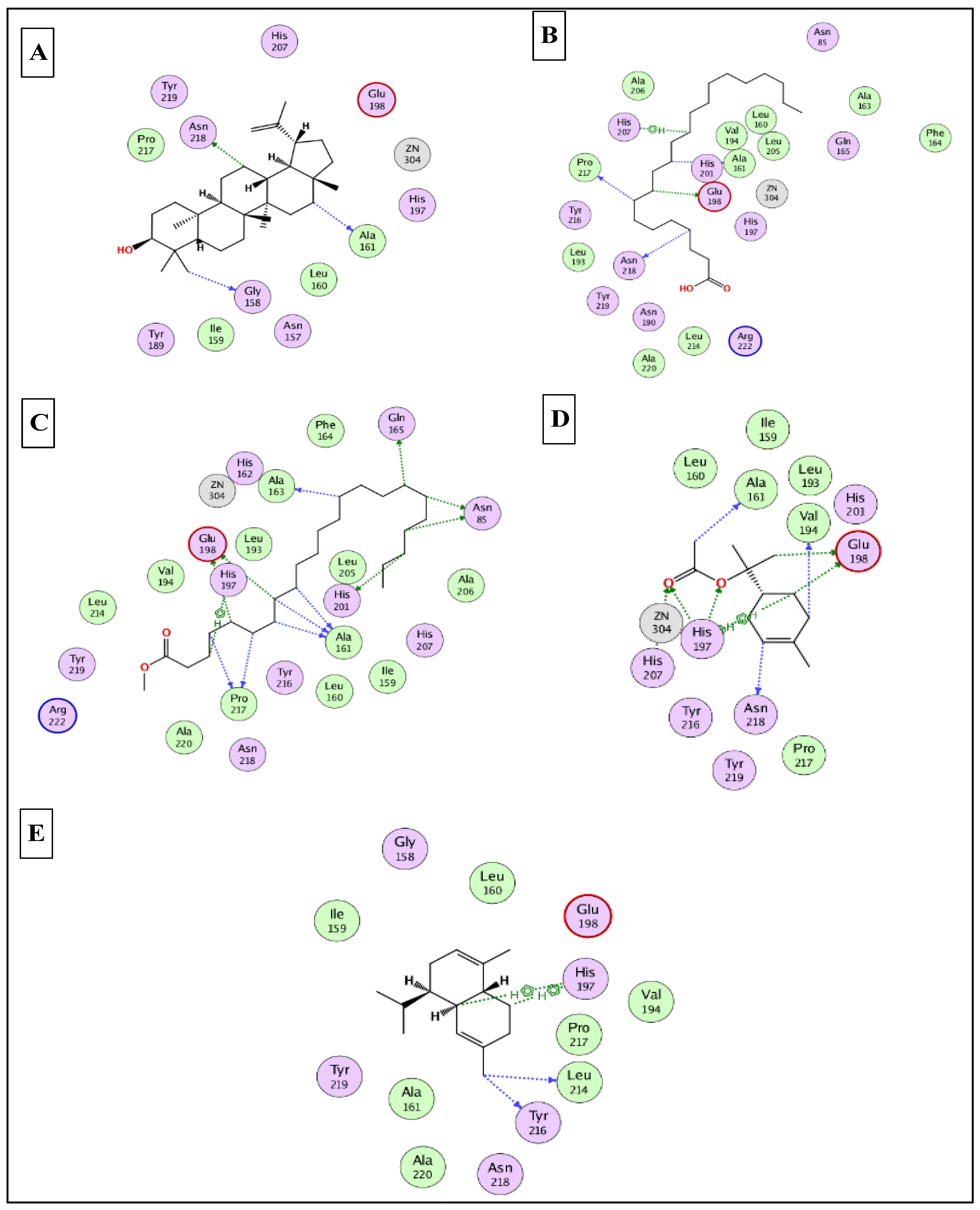
2.5. In Silico Molecular Docking Studies
3. Materials and Methods
3.1. Plant Material
3.2. Preparation of the N-Hexane Extracts of Various Organs
3.3. GC/MS Analysis
3.4. Identification of the N-Hexane Extract Components
3.5. Chemometric Analysis
3.6. Anti-Inflammatory Activity
3.7. Wound Healing Activity
3.7.1. Cytotoxicity Assay
3.7.2. Scratch Wound Assay
3.8. Statistical Analysis
3.9. In Silico Molecular Docking Studies
4. Conclusions
Author Contributions
Funding
Institutional Review Board Statement
Informed Consent Statement
Data Availability Statement
Acknowledgments
Conflicts of Interest
Sample Availability
References
- Elmaidomy, A.H.; Abdelmohsen, U.R.; Alsenani, F.; Aly, H.F.; Shams, S.G.E.; Younis, E.A.; Ahmed, K.A.; Sayed, A.M.; Owis, A.I.; Afifi, N.; et al. The Anti-Alzheimer Potential of Tamarindus Indica: An in Vivo Investigation Supported by in Vitro and in Silico Approaches. RSC Adv. 2022, 12, 11769–11785. [Google Scholar] [CrossRef] [PubMed]
- Chong, U.R.W.; Abdul-Rahman, P.S.; Abdul-Aziz, A.; Hashim, O.H.; Mat Junit, S. Tamarindus indica Extract Alters Release of Alpha Enolase, Apolipoprotein A-I, Transthyretin and Rab GDP Dissociation Inhibitor Beta from HepG2 Cells. PLoS ONE 2012, 7, e39476. [Google Scholar] [CrossRef] [PubMed] [Green Version]
- Havinga, R.M.; Hartl, A.; Putscher, J.; Prehsler, S.; Buchmann, C.; Vogl, C.R. Tamarindus indica L. (Fabaceae): Patterns of Use in Traditional African Medicine. J. Ethnopharmacol. 2010, 127, 573–588. [Google Scholar] [CrossRef] [PubMed]
- Lim, C.Y.; Mat Junit, S.; Abdulla, M.A.; Abdul Aziz, A. In Vivo Biochemical and Gene Expression Analyses of the Antioxidant Activities and Hypocholesterolaemic Properties of Tamarindus indica Fruit Pulp Extract. PLoS ONE 2013, 8, e70058. [Google Scholar] [CrossRef] [Green Version]
- Aly, S.H.; Elissawy, A.M.; Eldahshan, O.A.; Elshanawany, M.A.; Efferth, T.; Singab, A.N.B. The Pharmacology of the Genus Sophora (Fabaceae): An Updated Review. Phytomedicine 2019, 64, 153070. [Google Scholar] [CrossRef] [PubMed]
- Eldahshan, O.; Aly, S.; Elissawy, A.; Elshanawany, M.; Singab, A.N. Morphological and Genetic Characteristics of Sophora secundiflora and Sophora tomentosa (Fabaceae) Cultivated in Egypt. Taeckholmia 2019, 39, 103–129. [Google Scholar] [CrossRef]
- Abdelrahman, G.H.; Mariod, A.A. Wild Fruits: Composition, Nutritional Value and Products; Springer: Berlin/Heidelberg, Germany, 2019; ISBN 9783030318857. [Google Scholar]
- Kuru, P. Tamarindus Indica and Its Health Related Effects. Asian Pac. J. Trop. Biomed. 2014, 4, 676–681. [Google Scholar] [CrossRef] [Green Version]
- Martinello, F.; Soares, S.M.; Franco, J.J.; Santos, A.C.; Sugohara, A.; Garcia, S.B.; Curti, C.; Uyemura, S.A. Hypolipemic and Antioxidant Activities from Tamarindus indica L. Pulp Fruit Extract in Hypercholesterolemic Hamsters. Food Chem. Toxicol. Int. J. Publ. Br. Ind. Biol. Res. Assoc. 2006, 44, 810–818. [Google Scholar] [CrossRef] [PubMed]
- Bhadoriya, S.S.; Mishra, V.; Raut, S.; Ganeshpurkar, A.; Jain, S.K. Anti-Inflammatory and Antinociceptive Activities of a Hydroethanolic Extract of Tamarindus indica Leaves. Sci. Pharm. 2012, 80, 685–700. [Google Scholar] [CrossRef] [Green Version]
- Maiti, R.; Jana, D.; Das, U.K.; Ghosh, D. Antidiabetic Effect of Aqueous Extract of Seed of Tamarindus indica in Streptozotocin-Induced Diabetic Rats. J. Ethnopharmacol. 2004, 92, 85–91. [Google Scholar] [CrossRef]
- De Caluwé, E.; Halamová, K.; Van Damme, P. Tamarind (Tamarindus indica L.): A Review of Traditional Uses, Phytochemistry and Pharmacology. ACS Symp. Ser. 2009, 1021, 85–110. [Google Scholar] [CrossRef]
- Naeem, N.; Nadeem, F.; Azeem, M.W.; Dharmadasa, R.M. Tamarindus indica—A Review of Explored Potentials. Int. J. Chem. Biochem. Sci. 2017, 12, 98–106. [Google Scholar]
- Escalona-Arranz, J.; Péres-Roses, R.; Urdaneta-Laffita, I.; Camacho-Pozo, M.; Rodríguez-Amado, J.; Licea-Jiménez, I. Antimicrobial Activity of Extracts from Tamarindus indica L. Leaves. Pharmacogn. Mag. 2010, 6, 242–247. [Google Scholar] [CrossRef] [PubMed] [Green Version]
- Al-Fatimi, M.; Wurster, M.; Schröder, G.; Lindequist, U. Antioxidant, Antimicrobial and Cytotoxic Activities of Selected Medicinal Plants from Yemen. J. Ethnopharmacol. 2007, 111, 657–666. [Google Scholar] [CrossRef] [PubMed]
- Meléndez, P.A.; Capriles, V.A. Antibacterial Properties of Tropical Plants from Puerto Rico. Phytomedicine 2006, 13, 272–276. [Google Scholar] [CrossRef]
- Komakech, R.; Kim, Y.; Matsabisa, G.M.; Kang, Y. Anti-Inflammatory and Analgesic Potential of Tamarindus indica Linn. (Fabaceae): A Narrative Review. Integr. Med. Res. 2019, 8, 181–186. [Google Scholar] [CrossRef]
- Meher, B.; Dash, D.K.; Kumar Dash, D.; Roy, A. A Review on: Phytochemistry, Pharmacology and Traditional Uses of Tamarindus indica L. World J. Pharm. Pharm. Sci. 2014, 3, 229. [Google Scholar]
- Yerima, M.; Anuka, J.A.; Salawu, O.A.; Abdu-Aguye, I. Antihyperglycaemic Activity of the Stem-Bark Extract of Tamarindus indica L. on Experimentally Induced Hyperglycaemic and Normoglycaemic Wistar Rats. Pak. J. Biol. Sci. PJBS 2014, 17, 414–418. [Google Scholar] [CrossRef] [Green Version]
- Das, S.S.; Dey, M.; Ghosh, A.K. Determination of Anthelmintic Activity of the Leaf and Bark Extract of Tamarindus indica Linn. Indian J. Pharm. Sci. 2011, 73, 104–107. [Google Scholar] [CrossRef] [Green Version]
- Okur, M.E.; Karadağ, A.E.; Okur, N.Ü.; Özhan, Y.; Sipahi, H.; Ayla, Ş.; Daylan, B.; Demirci, B.; Demirci, F. In Vivo Wound Healing and in Vitro Anti-Inflammatory Activity Evaluation of Phlomis russeliana Extract Gel Formulations. Molecules 2020, 25, 2695. [Google Scholar] [CrossRef]
- Bakr, R.O.; Amer, R.I.; Attia, D.; Abdelhafez, M.M.; Al-Mokaddem, A.K.; El Gendy, A.N.; El-Fishawy, A.M.; Fayed, M.A.A.; Gad, S.S. In-Vivo Wound Healing Activity of a Novel Composite Sponge Loaded with Mucilage and Lipoidal Matter of Hibiscus Species. Biomed. Pharmacother. 2021, 135, 111225. [Google Scholar] [CrossRef] [PubMed]
- Imran, M.; Sharma, J.N.; Kamal, M.; Asif, M. Standardization and Wound-Healing Activity of Petroleum, Ethanolic and Aqueous Extracts of Ficus benghalensis Leaves. Pharm. Chem. J. 2021, 54, 1057–1062. [Google Scholar] [CrossRef]
- Agyare, C.; Boakye, Y.D.; Bekoe, E.O.; Hensel, A.; Dapaah, S.O.; Appiah, T. Review: African Medicinal Plants with Wound Healing Properties. J. Ethnopharmacol. 2016, 177, 85–100. [Google Scholar] [CrossRef] [PubMed]
- Haque, S.D.; Saha, S.K.; Salma, U.; Nishi, M.K.; Rahaman, M.S. Antibacterial Effect of Aloe Vera (Aloe barbadensis) Leaf Gel against Staphylococcus aureus, Pseudomonas aeruginosa, Escherichia coli and Klebsiella pneumoniae. Mymensingh Med. J. 2019, 28, 490–496. [Google Scholar] [CrossRef] [PubMed]
- Shedoeva, A.; Leavesley, D.; Upton, Z.; Fan, C. Wound Healing and the Use of Medicinal Plants. Evid.-Based Complement. Altern. Med. 2019, 2019, 2684108. [Google Scholar] [CrossRef] [PubMed] [Green Version]
- Ads, E.N.; Hassan, S.I.; Rajendrasozhan, S.; Hetta, M.H.; Aly, S.H.; Ali, M.A. Isolation, Structure Elucidation and Antimicrobial Evaluation of Natural Pentacyclic Triterpenoids and Phytochemical Investigation of Different Fractions of Ziziphus spina-christi (L.) Stem Bark Using LCHRMS Analysis. Molecules 2022, 27, 1805. [Google Scholar] [CrossRef]
- El-Nashar, H.A.S.; Aly, S.H.; Ahmadi, A.; El-Shazly, M. The Impact of Polyphenolics in the Management of Breast Cancer: Mechanistic Aspects and Recent Patents. Recent Pat. Anticancer Drug Discov. 2021, 17, 358–379. [Google Scholar] [CrossRef]
- Aly, S.H.; Elgindi, M.R.; Singab, A.E.N.B.; Mahmoud, I.I. Hyophorbe verschaffeltii DNA Profiling, Chemical Composition of the Lipophilic Fraction, Antimicrobial, Anti-Inflammatory and Cytotoxic Activities. Res. J. Pharm. Biol. Chem. Sci. 2016, 7, 120–130. [Google Scholar]
- Aly, S.H.; Elissawy, A.M.; Fayez, A.M.; Eldahshan, O.A.; Elshanawany, M.A.; Singab, A.N.B. Neuroprotective Effects of Sophora secundiflora, Sophora tomentosa Leaves and Formononetin on Scopolamine-Induced Dementia. Nat. Prod. Res. 2021, 35, 5848–5852. [Google Scholar] [CrossRef]
- Saber, F.R.; Aly, S.H.; Khallaf, M.A.; El-Nashar, H.A.S.; Fahmy, N.M.; El-Shazly, M.; Radha, R.; Prakash, S.; Kumar, M.; Taha, D.; et al. Hyphaene thebaica (Areceaeae) as a Promising Functional Food: Extraction, Analytical Techniques, Bioactivity, Food, and Industrial Applications. Food Anal. Methods 2022, 1–21. [Google Scholar] [CrossRef]
- Raslan, M.A.; Afifi, A.H. In Vitro Wound Healing Properties, Antioxidant Activities, HPLC-ESI-MS/MS Profile and Phytoconstituents of the Stem Aqueous Methanolic Extract of Dracaena reflexa Lam. Biomed. Chromatogr. 2022, 36, e5352. [Google Scholar] [CrossRef] [PubMed]
- Labib, R.M.; Ayoub, I.M.; Michel, H.E.; Mehanny, M.; Kamil, V.; Hany, M.; Magdy, M.; Moataz, A.; Maged, B.; Mohamed, A. Appraisal on the Wound Healing Potential of Melaleuca alternifolia and Rosmarinus officinalis L. Essential Oil-Loaded Chitosan Topical Preparations. PLoS ONE 2019, 14, e0219561. [Google Scholar] [CrossRef] [PubMed]
- El-Nashar, H.A.S.; Mostafa, N.M.; Eldahshan, O.A.; Singab, A.N.B. Chemical Composition, Cytotoxic and Anti-Arthritic Activities of Hexane Extracts of Certain Schinus Species. J. Pharm. Pharmacol. 2021, 9, 378–386. [Google Scholar] [CrossRef]
- Aly, S.H.; Eldahshan, O.A.; Al-Rashood, S.T.; Binjubair, F.A.; El-Hassab, M.A.; Eldehna, W.M.; Acqua, S.D.; Zengin, G. Chemical Constituents, Antioxidant, and Enzyme Inhibitory Activities Supported by In-Silico Study of n-Hexane Extract and Essential Oil of Guava Leaves. Molecules 2022, 27, 8979. [Google Scholar] [CrossRef]
- Ali, A.; Garg, P.; Goyal, R.; Kaur, G.; Li, X.; Negi, P.; Valis, M.; Kuca, K.; Kulshrestha, S. A Novel Herbal Hydrogel Formulation of Moringa oleifera for Wound Healing. Plants 2020, 10, 25. [Google Scholar] [CrossRef]
- Ayoub, I.M.; Korinek, M.; El-Shazly, M.; Wetterauer, B.; El-Beshbishy, H.A. Activity of Chasmanthe aethiopica Leaf Extract and Its Profiling Using LC/MS and GLC/MS. Plants 2021, 10, 1118. [Google Scholar] [CrossRef]
- Sakib, S.A.; Tareq, A.M.; Islam, A.; Rakib, A.; Islam, M.N.; Uddin, M.A.; Rahman, M.M.; Seidel, V.; Emran, T. Bin Anti-Inflammatory, Thrombolytic and Hair-Growth Promoting Activity of the n-Hexane Fraction of the Methanol Extract of Leea indica Leaves. Plants 2021, 10, 1081. [Google Scholar] [CrossRef]
- Carullo, G.; Sciubba, F.; Governa, P.; Mazzotta, S.; Frattaruolo, L.; Grillo, G.; Cappello, A.R.; Cravotto, G.; Di Cocco, M.E.; Aiello, F. Mantonico and Pecorello Grape Seed Extracts: Chemical Characterization and Evaluation of In Vitro Wound-Healing and Anti-Inflammatory Activities. Pharmaceuticals 2020, 13, 97. [Google Scholar] [CrossRef]
- Ibrahim, N.A.; El-Gengaihi, S.; El-Hamidi, A.; Bashandy, S.A.E. Chemical and Biological Evaluation of Tamarindus indica L. Growing in Sudan. Acta Hortic. 1995, 390, 51–58. [Google Scholar] [CrossRef]
- Carasek, E.; Pawliszyn, J. Screening of Tropical Fruit Volatile Compounds Using Solid-Phase Microextraction (SPME) Fibers and Internally Cooled SPME Fiber. J. Agric. Food Chem. 2006, 54, 8688–8696. [Google Scholar] [CrossRef]
- Gad, H.A.; Mukhammadiev, E.A.; Zengen, G.; Musayeib, N.M.A.; Hussain, H.; Bin Ware, I.; Ashour, M.L.; Mamadalieva, N.Z. Chemometric Analysis Based on GC-MS Chemical Profiles of Three Stachys Species from Uzbekistan and Their Biological Activity. Plants 2022, 11, 1215. [Google Scholar] [CrossRef] [PubMed]
- Rimbau, V.; Cerdan, C.; Vila, R.; Iglesias, J. Antiinflammatory Activity of Some Extracts from Plants Used in the Traditional Medicine of North-African Countries (II). Phyther. Res. 1999, 13, 128–132. [Google Scholar] [CrossRef]
- Hivrale, M.G.; Bandawane, D.D.; Mali, A.A. Anti-Inflammatory and Analgesic Activities of Petroleum Ether and Ethyl Acetate Fractions of Tamarindus indica Seeds. Orient. Pharm. Exp. Med. 2013, 13, 319–326. [Google Scholar] [CrossRef]
- Dias, A.M.A.; Rey-Rico, A.; Oliveira, R.A.; Marceneiro, S.; Alvarez-Lorenzo, C.; Concheiro, A.; Júnior, R.N.C.; Braga, M.E.M.; De Sousa, H.C. Wound Dressings Loaded with an Anti-Inflammatory Jucá (Libidibia ferrea) Extract Using Supercritical Carbon Dioxide Technology. J. Supercrit. Fluids 2013, 74, 34–45. [Google Scholar] [CrossRef] [Green Version]
- Lucetti, D.L.; Lucetti, E.C.; Bandeira, M.A.M.; Veras, H.N.; Silva, A.H.; Leal, L.K.A.; Lopes, A.A.; Alves, V.C.; Silva, G.S.; Brito, G.A.; et al. Anti-Inflammatory Effects and Possible Mechanism of Action of Lupeol Acetate Isolated from Himatanthus drasticus (Mart.) Plumel. J. Inflamm. 2010, 7, 60. [Google Scholar] [CrossRef] [Green Version]
- Skehan, P.; Storeng, R.; Scudiero, D.; Monks, A.; McMahon, J.; Vistica, D.; Warren, J.T.; Bokesch, H.; Kenney, S.; Boyd, M.R. New Colorimetric Cytotoxicity Assay for Anticancer—Drug Screening. J. Natl. Cancer Inst. 1990, 82, 1107–1112. [Google Scholar] [CrossRef]
- Adeniyi, O.V.; Olaifa, F.E.; Emikpe, B.O.; Oyagbemi, A.A. Experimental Evaluation of the Wound-Healing and Antioxidant Activities of Tamarind (Tamarindus indica) Pulp and Leaf Meal in the African Catfish (Clarias gariepinus). Acta Vet. Eurasia 2018, 44, 63–72. [Google Scholar] [CrossRef]
- Attah, M.O.; Ishaya, H.B.; Chiroma, M.S.; Amaza, D.; Balogun, S.U.; Jacks, T. Effect of Tamarindus indica (Linn) on the Rate of Wound Healing in Adult Rabbits. IOSR J. Dent. Med. Sci. 2015, 14, 2279–2861. [Google Scholar] [CrossRef]
- Poljšak, N.; Kočevar Glavač, N. Tilia Sp. Seed Oil—Composition, Antioxidant Activity and Potential Use. Appl. Sci. 2021, 11, 4932. [Google Scholar] [CrossRef]
- Hernandez, G.R.; Hernandez Garcia, D.Y.; Sanchez, M.L. Healing Cream Tournefortia hirsutissima L. Med. Aromat. Plants 2017, 6, 4–6. [Google Scholar] [CrossRef]
- Hata, K.; Hori, K.; Takahashi, S. Role of P38 MAPK in Lupeol-Induced B16 2F2 Mouse Melanoma Cell Differentiation. J. Biochem. 2003, 134, 441–445. [Google Scholar] [CrossRef] [PubMed]
- Geetha, T.; Varalakshmi, P. Anti-Inflammatory Activity of Lupeol and Lupeol Linoleate in Rats. J. Ethnopharmacol. 2001, 76, 77–80. [Google Scholar] [CrossRef] [PubMed]
- Pereira Beserra, F.; Xue, M.; Maia, G.L.d.A.; Leite Rozza, A.; Helena Pellizzon, C.; Jackson, C.J. Lupeol, a Pentacyclic Triterpene, Promotes Migration, Wound Closure, and Contractile Effect In Vitro: Possible Involvement of PI3K/Akt and P38/ERK/MAPK Pathways. Molecules 2018, 23, 2819. [Google Scholar] [CrossRef] [PubMed]
- Patel, S.; Srivastava, S.; Singh, M.R.; Singh, D. Preparation and Optimization of Chitosan-Gelatin Films for Sustained Delivery of Lupeol for Wound Healing. Int. J. Biol. Macromol. 2018, 107, 1888–1897. [Google Scholar] [CrossRef] [PubMed]
- Beserra, F.P.; Vieira, A.J.; Gushiken, L.F.S.; De Souza, E.O.; Hussni, M.F.; Hussni, C.A.; Takahira, R.K.; Nóbrega, R.H.; Martinez, E.R.M.; Jackson, C.J.; et al. Lupeol, a Dietary Triterpene, Enhances Wound Healing in Streptozotocin-Induced Hyperglycemic Rats with Modulatory Effects on Inflammation, Oxidative Stress, and Angiogenesis. Oxidative Med. Cell. Longev. 2019, 2019, 3182627. [Google Scholar] [CrossRef] [PubMed]
- Bopage, N.S.; Kamal Bandara Gunaherath, G.M.; Jayawardena, K.H.; Wijeyaratne, S.C.; Abeysekera, A.M.; Somaratne, S. Dual Function of Active Constituents from Bark of Ficus Racemosa L. in Wound Healing. BMC Complement. Altern. Med. 2018, 18, 29. [Google Scholar] [CrossRef] [PubMed]
- Malinowska, M.; Miroslaw, B.; Sikora, E.; Ogonowski, J.; Wojtkiewicz, A.M.; Szaleniec, M.; Pasikowska-Piwko, M.; Eris, I. New Lupeol Esters as Active Substances in the Treatment of Skin Damage. PLoS ONE 2019, 14, e0214216. [Google Scholar] [CrossRef]
- Bopage, N.S.; Jayawardena, K.H.; Wijeyaratne, C.; Abeysekera, A.M.; Gunaherath, G.M.K.B. Wound Healing Activity of Some Lupeol Derivatives Using. Life Sci. 2016, 3–5. [Google Scholar]
- Poljšak, N.; Kreft, S.; Kočevar Glavač, N. Vegetable Butters and Oils in Skin Wound Healing: Scientific Evidence for New Opportunities in Dermatology. Phyther. Res. 2020, 34, 254–269. [Google Scholar] [CrossRef]
- Lewinska, A.; Zebrowski, J.; Duda, M.; Gorka, A.; Wnuk, M. Fatty Acid Profile and Biological Activities of Linseed and Rapeseed Oils. Molecules 2015, 20, 22872–22880. [Google Scholar] [CrossRef] [Green Version]
- Xu, A.L.; Xue, Y.-Y.; Tao, W.-T.; Wang, S.-Q.; Xu, H.-Q. Oleanolic Acid Combined with Olaparib Enhances Radiosensitization in Triple Negative Breast Cancer and Hypoxia Imaging with (18)F-FETNIM Micro PET/CT. Biomed. Pharmacother. 2022, 150, 113007. [Google Scholar] [CrossRef] [PubMed]
- Lin, T.-K.; Zhong, L.; Santiago, J.L. Anti-Inflammatory and Skin Barrier Repair Effects of Topical Application of Some Plant Oils. Int. J. Mol. Sci. 2017, 19, 70. [Google Scholar] [CrossRef] [PubMed] [Green Version]
- Sánchez-Quesada, C.; López-Biedma, A.; Toledo, E.; Gaforio, J.J. Squalene Stimulates a Key Innate Immune Cell to Foster Wound Healing and Tissue Repair. Evid.-Based Complement. Altern. Med. 2018, 2018, 9473094. [Google Scholar] [CrossRef]
- Abd El-Ghffar, E.A.; El-Nashar, H.A.S.; Eldahshan, O.A.; Singab, A.N.B. GC-MS Analysis and Hepatoprotective Activity of the n-Hexane Extract of Acrocarpus fraxinifolius Leaves against Paracetamol-Induced Hepatotoxicity in Male Albino Rats. Pharm. Biol. 2017, 55, 444–449. [Google Scholar] [CrossRef]
- Rostad, C.E.; Pereira, W.E. Kovats and Lee Retention Indices Determined by Gas Chromatography/Mass Spectrometry for Organic Compounds of Environmental Interest. J. High Resolut. Chromatogr. 1986, 9, 328–334. [Google Scholar] [CrossRef]
- Ivanov, I.; Dincheva, I.; Badjakov, I.; Petkova, N.; Denev, P.; Pavlov, A. GC-MS Analysis of Unpolar Fraction from Ficus carica L. (Fig) Leaves. Int. Food Res. J. 2018, 25, 282–286. [Google Scholar]
- de Carvalho, F.M.d.A.; Schneider, J.K.; de Jesus, C.V.F.; de Andrade, L.N.; Amaral, R.G.; David, J.M.; Krause, L.C.; Severino, P.; Soares, C.M.F.; Bastos, E.C.; et al. Brazilian Red Propolis: Extracts Production, Physicochemical Characterization, and Cytotoxicity Profile for Antitumor Activity. Biomolecules 2020, 10, 726. [Google Scholar] [CrossRef]
- El-Nashar, H.A.S.; Eldehna, W.M.; Al-Rashood, S.T.; Alharbi, A.; Eskandrani, R.O.; Aly, S.H. GC/MS Analysis of Essential Oil and Enzyme Inhibitory Activities of Syzygium cumini (Pamposia) Grown in Docking Studies. Molecules 2021, 26, 6984. [Google Scholar] [CrossRef]
- Aly, S.H.; Elissawy, A.M.; Eldahshan, O.A.; Elshanawany, M.A.; Singab, A.N.B. Phytochemical Investigation Using GC/MS Analysis and Evaluation of Antimicrobial and Cytotoxic Activities of the Lipoidal Matter of Leaves of Sophora secundiflora and Sophora tomentosa. Arch. Pharm. Sci. Ain Shams Univ. 2020, 4, 207–214. [Google Scholar] [CrossRef]
- Aly, S.H.; Elissawy, A.M.; Eldahshan, O.A.; Elshanawany, M.A.; Singab, A.N.B. Variability of the Chemical Composition of the Essential Oils of Flowers and the Alkaloid Contents of Leaves of Sophora secundiflora and Sophora tomentosa. J. Essent. Oil-Bear. Plants 2020, 23, 442–452. [Google Scholar] [CrossRef]
- Gad, H.A.; Ayoub, I.M.; Wink, M. Phytochemical Profiling and Seasonal Variation of Essential Oils of Three Callistemon Species Cultivated in Egypt. PLoS One 2019, 14, e0219571. [Google Scholar] [CrossRef] [PubMed] [Green Version]
- Gad, H.A.; Mamadalieva, R.Z.; Khalil, N.; Zengin, G.; Najar, B.; Khojimatov, O.K.; Al Musayeib, N.M.; Ashour, M.L.; Mamadalieva, N.Z. GC-MS Chemical Profiling, Biological Investigation of Three Salvia Species Growing in Uzbekistan. Molecules 2022, 27, 5365. [Google Scholar] [CrossRef] [PubMed]
- Brereton, R.G. Applied Chemometrics for Scientists; John Wiley & Sons: Chichester, UK, 2007; pp. 1–379. [Google Scholar]
- Yoo, M.-S.; Shin, J.-S.; Choi, H.-E.; Cho, Y.-W.; Bang, M.-H.; Baek, N.-I.; Lee, K.-T. Fucosterol Isolated from Undaria pinnatifida Inhibits Lipopolysaccharide-Induced Production of Nitric Oxide and Pro-Inflammatory Cytokines via the Inactivation of Nuclear Factor-ΚB and P38 Mitogen-Activated Protein Kinase in RAW264.7 Macrophages. Food Chem. 2012, 135, 967–975. [Google Scholar] [CrossRef] [PubMed]
- Oliveira, T.; Figueiredo, C.A.; Brito, C.; Stavroullakis, A.; Prakki, A.; Da Silva Velozo, E.; Nogueira-Filho, G. Effect of Allium cepa L. on Lipopolysaccharide-Stimulated Osteoclast Precursor Cell Viability, Count, and Morphology Using 4′,6-Diamidino-2-Phenylindole-Staining. Int. J. Cell Biol. 2014, 2014, 535789. [Google Scholar] [CrossRef] [PubMed] [Green Version]
- Main, K.A.; Mikelis, C.M.; Doçi, C.L. In Vitro Wound Healing Assays to Investigate Epidermal Migration. In Methods in Molecular Biology; Springer: New York, NY, USA, 2019; Volume 2109, pp. 147–154. [Google Scholar]
- Martinotti, S.; Ranzato, E. Scratch Wound Healing Assay. In Methods in Molecular Biology; Springer: Berlin/Heidelberg, Germany, 2019; Volume 2109, pp. 225–229. [Google Scholar]
- Jonkman, J.E.N.; Cathcart, J.A.; Xu, F.; Bartolini, M.E.; Amon, J.E.; Stevens, K.M.; Colarusso, P. An Introduction to the Wound Healing Assay Using Live-Cell Microscopy. Cell Adhes. Migr. 2014, 8, 440–451. [Google Scholar] [CrossRef]
- Saitoh, M.; Kunitomo, J.; Kimura, E.; Hayase, Y.; Kobayashi, H.; Uchiyama, N.; Kawamoto, T.; Tanaka, T.; Mol, C.D.; Dougan, D.R.; et al. Design, Synthesis and Structure-Activity Relationships of 1,3,4-Oxadiazole Derivatives as Novel Inhibitors of Glycogen Synthase Kinase-3β. Bioorg. Med. Chem. 2009, 17, 2017–2029. [Google Scholar] [CrossRef]
- Tauro, M.; Laghezza, A.; Loiodice, F.; Piemontese, L.; Caradonna, A.; Capelli, D.; Montanari, R.; Pochetti, G.; Di Pizio, A.; Agamennone, M.; et al. Catechol-Based Matrix Metalloproteinase Inhibitors with Additional Antioxidative Activity. J. Enzym. Inhib. Med. Chem. 2016, 31, 25–37. [Google Scholar] [CrossRef] [Green Version]
- Xue, F.; Huang, J.; Ji, H.; Fang, J.; Li, H.; Martásek, P.; Roman, L.J.; Poulos, T.L.; Silverman, R.B. Structure-Based Design, Synthesis, and Biological Evaluation of Lipophilic-Tailed Monocationic Inhibitors of Neuronal Nitric Oxide Synthase. Bioorg. Med. Chem. 2010, 18, 6526–6537. [Google Scholar] [CrossRef] [Green Version]
- Vilar, S.; Cozza, G.; Moro, S. Medicinal Chemistry and the Molecular Operating Environment (MOE): Application of QSAR and Molecular Docking to Drug Discovery. Curr. Top. Med. Chem. 2008, 8, 1555–1572. [Google Scholar] [CrossRef]


| No. | Rt (min) | Compound Name | RIExp.a | RILit b | Molecular Formula | Peak Area (%) | |||
|---|---|---|---|---|---|---|---|---|---|
| TIB | TIL | TIS | TIF | ||||||
| 1 | 6.94 | α-pinene | 930 | 930 | C10H16 | - | - | - | 0.38 |
| 2 | 8.16 | α-Sabinene | 971 | 971 | C10H16 | - | - | - | 0.57 |
| 3 | 9.90 | D-Limonene | 1028 | 1028 | C10H16 | - | - | - | 1.35 |
| 4 | 9.97 | 1,8-Cineole | 1030 | 1030 | C10H18O | - | - | - | 0.61 |
| 5 | 13.93 | Isoborneol | 1156 | 1156 | C10H18O | - | - | - | 0.87 |
| 6 | 19.00 | α-Terpinyl acetate | 1328 | 1327 | C12H20O2 | 0.03 | - | - | 7.36 |
| 7 | 23.59 | α-Muurolene | 1494 | 1494 | C15H24 | 0.04 | - | - | 7.52 |
| 8 | 27.69 | 2-Methylhexadecane | 1668 | 1666 | C17H36 | 0.03 | - | - | 5.70 |
| 9 | 31.07 | Hexadecanal; Palmitaldehyde | 1810 | 1811 | C16H32O | - | - | - | 0.36 |
| 10 | 31.25 | Methyl pentadecanoate | 1819 | 1820 | C16H32O2 | - | - | - | 4.53 |
| 11 | 31.52 | Neophytadiene | 1833 | 1837 | C20H38 | - | 0.2 | - | - |
| 12 | 31.67 | Hexahydrofarnesyl acetone | 1841 | 1842 | C18H36O | - | 0.09 | - | - |
| 13 | 32.43 | Heptadecan-2-one | 1880 | 1892 | C17H34O | 0.03 | 0.07 | - | - |
| 14 | 33.37 | Methyl palmitate | 1928 | 1928 | C17H34O2 | 0.11 | - | - | - |
| 15 | 34.42 | α-Eicosene | 1980 | 1986 | C20H40 | - | - | - | 3.44 |
| 16 | 35.62 | Octadecanal | 2040 | 2034 | C18H36O | - | - | 0.58 | - |
| 17 | 36.73 | n-Heneicosane | 2098 | 2100 | C21H44 | 0.05 | - | - | - |
| 18 | 36.85 | Methyl linolenate | 2104 | 2108 | C19H32O2 | 0.07 | - | - | - |
| 19 | 37.06 | Phytol | 2116 | 2116 | C20H40O | - | 1.97 | - | - |
| 20 | 37.32 | Linolenic acid | 2130 | 2134 | C18H30O2 | - | - | 1.23 | 3.26 |
| 21 | 37.88 | Ethyl linolate | 2160 | 2164 | C20H36O2 | 0.08 | - | - | - |
| 22 | 38.48 | Stearic acid=n-Octadecanoic acid | 2192 | 2180 | C18H36O2 | - | - | 0.64 | |
| 23 | 38.59 | n-Docosane | 2198 | 2200 | C22H46 | 0.07 | - | 0.82 | - |
| 24 | 38.89 | Phytol acetate | 2215 | 2218 | C22H42O2 | - | - | 0.67 | - |
| 25 | 39.17 | Methyl nonadecanoate | 2231 | 2230 | C20H40O2 | - | - | 0.59 | - |
| 26 | 40.12 | Heneicosan-3-one | 2285 | 2283 | C21H42O | - | - | 0.54 | - |
| 27 | 40.20 | 1-Eicosanol | 2289 | 2292 | C20H42O | 0.09 | - | - | - |
| 28 | 40.36 | n- Tricosane | 2298 | 2300 | C23H48 | 0.17 | 0.04 | - | - |
| 29 | 40.65 | Methyl cis-11-eicosenoate | 2315 | 2302 | C21H40O2 | - | - | 1.83 | - |
| 30 | 41.15 | Methyl eicosanoate | 2344 | 2339 | C21H42O2 | - | - | 0.77 | - |
| 31 | 41.41 | 4,8,12,16-Tetramethylheptadecan-4-olide | 2360 | 2364 | C21H40O2 | - | 0.07 | 0.40 | - |
| 32 | 41.57 | n-Eicosanoic acid | 2369 | 2380 | C20H40O2 | - | - | 1.01 | - |
| 33 | 42.07 | 2,2′-Methylene-bis-(6-tert butyl-4-methylphenol) | 2398 | 2398 | C23H32O2 | 0.10 | 0.06 | - | - |
| 34 | 42.24 | n-Tetracosane | 2409 | 2400 | C24H50 | - | - | 0.53 | - |
| 35 | 42.48 | Docosanal | 2424 | 2426 | C22H44O | - | - | 0.64 | 1.48 |
| 36 | 42.61 | Methyl heneicosanoate | 2431 | 2430 | C22H44O2 | 0.07 | - | - | - |
| 37 | 42.85 | 1-Docosanol | 2446 | 2456 | C22H46O | - | - | 0.54 | - |
| 38 | 43.1 | 2-Methyltetracosane | 2461 | 2465 | C25H52 | - | - | 1.28 | - |
| 39 | 43.48 | 1-Pentatricosene | 2484 | 2485 | C35H70 | - | - | 1.77 | - |
| 40 | 43.62 | Cyclogallipharaol | 2493 | 2499 | C21H36O | 0.35 | - | 0.81 | 0.72 |
| 41 | 43.70 | n- Pentacosane | 2498 | 2500 | C25H52 | 0.24 | 0.23 | 2.46 | - |
| 42 | 43.87 | Heneicosyl acetate | 2509 | 2509 | C23H46O2 | - | - | 2.94 | - |
| 43 | 43.96 | Palmitic acid β-monoglyceride | 2514 | 2519 | C19H38O4 | 0.07 | - | - | - |
| 44 | 44.230 | Methyl docosanoate | 2531 | 2531 | C23H46O2 | 0.08 | - | - | - |
| 45 | 44.305 | 11-Methylpentacosane | 2536 | 2529 | C26H54 | - | - | 4.22 | - |
| 46 | 44.675 | cis-13,16-Docasadienoic acid | 2560 | 2566 | C22H40O2 | - | - | 3.70 | 3.76 |
| 47 | 44.84 | n-Docosanoic acid | 2570 | 2569 | C22H44O2 | - | - | 10.49 | - |
| 48 | 45.18 | Ethyl docosanoate | 2592 | 2593 | C24H48O2 | 1.44 | |||
| 49 | 45.275 | n-Hexacosane | 2598 | 2600 | C26H54 | 0.11 | 0.20 | 2.47 | - |
| 50 | 45.43 | Methyl tricosanoate | 2608 | 2615 | C24H48O2 | - | - | 7.09 | - |
| 51 | 45.62 | Erucylamide | 2621 | 2625 | C22H43NO | - | - | 1.32 | - |
| 52 | 45.83 | Tetracosanal | 2635 | 2632 | C24H48O | - | - | 5.42 | - |
| 53 | 46.19 | 2-Methylhexacosane | 2658 | 2662 | C27H56 | - | - | 4.31 | - |
| 54 | 46.793 | 1-Heptacosene | 2698 | 2694 | C27H54 | 0.58 | - | - | - |
| 55 | 46.805 | n-Heptacosane | 2699 | 2700 | C27H56 | - | 1.37 | 4.17 | 6.67 |
| 56 | 47.310 | Methyl tetracosanoate | 2733 | 2731 | C25H50O2 | 0.14 | - | 2.17 | - |
| 57 | 47.77 | 2-Methylheptacosane | 2765 | 2761 | C28H58 | - | - | 0.59 | - |
| 58 | 48.02 | 16-Acetoxycarterochaetol | 2782 | 2787 | C22H36O3 | - | - | 0.26 | - |
| 59 | 48.260 | n-Octacosane | 2798 | 2800 | C28H58 | 0.09 | 0.44 | 4.63 | - |
| 60 | 48.800 | Squalene | 2836 | 2835 | C30H50 | 0.24 | 1.29 | 1.82 | 4.17 |
| 61 | 48.865 | n-Hexacosanal | 2841 | 2833 | C26H52O | 0.04 | 0.09 | - | - |
| 62 | 48.94 | n-Hexacosanol | 2846 | 2848 | C26H54O | - | - | 0.25 | - |
| 63 | 49.26 | 2-Methyloctacosane | 2869 | 2860 | C29H60 | - | - | 1.32 | - |
| 64 | 49.44 | 1-Nonacosene | 2882 | 2884 | C29H58 | - | - | 0.67 | - |
| 65 | 49.730 | n-Nonacosane | 2902 | 2900 | C29H60 | 1.26 | 4.13 | 1.69 | 5.42 |
| 66 | 50.175 | 15-Methylnonacosane | 2935 | 2931 | C30H62 | 0.03 | - | - | - |
| 67 | 50.285 | Methyl hexacosanoate | 2943 | 2940 | C27H54O2 | - | 0.17 | - | - |
| 68 | 50.515 | 2-Methylnonacosane | 2960 | 2960 | C30H62 | 0.04 | - | - | - |
| 69 | 51.040 | n-Triacontane | 2998 | 3000 | C30H62 | 0.04 | 0.37 | - | - |
| 70 | 51.130 | Benzyl icosanoate | 3005 | 3003 | C27H46O2 | 0.02 | - | - | - |
| 71 | 51.210 | 1-Heptacosanol | 3011 | 3016 | C27H56O | - | 0.06 | - | - |
| 72 | 51.655 | n-Octacosanal | 3045 | 3039 | C28H56O | - | 0.29 | - | 0.44 |
| 73 | 51.850 | 2-Methyltriacontane | 3059 | 3060 | C31H64 | - | 0.09 | - | - |
| 74 | 52.370 | n-Hentriacontane | 3099 | 3100 | C31H64 | 0.08 | 1.73 | 0.61 | 3.32 |
| 75 | 52.475 | Octacosanol | 3107 | 3110 | C28H58O | 1.02 | - | - | 1.89 |
| 76 | 52.72 | 13-Methylhentriacontane | 3126 | 3130 | C32H66 | - | 0.15 | 0.85 | - |
| 77 | 52.835 | Campesterol | 3134 | 3131 | C28H48O | - | 0.05 | - | - |
| 78 | 53.015 | Cholest-3,5-diene | 3148 | - | C27H44 | 0.07 | 0.09 | - | - |
| 79 | 53.085 | α-Tocopherol | 3154 | 3149 | C29H50O2 | 0.03 | 1.01 | - | - |
| 80 | 53.155 | Ergosta-5,8,22-trien-3-ol, (3β,22E)- | 3159 | 3158 | C28H44O | - | 0.40 | - | - |
| 81 | 53.340 | Stigmasterol | 3173 | 3170 | C29H48O | 0.11 | 0.25 | - | - |
| 82 | 53.475 | β-Sitosterol | 3183 | 3197 | C29H50O | - | 0.10 | - | - |
| 83 | 53.710 | n-Dotriacontane | 3201 | 3200 | C32H66 | 0.26 | 0.23 | - | - |
| 84 | 53.925 | Cholest-5-ene, 3β-methoxy | 3216 | 3216 | C28H48O | - | 0.08 | - | - |
| 85 | 54.375 | Triacontanal | 3248 | 3251 | C30H60O | - | 0.47 | - | 0.69 |
| 86 | 54.575 | 5α-Stigmast-22-en-3β-ol | 3261 | 3253 | C29H50O | 1.09 | 0.84 | 0.72 | 1.39 |
| 87 | 55.070 | Chondrillasterol | 3296 | 3295 | C29H48O | 1.42 | 0.07 | - | - |
| 88 | 55.130 | n-Tritriacontane | 3300 | 3300 | C33H68 | - | 0.41 | - | - |
| 89 | 55.230 | Lanosterol | 3306 | 3302 | C30H50O | 0.25 | 0.13 | - | - |
| 90 | 55.310 | 1-Triacontanol | 3311 | 3306 | C33H68O | 0.38 | - | - | - |
| 91 | 55.440 | 5α-Stigmastan-3β-ol | 3320 | 3325 | C29H52O | 0.04 | 0.14 | - | - |
| 92 | 55.655 | β-Amyrin | 3332 | 3337 | C30H50O | 0.11 | - | - | - |
| 93 | 56.015 | γ-Sitosterol | 3353 | 3351 | C29H50O | 4.17 | 8.83 | 3.79 | 11.28 |
| 94 | 56.200 | 2-Methyldotriacontane | 3364 | 3259 | C33H68 | - | 0.52 | - | 0.76 |
| 95 | 56.335 | β-Amyrone | 3372 | 3327 | C30H50O | 1.78 | 2.05 | - | - |
| 96 | 56.445 | α-Amyrin | 3379 | 3376 | C30H50O | - | 0.33 | - | - |
| 97 | 56.740 | n-Tetratriacontane | 3397 | 3403 | C34H70 | 8.16 | 1.21 | - | 3.38 |
| 98 | 57.020 | β-Amyrin acetate | 3446 | 3437 | C32H52O2 | - | 0.08 | - | - |
| 99 | 57.195 | Lupenone | 3488 | 3384 | C30H48O | 9.36 | 13.08 | - | - |
| 100 | 57.550 | Lupeol acetate | 3518 | 3525 | C32H52O2 | 23.02 | 1.68 | - | - |
| 101 | 57.68 | Lupeol | 3527 | 3500 | C30H50O | 23.61 | 22.78 | - | 3.76 |
| 102 | 58.015 | (17E)-Cholesta-17,24-diene-3,6-diol | 3550 | - | C27H44O2 | 0.29 | 8.23 | - | - |
| 103 | 58.170 | Hexadecanoic acid, 3,7,11,15-tetramethyl-2-hexadecenyl ester | 3561 | 3567 | C36H70O2 | 0.04 | - | - | - |
| 104 | 58.295 | 24-Methylenecycloartan-3-one | 3570 | - | C31H50O | 0.45 | - | - | - |
| 105 | 58.605 | 24-Methylenecycloartanol | 3591 | - | C31H52O | 5.87 | 0.18 | - | - |
| 106 | 58.890 | 3β-Hydroxystigmast-5-en-7-one | 3611 | 3609 | C29H48O2 | - | 1.03 | - | - |
| 107 | 59.095 | Betulinaldehyde | 3625 | 3628 | C30H48O2 | - | 3.70 | - | - |
| 108 | 59.440 | Germanicol | 3649 | - | C30H50O | - | 0.80 | - | - |
| 109 | 61.000 | Betulin | 3757 | 3760 | C30H50O2 | 1.78 | - | - | - |
| 110 | 62.04 | Ursane-3,12-diol | 3829 | - | C30H52O2 | - | 0.59 | - | - |
| 111 | 62.11 | 1-Heptatriacontanol | 3834 | - | C37H76O | 4.25 | - | - | - |
| 112 | 62.39 | Stigmastane-3,6-dione | 3853 | 3601 | C29H48O2 | 1.16 | - | - | - |
| 113 | 62.79 | 9,19-Cyclolanost-23-ene-3,25-diol, (3β,23E)- | 3881 | - | C32H52O3 | - | 0.70 | - | - |
| Monoterpene Hydrocarbon | - | - | - | 2.30 | |||||
| Oxygenated Monoterpene | 0.03 | - | - | 8.84 | |||||
| Sesquiterpene Hydrocarbon | 0.04 | - | - | 7.52 | |||||
| Oxygenated Sesquiterpene | - | 0.09 | - | - | |||||
| Diterpenoids | - | 2.24 | 1.33 | - | |||||
| Triterpenoids | 61.06 | 47.41 | 1.82 | 7.93 | |||||
| Steroids | 13.47 | 11.86 | 4.51 | 12.67 | |||||
| Fatty acids and fatty acids derivatives | 0.97 | 8.40 | 35.22 | 11.55 | |||||
| Straight-chain Hydrocarbons and derivatives | 12.77 | 12.1 | 40.36 | 33.55 | |||||
| Others | 4.73 | 1.07 | 0.81 | 0.72 | |||||
| Total identified compounds % | 93.07 | 83.17 | 84.05 | 85.08 | |||||
| Sample | %NO Inhibition | |
|---|---|---|
| 10 μg/mL | 100 μg/mL | |
| TIB | 7.53 ±1.69 b | 51.44 ± 1.17 b |
| TIL | 53.97 ± 5.89 b | 98.00 ± 1.90 b |
| TIS | 19.54 ± 1.19 b | 85.47 ± 0.22 a |
| TIF | 26.66 ± 3.44 b | 83.69 ± 2.39 a |
| L-NAME (1 mM) | 84.64 ± 1.04 a | |
| Time (h) | Wound Width (mm) | ||||
|---|---|---|---|---|---|
| TIB (10 μg/mL) | TIL (10 μg/mL) | TIS (10 μg/mL) | TIF (10 μg/mL) | Control | |
| 0 | 2.68 ± 0.02 a | 2.75 ± 0.02 a | 2.72 ± 0 a | 2.74 ± 0.04 a | 2.73 ± 0.03 a |
| 24 | 1.09 ± 0.04 a | 1.12 ± 0.18 a | 1.09 ± 0.28 a | 1.41 ± 0.35 a | 1.37 ± 0.15 a |
| 48 | 0 a | 0.09 ± 0.16 a | 0.13 ± 0.12 a | 0.27 ± 0.12 a | 0 a |
| 72 | 0 | 0 | 0 | 0 | 0 |
| Compound Name | Docking Scores (Kcal/mol) | ||
|---|---|---|---|
| Glycogen Synthase Kinase 3-β (GSK3-β) 3F88 | Nitric Oxide Reductase (iNOS) 3N2R | Matrix Metalloproteinases 8 (MMP-8) 5H8X | |
| Lupeol | −12.5 | −13.7 | −9.8 |
| n-docosanoic acid | −11.7 | −12.6 | −11.9 |
| methyl tricosanoate | −11.2 | −11.8 | −13.1 |
| α-terpinyl acetate | −11.8 | −10.1 | −12.5 |
| α-muurolene | −10.4 | −11.8 | −10.1 |
| Gamma- sitosterol | −10.2 | −11.9 | −8.2 |
| Lupenone | −10.1 | −10.3 | −7.6 |
| Lupeol acetate | −11.3 | −9.5 | −7.7 |
| n-tetratriacontane | −9.8 | −7.6 | −7.5 |
| Betulinaldehyde | −8.7 | −8.1 | −7.4 |
| β-Amyrone | −9.2 | −6.5 | −7.1 |
| 24-methylenecycloartanol | −8.3 | −7.2 | −6.8 |
| methyl tricosanoate | −7.8 | −7.4 | −7.5 |
| n-hexacosane | −8.2 | −7.8 | −7.3 |
| cis-13,16-docasadienoic acid | −7.5 | −8.3 | −6.5 |
| n-tetratriacontane | −7.9 | −9.1 | −5.9 |
| methyl pentadecanoate | −7.3 | −7.8 | −7.6 |
| Squalene | −6.8 | −7.8 | −7.2 |
| Linolenic acid | −8.1 | −8.5 | −7.9 |
| n- pentacosane | −7.3 | −7.7 | −7.4 |
Disclaimer/Publisher’s Note: The statements, opinions and data contained in all publications are solely those of the individual author(s) and contributor(s) and not of MDPI and/or the editor(s). MDPI and/or the editor(s) disclaim responsibility for any injury to people or property resulting from any ideas, methods, instructions or products referred to in the content. |
© 2022 by the authors. Licensee MDPI, Basel, Switzerland. This article is an open access article distributed under the terms and conditions of the Creative Commons Attribution (CC BY) license (https://creativecommons.org/licenses/by/4.0/).
Share and Cite
Aly, S.H.; El-Hassab, M.A.; Elhady, S.S.; Gad, H.A. Comparative Metabolic Study of Tamarindus indica L.’s Various Organs Based on GC/MS Analysis, In Silico and In Vitro Anti-Inflammatory and Wound Healing Activities. Plants 2023, 12, 87. https://doi.org/10.3390/plants12010087
Aly SH, El-Hassab MA, Elhady SS, Gad HA. Comparative Metabolic Study of Tamarindus indica L.’s Various Organs Based on GC/MS Analysis, In Silico and In Vitro Anti-Inflammatory and Wound Healing Activities. Plants. 2023; 12(1):87. https://doi.org/10.3390/plants12010087
Chicago/Turabian StyleAly, Shaza H., Mahmoud A. El-Hassab, Sameh S. Elhady, and Haidy A. Gad. 2023. "Comparative Metabolic Study of Tamarindus indica L.’s Various Organs Based on GC/MS Analysis, In Silico and In Vitro Anti-Inflammatory and Wound Healing Activities" Plants 12, no. 1: 87. https://doi.org/10.3390/plants12010087