Article

Insight into Population Structure and Evolutionary Analysis of the Emerging Tomato Brown Rugose Fruit Virus

1
Department of Plant Protection, Faculty of Agriculture, Bolu Abant İzzet Baysal University, Bolu 14030, Turkey
2
Directorate of Central Plant Protection Research Institute, Gayret Mah. Fatih Sultan Mehmet Bulv., Yenimahalle, Ankara 06172, Turkey
3
Department of Plant Protection, Faculty of Agriculture, Universitas Gadjah Mada, Jl. Flora No. 1, Sleman, Yogyakarta 55281, Indonesia
4
Department of Plant Protection, Faculty of Agriculture, University of Zanjan, Zanjan 45371, Iran
*
Authors to whom correspondence should be addressed.
Plants 2022, 11(23), 3279; https://doi.org/10.3390/plants11233279
Submission received: 18 October 2022 / Revised: 24 November 2022 / Accepted: 24 November 2022 / Published: 28 November 2022
(This article belongs to the Topic Plant Virus)

Abstract

:
A total of 112 symptomatic tomatoes (Solanum lycopersicum L.) and 83 symptomatic pepper (Capsicum spp.) samples were collected in Ankara, Eskişehir, Bartın, and Zonguldak provinces of Turkey during 2020–2021. Six tomatoes and one pepper sample (3.6%) tested positive for tomato brown rugose fruit virus (ToBRFV, genus Tobamovirus) infection by DAS-ELISA and RT-PCR. ToBRFV-positive tomato and pepper plants were removed from greenhouses as soon as possible, and the greenhouses and tools were disinfected completely. Phylogenetic analysis on the complete CP sequences suggested the clustering of 178 GenBank isolates and 7 novel isolates into three groups. A study using DnaSP software showed very low genetic variation among current global ToBRFV isolates. All four ORFs of the virus genome were under strong negative evolutionary constraints, with a ω value range of 0.0869–0.2066. However, three neutrality tests indicated that most populations of the newly identified ToBRFV are currently expanding by assigning statistically significant negative values to them. The very low FST values (0.25 or less) obtained by all comparisons of the isolates from Europe, the Middle East, China, and America concluded that there is no clear genetic separation among currently known isolates from different geographic origins. The divergence time of ToBRFV was estimated to be in the middle of the course of the evolution of 11 tested tobamoviruses. The time to the most recent common ancestors (TMRCAs) of ToBRFV were calculated to be 0.8 and 1.87 with the genetically closest members of Tobamovirus. The results of this study could improve our understanding on the population structure of the emerging ToBRFV.

1. Introduction

Tomato (Solanum lycopersicum L.) is one of the most significant and widely produced horticulture crops in the world. Like other cultivated vegetables, tomato is exposed to different types of viruses, which often lead to changes not only in local crop management practice but also wider quarantine policy, particularly when unexpected and sudden outbreaks occur. Tomato brown rugose fruit virus (ToBRFV) has been one of such viruses that have caused the most devastating epidemic diseases on tomato crops globally in recent years [1]. The virus was also known to infect and induce yield losses on pepper (Capsicum spp., family Solanaceae) at a lesser degree [2,3].
ToBRFV is a member of the genus Tobamovirus, which, unlike other members of the family Virgaviridae, contains only a single genomic RNA [4]. The nearly 6400 nt single-stranded positive-sense RNA (+ssRNA) genome of ToBRFV has four open reading frames (ORFs) that express the following: two replication-related protein complexes of ca. 126 and 183 kDa by ORF1a and ORF1b, respectively (the second protein is synthesised by partial stop codon suppression); the movement protein (MP) of ca. 30 kDa by ORF2; and the 17.5 kDa coat protein (CP) encoded by 30-coterminal sub-genomic RNAs by ORF3 [5].
ToBRFV infection causes symptoms such as interveinal discolouration, distortion, and mosaics on leaves, deformation and necrosis on young leaves, necrosis and deformation in sepals, and browning, deformation, embossing, and necrosis in young fruits [6]. Typical of tobamoviruses, physical contact with infected crops is enough to mechanically transmit this virus to healthy plants [5]. It can also be dispersed over long distances via contaminated seeds, infected fruits, and other plant parts. Even worse, due to its highly stable properties, a ToBRFV particle can stay in a greenhouse long after post-harvesting, on equipment, and other surfaces, including clothing, wires, plastic, concrete, and soil [7,8].
It is no wonder that the easily transmissible ToBRFV has been found in many countries since its relatively recent first detection in Jordan [5] and in Israel on tomatoes bearing the resistance gene Tm-22 [9]. Up to now, ToBRFV infection has been reported in Mexico [2], the United States of America (California) [10], Germany [11], Italy [12], Palestine [13], Turkey [14], the United Kingdom [15], China [16], Greece [17], Holland [18], Spain [19], Iran [20,21], and France [22]. Despite stricter quarantine measures, the virus spread is likely still ongoing due to ToBRFV’s capacity to travel via imported fruits and seeds; thus, these reports maybe understated, and the virus is becoming a worldwide concern in the future.
Because of their huge population size, quasispecies, the absence of proofreading systems for genomes, and rapid generation rates, allowing for genetic heterogeneity, RNA viruses have a high potential to develop and adapt swiftly under natural selective pressures [23]. The high frequency of mutation, recombination, and reassortment in virus genomes promotes the development of novel forms, which quickly spread across the viral population when the genetic variation leads in a functional gain [24]. Therefore, understanding the population structure of a virus and examining the diversity are very important in terms of pathogen control.
The genome of ToBRFV isolates from different regions was known to be closely linked, implying that they descended from a single common origin [7]. However, research relevant to the population of ToBRFV is limited. A study and genomic comparison of several tobamoviruses by [25] suggested that a host-shifting occurrence of the ToBRFV variants happened with a comparatively low mutation rate in a very short period. Phylogenetic analyses have also been conducted, but no general agreement on isolates clustering was achieved [8,26,27,28,29]. Therefore, the genome sequences of all isolates available in NCBI GenBank were retrieved and analysed, together with the coat protein region of the novel Turkish isolates reported in this study, to provide the first insight into the global population structure of the emerging ToBRFV.

2. Results

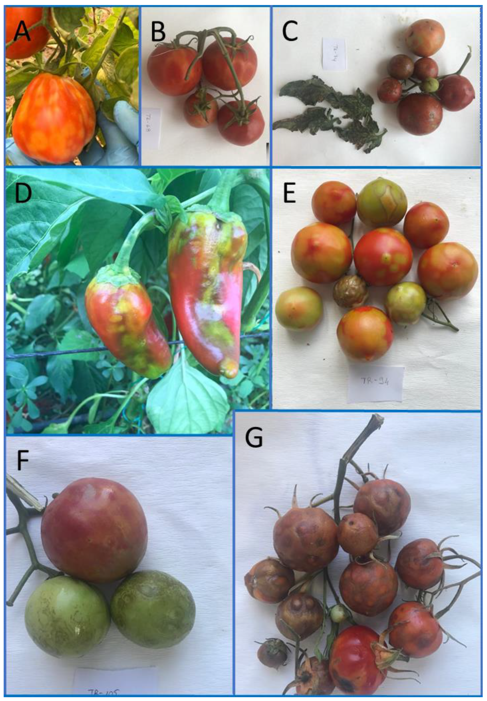
2.1. Incidence and Sequencing

Upon testing by DAS-ELISA and RT-PCR, only seven samples: six tomatoes (A 4, E 1, and B 1) and one pepper (A 1) were positive for ToBRFV infection, giving a low disease incidence of 3.6%. All infected plants showed severe symptoms, including leaf mosaic and spots and blisters on fruits (Figure 1).
The targeted genome regions of seven isolates were sequenced and NCBI GenBank accession codes were provided for them (Table S1). The recovered 824 bp region (201 bp partial MP, 480 bp complete CP, and 143 bp 3`-UTR) encodes 274 amino acids (aa) and is homologous to nt 5514-6337 of the complete genome sequence of the ToBRFV reference isolate NC_028478.

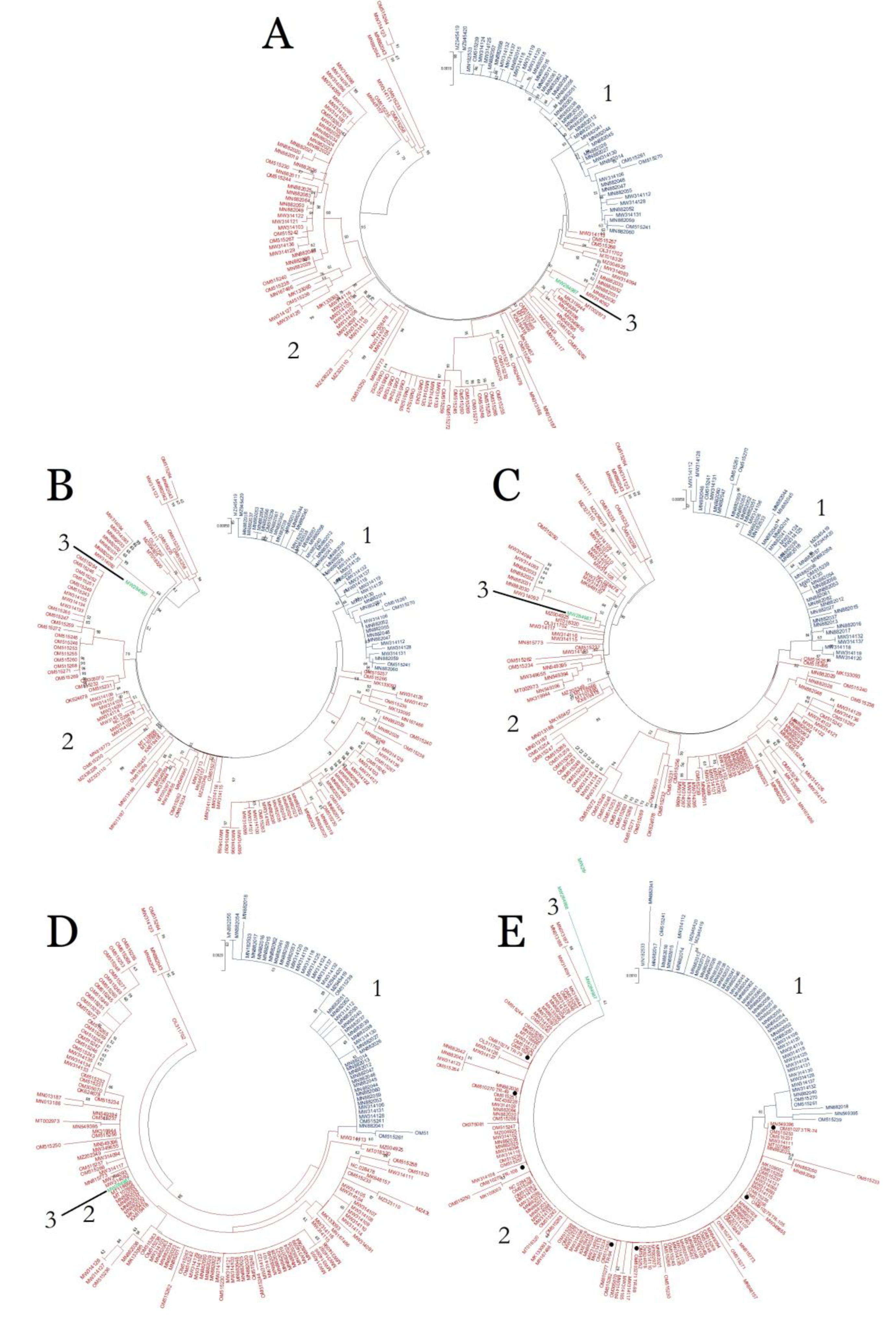
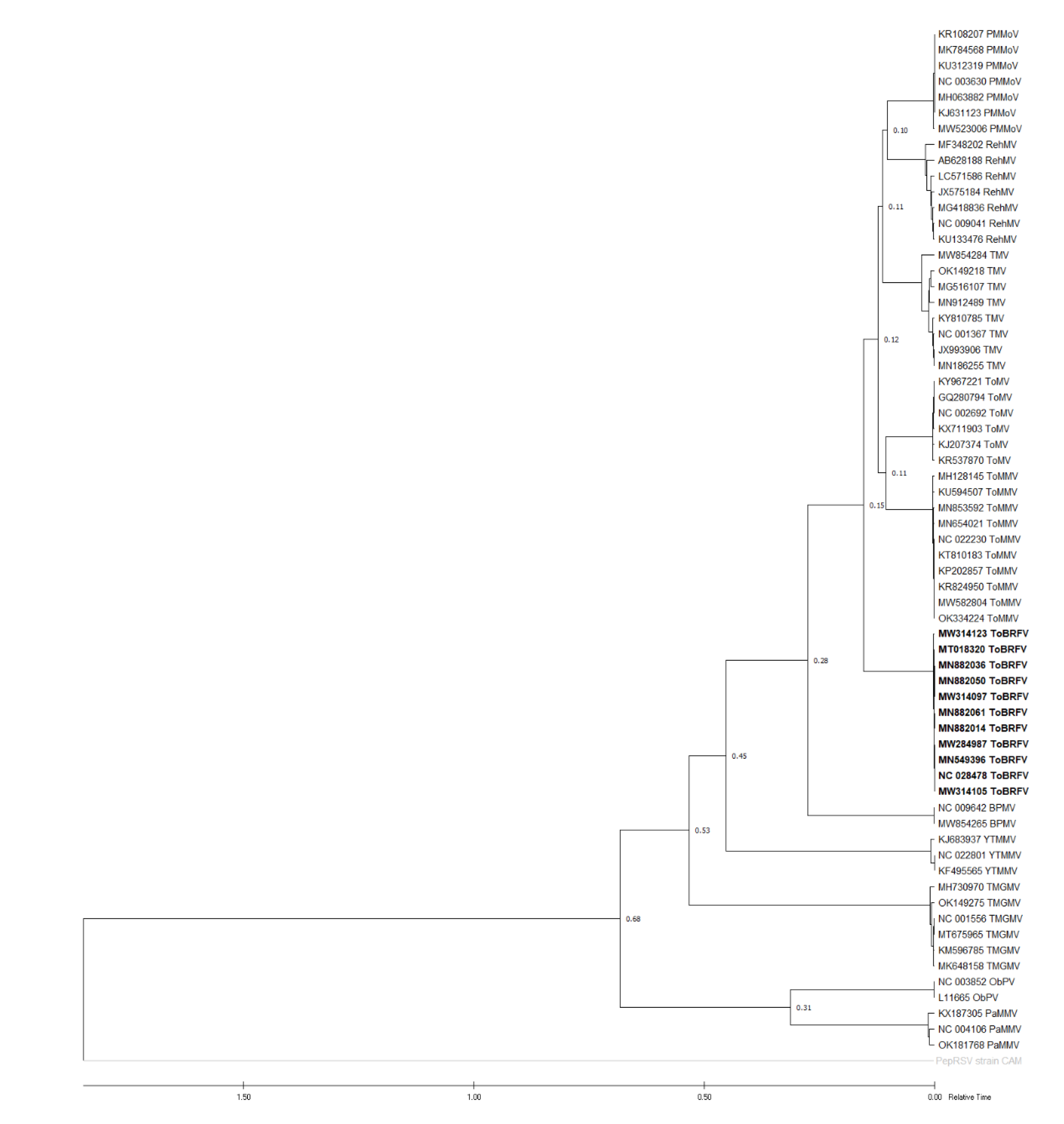
2.2. Recombination and Phylogenetic Analyses

RDP4 scan on the complete genome sequences alignment of 174 isolates did not detect any recombination event. Evidence of recombination was also not found on ORF4/CP alignment of 185 isolates, which included 4 other GenBank isolates and 7 novel isolates.
Phylogenetic analysis of the ORF4/CP alignment of 185 isolates by MEGA X suggested that ToBRFV phylogeny can be divided into three groups (Figure 2). The vast majority of the isolates were distributed into groups 1 and 2, with 134 and 49 isolates, respectively. Two French isolates: 22006291-H (MW284987) and 22006291-L (MW284988) were positioned in group 3 (Figure 2).
Further analysis on the complete genome and the other three ORF alignments deduced that the clustering of the 49 isolates in group 1 was fully intact in the complete genome, ORF1, ORF2, and ORF3 phylogenetic trees, and showed very close relation to these isolates (Figure 2). On the other hand, 134 isolates in group 2 were dispersed into different groups in the complete genome, ORF1, ORF2, and ORF3 phylogenetic trees (Figure 2).

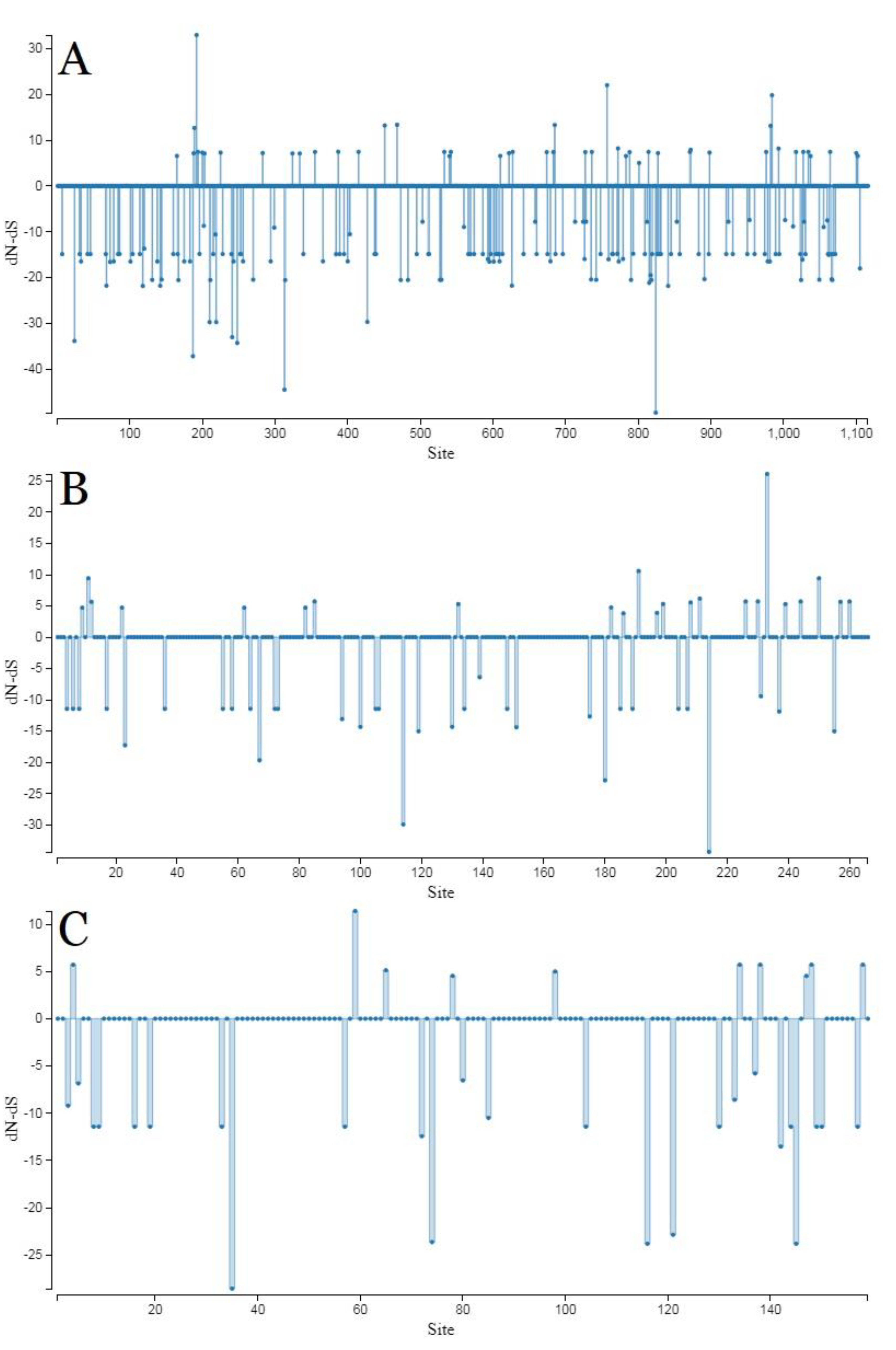
2.3. Genetic Variation and Selection Pressure Analyses

Analysis by DnaSP provided quantitative estimation of low genetic variation among current global ToBRFV isolates. All tested populations obtained very low π (<0.1) and k values. CP was shown to be the most conserved region, demonstrated by the lowest Hd, S, η, k, and π values among analysed ORFs which were all obtained by the gene. In addition to this, analysis on the CP sequences of 185 isolates found only 34 distinct haplotypes (h) (Table 1).
The ToBRFV genome was under strong purifying pressure, as shown by the low dN/dS ratio (ω < 0.2) assigned to all of the analysed ORFs and geographic regions. Group 2 and “Middle East+China” populations consistently obtained significantly higher ω values than other populations in both comparisons based on the complete genome and CP sequences, in the phylogroup and region categories, respectively (Table 1). On the basis of the ORFs (ORF2, ORF3, ORF4), there were no codons under positive selection by the SLAC method in HyPhy software package, which was implemented in Datamonkey webserver with a p-value threshold (p ≤ 0.1) (Figure 3). These data showed that ToBRFV was under strong negative evolutionary constraints on the ORFs.

2.4. Neutrality Tests

Neutrality tests using three parameters consistently gave negative values to all tested populations in the analysis of the complete genome, four ORFs, and geographic regions. All tests on the different genome regions obtained significant values and indicated there were enough data to support the statistical calculations (Table 2).

2.5. Gene Flow and Genetic Separation among Populations

Gene flow study by DnaSP assigned high KS*, KST*, Z*, and Snn metrics, with a significant p-value in comparisons between groups 1 and 2, in both analyses of the complete genome and CP sequences, indicated that the clustering of these groups had been conducted correctly. Additionally, in both analyses based on the complete genome and CP sequences, FST values for group 1 vs. 2 were estimated to be > 0.25, whereas for all isolates vs. group 2 were < 0.25 (0.0281 and 0.0748, respectively) (Table 3).
In the region category, all comparisons obtained relatively low KS*, KST*, Z*, and Snn metrics with significant and non-significant p-values. Additionally, FST values < 0.25, which indicated frequent gene flow and no genetic differentiation over time, were also assigned to all comparisons based on the complete genome and CP sequences (Table 3).

2.6. Molecular Dating

Eleven ToBRFV isolates from different phylogroups: nine, group 1; one, group 2; and one, group 3, were compared with ten other tobamovirus isolates from various countries and hosts available in GenBank. The constructed ML showed that a subgroup consisting of ToBRFV, ToMMV, ToMV, TMV, RehMV, and PMMoV populations was split with the BPMV population at around the middle time over the course of the evolution of 11 tested tobamoviruses (Figure 4). Similarly, percentage identity analysis by SDT v1.2 deduced that ToBRFV shared higher nt and aa identities with ToMMV, ToMV, TMV, RehMV, and BPMV than other compared tobamoviruses (Table 4). Because there is no available report on MRCA based on the complete genome sequences of tobamoviruses, the divergence time of ToBRFV was calculated using the ratios of ToBRFV and other patristic distances. Using this method, the divergence time of ToBRFV with BPMV was estimated to be 1.87 and the divergence time with the ToMMV, ToMV, TMV, RehMV, and PMMoV groups was estimated to be 0.8. TMRCAs of other tobamoviruses with ToBRFV are presented in Table 5.

3. Discussion

The naturally stable and easily mechanically transmitted tobamoviruses have always been serious problems to tomato, pepper, and other Solanaceae cultivation. Therefore, cultivars carrying the resistance genes Tm-1, Tm-2, and Tm-22 became the most effective measures to control members of the genus, such as ToMV and TMV [30,31,32]. ToBRFV then came along relatively recently and broke all those widely implemented highly resistant (HR) genes and was poised to become a dominant species [9,33,34]. However, the tolerance of some genotypes of Solanum pimpinellifolium [35] and the resistance of Solanum ochrantum [36] to the virus had been demonstrated. The knowledge on the genetic diversity and population structure of ToBRFV reported in the current study could also contribute to further the biological arms race to identify potential HR genes and their longevity.
With a low disease incidence of 3.6%, ToBRFV was shown to have limited spread to other regions after its initial detection in Southern Turkey [14]. The appearance of spots on fruits (Figure 1) seems to be due to discolouration only, different than those caused by tomato spotted wilt virus (TSWV, genus Orthotospovirus), which mostly have dark halos around the discoloured areas. In addition to that, TSWV infections usually do not lead to severe blisters on fruits [37]. Tomato and pepper plants in greenhouses where ToBRFV was found were promptly eradicated and the greenhouses and tools were thoroughly sterilised using disinfectant. These greenhouses were barred from planting anything for one year.
When full genome sequence data were absent, the phylogenetic relationships among the isolates of tobamoviruses were often established using full CP sequences [38,39,40]. Therefore, the three groups of ToBRFV phylogeny, based on 480 bp full CP gene nt sequences, were confirmed in this study. The clustering of the 49 isolates of group 1 survived phylogenetic analyses on the complete genome, and three other ORFs indicated a single recent common ancestor for all group 1 isolates. Meanwhile, the members of group 2 were dispersed into different groups in four other trees. Isolate no. MW284987, which belongs to group 3 in the CP tree, was always positioned among the members of group 2 in four other trees.
Phylogenetic analysis on the complete CP gene sequence positioned all seven novel isolates in group 2, indicating high identity among these isolates even with the TR-79 (OM810274) isolate, which was from pepper. Further observations also did not find an association between isolates clustering with the host species or origin. Interestingly, one of the previously registered Turkish isolates, TBRFV-Ant-Tom (MT107885), formed a monophyletic subcluster separated from all novel isolates, suggesting multiple recent intrusions into the country.
In line with phylogenetic analysis, a genetic variation study using DnaSP software also determined high identity among current global ToBRFV isolates. The CP gene was deduced as the most conserved region with the lowest genetic variation according to all parameters, thereby supporting the suitability of CP sequences to help to resolve ToBRFV phylogeny.
Vigorous purifying selections were observed in the complete genome and all four ORF sequences (ω = 0.0869–0.2066). Due to the SLAC result, all of the codons/sites were under negative selection, whereby it seems probable that negative sites have an important role in infection intensity and virus transmission. In CP, group 1 isolates were under more intense negative evolutionary constraints compared to isolates in group 2. However, group 1, together with group 2 and all regional populations, were all shown to be under weak purifying selections and assigned negative values by neutrality tests. Therefore, these populations are experiencing expanding or bottleneck selections, with the growth likely due to low-frequency polymorphism (new mutations); thus, further division into subgroups or even new groups can be anticipated from these populations.
The high KS*, KST*, Z*, and Snn values, supported by significant p-values, showed that there is a large genetic variation among group 1 and group 2. In line with this, FST values for group 1 vs. 2, in both the complete genome and CP, were consistently more than 0.25 (0.3987 and 0.6758, respectively), indicating strong genetic separation between them. Therefore, these results corroborated the results of phylogenetic analysis.
Tomato and pepper have been cultivated for thousands of years in their native South America and for hundreds of years in other regions since their introduction. It is interesting that such an evolutionarily successful ToBRFV, of which natural hosts are mostly restricted to two plant species, has just been detected recently, much later than other important tobamoviruses infecting Solanaceae. Therefore, its origin and genetic relation with the other members of Tobamovirus become necessary to understand.
So far, South America is thought to be the origin of the virus, mainly due to the position of one Peruvian isolate, 36783571_2 (MW314111), in a phylogenetic tree based on complete genome sequences [8]. Likewise, the complete genome tree constructed in the current study also positioned 36783571_2 in an early diverging cluster, together with three isolates from Peru: 40732089_3 (OM515235), 41108421 (OM515258), and 36364500_1 (OM515233), and four isolates from Netherlands: 39563433_3 (MW314123), 39563361_A (MN882042), 39563361_B (MN882043), and 41903353 (OM515264) (Figure 2). This showed that the more complete genome sequences of isolates from South America and other regions are clearly needed to complement the already abundant European isolates in order to resolve the origin of ToBRFV, as also stated by [8]. In line with genetic variation and gene flow analyses, a quantitative measurement using Fixation index also concluded high identity among global isolates, and that there is no clear genetic separation among isolates from different origins at present, as demonstrated by FST values between three regions: Europe, “Middle East+China”, and America were all 0.25 or less (Table 3).
Analysis on CP nt sequences estimated that the divergence between ribgrass mosaic virus (RMV) and a subgroup that consists of PMMoV, TMGMV, ToMV, and TMV happened 1987–5268 years ago [41]. Another molecular dating analysis based on replicase gene sequences showed that the group contains BPMV, ToMV, TMV, RehMV, and PMMoV, which diverged from another group that consists of PaMMV, ObPV, TMGMV along the tobamovirus evolution timeline [42], very similar to analysis based on the complete genome by [43] and this current study. However, the estimated TMRCAs of tobamoviruses based on the complete genome sequences comparison were not available. Therefore, a simple estimation of the TMRCA of ToBRFV was conducted using the ratios of the virus patristic distance with other tested tobamoviruses provided by MEGA X software. TMRCA of ToBRFV was determined to be the closest with BPMV, with a ratio of 1.87. The constructed TimeTree pictured ToBRFV emergence as a distinct species was not recent, around the middle of the course of genus Tobamovirus evolution (Figure 4).

4. Materials and Methods

4.1. Samples Collection

Surveys were conducted to detect the possible spread of ToBRFV into greenhouses located in Ankara (A), Eskişehir (E), Bartın (B), and Zonguldak (Z) provinces, which represented the centre, northwest, and north regions of Turkey, respectively. Leaf samples were taken from 195 plants: 112 tomato (A 37, E 20, B 42, and Z 13) and 83 pepper (A 14, E 26, B 7, and Z 36), showing viral symptoms such as leaf mosaic, leaf yellowing, spots on fruits, blisters on fruits, plant malformation, and stunting. An individual sample was put into a plastic bag, labelled, then kept at −20 °C until further use.

4.2. Serological Test

The collected samples were tested for ToBRFV infection using the double-antibody sandwich enzyme-linked immunosorbent assay (DAS-ELISA) technique as described by the manufacturer (Loewe, Germany). A Multiscan FC Microplate Photometer plate reader (Thermo Scientific, USA) was used to evaluate colour development in the plates at a wavelength of 405 nm. A sample was considered positive if its average absorbance value was at least twice that of the negative control.

4.3. RNA Isolation, RT-PCR, and Sequencing

Total RNA was extracted from leaves of all collected samples using the “NucleoSpin RNA Plant Mini kit” according to the manufacturer’s protocol (Macherey-Nagel, Duren, Germany). The concentration and purity of the obtained total RNA were determined with the help of a spectrophotometer (NanoDrop 2000, ThermoFisher Scientific, Waltham, MA, USA); total RNA concentrations of all samples were diluted to 50 ng.
The 25 μL one-step RT-PCR reaction mixture was composed of 6 μL 5X Go Taq Flexi Green buffer, 1.2 μL 25 mM MgCl2, 0.7 μL 10 mM dNTPs, 1 μL each of 10 μM forward and reverse primer, 0.1 μL Reverse Transcriptase (ProtoScript® II Reverse Transcriptase-M-MuLV), 0.1 μL RNase (RNase Inhibitor, Murine 1 U/μL), 0.25 μL Taq DNA Polymerase (GoTaq® G2 Flexi DNA Polymerase 5 U/μL), and 2 μL of 50 ng/μL total RNA. ToBRFV-F (5503) and ToBRFV-R (6344) primer pairs [10] to amplify 842 bp of partial MP (209 bp), complete CP (480 bp), and 3′-UTR (153 bp) regions were applied in the PCR step. An RT-PCR amplification program of 20 min at 42 °C, 5 min at 94 °C, 30 cycles of 60 s at 94 °C, 60 s at 48 °C, and 60 s at 72 °C, followed by a final extension for 10 min at 72 °C, was performed using a thermal cycler (Biometra, Analytik Jena, Germany).
RT-PCR products were mixed with loading buffer (ThermoFisher Scientific, Waltham, MA, USA) (7:1), then loaded into ready-to-use agarose gel stained with Pronosafe DNA fluorescent marking (Condalab, Madrid, Spain), and then run in an electrophoresis apparatus (ThermoFisher Scientific, Waltham, MA, USA) at 100 V for 1 h. The appearance of the targeted specific band was observed under a UV transilluminator (Vilber, Marne-la-Vallée, France). The amplicons of positive tested samples were TA cloned in pGEM-T Easy vector (Promega Corp., Madison, WI, USA), and then shipped to a commercial firm (BM Laboratory Systems, Ankara, Turkey) for bidirectional Sanger sequencing.

4.4. Recombination Analysis and Phylogeny

In September 2022, all 174 isolates with the complete genome sequences available in NCBI GenBank were retrieved and aligned together using the ClustalW algorithm implemented in MEGA X v.10.2.4 program [44]. The 5′ and 3′-UTR regions were trimmed to create the “complete genome” alignment (6117 bp). Separately, to assess the phylogeny and genetic structure of each ORF, alignment was also trimmed according to ORF1/small replicase (3351 bp), ORF2/RdRp (1422 bp), ORF3/MP (801 bp), and ORF4/CP (480 bp) sequences, respectively. The complete CP sequences of four other GenBank isolates and also novel isolates obtained in this study were added to ORF4/CP alignment (Table S1).
The presence of any phylogenetic anomaly in the alignments was scanned using Recombination Detection Program 4 (RDP4) with its full suite of options: RDP, Chimaera, MaxChi, Bootscan, Siscan, GENECONV, and 3Seq, in default parameters [45]. Anomalies detected by fewer than five algorithms with a Bonferroni-corrected p-value of < 0.05 were ignored.
The best substitution model to construct a Neighbor-Joining (NJ) phylogenetic tree based on each alignment was assessed by MEGA X to be T92 [46] plus uniform rates among sites, with 1000 bootstrap replicates. The percentage identities of the nucleotide (nt) and amino acid (aa) sequences of the complete genome of ToBRFV were calculated using Sequence Demarcation Tool (SDT) v1.2 software [47].

4.5. Population Structure of ToBRFV

The number of haplotypes (h), haplotype diversity (Hd), the number of variable sites (S), the total number of mutations (η), the average number of nt differences between sequences (k), and nt diversity (per site) (π) were analysed using the DnaSP v.6.12.03 program [48] to determine genetic variation in the complete genome, ORF1, ORF2, ORF3 (MP), and ORF 4 (CP) sequences of different populations. Additionally, transcriptional selection (dN/dS ratio = ω) was also estimated. When ω is > 1, equal = 1, and < 1, the related genome region is evaluated to be under positive (diversifying), neutral, or negative (purifying) selection, respectively. To evaluate the dS/dN, individual codon positions for each ORF (ORF1, ORF2, ORF3, ORF4) under natural selection were used for single likelihood ancestor counting algorithm (SLAC) (implemented in MEGA X software and available free online on Datamonkey webserver [49].
To calculate the neutrality, Fu and Li’s D*, Fu and Li’s F* [50], and Tajima’s D [51] metrics were used with a window length of 100 sites and a step size of 25 sites. The genetic differentiation and gene flow among populations were assessed using the KS*, KST*, Z*, Snn, and FST (fixation index) [52]. For panmictic populations, the FST value was 0, while a ratio higher than 0.25 implies expanding genetic separation [53,54].

4.6. Molecular Dating Analysis

Lineages of tobamoviruses were determined to be congruent with those of their hosts [43]. Therefore, the divergence times of the ToBRFV population with 10 other Asterids-infecting tobamoviruses that are known to have close genetic relationship [43]: pepper mild mottle virus (PMMoV), rehmannia mosaic virus (RheMV), tobacco mosaic virus (TMV), tomato mosaic virus (ToMV), tomato mottle mosaic virus (ToMMV), bell pepper mottle virus (BPMV), yellow tailflower mild mottle virus (YTMMV), tobacco mild green mosaic virus (TMGMV), obuda pepper virus (ObPV), paprika mild mottle virus (PaMMV), and a Tobravirus species: pepper ringspot virus (PepRSV) as outgroup, were estimated based on the age evaluation of internal nodes [44]. TimeTree was reconstructed using the fast-dating RelTime-ML computational method under the Tamura 3-parameter model (Tamura, 1992) implemented in MEGA X software, with a default calibration of TMRCA (time to most recent common ancestors) [55].

5. Conclusions

The clustering of ToBRFV isolates into three phylogroups was confirmed. Currently known isolates were mostly sampled from Europe, and some were from the Middle East; thus, there was a very high genetic identity among them. On the basis of the few available data, the South American isolates, when sampled more in the future, may provide more variation and explain better the origin and ancestor of the global ToBRFV population.

Supplementary Materials

The following supporting information can be downloaded at: https://www.mdpi.com/article/10.3390/plants11233279/s1, Table S1: Tomato brown rugose fruit virus (ToBRFV) isolates tested in this study.

Author Contributions

Conceptualization, A.Ç. and A.F.M.; methodology, A.Ç., A.F.M., and A.I.S.; formal analysis, A.Ç. and A.I.S.; investigation, S.C. and A.F.M.; resources, S.C.; writing—original draft, A.Ç. and A.I.S.; writing—review and editing, A.F.M., S.C., and D.K.; funding acquisition, S.C., A.F.M., and A.I.S. All authors have read and agreed to the published version of the manuscript.

Funding

This research was funded by the General Directorate of Agricultural Research and Policies (TAGEM), Turkey, project no. TAGEM/BSAD/Ü/21/A2/P1/5064. Permission to publish new data related to Turkey was obtained from the Ministry of Agriculture and Forestry of Republic of Turkey, permit no. E-36871779-320.03.05-4502631. Adyatma Irawan Santosa was supported by the grant no. 3612/UN1/PN/PN/PT.01.03/2022 from the Faculty of Agriculture, Universitas Gadjah Mada.

Ethical Approval

This article does not contain any studies with human participants or animals performed by any of the authors.

Data Availability Statement

Partial genome sequences of seven novel Turkish ToBRFV isolates are available in GenBank, reference number OM810270, OM810271, OM810273, OM810274, OM810277, OM810278, and OM810279.

Conflicts of Interest

The authors have no conflict of interest to declare that are relevant to the content of this article.

References

  1. Panno, S.; Caruso, A.G.; Barone, S.; Lo Bosco, G.; Rangel, E.A.; Davino, S. Spread of tomato brown rugose fruit virus in Sicily and evaluation of the spatiotemporal dispersion in experimental conditions. Agronomy 2020, 10, 834. [Google Scholar] [CrossRef]
  2. Cambrón-Crisantos, J.M.; Rodríguez-Mendoza, J.; Valencia-Luna, J.B.; Alcasio Rangel, S.; de Jesús García-Ávila, C.; López-Buenfil, J.A.; Ochoa-Martínez, D.L. First report of Tomato brown rugose fruit virus (ToBRFV) in Michoacan. Rev. Mex. Fitopatol. 2019, 37, 185–192. [Google Scholar] [CrossRef] [Green Version]
  3. Abou Kubaa, R.; Choueiri, E.; Heinoun, K.; Cillo, F.; Saponari, M. First report of tomato brown rugose fruit virus infecting sweet pepper in Syria and Lebanon. J. Plant Pathol. 2022, 104, 425. [Google Scholar] [CrossRef]
  4. King, A.M.; Lefkowitz, E.; Adams, M.J.; Carstens, E.B. Virus Taxonomy: Ninth Report of the International Committee on Taxonomy of Viruses; Elsevier: Amsterdam, The Netherlands, 2011. [Google Scholar]
  5. Salem, N.; Mansour, A.; Ciuffo, M.; Falk, B.W.; Turina, M. A new tobamovirus infecting tomato crops in Jordan. Arch. Virol. 2016, 161, 503–506. [Google Scholar] [CrossRef] [PubMed]
  6. Davino, S.; Caruso, A.G.; Bertacca, S.; Barone, S.; Panno, S. Tomato brown rugose fruit virus: Seed transmission rate and efficacy of different seed disinfection treatments. Plants 2020, 9, 1615. [Google Scholar] [CrossRef] [PubMed]
  7. Oladokun, J.O.; Halabi, M.H.; Barua, P.; Nath, P.D. Tomato brown rugose fruit disease: Current distribution, knowledge and future prospects. Plant Pathol. 2019, 68, 1579–1586. [Google Scholar] [CrossRef] [Green Version]
  8. Van de Vossenberg, B.T.L.H.; Dawood, T.; Wozny, M.; Botermans, M. First expansion of the public Tomato brown rugose fruit Virus (ToBRFV) Nextstrain Build; Inclusion of New Genomic and Epidemiological Data. PhytoFrontiers 2021, 1, 359–363. [Google Scholar] [CrossRef]
  9. Luria, N.; Smith, E.; Reingold, V.; Bekelman, I.; Lapidot, M.; Levin, I.; Elad, N.; Tam, Y.; Sela, N.; Abu-Ras, A.; et al. A new Israeli Tobamovirus isolate infects tomato plants harboring Tm-22 resistance genes. PLoS ONE 2017, 12, e0170429. [Google Scholar] [CrossRef] [Green Version]
  10. Ling, K.S.; Tian, T.; Gurung, S.; Salati, R.; Gilliard, A. First report of tomato brown rugose fruit virus infecting greenhouse tomato in the United States. Plant Dis. 2019, 103, 1439. [Google Scholar] [CrossRef]
  11. Menzel, W.; Knierim, D.; Winter, S.; Hamacher, J.; Heupel, M. First report of tomato brown rugose fruit virus infecting tomato in Germany. New Dis. Rep. 2019, 39, 1. [Google Scholar] [CrossRef]
  12. Panno, S.; Caruso, A.G.; Davino, S. First report of tomato brown rugose fruit virus on tomato crops in Italy. Plant Dis. 2019, 103, 1443. [Google Scholar] [CrossRef]
  13. Alkowni, R.; Alabdallah, O.; Fadda, Z. Molecular identification of tomato brown rugose fruit virus in tomato in Palestine. J. Plant Pathol. 2019, 101, 719–723. [Google Scholar] [CrossRef]
  14. Fidan, H.; Sarikaya, P.; Calis, O. First report of Tomato brown rugose fruit virus on tomato in Turkey. New Dis. Rep. 2019, 39, 18. [Google Scholar] [CrossRef] [Green Version]
  15. Skelton, A.; Buxton-Kirk, A.; Ward, R.; Harju, V.; Frew, L.; Fowkes, A.; Long, M.; Negus, A.; Forde, S.; Adams, I.P.; et al. First report of Tomato brown rugose fruit virus in tomato in the United Kingdom. New Dis. Rep. 2019, 40, 12. [Google Scholar] [CrossRef] [Green Version]
  16. Yan, Z.Y.; Ma, H.Y.; Han, S.L.; Geng, C.; Tian, Y.P.; Li, X.D. First report of Tomato brown rugose fruit virus infecting tomato in China. Plant Dis. 2019, 103, 2973. [Google Scholar] [CrossRef]
  17. Beris, D.; Malandraki, I.; Kektsidou, O.; Theologidis, I.; Vassilakos, N.; Varveri, C. First report of tomato brown rugose fruit virus infecting tomato in Greece. Plant Dis. 2020, 104, 2035. [Google Scholar] [CrossRef] [Green Version]
  18. Van de Vossenberg, B.T.L.H.; Visser, M.; Bruinsma, M.; Koenraadt, H.M.; Westenberg, M.; Botermans, M. Real-time tracking of Tomato brown rugose fruit virus (ToBRFV) outbreaks in the Netherlands using Nextstrain. PLoS ONE 2020, 15, e0234671. [Google Scholar] [CrossRef]
  19. Alfaro-Fernández, A.; Castillo, P.; Sanahuja, E.; Rodríguez-Salido, M.C.; Font, M.I. First report of tomato brown rugose fruit virus in tomato in Spain. Plant Dis. 2021, 105, 515. [Google Scholar] [CrossRef]
  20. Esmaeilzadeh, F.; Koolivand, D. Occurrence of tomato brown rugose fruit virus in tomato in Iran. J. Plant Pathol. 2022, 104, 457. [Google Scholar] [CrossRef]
  21. Esmaeilzadeh, F.; Koolivand, D. First report of tomato brown rugose fruit virus infecting bell pepper in Iran. J. Plant Pathol. 2022, 104, 893. [Google Scholar] [CrossRef]
  22. Skelton, A.; Gentit, P.; Porcher, L.; Visage, M.; Fowkes, A.; Adams, I.P.; Harju, V.; Webster, G.; Pufal, H.; McGreig, S.; et al. First report of Tomato brown rugose fruit virus in tomato in France. New Dis. Rep. 2022, 45, e12061. [Google Scholar] [CrossRef]
  23. Hanssen, I.M.; Mumford, R.; Blystad, D.R.; Cortez, I.; Hasiów-Jaroszewska, B.; Hristova, D.; Pagán, I.; Pereira, A.M.; Peters, J.; Pospieszny, H.; et al. Seed transmission of Pepino mosaic virus in tomato. Eur. J. Plant Pathol. 2010, 126, 145–152. [Google Scholar] [CrossRef]
  24. Moya, A.; Holmes, E.C.; González-Candelas, F. The population genetics and evolutionary epidemiology of RNA viruses. Nat. Rev. Microbiol. 2004, 2, 279–288. [Google Scholar] [CrossRef] [PubMed]
  25. Maayan, Y.; Pandaranayaka, E.P.; Srivastava, D.A.; Lapidot, M.; Levin, I.; Dombrovsky, A.; Harel, A. Using genomic analysis to identify tomato Tm-2 resistance-breaking mutations and their underlying evolutionary path in a new and emerging tobamovirus. Arch. Virol. 2018, 163, 1863–1875. [Google Scholar] [CrossRef]
  26. Chanda, B.; Rivera, Y.; Nunziata, S.O.; Galvez, M.E.; Gilliard, A.; Ling, K.S. Complete genome sequence of a tomato brown rugose fruit virus isolated in the United States. Microbiol. Resour. Announc. 2020, 9, e00630-20. [Google Scholar] [CrossRef]
  27. Ma, Z.; Zhang, H.; Ding, M.; Zhang, Z.; Yang, X.; Zhou, X. Molecular characterization and pathogenicity of an infectious cDNA clone of tomato brown rugose fruit virus. Phytopathol. Res. 2021, 3, 14. [Google Scholar] [CrossRef]
  28. Yan, Z.Y.; Zhao, M.S.; Ma, H.Y.; Liu, L.Z.; Yang, G.L.; Geng, C.; Tian, Y.; Li, X.D. Biological and molecular characterization of tomato brown rugose fruit virus and development of quadruplex RT-PCR detection. J. Integr. Agric. 2021, 20, 1871–1879. [Google Scholar] [CrossRef]
  29. Zhang, S.; Griffiths, J.S.; Marchand, G.; Bernards, M.A.; Wang, A. Tomato brown rugose fruit virus: An emerging and rapidly spreading plant RNA virus that threatens tomato production worldwide. Mol. Plant Pathol. 2022, 23, 1262–1277. [Google Scholar] [CrossRef]
  30. Soost, R.K. Hybrid tomato resistant to tobacco mosaic virus: Inheritance for Resistance in Derivatives of a Complex Species Hybrid. J. Hered. 1963, 54, 241–244. [Google Scholar] [CrossRef]
  31. Alexander, L.J. Transfer of a dominant type of resistance to the four known Ohio pathogenic strains of tobacco mosaic virus (TMV) from Lycopersicon peruvianum to L. esculentum. Phytopathology 1963, 53, 869. [Google Scholar]
  32. Pfitzner, A.J.P. Resistance to Tobacco mosaic virus and Tomato mosaic virus in tomato. In Natural Resistance Mechanisms of Plants to Viruses; Loebenstein, G., Carr, J.P., Eds.; Springer: Dordrecht, The Netherlands, 2006; pp. 399–413. [Google Scholar]
  33. Dombrovsky, A.; Smith, E. Seed transmission of tobamoviruses: Aspects of global disease distribution. In Seed Biology; Jimenez-Lopez, J.C., Ed.; IntechOpen: London, UK, 2017; pp. 234–260. [Google Scholar]
  34. Fidan, H.; Sarikaya, P.; Yildiz, K.; Topkaya, B.; Eekis, G.; Calis, O. Robust molecular detection of the new Tomato brown rugose fruit virus in infected tomato and pepper plants from Turkey. J. Integr. Agric. 2021, 20, 2170–2179. [Google Scholar] [CrossRef]
  35. Zinger, A.; Lapidot, M.; Harel, A.; Doron-Faigenboim, A.; Gelbart, D.; Levin, I. Identification and mapping of tomato genome loci controlling tolerance and resistance to tomato brown rugose fruit virus. Plants 2021, 10, 179. [Google Scholar] [CrossRef] [PubMed]
  36. Jewehan, A.; Salem, N.; Tóth, Z.; Salamon, P.; Szabó, Z. Screening of Solanum (sections Lycopersicon and Juglandifolia) germplasm for reactions to the tomato brown rugose fruit virus (ToBRFV). J. Plant Dis. Prot. 2022, 129, 117–123. [Google Scholar] [CrossRef]
  37. Morca, A.F.; Çelik, A.; Coşkan, S.; Santosa, A.I.; Akbaş, B. Population analysis on tomato spotted wilt virus isolates inducing various symptoms on tomato, pepper, and Chenopodium album in Turkey. Physiol. Mol. Plant Pathol. 2022, 118, 101786. [Google Scholar] [CrossRef]
  38. Gibbs, A.J. Tobamovirus classification. In The Plant Viruses; Van Regenmortel, M.H.V., Fraenkel-Conrat, H., Eds.; Springer: Boston, MA, USA, 1986; pp. 167–180. [Google Scholar]
  39. Duarte, L.M.L.; Alexandre, M.A.V.; Rivas, E.B.; Cattai, M.B.; Soares, R.M.; Harakava, R.; Fernandes, F.M.C. Phylogenetic analysis of Tomato mosaic virus from Hemerocallis sp. and Impatiens hawkeri. Summa Phytopathol. 2007, 33, 409–413. [Google Scholar] [CrossRef]
  40. Listhihani, L.; Hidayat, S.H.; Wiyono, S.; Damayanti, T.A. Characteristic of Tobacco mosaic virus isolated from cucumber and tobacco collected from East Java, Indonesia. Biodiversitas 2019, 20, 2937–2942. [Google Scholar] [CrossRef]
  41. Pagán, I.; Firth, C.; Holmes, E.C. Phylogenetic analysis reveals rapid evolutionary dynamics in the plant RNA virus genus Tobamovirus. J. Mol. Evol. 2010, 71, 298–307. [Google Scholar] [CrossRef]
  42. Stobbe, A.H.; Melcher, U.; Palmer, M.W.; Roossinck, M.J.; Shen, G. Co-divergence and host-switching in the evolution of tobamoviruses. J. Gen. Virol. 2012, 93, 408–418. [Google Scholar] [CrossRef]
  43. Gibbs, A.J.; Wood, J.; Garcia-Arenal, F.; Ohshima, K.; Armstrong, J.S. Tobamoviruses have probably co-diverged with their eudicotyledonous hosts for at least 110 million years. Virus Evol. 2015, 1, vev019. [Google Scholar] [CrossRef] [Green Version]
  44. Kumar, S.; Stecher, G.; Li, M.; Knyaz, C.; Tamura, K. MEGA X: Molecular Evolutionary Genetics Analysis across computing platforms. Mol. Biol. Evol. 2018, 22, 1547–1549. [Google Scholar] [CrossRef]
  45. Martin, D.P.; Murrell, B.; Golden, M.; Khoosal, A.; Muhire, B. RDP4: Detection and analysis of recombination patterns in virus genomes. Virus Evol. 2015, 1, vev003. [Google Scholar] [CrossRef] [PubMed]
  46. Tamura, K. Estimation of the number of nucleotide substitutions when there are strong transition-transversion and G + C-content biases. Mol. Biol. Evol. 1992, 9, 678–687. [Google Scholar] [CrossRef] [PubMed] [Green Version]
  47. Muhire, B.M.; Varsani, A.; Martin, D.P. (SDT: A virus classification tool based on pairwise sequence alignment and identity calculation. PLoS ONE 2014, 9, e108277. [Google Scholar] [CrossRef]
  48. Rozas, J.; Ferrer-Mata, A.; Sánchez-DelBarrio, J.C.; Guirao-Rico, S.; Librado, P.; Ramos-Onsins, S.E.; Sánchez-Gracia, A. DnaSP 6: DNA sequence polymorphism analysis of large data sets. Mol. Biol. Evol. 2017, 34, 3299–3302. [Google Scholar] [CrossRef]
  49. Datamonkey. Available online: http://www.datamonkey.org (accessed on 25 October 2022).
  50. Fu, Y.X.; Li, W.H. Statistical tests of neutrality of mutations. Genetics 1993, 133, 693–709. [Google Scholar] [CrossRef] [PubMed]
  51. Tajima, F. Statistical method for testing the neutral mutation hypothesis by DNA polymorphism. Genetics 1989, 123, 585–595. [Google Scholar] [CrossRef] [PubMed]
  52. Hudson, R.R. A new statistic for detecting genetic differentiation. Genetics 2000, 155, 2011–2014. [Google Scholar] [CrossRef] [PubMed]
  53. Çelik, A.; Santosa, A.I.; Gibbs, A.J.; Ertunç, F. Prunus necrotic ringspot virus in Turkey: An immigrant population. Arch. Virol. 2022, 167, 553–562. [Google Scholar] [CrossRef] [PubMed]
  54. Coşkan, S.; Morca, A.F.; Akbaş, B.; Çelik, A.; Santosa, A.I. Comprehensive surveillance and population study on plum pox virus in Ankara Province of Turkey. J. Plant Dis. Prot. 2022, 129, 981–991. [Google Scholar] [CrossRef]
  55. Mello, B. Estimating TimeTrees with MEGA and the TimeTree resource. Mol. Biol. Evol. 2018, 35, 2334–2342. [Google Scholar] [CrossRef]
Figure 1. Different symptoms on plants infected by ToBRFV Turkish isolates. (A) Spots on fruit and leaf mosaic on tomato infected by TR-49; (B) spots on tomato fruits infected by TR-68; (C) leaf and fruit malformation on tomatoes infected by TR-74; (D) yellow spots and severe malformation on peppers infected by TR-79; (E) spots and blisters on tomato fruits infected by TR-94; (F) spots on tomato fruits infected by TR-105; and (G) severe blisters on tomato fruits infected by TR-108.
Plants 11 03279 g001
Figure 2. Neighbor-Joining phylogenetic analysis using Tamura 3-parameter model (TN92) with uniform rates among sites (1000 bootstrap replicates, only >50% values were shown) by MEGA X software to show the distribution of 174 isolates in each phylogenetic tree based on (A) the complete genome, (B) ORF1, (C) ORF2, (D) ORF3 (MP), and 185 isolates for (E) ORF4 (CP). The three phylogrouping systems (1: Blue; 2: Red; and 3: Green) were based on CP tree. Black dots are seven Turkish isolates identified in the present study.
Plants 11 03279 g002
Figure 3. SLAC site graph to identify positive and negative codons/sites for ORF2 (A), ORF3 (B), and ORF4 (C).
Plants 11 03279 g003
Figure 4. Divergence time estimation of 11 tobamoviruses with Asterids as main hosts: pepper mild mottle virus (PMMoV), rehmannia mosaic virus (RheMV), tobacco mosaic virus (TMV), tomato mosaic virus (ToMV), tomato mottle mosaic virus (ToMMV), tomato brown rugose fruit virus (ToBRFV), bell pepper mottle virus (BPMV), yellow tailflower mild mottle virus (YTMMV), tobacco mild green mosaic virus (TMGMV), obuda pepper virus (ObPV), and paprika mild mottle virus (PaMMV) by RelTime-ML in MEGA X. An isolate of pepper ringspot virus (PepRSV, genus Tobravirus) was used as out-group.
Plants 11 03279 g004
Table 1. Genetic diversity and polymorphism analyses of the complete genome, ORF1, ORF2, ORF3 (MP), and ORF4 (CP) of ToBRFV from different phylogroups and regions.
PopulationNhHdSηkπdSdNω
Complete genome1741230.99437739017.3960.00280.00880.00110.125
Phylogroup
Group 149350.98562635.6390.00090.00290.00040.1379
Group 2124870.99132633317.7190.00290.00870.00120.1379
Region 1
Europe130860.99123524117.3720.00280.00890.00110.1236
ME and China34280.98813213412.8820.00210.00590.00090.1525
America10101.000495014.6670.00240.00810.00080.0988
ORF11741120.98928329212.7890.00260.00850.00090.1059
ORF21741030.9842042058.2360.00250.00810.00080.0988
ORF3 (MP)174520.93360633.6740.00460.01210.00250.2066
ORF4 (CP)185340.64639400.9940.00210.00690.00060.0869
Phylogroup
Group 14970.267770.3250.00070.00250.00010.04
Group 2134250. 42129290.6370.00130.00350.00070.2
Region 1
Europe131210.66823240.9870.00210.00720.00050.0694
ME and China42100.45812120.7070.00150.00330.00090.2727
America1250.576771.1670.00240.00880.00050.0568
N: number of isolates, h: number of haplotypes, Hd: haplotype diversity, S: number of variable sites, η: total number of mutations, k: average number of nucleotide differences between sequences, π: nucleotide diversity (per site), dN: non-synonymous nucleotide diversity, dS: synonymous nucleotide diversity, and ω: dN/dS. 1 Isolate origin—Europe: Belgium, France, Germany, Greece, Italy, Netherlands, and UK; Middle East and China: Cyprus, Egypt, Iran, Israel, Jordan, Palestine, Turkey, and China; and America: Canada, Mexico, Peru, and USA.
Table 2. Results from demography test statistics between sequences of the complete genome, ORF1, ORF2, ORF3 (MP), and ORF4 (CP) of ToBRFV populations.
PopulationFu and Li’s D *Fu and Li’s F *Tajima’s D
Complete genome−5.56444 **−4.81135 **−2.40531 **
Phylogroup
Group 1−2.18556 ns−2.57257 *−2.10472 *
Group 2−4.89980 **−4.48389 **−2.36401 **
Region 1
Europe−3.60441 **−3.43963 **−1.99775 *
ME and China−2.48265 ns−2.86841 *−2.28997 **
America−1.10835 ns−1.17368 ns−0.83168 ns
ORF1−5.33498 **−4.69009 **−2.41119 **
ORF2−5.63445 **−4.93305 **−2.46251 **
ORF3 (MP)−3.27960 **−3.27892 **−2.03581 *
ORF4 (CP)−6.29855 **−5.63807 **−2.51446 ***
Phylogroup
Group 1−3.41067 **−3.51604 **−2.10661 *
Group 2−5.37507 **−5.10880 **−2.58167 ***
Region 1
Europe−4.81828 **−4.56208 **−2.23084 **
ME and China−3.17348 *−3.39447 **−2.27783 **
America−2.38877 *−2.57986 *−1.94368 *
* 0.01 < p-value > 0.05; ** 0.001 < p-value > 0.01; *** p-value < 0.001; and ns: not significant. 1 Isolate origin—Europe: Belgium, France, Germany, Greece, Italy, Netherlands, and UK; Middle East and China: Egypt, Iran, Israel, Jordan, Palestine, Turkey, and China; and America: Canada, Mexico, Peru, and USA.
Table 3. Genetic differentiation estimates for lineages of ToBRFV, based on the complete genome and CP gene sequences comparison.
ComparisonαKS*αKST *p-ValueαZ *p-ValueSnnp-ValueβFST
Complete genome
Phylogroup
All (n = 174)/Group 1(n = 49)2.57810.04210.0000 ***8.94950.0000 ***0.64050.7180 ns0.2551
All (n = 174)/Group 2 (n = 124)2.78920.00660.0000 ***9.66740.0000 ***0.31441.0000 ns0.0281
Group 1 (n = 49)/Group 2 (n = 124)2.51640.09880.0000 ***8.13170.0000 ***1.00000.0000 ***0.3987
Region 1
Europe (n = 130)/ME and China (n = 34)2.69010.02790.0000 ***8.38880.0000 ***0.94150.0000 ***0.1291
Europe (n = 130)/America (n = 10)2.72920.01510.0000 ***8.13490.0000 ***0.99290.0000 ***0.1836
ME and China (n = 34)/America (n = 10)2.52160.02950.0000 ***5.70250.0000 ***0.98860.0000 ***0.1293
ORF4 (CP)
Phylogroup
All (n = 185)/Group 1 (n = 49)0.49930.16870.0000 ***9.16450.0000 ***0.76420.0000 ***0.4498
All (n = 185)/Group 2 (n = 134)0.48990.03690.0000 ***9.94510.0000 ***0.50810.6070 ns0.0748
Group 1 (n = 49)/Group 2 (n = 134)0.32980.40910.0000 ***8.50450.0000 ***1.00000.0000 ***0.6758
Region 1
Europe (n = 131)/ME and China (n = 42)0.53960.04940.0000 ***8.68610.0000 ***0.71490.0000 ***0.1451
Europe (n = 131)/America (n = 12)0.58070.01750.0120 *8.32620.0220 *0.86740.0210 *0.1124
ME and China (n = 42)/America (n = 12)0.45110.00650.2500 ns6.42690,2870 ns0.65800.3070 ns−0.0005
* 0.01 < p-value > 0.05 and *** p-value < 0.001. α KS*, KST*, Z, and Snn are test statistics of genetic differentiation; β FST is a coefficient of gene differentiation, which measures inter-population diversity; ns: not significant. 1 Isolate origin—Europe: Belgium, France, Germany, Greece, Italy, Netherlands, and UK; Middle East and China: Egypt, Iran, Israel, Jordan, Palestine, Turkey, and China; and America: Canada, Mexico, Peru, and USA.
Table 4. Identity percentage of the complete genome nucleotide and amino acid sequences of ToBRFV with other Asterids-infecting tobamoviruses.
ToBRFV vs.Identity (%)
NucleotideAmino Acid
PMMoV69–69.661.8–62.3
RehMV80.1–81.584–85.9
TMV81.2–81.785.3–86.7
ToMV81–81.485.5–86.4
ToMMV80.7–81–184.4–85.5
BPMV75.4–76.171.4–72.1
YTMMV64.1–64.557.9–58.4
TMGMV65.1–65.562.5–63.1
ObPV65–65.162.9–64
PaMMV65.3–65.763.7–64.1
Table 5. Estimated ToBRFV TMRCA based on the ratio of the patristic distances within the ten Asterids-infecting tobamoviruses’ complete genome sequence maximum likelihood tree.
SpeciesMean Patristic DistanceRatios of ToBRFV and Other Patristic Distances
PaMMV0.684.53
ObPV0.684.53
TMGMV0.533.53
YTMMV0.453
BPMV0.281.87
ToBRFV0.151
ToMMV0.120.8
ToMV0.120.8
TMV0.110.73
RehMV0.100.67
PMMoV0.100.67
Publisher’s Note: MDPI stays neutral with regard to jurisdictional claims in published maps and institutional affiliations.

