
Proteomic and Genomic Studies of Micronutrient Deficiency and Toxicity in Plants
 and
and
Abstract
:1. Introduction
2. Micronutrients and Their Roles
2.1. Boron
2.2. Chlorine
2.3. Copper
2.4. Iron
2.5. Manganese
2.6. Molybdenum
2.7. Zinc
3. Molecular Approaches for Understanding Micronutrient Stress Mechanisms in Plants
3.1. Boron
3.2. Chlorine
3.3. Copper
3.4. Iron
3.5. Manganese
3.6. Molybdenum
3.7. Zinc
4. Conclusions
Author Contributions
Funding
Institutional Review Board Statement
Informed Consent Statement
Data Availability Statement
Conflicts of Interest
References
- Das, S.K. Role of Micronutrient in Rice Cultivation and Management Strategy in Organic Agriculture—A Reappraisal. Agric. Sci. 2014, 5, 765–769. [Google Scholar] [CrossRef]
- Ali, A.; Bhat, B.A.; Rather, G.A.; Malla, B.A.; Ganie, S.A. Proteomic Studies of Micronutrient Deficiency and Toxicity. In Plant Micronutrients; Springer: Cham, Switzerland, 2020; pp. 257–284. [Google Scholar] [CrossRef]
- Arigony, A.L.V.; de Oliveira, I.M.; Machado, M.; Bordin, D.L.; Bergter, L.; Prá, D.; Henriques, J.A.P. The Influence of Micronutrients in Cell Culture: A Reflection on Viability and Genomic Stability. BioMed Res. Int. 2013, 2013, 597282. [Google Scholar] [CrossRef] [PubMed]
- Waraich, E.A.; Ahmad, R.; Ashraf, M.Y. Role of mineral nutrition in alleviation of drought stress in plants. Aust. J. Crop Sci. 2011, 5, 764–777. [Google Scholar]
- Vasu, D.; Singh, S.; Sahu, N.; Tiwary, P.; Chandran, P.; Duraisami, V.; Ramamurthy, V.; Lalitha, M.; Kalaiselvi, B. Assessment of spatial variability of soil properties using geospatial techniques for farm level nutrient management. Soil Tillage Res. 2017, 169, 25–34. [Google Scholar] [CrossRef]
- Kerry, R.G.; Mahapatra, G.P.; Patra, S.; Sahoo, S.L.; Pradhan, C.; Padhi, B.K.; Rout, J.R. Proteomic and genomic responses of plants to nutritional stress. BioMetals 2018, 31, 161–187. [Google Scholar] [CrossRef]
- Luo, Z.-B.; Wu, C.; Zhang, C.; Li, H.; Lipka, U.; Polle, A. The role of ectomycorrhizas in heavy metal stress tolerance of host plants. Environ. Exp. Bot. 2013, 108, 47–62. [Google Scholar] [CrossRef]
- Broadley, M.R.; White, P.J.; Hammond, J.P.; Zelko, I.; Lux, A. Zinc in plants. New Phytol. 2007, 173, 677–702. [Google Scholar] [CrossRef]
- Hansch, R.; Mendel, R.R. Physiological functions of mineral micronutrients (Cu, Zn, Mn, Fe, Ni, Mo, B, Cl). Curr. Opin. Plant Biol. 2009, 12, 259–266. [Google Scholar] [CrossRef]
- Richter, S.; Lamppa, G.K. Structural properties of the chloroplast stromal processing peptidase required for its function in transit peptide removal. J. Biol. Chem. 2003, 278, 39497–39502. [Google Scholar] [CrossRef]
- Supuran, C.T. Carbonic anhydrases—An overview. Cur. Pharm. Des. 2008, 14, 603–614. [Google Scholar] [CrossRef]
- Zelko, I.N.; Mariani, T.J.; Folz, R.J. Superoxide dismutase multigene family: A comparison of the CuZn-SOD (SOD1), Mn-SOD (SOD2), and EC-SOD (SOD3) gene structures, evolution, and expression. Free Radic. Biol. Med. 2002, 33, 337–349. [Google Scholar] [CrossRef]
- Thompson, C.E.; Salzano, F.M.; de Souza, O.N.; Freitas, L.B. Sequence and structural aspects of the functional diversification of plant alcohol dehydrogenases. Gene 2007, 396, 108–115. [Google Scholar] [CrossRef]
- Serero, A.; Giglione, C.; Meinnel, T. Distinctive features of the two classes of eukaryotic peptide deformylases. J Mol. Biol. 2001, 314, 695–708. [Google Scholar] [CrossRef] [PubMed]
- Snaith, S.M.; Levvy, G.A. Alpha-mannosidase as a zinc-dependent enzyme. Nature 1968, 218, 91–92. [Google Scholar] [CrossRef] [PubMed]
- Maidment, J.M.; Moore, D.; Murphy, G.P.; Murphy, G.; Clark, I.M. Matrix metalloproteinase homologues from Arabidopsis thaliana. Expression and activity. J. Biol. Chem. 1999, 274, 34706–34710. [Google Scholar] [CrossRef]
- Pilon, M.; Abdel-Ghany, S.E.; Cohu, C.M.; Gogolin, K.A.; Ye, H. Copper cofactor delivery in plant cells. Curr. Opin. Plant Biol. 2006, 9, 1–8. [Google Scholar] [CrossRef]
- Puig, S.; Askeland, E.; Thiele, D.J. Coordinated remodeling of cellular metabolism during iron deficiency through targeted mRNA degradation. Cell 2005, 120, 99–110. [Google Scholar] [CrossRef]
- Yruela, I. Copper in plants: Acquisition, transport and interactions. Funct. Plant Biol. 2009, 36, 409–443. [Google Scholar] [CrossRef]
- Santagostini, L.; Gullotti, M.; De Gioia, L.; Fantucci, P.; Franzini, E.; Marchesini, A.; Monzani, E.; Casella, L. Probing the location of the substrate binding site of ascorbate oxidase near type 1 copper: An investigation through spectroscopic, inhibition and docking studies. Int. J. Biochem. Cell Biol. 2004, 36, 881–892. [Google Scholar] [CrossRef]
- Marusek, C.M.; Trobaugh, N.M.; Flurkey, W.H.; Inlow, J.K. Comparative analysis of polyphenol oxidase from plant and fungal species. J. Inorg. Biochem. 2006, 100, 108–123. [Google Scholar] [CrossRef]
- Peiffer, W.E.; Ingle, R.T.; Ferguson-Miller, S. Structurally unique plant cytochrome c oxidase isolated from wheat germ, a rich source of plant mitochondrial enzymes. Biochemistry 1990, 29, 8696–8701. [Google Scholar] [CrossRef] [PubMed]
- Jansson, H.; Okvist, M.; Jacobson, F.; Ejdeback, M.; Hansson, O.; Sjolin, L. The crystal structure of the spinach plastocyanin double mutant G8D/L12E gives insight into its low reactivity towards photosystem 1 and cytochrome f. Biochim. Biophys. Acta. 2003, 1607, 203–210. [Google Scholar] [CrossRef] [PubMed]
- Domenech, J.; Palacios, O.; Villarreal, L.; Gonzalez-Duarte, P.; Capdevila, M.; Atrian, S. MTO: The second member of a Drosophila dual copper-thionein system. FEBS Lett. 2003, 533, 72–78. [Google Scholar] [CrossRef]
- Rodriguez, F.I.; Esch, J.J.; Hall, A.E.; Binder, B.M.; Schaller, G.E.; Bleecker, A.B. A copper cofactor for the ethylene receptor ETR1 from Arabidopsis. Science 1999, 283, 996–998. [Google Scholar] [CrossRef]
- Kuper, J.; Llamas, A.; Hecht, H.J.; Mendel, R.R.; Schwarz, G. Structure of the molybdopterin-bound Cnx1G domain links molybdenum and copper metabolism. Nature 2004, 430, 803–806. [Google Scholar] [CrossRef]
- Jeong, J.; Connolly, E.L. Iron uptake mechanisms in plants: Functions of the FRO family of ferric reductases. Plant Sci. 2009, 176, 709–714. [Google Scholar] [CrossRef]
- Adamski, J.M.; Danieloski, R.; Deuner, S.; Braga, E.J.; de Castro, L.A.; Peters, J.A. Responses to excess iron in sweet potato: Impacts on growth, enzyme activities, mineral concentrations, and anatomy. Acta Physiol. Plant. 2012, 34, 1827–1836. [Google Scholar] [CrossRef]
- Moeder, W.; Del Pozo, O.; Navarre, D.A.; Martin, G.B.; Klessig, D.F. Aconitase plays a role in regulating resistance to oxidative stress and cell death in Arabidopsis and Nicotiana benthamiana. Plant Mol. Biol. 2007, 63, 273–287. [Google Scholar] [CrossRef]
- Figueroa, P.; Leon, G.; Elorza, A.; Holuigue, L.; Araya, A.; Jordana, X. The four subunits of mitochondrial respiratory complex II are encoded by multiple nuclear genes and targeted to mitochondria in Arabidopsis thaliana. Plant Mol. Biol. 2002, 50, 725–734. [Google Scholar] [CrossRef]
- Rasmusson, A.G.; Geisler, D.A.; Moller, I.M. The multiplicity of dehydrogenases in the electron transport chain of plant mitochondria. Mitochondrion 2008, 8, 47–60. [Google Scholar] [CrossRef]
- Dai, S.; Friemann, R.; Glauser, D.A.; Bourquin, F.; Manieri, W.; Schurmann, P.; Eklund, H. Structural snapshots along the reaction pathway of ferredoxin-thioredoxin reductase. Nature 2007, 448, 92–96. [Google Scholar] [CrossRef] [PubMed]
- Hesberg, C.; Hansch, R.; Mendel, R.R.; Bittner, F. Tandem orientation of duplicated xanthine dehydrogenase genes from Arabidopsis thaliana: Differential gene expression and enzyme activities. J Biol. Chem. 2004, 279, 13547–13554. [Google Scholar] [CrossRef] [PubMed]
- Koshiba, T.; Saito, E.; Ono, N.; Yamamoto, N.; Sato, M. Purification and properties of flavin- and molybdenum-containing aldehyde oxidase from coleoptiles of maize. Plant Physiol. 1996, 110, 781–789. [Google Scholar] [CrossRef]
- Voss, I.; Koelmann, M.; Wojtera, J.; Holtgrefe, S.; Kitzmann, C.; Backhausen, J.E.; Scheibe, R. Knockout of major leaf ferredoxin reveals new redox-regulatory adaptations in Arabidopsis thaliana. Physiol. Plant. 2008, 133, 584–598. [Google Scholar] [CrossRef]
- Maschner, H. Mineral Nutrition of Higher Plants; Academic Press Inc.: San Diego, CA, USA, 1995; pp. 313–404. [Google Scholar]
- Zamocky, M.; Regelsberger, G.; Jakopitsch, C.; Obinger, C. The molecular peculiarities of catalase-peroxidases. FEBS Lett. 2001, 492, 177–182. [Google Scholar] [CrossRef]
- De Paula, J.C.; Peiffer, W.E.; Ingle, R.T.; Centeno, J.A.; Ferguson-Miller, S.; Babcock, G.T. Hemes a and a3 environments of plant cytochrome c oxidase. Biochemistry 1990, 29, 8702–8706. [Google Scholar] [CrossRef]
- Campbell, W.H. Nitrate reductase structure, function and regulation: Bridging the gap between biochemistry and physiology. Annu. Rev. Plant Physiol Plant Mol. Biol. 1999, 50, 277–303. [Google Scholar] [CrossRef] [PubMed]
- Swamy, U.; Wang, M.; Tripathy, J.N.; Kim, S.K.; Hirasawa, M.; Knaff, D.B.; Allen, J.P. Structure of spinach nitrite reductase: Implications for multi-electron reactions by the iron–sulfur: Siroheme cofactor. Biochemistry 2005, 44, 16054–16063. [Google Scholar] [CrossRef]
- Li, L.; Chang, Z.; Pan, Z.; Fu, Z.Q.; Wang, X. Modes of heme binding and substrate access for cytochrome P450 CYP74A revealed by crystal structures of allene oxide synthase. Proc. Natl. Acad. Sci. USA 2008, 105, 13883–13888. [Google Scholar] [CrossRef]
- Garrocho-Villegas, V.; Gopalasubramaniam, S.K.; Arredondo- Peter, R. Plant hemoglobins: What we know six decades after their discovery. Gene 2007, 398, 78–85. [Google Scholar] [CrossRef]
- Munoz, I.G.; Moran, J.F.; Becana, M.; Montoya, G. Crystallization and preliminary X-ray diffraction studies of the eukaryotic iron superoxide dismutase (FeSOD) from Vigna unguiculata. Acta Crystallogr. D 2003, 59, 1070–1072. [Google Scholar] [CrossRef] [PubMed]
- Peariso, A.M.; Nicholson, K.M.; Benjamin Jones, R.; Green-Church, K.B.; Funk, M.O. Electrospray ionization mass spectrometry of soybean lipoxygenases: N-terminal acetylation, chemical modification, and solution conformation. Proteins 2008, 70, 650–658. [Google Scholar] [CrossRef] [PubMed]
- Moore, A.L.; Albury, M.S.; Crichton, P.G.; Affourtit, C. Function of the alternative oxidase: Is it still a scavenger? Trends Plant Sci. 2002, 7, 478–481. [Google Scholar] [CrossRef]
- Harrison, P.M.; Arosio, P. The ferritins: Molecular properties, iron storage function and cellular regulation. Biochim. Biophys. Acta. 1996, 1275, 161–203. [Google Scholar] [CrossRef]
- Burnell, J.N. The biochemistry of manganese in plants. In Manganese in Soils and Plants; Springer: Dordrecht, The Netherlands, 1988; pp. 125–137. [Google Scholar] [CrossRef]
- Millaleo, R.; Reyes-Díaz, M.; Ivanov, A.G.; Mora, M.L.; Alberdi, M. Manganese as essential and toxic element for plants: Transport, accumulation and resistance mechanisms. J. Soil Sci. Plant Nutr. 2010, 10, 470–481. [Google Scholar] [CrossRef]
- Marschner, H. Mineral Nutrition of Higher Plants; Academic Press: London, UK, 1995; p. 889. [Google Scholar]
- Werner, A.K.; Sparkes, I.A.; Romeis, T.; Witte, C.P. Identification, biochemical characterization, and subcellular localization of allantoate amidohydrolases from Arabidopsis and soybean. Plant Physiol. 2008, 146, 418–430. [Google Scholar] [CrossRef]
- Khan, A.S.; Van Driessche, E.; Kanarek, L.; Beeckmans, S. The purification and physicochemical characterization of maize (Zea mays L.) isocitrate lyase. Arch. Biochem. Biophys. 1992, 297, 9–18. [Google Scholar] [CrossRef]
- Holyoak, T.; Sullivan, S.M.; Nowak, T. Structural insights into the mechanism of PEPCK catalysis. Biochemistry 2006, 45, 8254–8263. [Google Scholar] [CrossRef]
- Reguera, M.; Wimmer, M.; Bustos, P.; Goldbach, H.E.; Bolanos, L.; Bonilla, I. Ligands of boron in Pisum sativum nodules are involved in regulation of oxygen concentration and rhizobial infection. Plant Cell Environ. 2010, 33, 1039–1048. [Google Scholar] [CrossRef] [Green Version]
- Matas, M.A.; Gonzalez-Fontes, A.; Camacho-Cristobal, J.J. Effect of boron supply on nitrate concentration and its reduction in roots and leaves of tobacco plants. Biol. Plant. 2009, 53, 120–124. [Google Scholar] [CrossRef]
- Moran, N. Osmoregulation of leaf motor cells. FEBS Letter. 2007, 581, 2337–2347. [Google Scholar] [CrossRef] [PubMed]
- Kohorn, B.D. Plasma membrane-cell wall contacts. Plant Physiol. 2000, 124, 31–38. [Google Scholar] [CrossRef] [PubMed]
- Schwarz, G.; Mendel, R.R. Molybdenum cofactor biosynthesis and molybdenum enzymes. Annu. Rev. Plant Biol. 2006, 57, 623–647. [Google Scholar] [CrossRef] [PubMed]
- Lang, C.; Popko, J.; Wirtz, M.; Hell, R.; Herschbach, C.; Kreuzwieser, J.; Rennenberg, H.; Mendel, R.R.; Hansch, R. Sulphite oxidase as key enzyme for protecting plants against Sulphur dioxide. Plant Cell Environ. 2007, 30, 447–455. [Google Scholar] [CrossRef]
- Kusunoki, M. Mono-manganese mechanism of the photosystem II water splitting reaction by a unique Mn4Ca cluster. Biochim. Biophys. Acta. 2007, 1767, 484–492. [Google Scholar] [CrossRef]
- Hossain, Z.; Komatsu, S. Contribution of proteomic studies towards understanding plant heavy metal stress response. Front. Plant Sci. 2013, 3, 310. [Google Scholar] [CrossRef]
- Kumar, A.; Choudhary, A.K.; Pooniya, V.; Suri, V.K.; Singh, U. Soil Factors Associated with Micronutrient Acquisition in Crops-Biofortification Perspective. In Biofortification of Food Crops; Springer: Berlin/Heidelberg, Germany, 2016; pp. 159–176. [Google Scholar] [CrossRef]
- Yang, L.T.; Lu, Y.B.; Zhang, Y.; Guo, P.; Chen, L.S. Proteomicprofile of Citrus grandis roots under long-term boron-deficiencyrevealed by iTRAQ. Trees 2016, 30, 1057–1071. [Google Scholar] [CrossRef]
- Chen, M.; Mishra, S.; Heckathorna, S.; Frantzb, J.M.; Krause, C. Proteomic analysis of Arabidopsis thaliana leaves inresponse to acute boron deficiency and toxicity revealseffects on photosynthesis, carbohydrate metabolism, andprotein synthesis. J. Plant Physiol. 2014, 171, 235–242. [Google Scholar] [CrossRef]
- Roy, S.K.; Kwon, S.J.; Cho, S.W.; Kamal, A.H.M.; Kim, S.W.; Sarker, K.; Oh, M.W.; Lee, M.S.; Chung, K.Y.; Xin, Z.; et al. Leaf proteome characterization in the context of physiological and morphological changes in response to copper stress in sorghum. Biometals 2016, 29, 495–513. [Google Scholar] [CrossRef]
- Chen, C.; Song, Y.; Zhuang, K.; Li, L.; Xia, Y.; Shen, Z. Proteomic analysis of copperbinding proteins in excess copper-stressed roots of two rice (Oryza sativa L.) varieties with different Cu tolerances. PLoS ONE 2015, 10, e0125367. [Google Scholar] [CrossRef]
- Qin, R.; Ning, C.; Bjorn, L.O.; Li, S. Proteomic analysis of Allium cepa var. agrogarum L. roots under copper stress. Plant Soil 2015, 401, 197–212. [Google Scholar] [CrossRef]
- Li, W.; Lan, P. The understanding of the plant iron deficiency responses in strategy I plants and the role of ethylene in this process by omic approaches. Front. Plant Sci. 2017, 8, 1–15. [Google Scholar] [CrossRef] [PubMed]
- Hopff, D.; Wienkoopb, S.; Luthjea, S. The plasma membrane proteome of maize roots grown under low and high iron conditions. J. Proteom. 2013, 91, 605–618. [Google Scholar] [CrossRef] [PubMed]
- Donnini, S.; Prinsi, B.; Negri, A.S.; Vigani, G.; Espen, L.; Zocchi, G. Proteomic characterization of iron deficiency responses in Cucumis sativus L. roots. BMC Plant Biol. 2010, 10, 268. [Google Scholar] [CrossRef] [PubMed]
- You, X.; Yang, L.T.; Lu, Y.B.; Li, H.; Zhang, S.Q.; Chen, L.A. Proteomic changes of citrus roots in response to long-term manganese toxicity. Trees 2014, 28, 1383–1399. [Google Scholar] [CrossRef]
- Elucini, L.; Ebernardo, L. Comparison of proteome response to saline and zinc stress in lettuce. Front. Plant Sci. 2015, 6, 240. [Google Scholar] [CrossRef]
- Lv, Q.; Wang, L.; Wang, J.-Z.; Li, P.; Chen, Y.-L.; Du, J.; He, Y.-K.; Bao, F. SHB1/HY1 Alleviates Excess Boron Stress by Increasing BOR4 Expression Level and Maintaining Boron Homeostasis in Arabidopsis Roots. Front. Plant Sci. 2017, 8, 790. [Google Scholar] [CrossRef] [PubMed]
- Neto, J.B.D.A.; Hurtado-Perez, M.C.; Wimmer, M.A.; Frei, M. Genetic factors underlying boron toxicity tolerance in rice: Genome-wide association study and transcriptomic analysis. J. Exp. Bot. 2017, 68, 687–700. [Google Scholar] [CrossRef] [Green Version]
- Huang, X.; Deng, F.; Yamaji, N.; Pinson, S.R.; Fujii-Kashino, M.; Danku, J.; Douglas, A.; Guerinot, M.L.; Salt, D.E.; Ma, J.F. A heavy metal P-type ATPase OsHMA4 prevents copper accumulation in rice grain. Nat. Commun. 2016, 7, 12138. [Google Scholar] [CrossRef]
- Chen, J.; Yang, L.; Yan, X.; Liu, Y.; Wang, R.; Fan, T.; Ren, Y.; Tang, X.; Xiao, F.; Liu, Y.; et al. Zinc-Finger Transcription Factor ZAT6 Positively Regulates Cadmium Tolerance through the Glutathione-Dependent Pathway in Arabidopsis. Plant Physiol. 2016, 171, 707–719. [Google Scholar] [CrossRef]
- Fu, L.; Zhu, Q.; Sun, Y.; Du, W.; Pan, Z.; Peng, S. Physiological and Transcriptional Changes of Three Citrus Rootstock Seedlings under Iron Deficiency. Front. Plant Sci. 2017, 8, 1104. [Google Scholar] [CrossRef] [PubMed]
- Satbhai, S.B.; Setzer, C.; Freynschlag, F.; Slovak, R.; Kerdaffrec, E.; Busch, W. Natural allelic variation of FRO2 modulates Arabidopsis root growth under iron deficiency. Nat. Commun. 2017, 8, 15603. [Google Scholar] [CrossRef]
- Celma, J.R.; Tsai, Y.H.; Wen, T.N.; Wu, Y.C.; Curie, C.; Schmidt, W. Systems-wide analysis of manganese deficiencyinducedchanges in gene activity of Arabidopsis roots. Sci. Rep. 2016, 6, 35846. [Google Scholar] [CrossRef] [PubMed]
- Zhou, C.P.; Qi, Y.P.; You, X.; Yang, L.T.; Guo, P.; Ye, X.; Zhou, X.X.; Ke, F.J.; Chen, L.S. Leaf cDNA-AFLP analysis of two citrus species differing in manganese tolerance in response to long-term manganese-toxicity. BMC Genom. 2013, 14, 621. [Google Scholar] [CrossRef] [PubMed]
- Liu, C.; Cheng, Y.-J.; Wang, J.-W.; Weigel, D. Prominent topologically associated domains differentiate global chromatin packing in rice from Arab. Nat. Plants 2017, 3, 742–748. [Google Scholar] [CrossRef]
- Miwa, K.; Fujiwara, T. Boron transport in plants: Co-ordinated regulation of transporters. Ann. Bot. 2010, 105, 1103–1108. [Google Scholar] [CrossRef]
- Sang, W.; Huang, Z.-R.; Qi, Y.-P.; Yang, L.-T.; Guo, P.; Chen, L.-S. Two-dimensional gel electrophoresis data in support of leaf comparative proteomics of two citrus species differing in boron-tolerance. Data Brief 2015, 4, 44–46. [Google Scholar] [CrossRef]
- Boyko, A.; Golubov, A.; Bilichak, A.; Kovalchuk, I. Chlorine Ions but not Sodium Ions Alter Genome Stability of Arabidopsis thaliana. Plant Cell Physiol. 2010, 51, 1066–1078. [Google Scholar] [CrossRef] [Green Version]
- Shabbir, Z.; Sardar, A.; Shabbir, A.; Abbas, G.; Shamshad, S.; Khalid, S.; Natasha; Murtaza, G.; Dumat, C.; Shahid, M. Copper uptake, essentiality, toxicity, detoxification and risk assessment in soil-plant environment. Chemosphere 2020, 259, 127436. [Google Scholar] [CrossRef]
- Merchant, S.S. The Elements of Plant Micronutrients. Plant Physiol. 2010, 154, 512–515. [Google Scholar] [CrossRef]
- Vatansever, R.; Filiz, E.; Ozyigit, I.I. In silico analysis of Mn transporters (NRAMP1) in various plant species. Mol. Biol. Rep. 2016, 43, 151–163. [Google Scholar] [CrossRef] [PubMed]
- Rana, M.S.; Bhantana, P.; Sun, X.-C.; Imran, M.; Shaaban, M.; Moussa, M.G.; Saleem, M.H.; Elyamine, A.M.; Binyamin, R.; Alam, M.; et al. Molybdenum as an Essential Element for Crops: An Overview. Int. J. Sci. Res. 2020, 24, 18535–18547. [Google Scholar] [CrossRef]
- Presta, L.; Fondi, M.; Emiliani, G.; Fani, R. Molybdenum Cofactors and Their Role in the Evolution of MetabolicPathways; Springer: Berlin/Heidelberg, Germany, 2015; pp. 3–79. [Google Scholar] [CrossRef]
- Gupta, N.; Ram, H.; Kumar, B. Mechanism of Zinc absorption in plants: Uptake, transport, translocation and accumulation. Rev. Environ. Sci. Bio./Technol. 2016, 15, 89–109. [Google Scholar] [CrossRef]
- Komatsu, S.; Hossain, Z. Preface—Plant Proteomic Research. Int. J. Mol. Sci. 2017, 18, 88. [Google Scholar] [CrossRef] [PubMed]
- Gutierrez-Carbonell, E.; Takahashi, D.; Lüthje, S.; González-Reyes, J.A.; Mongrand, S.; Contreras-Moreira, B.; Abadía, A.; Uemura, M.; Abadía, J.; López-Millán, A.F. A Shotgun Proteomic Approach Reveals That Fe Deficiency Causes Marked Changes in the Protein Profiles of Plasma Membrane and Detergent-Resistant Microdomain Preparations from Beta vulgaris Roots. J. Proteome Res. 2016, 15, 2510–2524. [Google Scholar] [CrossRef] [PubMed]
- Koussounadis, A.; Langdon, S.P.; Um, I.H.; Harrison, D.; Smith, V.A. Relationship between differentially expressed mRNA and mRNA-protein correlations in a xenograft model system. Sci. Rep. 2015, 5, srep10775. [Google Scholar] [CrossRef]
- Tamburino, R.; Vitale, M.; Ruggiero, A.; Sassi, M.; Sannino, L.; Arena, S.; Costa, A.; Batelli, G.; Zambrano, N.; Scaloni, A.; et al. Chloroplast proteome response to drought stress and recovery in tomato (Solanum lycopersicum L.). BMC Plant Biol. 2017, 17, 40. [Google Scholar] [CrossRef] [Green Version]
- Holman, J.D.; Dasari, S.; Tabb, D.L. Informatics of protein and posttranslational modification detection via shotgun proteomics. In Proteomics for Biomarker Discovery; Humana Press: Totowa, NJ, USA, 2013; pp. 167–179. [Google Scholar] [CrossRef]
- Mourato, M.P.; Moreira, I.N.; Leitão, I.; Pinto, F.R.; Sales, J.R.; Martins, L.L. Effect of Heavy Metals in Plants of the Genus Brassica. Int. J. Mol. Sci. 2015, 16, 17975–17998. [Google Scholar] [CrossRef]
- Yang, J.; Li, M.; Xie, X.; Han, G.; Sui, N.; Wang, B. Deficiency of phytochrome B alleviates chilling-induced photoinhibition in rice. Am. J. Bot. 2013, 100, 1860–1870. [Google Scholar] [CrossRef]
- Shah, A.; Wu, X.; Ullah, A.; Fahad, S.; Muhammad, R.; Yan, L.; Jiang, C. Deficiency and toxicity of boron: Alterations in growth, oxidative damage and uptake by citrange orange plants. Ecotoxicol. Environ. Saf. 2017, 145, 575–582. [Google Scholar] [CrossRef]
- Atik, A.E.; Bozdağ, G.O.; Akinci, E.; Kaya, A.; Koç, A.; Yalçin, T.; Karakaya, H. Proteomic changes during boron tolerance in barley (Hordeum vulgare) and the role of vacuolar proton-translocating ATPase subunit E. Turk. J. Bot. 2011, 35, 379–388. [Google Scholar] [CrossRef]
- Sang, W.; Huang, Z.-R.; Yang, L.-T.; Guo, P.; Ye, X.; Chen, L.-S. Effects of High Toxic Boron Concentration on Protein Profiles in Roots of Two Citrus Species Differing in Boron-Tolerance Revealed by a 2-DE Based MS Approach. Front. Plant Sci. 2017, 8, 180. [Google Scholar] [CrossRef] [PubMed]
- Guo, P.; Qi, Y.-P.; Yang, L.-T.; Ye, X.; Huang, J.-H.; Chen, L.-S. Long-Term Boron-Excess-Induced Alterations of Gene Profiles in Roots of Two Citrus Species Differing in Boron-Tolerance Revealed by cDNA-AFLP. Front. Plant Sci. 2016, 7, 898. [Google Scholar] [CrossRef] [PubMed]
- Tejada-Jiménez, M.; Galván, A.; Fernández, E.; Llamas, Á. Homeostasis of the micronutrients Ni, Mo and Cl with specific biochemical functions. Curr. Opin. Plant Biol. 2009, 12, 358–363. [Google Scholar] [CrossRef] [PubMed]
- Isayenkov, S.; Isner, J.C.; Maathuis, F.J. Vacuolar ion channels: Roles in plant nutrition and signalling. FEBS Lett. 2010, 584, 1982–1988. [Google Scholar] [CrossRef]
- Demes, E.; Besse, L.; Cubero-Font, P.; Satiat-Jeunemaitre, B.; Thomine, S.; De Angeli, A. Dynamic measurement of cytosolic pH and [NO3−] uncovers the role of the vacuolar transporter AtCLCa in cytosolic pH homeostasis. Proc. Natl. Acad. Sci. USA 2020, 117, 15343–15353. [Google Scholar] [CrossRef]
- Li, B.; Tester, M.; Gilliham, M. Chloride on the Move. Trends Plant Sci. 2017, 22, 236–248. [Google Scholar] [CrossRef]
- Castruita, M.; Casero, D.; Karpowicz, S.J.; Kropat, J.; Vieler, A.; Hsieh, S.I.; Yan, W.; Cokus, S.; Loo, J.A.; Benning, C.; et al. Systems Biology Approach in Chlamydomonas Reveals Connections between Copper Nutrition and Multiple Metabolic Steps. Plant Cell 2011, 23, 1273–1292. [Google Scholar] [CrossRef]
- Printz, B.; Lutts, S.; Hausman, J.-F.; Sergeant, K.; Printz, B.; Lutts, S.; Hausman, J.-F.; Sergeant, K. Copper Trafficking in Plants and Its Implication on Cell Wall Dynamics. Front. Plant Sci. 2016, 7, 601. [Google Scholar] [CrossRef]
- Billard, V.; Maillard, A.; Garnica, M.; Cruz, F.; Garcia-Mina, J.-M.; Yvin, J.-C.; Ourry, A.; Etienne, P. Zn deficiency in Brassica napus induces Mo and Mn accumulation associated with chloroplast proteins variation without Zn remobilization. Plant Physiol. Biochem. 2015, 86, 66–71. [Google Scholar] [CrossRef]
- Shahbaz, M.; Ravet, K.; Peers, G.; Pilon, M. Prioritization of copper for the use in photosynthetic electron transport in developing leaves of hybrid poplar. Front. Plant Sci. 2015, 6, 407. [Google Scholar] [CrossRef] [PubMed]
- Zhu, W.; Zuo, R.; Zhou, R.; Huang, J.; Tang, M.; Cheng, X.; Liu, Y.; Tong, C.; Xiang, Y.; Dong, C.; et al. Vacuolar Iron Transporter BnMEB2 Is Involved in Enhancing Iron Tolerance of Brassica napus. Front. Plant Sci. 2016, 7, 1353. [Google Scholar] [CrossRef] [PubMed]
- Li, Y.; Mu, J.; Chen, D.; Han, F.; Xu, H.; Kong, F.; Xie, F.; Feng, B. Production of biomass and lipid by the microalgae Chlorella protothecoides with heterotrophic-Cu (II) stressed (HCuS) coupling cultivation. Bioresour. Technol. 2013, 148, 283–292. [Google Scholar] [CrossRef] [PubMed]
- Al-Obaidi, J.R. Micro- and Macronutrient Signalling in Plant Cells: A Proteomic Standpoint Under Stress Conditions. In Plant Micronutrients; Springer: Cham, Switzerland, 2020; pp. 241–255. [Google Scholar] [CrossRef]
- Zhang, H.; Xia, Y.; Chen, C.; Zhuang, K.; Song, Y.; Shen, Z. Analysis of Copper-Binding Proteins in Rice Radicles Exposed to Excess Copper and Hydrogen Peroxide Stress. Front. Plant Sci. 2016, 7, 1216. [Google Scholar] [CrossRef] [PubMed]
- Liu, Q.; Luo, L.; Wang, X.; Shen, Z.; Zheng, L. Comprehensive Analysis of Rice Laccase Gene (OsLAC) Family and Ectopic Expression of OsLAC10 Enhances Tolerance to Copper Stress in Arabidopsis. Int. J. Mol. Sci. 2017, 18, 209. [Google Scholar] [CrossRef]
- Leng, X.; Jia, H.; Sun, X.; Shangguan, L.; Mu, Q.; Wang, B.; Fang, J. Comparative transcriptome analysis of grapevine in response to copper stress. Sci. Rep. 2015, 5, 17749. [Google Scholar] [CrossRef]
- Gielen, H.; Remans, T.; Vangronsveld, J.; Cuypers, A. Toxicity responses of Cu and Cd: The involvement of miRNAs and the transcription factor SPL7. BMC Plant Biol. 2016, 16, 145. [Google Scholar] [CrossRef]
- Kobayashi, T.; Nishizawa, N.K. Iron Uptake, Translocation, and Regulation in Higher Plants. Annu. Rev. Plant Biol. 2012, 63, 131–152. [Google Scholar] [CrossRef]
- Clemens, S.; Weber, M. The essential role of coumarin secretion for Fe acquisition from alkaline soil. Plant Signal. Behav. 2015, 11, e1114197. [Google Scholar] [CrossRef]
- Chen, H.; Lu, C.; Jiang, H.; Peng, J. Global Transcriptome Analysis Reveals Distinct Aluminum-Tolerance Pathways in the Al-Accumulating Species Hydrangea macrophylla and Marker Identification. PLoS ONE 2015, 10, e0144927. [Google Scholar] [CrossRef]
- Li, Q.; Chen, L.; Yang, A. The Molecular Mechanisms Underlying Iron Deficiency Responses in Rice. Int. J. Mol. Sci. 2019, 21, 43. [Google Scholar] [CrossRef] [PubMed]
- Singh, S.; Parihar, P.; Singh, R.; Singh, V.P.; Prasad, S.M. Heavy Metal Tolerance in Plants: Role of Transcriptomics, Proteomics, Metabolomics, and Ionomics. Front. Plant Sci. 2016, 6, 1143. [Google Scholar] [CrossRef] [PubMed]
- Chungopast, S.; Duangkhet, M.; Tajima, S.; Ma, J.F.; Nomura, M. Iron-induced nitric oxide leads to an increase in the expression of ferritin during the senescence of Lotus japonicus nodules. J. Plant Physiol. 2016, 208, 40–46. [Google Scholar] [CrossRef] [PubMed]
- Gupta, D.S.; McPhee, K.; Kumar, S. Development of Molecular Markers for Iron Metabolism Related Genes in Lentil and Their Expression Analysis under Excess Iron Stress. Front. Plant Sci. 2017, 8, 579. [Google Scholar] [CrossRef]
- Finatto, T.; de Oliveira, A.C.; Chaparro, C.; da Maia, L.C.; Farias, D.R.; Woyann, L.G.; Mistura, C.C.; Soares-Bresolin, A.P.; Llauro, C.; Panaud, O.; et al. Abiotic stress and genome dynamics: Specific genes and transposable elements response to iron excess in rice. Rice 2015, 8, 13. [Google Scholar] [CrossRef] [PubMed] [Green Version]
- Schmidt, S.B.; Jensen, P.E.; Husted, S. Manganese Deficiency in Plants: The Impact on Photosystem II. Trends Plant Sci. 2016, 21, 622–632. [Google Scholar] [CrossRef]
- Hsieh, S.I.; Castruita, M.; Malasarn, D.; Urzica, E.; Erde, J.; Page, M.D.; Yamasaki, H.; Casero, D.; Pellegrini, M.; Merchant, S.S.; et al. The proteome of copper, iron, zinc, and manganese micronutrient deficiency in Chlamydomonas reinhardtii. Mol. Cell. Proteom. 2013, 12, 65–86. [Google Scholar] [CrossRef]
- Mou, D.; Yao, Y.; Yang, Y.; Zhang, Y.; Tian, C.; Achal, V. Plant high tolerance to excess manganese related with root growth, manganese distribution and antioxidative enzyme activity in three grape cultivars. Ecotoxicol. Environ. Saf. 2011, 74, 776–786. [Google Scholar] [CrossRef]
- Li, Q.; Li, Y.; Wu, X.; Zhou, L.; Zhu, X.; Fang, W. Metal transport protein 8 in Camellia sinensis confers superior manganese tolerance when expressed in yeast and Arabidopsis thaliana. Sci. Rep. 2017, 7, 39915. [Google Scholar] [CrossRef]
- Chen, Z.; Sun, L.; Liu, P.; Liu, G.; Tian, J.; Liao, H. Malate synthesis and secretion mediated by a manganese-enhanced malate dehydrogenase confers superior manganese tolerance in Stylosanthesguianensis. Plant Physiol. 2015, 167, 176–188. [Google Scholar] [CrossRef]
- Liu, P.; Huang, R.; Hu, X.; Jia, Y.; Li, J.; Luo, J.; Liu, Q.; Luo, L.; Liu, G.; Chen, Z. Physiological responses and proteomic changes reveal insights into Stylosanthes response to manganese toxicity. BMC Plant Biol. 2019, 19, 212. [Google Scholar] [CrossRef] [PubMed]
- Führs, H.; Behrens, C.; Gallien, S.; Heintz, D.; Van Dorsselaer, A.; Braun, H.-P.; Horst, W.J. Physiological and proteomic characterization of manganese sensitivity and tolerance in rice (Oryza sativa) in comparison with barley (Hordeum vulgare). Ann. Bot. 2010, 105, 1129–1140. [Google Scholar] [CrossRef] [PubMed]
- Leplat, F.; Pedas, P.R.; Rasmussen, S.K.; Husted, S. Identification of manganese efficiency candidate genes in winter barley (Hordeum vulgare) using genome wide association mapping. BMC Genom. 2016, 17, 775. [Google Scholar] [CrossRef]
- Gao, L.; Sun, Y.; Wu, M.; Wang, D.; Wei, J.; Wu, B.; Wang, G.; Wu, W.; Jin, X.; Wang, X.; et al. Physiological and Proteomic Analyses of Molybdenum- and Ethylene-Responsive Mechanisms in Rubber Latex. Front. Plant Sci. 2018, 9, 621. [Google Scholar] [CrossRef] [PubMed]
- Gasber, A.; Klaumann, S.; Trentmann, O.; Trampczynska, A.; Clemens, S.; Schneider, S.; Sauer, N.; Feifer, I.; Bittner, F.; Mendel, R.R.; et al. Identification of an Arabidopsis solute carrier critical for intracellular transport and inter-organ allocation of molybdate. Plant Biol. 2011, 13, 710–718. [Google Scholar] [CrossRef] [PubMed]
- Nie, Z.; Hu, C.; Tan, Q.; Sun, X. Gene expression related to molybdenum enzyme biosynthesis in response to molybdenum deficiency in winter wheat. J. Soil Sci. Plant Nutr. 2016, 16, 979–990. [Google Scholar] [CrossRef]
- Fukao, Y.; Kobayashi, M.; Zargar, S.M.; Kurata, R.; Fukui, R.; Mori, I.C.; Ogata, Y. Quantitative Proteomic Analysis of the Response to Zinc, Magnesium, and Calcium Deficiency in Specific Cell Types of Arabidopsis Roots. Proteomes 2016, 4, 1. [Google Scholar] [CrossRef]
- Romeo, S.; Trupiano, D.; Ariani, A.; Renzone, G.; Scippa, G.S.; Scaloni, A.; Sebastiani, L. Proteomic analysis of Populus×euramericana (clone I-214) roots to identify key factors involved in zinc stress response. J. Plant Physiol. 2014, 171, 1054–1063. [Google Scholar] [CrossRef]
- Gutierrez-Carbonell, E.; Lattanzio, G.; Sagardoy, R.; Rodríguez-Celma, J.; Ruiz, J.J.R.; Matros, A.; Abadía, A.; Abadía, J.; López-Millán, A.-F. Changes induced by zinc toxicity in the 2-DE protein profile of sugar beet roots. J. Proteom. 2013, 94, 149–161. [Google Scholar] [CrossRef]
- Mustafa, G.; Komatsu, S. Toxicity of heavy metals and metal-containing nanoparticles on plants. Biochim. Biophys. Acta Proteins Proteom. 2016, 1864, 932–944. [Google Scholar] [CrossRef]
- Zargar, S.M.; Kurata, R.; Inaba, S.; Oikawa, A.; Fukui, R.; Ogata, Y.; Agrawal, G.K.; Rakwal, R.; Fukao, Y. Quantitative proteomics of Arabidopsis shoot microsomal proteins reveals a cross-talk between excess zinc and iron deficiency. Proteomics 2015, 15, 1196–1201. [Google Scholar] [CrossRef] [PubMed]
- Lin, Y.-F.; Hassan, Z.; Talukdar, S.; Schat, H.; Aarts, M.G.M. Expression of the ZNT1 Zinc Transporter from the Metal Hyperaccumulator Noccaeacaerulescens Confers Enhanced Zinc and Cadmium Tolerance and Accumulation to Arabidopsis thaliana. PLoS ONE 2016, 11, e0149750. [Google Scholar] [CrossRef]


| S. No. | Micronutrient | Year of Discovery | Ionic Form of Intake | Normal Value (ppm) | Deficient Value (ppm) | Toxic Value (ppm) |
|---|---|---|---|---|---|---|
| 1 | Boron (B) | Warington (1937) | H2BO3, H2BO3−, HBO32−, BO33− | 10–20 | 5–10 | 50–200 |
| 2 | Chlorine (Cl) | Broyer et al. (1954) | Cl− | 100–500 | Less than 100 | 500–1000 |
| 3 | Copper (Cu) | Lipman and Mackinney (Sachs 1931) | Cu2+ | 5–30 | 2–5 | 100–200 |
| 4 | Iron (Fe) | Sachs (1860) | Fe2+, Fe3+ | 100–500 | Less than 50 | More than 500 |
| 5 | Manganese (Mn) | Mchargue (1922) | Mn2+ | 20–300 | 15–20 | 300–500 |
| 6 | Molybdenum (Mo) | Arnon and Stout (1939) | MoO42− | 0.1–2.0 | 0.03–0.15 | More than 100 |
| 7 | Zinc (Zn) | Sommer and Lipman (1926) | Zn2+ | 27–150 | 10–20 | 100–400 |
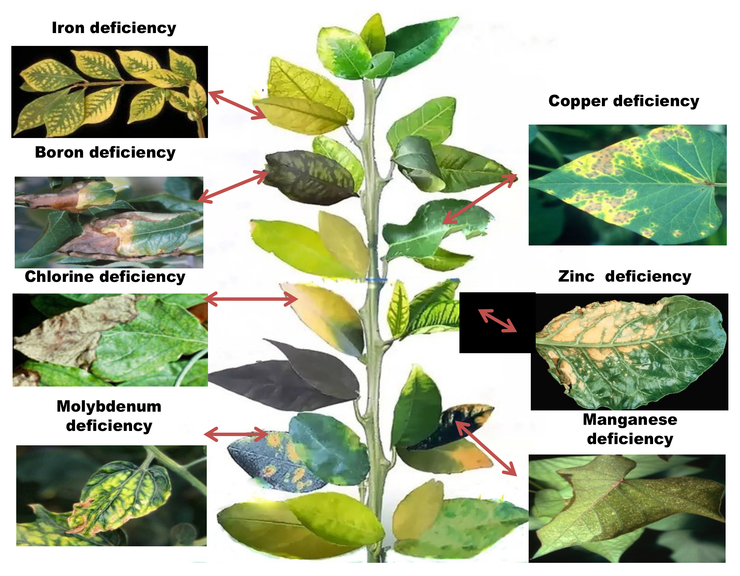
| S.No. | Micronutrients | Representative Constituents/Proteins | Functions | Symptoms of Deficiency | Probable Cause of Deficiency & Method of Correction | References |
|---|---|---|---|---|---|---|
| 1 | Zinc (Zn) | Cu–Zn superoxide, Peptide deformylase, enzyme carbonic anhydrase (CA), α-Mannosidase, Matrix metalloproteinase alcoholic dehydrogenase, and superoxide dismutase (SOD) | role in nitrogen metabolism and photosynthesis, controls the concentration of auxin in plants, increases seed viability and seedling vigor, protection against abiotic and biotic stresses | Reduced vigor, chlorotic leaves, white streaks parallel to leaf blade, slow growth, restricted RNA and protein synthesis | Low Zn in soil, high soil pH—lower soil pH, apply foliar spray or add Zn to soil. | [8,9,10,11,12,13,14,15,16] |
| 2 | Copper (Cu) | Plastocyanin Cu-Znsuperoxide, Ascorbate, Cu-metallothionein, biosynthesis oxidase, Mo-cofactor, dismutase, polyphenol or catechol oxidase, tyrosinase, laccase, Cytochrome-C oxidase, Ethylene receptor ascorbic oxidase and Polyphenol oxidase | saves plants from diseases, improves the fertility of male flower, concerned with the oxidation of iron in plants | Leaf tips dries, break down and dies, ragged leaves, reduced growth | Low soil Cu, high organic matter—apply foliar spray or add Cu to soil | [17,18,19,20,21,22,23,24,25,26] |
| 3 | Iron (Fe) | Aconitase, dismutase, Xanthine dehydrogenase, Ferredoxin, porphyrin NADH, Succinate, Leg hemoglobin, heme and heme enzymes peroxidase oxidase, dehydrogenase, Nitrate reductase oxidoreductases, Thioredoxin reductase, Cytochrome P450, Aldehyde oxidase, Catalase, Nitrite reductase, Lipoxygenase, Alternative oxidase, Fe-superoxide Ferritin and other functional metallic proteins | Present in haemoglobin of the leguminous root nodules, leg-haemoglobin and is involved in nitrogen fixation as a constituent of ferredoxin | Interveinal, creamy chlorosis on apical leaves, stunted shoots, reduced yield | Waterlogged soil, over fertilized, excess of elements like Mn—spraying plant with iron rich fertilizer, chelated iron powder or blood meal directly to the soil | [27,28,29,30,31,32,33,34,35,36,37,38,39,40,41,42,43,44,45,46] |
| 4 | Manganese (Mn) | Malic enzyme, Mn-superoxide, amidohydrolase, primary component of water-splitting enzyme related to photosystem II, PEP carboxylase, Allantoate, Isocitratelyase, dismutase PEP-carboxykinase | Involved in tricarboxylic acid cycle in oxidation and reduction reactions, activates several enzymes such as oxidoreductases, hydrolases and lyase, also autocatalyzes isocitrate dehydrogenase, malic dehydrogenase, glycocyaminase and D-alanyl synthase. | Reduced quality and yield, stunted plants, intervenial chlorosis in leaves, yellow cast in deficient areas. Death of basal leaves, decreased cold hardiness, growth of lateral roots stopped, inhibition of nitrate metabolism. | Low soil Mn, high soil pH due to over liming—lower soil pH, apply foliar spray or add Mn to soil | [47,48,49,50,51,52] |
| 5 | Boron (B) | Rhamnogalacturonan II | increases cell wall thickness and flower production, as well as retention, pollen tube elongation and germination, along with seed and fruit development. It also helps in the translocation of photosynthates. It inhibits IAA oxidation and gives drought tolerance to crops | Thick and leathery old leaves, shoot tip death, rosette leaves with short internodes, excess branching, short, twisted and/or ruptured petioles, vegetables with hollow heart, small and deformed/no fruits with cork spots chlorosis, stubby roots, inhibited nitrogen metabolism. | Low soil B especially on sandy soils or light textured soils—apply foliar spray or add B to soil | [53,54,55,56] |
| 6 | Molybdenum (Mo) | nitrogenase, sulphite oxidase, nitrate reductase Aldehyde oxidase and xanthine oxidase/dehydrogenase | aids in the synthesis of ascorbic acid, formation of pollens and anthers, acts as a remedy to excessive copper, manganese and zinc | stunted growth with twisted stems, leaves turning pale green, necrotic area in leaves along the mid rib between veins and along leaf edges | Low soil pH, low Mo content in soil—inoculate seed with Mo, apply foliar spray or add Mo to soil | [33,34,57,58] |
| 7 | Chlorine (Cl) | Oxygen-evolving complex Seismonastic movement | activates enzymes that are involved in starch utilization which affects germination and energy transfer. Inmoisture-stress conditions chlorine helps in the movement of water into cells and maintenance of that water. Chlorine also controls the opening and closing of stomata on leaf surfaces | stunted/restricted growth, stubby roots, interveinal chlorosis, non-succulent tissue, wilting | Low soil Cl especially in soils subjected to leaching—apply Cl containing fertilizer | [55,59] |
| S. No. | Micronutrients | Type | Symptoms of Excess Usage | References |
|---|---|---|---|---|
| 1 | Zinc (Zn) | Metal | Inhibited root growth, young leaf chlorosis, interveinal mild chlorosis in young leaves starting from base and spread towards apex followed by reddish brown coloration, rolling of leaves | [61] |
| 2 | Copper (Cu) | Metal | Reduced vigor, inhibited root growth/root damage, older leaves develop orange or pink coloration followed by severe rolling of leaf margins due to loss in turgidity | |
| 3 | Iron (Fe) | Metal | Reduced yield, bronzing and stippling of leaves, and, in some plants, acid is secreted from the roots | |
| 4 | Manganese (Mn) | Metal | Tissue injury, leaf sheath and lower parts of stem in cereal normally consist of minute brown spots, legumes develop brown or purple spots over leaf margin, deficiency symptoms of other nutrients | |
| 5 | Boron (B) | Metalloid | Toxicity results in dark brown speckles or necrosis on the edge of older leaves, cupped and wrinkled young leaves | |
| 6 | Molybdenum (Mo) | Metal | Leaf malformation, tints of golden yellow or blue color in leaves | |
| 7 | Chlorine (Cl) | Non metal | Death of leaf margin, leaves are reduced in size and number, have bronze or yellow coloration with brown or scorched leaf margins. |
| Metal | Plant Species | Plant Part | Extraction Method | Protein Name | Function | Regulation | References |
|---|---|---|---|---|---|---|---|
| Boron (B) | Citrus grandis | Root | iTRAQ | Alcohol dehydrogenase 1 | Energy metabolism | Down | [62] |
| Serine/arginine- rich 22 | Nucleic acid metabolic process | Down | |||||
| Clathrin light chain protein | Cellular cytoskeleton and transport | Up | |||||
| Peroxiredoxin IIF | Cellular response to stress | Down | |||||
| Phospholipase C2 | Signal transduction | Down | |||||
| Arabidopsis thaliana | Leaf | 2-DE, LC-MS/MS | Rubisco activase | Photosynthesis | Up | [63] | |
| Actin 7 | Metabolism | Down | |||||
| Fructose-bisphosphatealdolase | Energy metabolism | Up | |||||
| Glycolate oxidase | Photosynthesis | Down | |||||
| Coppe (Cu) | Sorghum bicolor | Leaf | 2-DE, MALDI-TOF MS | Thymidine kinase | Protein translation and synthesis | Down | [64] |
| Thaumatin-like protein | Stress and defense | Up | |||||
| Maturase K | Growth and development | Down | |||||
| Alcohol dehydrogenase | Oxidation– reduction process | Up | |||||
| Oryza sativa | Root | 2-DE, MALDI-TOF MS | Putativeperoxidase | Antioxidation and detoxification | Up | [65] | |
| Putative cold shock protein-1 | Transcriptionalregulation | Up | |||||
| Putative elongationfactor EF-2 | Protein synthesis | Down | |||||
| Glutamine synthetase shoot isozyme | Amino acid synthesis | Down | |||||
| Allium cepa | Root | 2-DE, MALDI-TOF MS | Glutaredoxin | Defense | Up | [66] | |
| Ran-binding protein 1 | Protein synthesis | Down | |||||
| Cinnamoyl-Co-A-reductase 1 | Cell wall synthesis | Down | |||||
| Proliferation-associated 2 g4 | Cell cycle and DNA replication | Up | |||||
| Iron (Fe) | Arabidopsis thaliana | Root | Itraq, LC-MS | Oxidoreductase | Hormone metabolism | Up | [67] |
| WAKL4, WAK- like receptor- like kinase | Signaling | Up | |||||
| FRO3, ferric chelate reductase 3 | Metal handling | Up | |||||
| SAPX, Stromal ascorbate peroxidase | Redox | Down | |||||
| IRT3, Iron regulated transporter3 | transport | Down | |||||
| Zea may | Root | LC-MS/MS | Aquaporin PIP2-2 | Transport proteins | Down | [68] | |
| Gibberellin receptor GID1L2 | Signaling proteins | Down | |||||
| Aldolase | Metabolism | Up | |||||
| Actin-2 | Cytoskeleton | Down | |||||
| Callreticulin2 | Protein folding | Up | |||||
| Cucumissativus | Root | 2-DE, ESI-LC-MS | Phosphoglycerate kinase | Glycolysis | Up | [69] | |
| Malate dehydrogenase | Carbohydrate-related metabolism | Up | |||||
| Alanine aminotransferase | Nitrogen-related metabolism | Up | |||||
| Xylan 1,4-beta-xylosidase | Metabolism of sucrose | Down | |||||
| Manganese (Mn) | Citrus grandis | Root | 2-DE, MALDI-TOF MS, LTQ-ESI-MS/MS | Acetohydroxyacidisomeroreductase | Protein metabolism | Down | [70] |
| Maturase K | Nucleic acid metabolism | Up | |||||
| Alcohol dehydrogenase | Carbohydrate metabolism | Up | |||||
| Iron- sUperoxide dismutase | Stress response | Down | |||||
| Valosin- containing protein | Cell transport | Down | |||||
| Zinc (Zn) | Lactuca sativa | Leaf | LC-MS/MS, MALDI-TOF | MPBQ/MSBQ transferase | Photosynthesis | Up | [71] |
| Cytosolic fructose-1,6-bisphosphate | Energy metabolism | Up | |||||
| Putative cellulose synthase | Cell wall metabolism | Down | |||||
| Phenylalanine ammonia-lyase | Phenylpropanoids biosynthesis | Down |
| Metals | Plant Species | Tissue | Method of Identification | Gene | Functions | Regulation | References |
|---|---|---|---|---|---|---|---|
| Boron (B) | Arabidopsis thaliana | Root | qRT-PCR | SHB1/HY1 | B-tolerance | Up | [72] |
| Oryza sativa | Root | Microarray, qRT-PCR | LOC_Os08g30740 | ABC transporter | Down | [73] | |
| LOC_Os10g30156 | Starch synthase | Down | |||||
| LOC_Os10g30080 | Xylosyltransferase | Down | |||||
| Citrus sinensis | Leaf | Illuminasequencing, qRT-PCR | Ciclev10017846m | Ubiquitin-protein ligase activity | Down | [74] | |
| Ciclev 10012377m | Transcription factor | Up | |||||
| Ciclev 10009779 m | Blue copper protein | Up | |||||
| Zinc (Zn) | Arabidopsis thaliana | Whole plant | qRT-PCR | PDR8 | Phytochelatin synthesis | Up | [75] |
| ABCCI | Metabolism | Down | |||||
| Iron (Fe) | Citrus sinensis | Whole plant | qRT-PCR | Cs3g19420 | Ethylene-responsive transcription factor | Up | [76] |
| Cs4g12540 | Ethylene synthesis | Down | |||||
| Cs9g02930 | Flavone synthase | Down | |||||
| Arabidopsis thaliana | Root | qRT-PCR | FRO2 | Iron homeostasis | Up | [77] | |
| Manganese (Mn) | Arabidopsis thaliana | Root | qRT-PCR and RNA sequencing | AT5G05960 | Bifunctional inhibitor | Down | [78] |
| AT4G39940 | Glucosinolate | Up | |||||
| AT1G15820 | metabolism Photosynthesis | Up | |||||
| Citrus sinensis | Leaf | cDNA-AFLP and qRT-PCR | TDF #065-1 | Energy metabolism | Up | [79] | |
| TDF #073-1 | Cell transport | Down | |||||
| TDF #103-2 | Stress responses | Down | |||||
| Copper (Cu) | Arabidopsis thaliana | Whole plant | qRT-PCR | OsLAC10 | Laccase activity | Up | [80] |
| AtLAC11 | Lignin biosynthesis | Up | |||||
| Oryza sativa | Whole plant | Western blotting, qRT-PCR | OsHMA4 | Copper accumulation | Down | [74] |
Publisher’s Note: MDPI stays neutral with regard to jurisdictional claims in published maps and institutional affiliations. |
© 2022 by the authors. Licensee MDPI, Basel, Switzerland. This article is an open access article distributed under the terms and conditions of the Creative Commons Attribution (CC BY) license (https://creativecommons.org/licenses/by/4.0/).
Share and Cite
Prusty, S.; Sahoo, R.K.; Nayak, S.; Poosapati, S.; Swain, D.M. Proteomic and Genomic Studies of Micronutrient Deficiency and Toxicity in Plants. Plants 2022, 11, 2424. https://doi.org/10.3390/plants11182424
Prusty S, Sahoo RK, Nayak S, Poosapati S, Swain DM. Proteomic and Genomic Studies of Micronutrient Deficiency and Toxicity in Plants. Plants. 2022; 11(18):2424. https://doi.org/10.3390/plants11182424
Chicago/Turabian StylePrusty, Suchismita, Ranjan Kumar Sahoo, Subhendu Nayak, Sowmya Poosapati, and Durga Madhab Swain. 2022. "Proteomic and Genomic Studies of Micronutrient Deficiency and Toxicity in Plants" Plants 11, no. 18: 2424. https://doi.org/10.3390/plants11182424