
Effect of Three Types of Ion Beam Irradiation on Gerbera (Gerbera hybrida) In Vitro Shoots with Mutagenesis Efficiency

 and
and
Abstract
:1. Introduction
2. Results
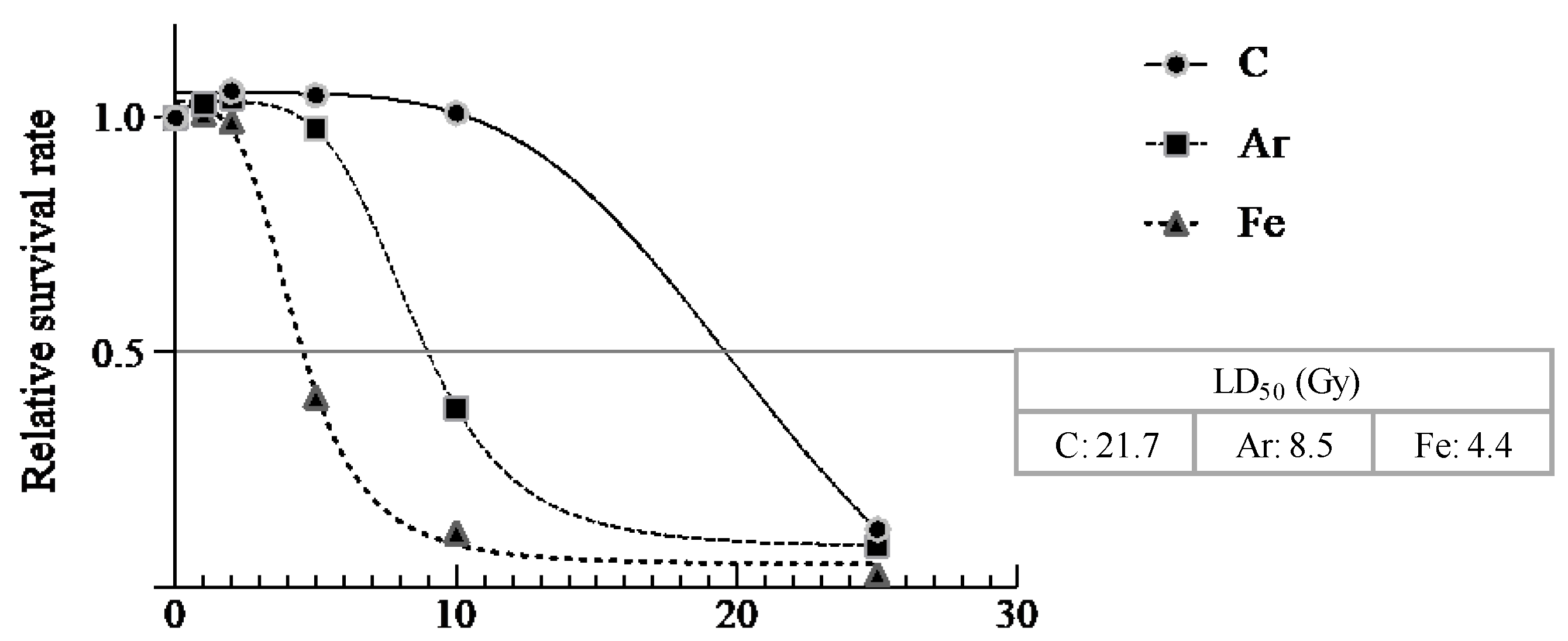
2.1. Survival Rate
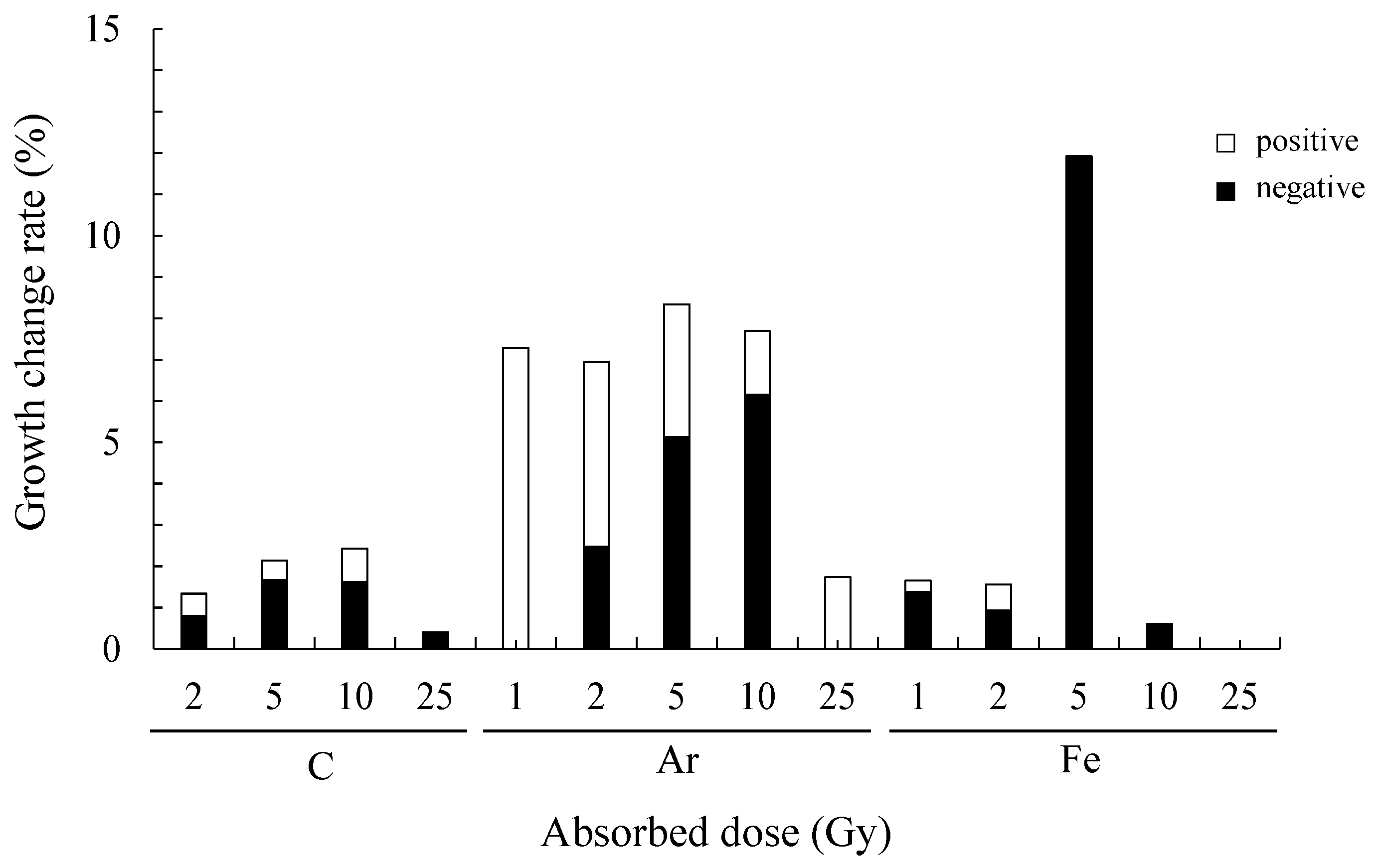
2.2. Growth Change Rate
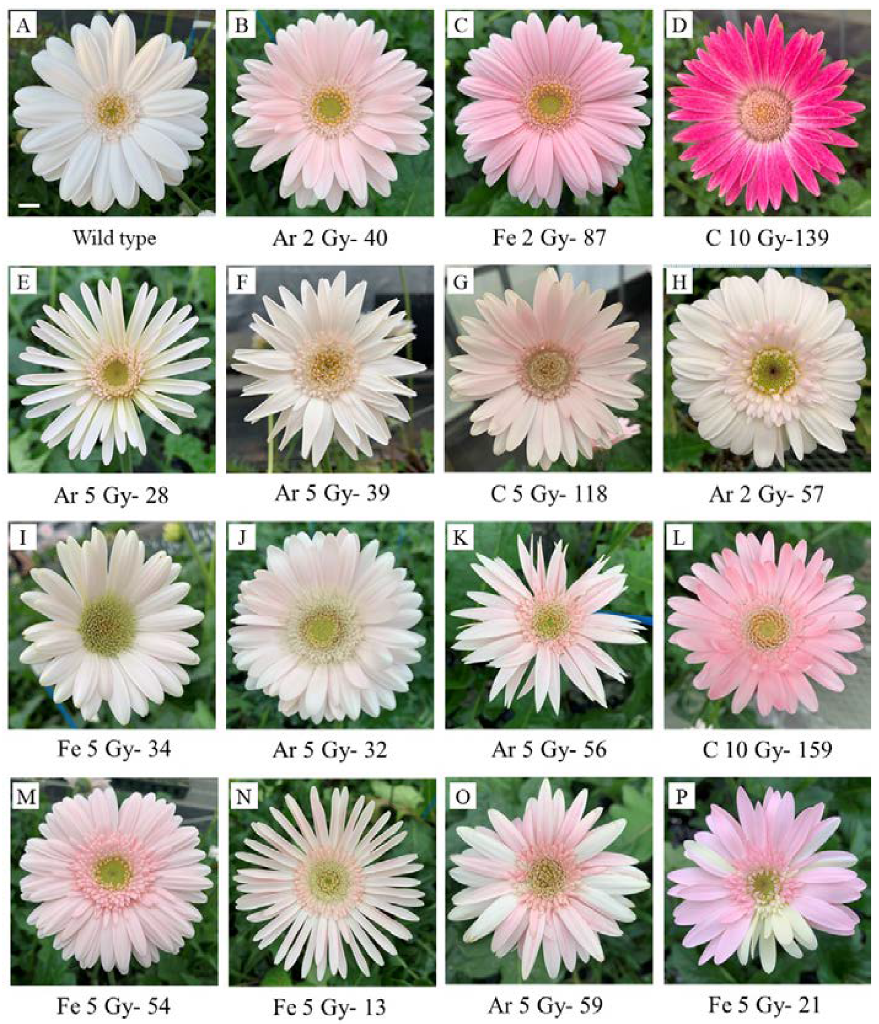
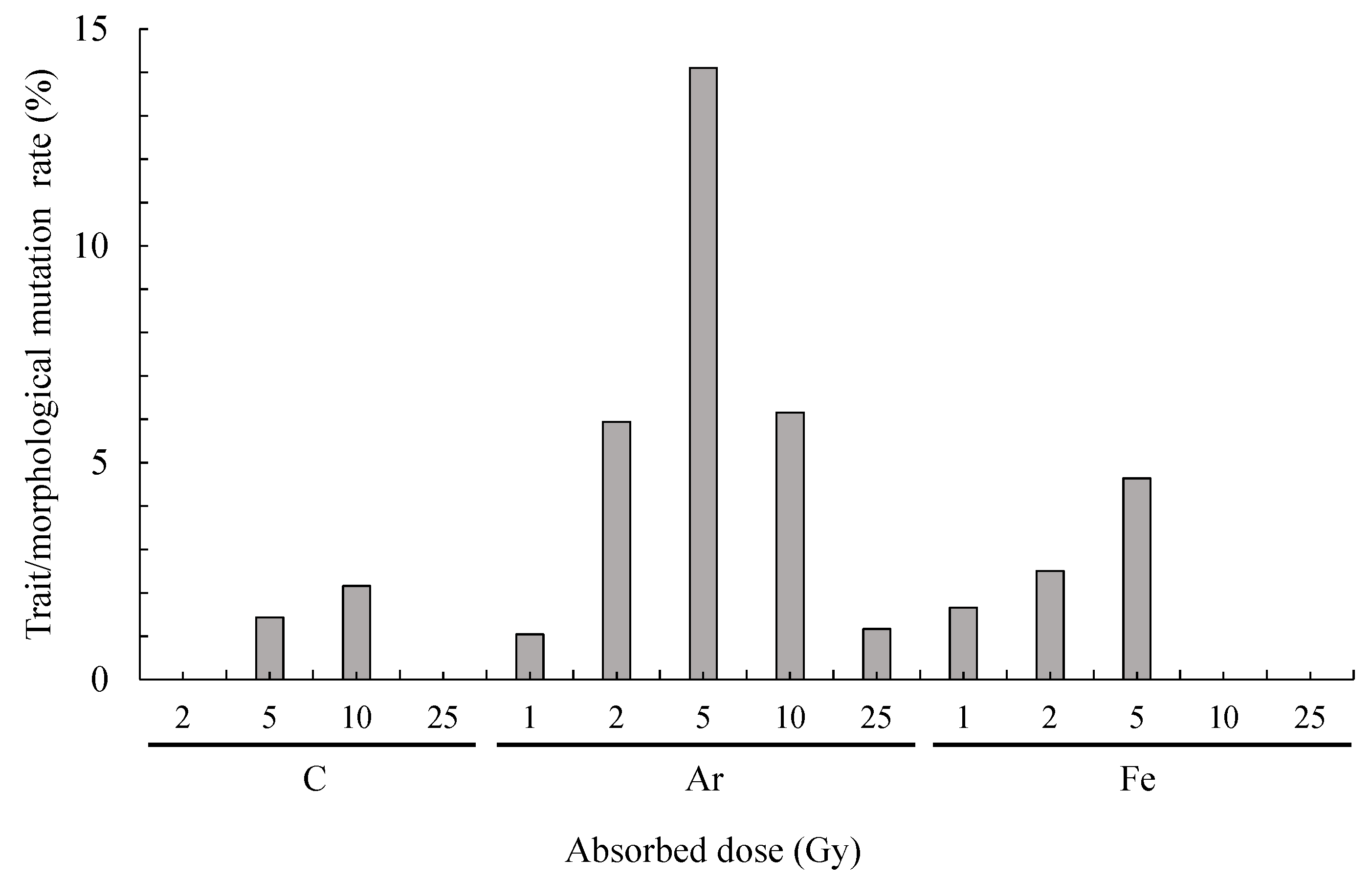
2.3. Trait/Morphological Mutation Rate
2.4. Genotype Analysis of Gerbera Mutants Obtained by Ion Beam Irradiation
3. Discussion
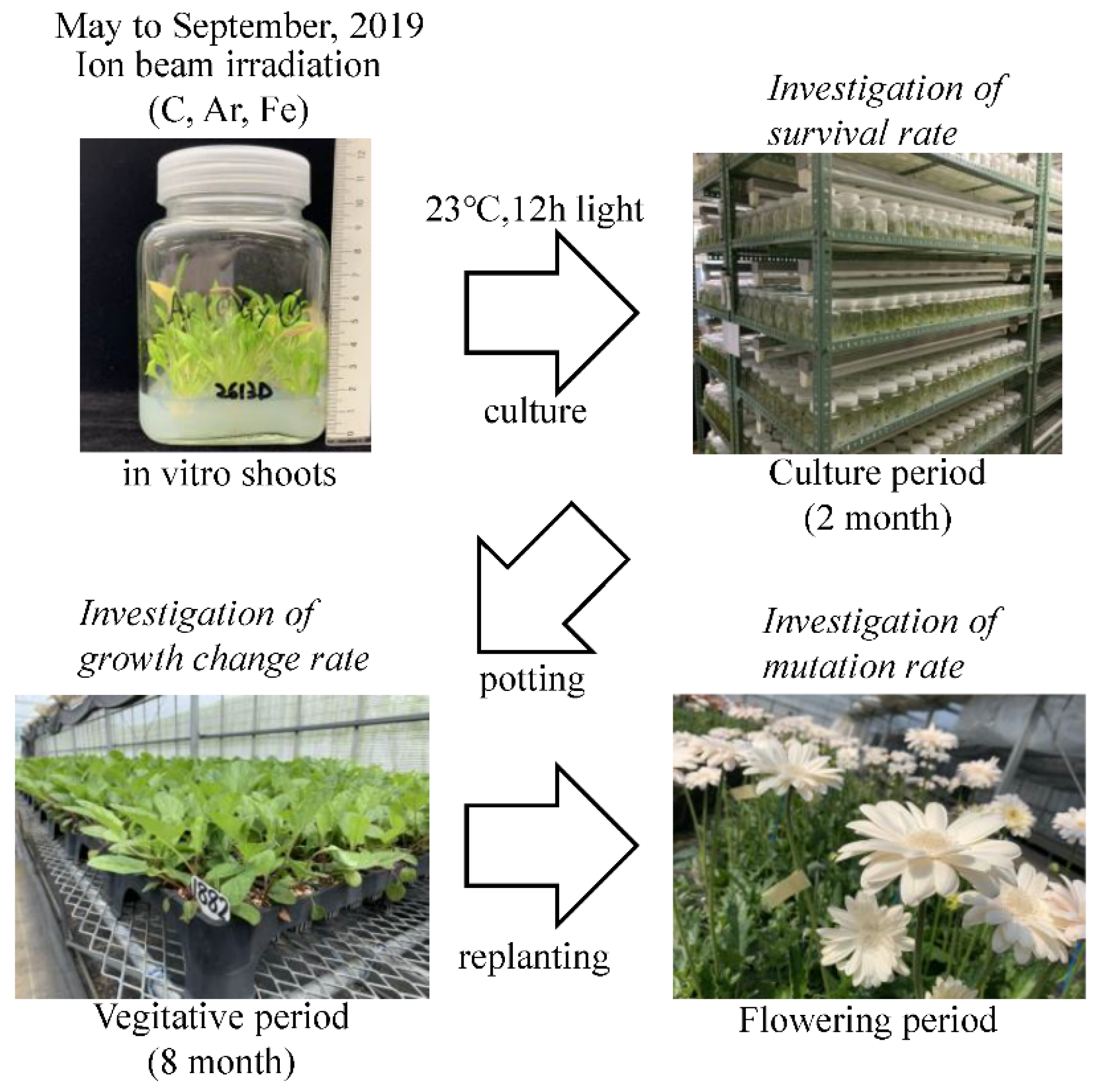
4. Materials and Methods
4.1. Plant Material
4.2. Procedure
4.3. Survival Rate
4.4. Growth Change Rate
4.5. Trait/Morphological Mutation Rate
4.6. Genotype Analysis of Gerbera Mutants Obtained by Ion Beam Irradiation
Author Contributions
Funding
Institutional Review Board Statement
Informed Consent Statement
Data Availability Statement
Acknowledgments
Conflicts of Interest
References
- Cardoso, J.C.; da Silva, J.A.T. Gerbera micropropagation. Biotechnol. Adv. 2013, 31, 1344–1357. [Google Scholar] [CrossRef]
- MAFF. The 94th Statistical Yearbook of Ministry of Agriculture, Forestry and Fisheries. 2020. Available online: https://www.maff.go.jp/e/data/stat/94th/index.html (accessed on 18 May 2021).
- Gerbera.org. Available online: http://www.gerbera.org (accessed on 14 May 2021).
- Bhatia, R.; Singh, K.P.; Sharma, T.R.; Jhang, T. Evaluation of the genetic fidelity of in vitro propagated gerbera (Gerbera jamesonii Bolus) using DNA-based markers. Plant Cell Tissue Organ Cult. 2011, 104, 131–135. [Google Scholar] [CrossRef]
- Chakabrarty, D.; Datta, S.K. Micropropagation of gerbera: Lipid peroxidation and antioxidante enzyme activities during acclimatization process. Acta Physiol. Plant. 2008, 30, 325–331. [Google Scholar] [CrossRef]
- Dowrick, G.J.; El-Bayoumi, A. The induction of mutations in chrysanthemum using X- and gamma radiation. Euphytica 1966, 15, 204–210. [Google Scholar] [CrossRef]
- Kazama, Y.; Ishii, K.; Hirano, T.; Wakana, T.; Yamada, M.; Ohbu, S.; Abe, T. Different mutational function of low- and high-linear energy transfer heavy-ion irradiation demonstrated by whole-genome resequencing of Arabidopsis mutants. Plant J. 2017, 92, 1020–1030. [Google Scholar] [CrossRef] [Green Version]
- Yamaguchi, H. Mutation breeding of ornamental plants using ion beams. Breed. Sci. 2018, 68, 71–78. [Google Scholar] [CrossRef] [PubMed] [Green Version]
- Miyazaki, K.; Suzuki, K.; Iwaki, K.; Kusumi, T.; Abe, T.; Yoshida, S.; Fukui, H. Flower pigment mutations induced by heavy ion beam irradiation in an interspecific hybrid of Torenia. Plant Biotechnol. 2006, 23, 163–167. [Google Scholar] [CrossRef] [Green Version]
- Sasaki, K.; Aida, R.; Niki, T.; Yamaguchi, H.; Narumi, T.; Nishijima, T.; Hayashi, Y.; Ryuto, H.; Fukunishi, N.; Abe, T.; et al. High efficiency improvement of transgenic torenia flowers by ion beam irradiation. Plant Biotechnol. 2008, 25, 81–89. [Google Scholar] [CrossRef] [Green Version]
- Kanaya, T.; Saito, H.; Hayashi, Y.; Fukunishi, N.; Ryuto, H.; Miyazaki, K.; Kusumi, T.; Abe, T.; Suzuki, K. Heavy-ion beam-induced sterile mutants of verbena (Verbena × hybrida) with an improved flowering habit. Plant Biotechnol. 2008, 25, 91–96. [Google Scholar] [CrossRef] [Green Version]
- Suzuki, K.; Yomo, Y.; Abe, T.; Katsumoto, Y.; Miyazaki, K.; Yoshida, S.; Kusumi, T. Isolation of Sterile Mutants of Verbena hybrida Using Heavy-Ion Beam Irradiation. RIKEN Accel. Prog. Rep. 2002, 35, 129. [Google Scholar]
- Sasaki, N.; Watanabe, A.; Asakawa, T.; Sasaki, M.; Hoshi, N.; Naito, Z.; Furusawa, Y.; Shimokawa, T.; Nishihara, M. Biological effects of ion beam irradiation on perennial gentian and apple. Plant Biotechnol. 2018, 35, 249–257. [Google Scholar] [CrossRef] [Green Version]
- Sugiyama, M.; Saito, H.; Ichida, H.; Hayashi, Y.; Ryuto, H.; Fukunishi, N.; Terakawa, T.; Abe, T. Biological effects of heavy-ion beam irradiation on cyclamen. Plant Biotechnol. 2008, 25, 101–104. [Google Scholar] [CrossRef] [Green Version]
- Hase, Y.; Okamura, M.; Takeshita, D.; Narumi, I.; Tanaka, A. Efficient induction of flower-color mutants by ion beam irradiation in petunia seedlings treated with high sucrose concentration. Plant Biotechnol. 2010, 27, 99–103. [Google Scholar] [CrossRef] [Green Version]
- Nakano, M.; Amano, J.; Watanabe, Y.; Nomizu, T.; Suzuki, M.; Mizunashi, K.; Mori, S.; Kuwayama, S.; Han, D.S.; Saito, H.; et al. Morphological variation in Tricyrtis hirta plants regenerated from heavy ion beam-irradiated embryogenic calluses. Plant Biotechnol. 2010, 27, 155–160. [Google Scholar] [CrossRef] [Green Version]
- Okamura, M.; Umemoto, N.; Onishi, N. Breeding glittering carnations by an efficient mutagenesis system. Plant Biotechnol. 2012, 29, 209–214. [Google Scholar] [CrossRef] [Green Version]
- Okamura, M.; Nakayama, M.; Umemoto, N.; Cano, E.A.; Hase, Y.; Nishizaki, Y.; Sasaki, N.; Ozeki, Y. Crossbreeding of a metallic color carnation and diversification of the peculiar coloration by ion-beam irradiation. Euphytica 2013, 191, 45–56. [Google Scholar] [CrossRef] [Green Version]
- Matsuyama, T.; Watanabe, M.; Murota, Y.; Nakata, N.; Kitamura, H.; Shimokawa, T.; Ebisuzaki, T.; Wada, S.; Sato, S.; Tabata, S. Efficient mutation induction using heavy-ion beam irradiation and simple T genomic screening with random primers in taro (Colocasia esculenta L. Schott). Sci. Hortic. 2020, 272, 109568. [Google Scholar] [CrossRef]
- Matsumura, A.; Nomizu, T.; Furutani, N.; Hayashi, K.; Minamiyama, Y.; Hase, Y. Ray florets color and shape mutants induced by 12C5+ ion beam irradiation in chrysanthemum. Sci. Hortic. 2010, 123, 558–561. [Google Scholar] [CrossRef]
- Okamura, M.; Yasuno, N.; Takano, M.; Tanaka, A.; Shikazono, N.; Hase, Y. Mutation generation in chrysanthemum plants regenerated from floral organ cultures irradiated with ion beams. TIARA Ann. Rep. 2002, 2001, 42–43. [Google Scholar]
- Okamura, M.; Hase, Y.; Furusawa, Y.; Tanaka, A. Tissue dependent somaclonal mutation frequencies and spectra enhanced by ion beam irradiation in chrysanthemum. Euphytica 2015, 202, 333–343. [Google Scholar] [CrossRef]
- Shirao, T.; Ueno, K.; Abe, T.; Matsuyama, T. Development of DNA markers for identifying chrysanthemum cultivars generated by ion-beam irradiation. Mol. Breed. 2013, 31, 729–735. [Google Scholar] [CrossRef]
- Tamaki, K.; Yamanaka, M.; Hayashi, Y.; Abe, T.; Koyama, Y. Effect of the cultivar characteristics on the appearance of flower color mutants by C-ion irradiation in Chrysanthemum. Hortic. Res. (Jpn.) 2017, 16, 117–123, (Abstract in English). [Google Scholar] [CrossRef]
- Tanokashira, Y.; Nagayoshi, S.; Hirano, T.; Abe, T. Effects of heavy-ion-beam irradiation on flower-color mutation in chrysanthemum. RIKEN Accel. Prog. Rep. 2015, 47, 297. [Google Scholar]
- Ueno, K.; Nagayoshi, S.; Imakiire, S.; Koriyama, K.; Minami, T.; Tanaka, A.; Hase, Y.; Matsumoto, T. Breeding of new chrysanthemum cultivar ‘Aladdin 2′ through stepwise improvements of cv. ‘Jimba’ using ion beam re-irradiation. Hortic. Res. (Jpn.) 2013, 12, 245–254. [Google Scholar] [CrossRef] [Green Version]
- Wakita, N.; Kazama, Y.; Hayashi, Y.; Ryuto, H.; Fukunishi, N.; Yamamoto, K.; Ijichi, S.; Abe, T. Induction of floral color mutation by C-ion irradiation in spray-type chrysanthemum. RIKEN Accel. Prog. Rep. 2009, 41, 230. [Google Scholar]
- Yamaguchi, H.; Shimizu, A.; Hase, Y.; Degi, K.; Tanaka, A.; Morishita, T. Mutation induction with ion beam irradiation of lateral buds of chrysanthemum and analysis of chimeric structure of induced mutants. Euphytica 2009, 165, 97–103. [Google Scholar] [CrossRef]
- Yamaguchi, H.; Shimizu, A.; Hase, Y.; Tanaka, A.; Shikazono, N.; Degi, K.; Morishita, T. Effects of ion beam irradiation on mutation induction and nuclear DNA content in chrysanthemum. Breed. Sci. 2010, 60, 398–404. [Google Scholar] [CrossRef] [Green Version]
- Hirano, T.; Kazama, Y.; Ishii, K.; Ohbu, S.; Shirakawa, Y.; Abe, T. Comprehensive identification of mutations induced by heavy-ion beam irradiation in Arabidopsis thaliana. Plant J. 2015, 82, 93–104. [Google Scholar] [CrossRef]
- Yamaguchi, H.; Watanabe, Y. Mutation Breeding; YokendoTokyo: Tokyo, Japan, 1983. (In Japanese) [Google Scholar]
- Beyaz, R.; Kahramanogullari, C.T.; Yildiz, C.; Darcin, E.S.; Yildiz, M. The effect of gamma radiation on seed germination and seedling growth of Lathyrus chrysanthus Boiss. under in vitro conditions. J. Environ. Radioact. 2016, 162–163, 129–133. [Google Scholar] [CrossRef] [PubMed]
- Charbaji, T.; Nabulsi, I. Effect of low doses of gamma irradiation on in vitro growth of grapevine. Plant Cell Tissue Organ Cult. 1999, 57, 129–132. [Google Scholar] [CrossRef]
- Wang, L.; Ma, R.; Yin, Y.; Jiao, Z. Role of carbon ion beams irradiation in mitigating cold stress in Arabidopsis thaliana. Ecotoxicol. Environ. Saf. 2018, 162, 341–347. [Google Scholar] [CrossRef]
- Li, Y.; Zheng, L.; Corke, F.; Smith, C.; Bevan, M.W. Control of final seed and organ size by the DA1 gene family in Arabidopsis thaliana. Gene Dev. 2008, 22, 1331–1336. [Google Scholar] [CrossRef] [Green Version]
- Wang, J.; Tang, M.; Chen, S.; Zheng, X.; Mo, H.; Li, S.; Wang, Z.; Zhu, K.; Ding, L.; Liu, S.; et al. Down-regulation of BnDA1, whose gene locus is associated with the seeds weight, improves the seeds weight and organ size in Brassica napus. Plant Biotechnol. J. 2017, 15, 1024–1033. [Google Scholar] [CrossRef] [PubMed] [Green Version]
- Ohmiya, A.; Kishimoto, S.; Aida, R.; Yoshioka, S.; Sumitomo, K. Carotenoid cleavage dioxygenase (CmCCD4a) contributes to white color formation in chrysanthemum petals. Plant Physiol. 2006, 142, 1193–1201. [Google Scholar] [CrossRef] [Green Version]
- Naing, A.H.; Lee, J.H.; Park, K.I.; Kim, K.-O.; Chung, M.Y.; Kim, C.K. Transcriptional control of anthocyanin biosynthesis genes and transcription factors associated with flower coloration patterns in Gerbera hybrida. 3 Biotech 2018, 8, 65. [Google Scholar] [CrossRef] [PubMed]
- Laneri, U.; Franconi, R.; Altavista, P. Somatic mutagenesis of Gerbera jamesonii hybr.: Irradiation and in vitro culture. Acta Hortic. 1990, 280, 395–402. [Google Scholar] [CrossRef]
- Edger, P.P.; Poorten, T.J.; VanBuren, R.; Hardigan, M.A.; Colle, M.; McKain, M.R.; Smith, R.D.; Teresi, S.J.; Nelson, A.D.L.; Wai, C.M.; et al. Origin and evolution of the octoploid strawberry genome. Nat. Genet. 2019, 51, 541–547. [Google Scholar] [CrossRef] [PubMed] [Green Version]
- Sato, S.; Nakamura, Y.; Kaneko, T.; Asamizu, E.; Kato, T.; Nakao, M.; Sasamoto, S.; Watanabe, A.; Ono, A.; Kawashima, K.; et al. Genome structure of the legume, Lotus japonicus. DNA Res. 2008, 15, 227–239. [Google Scholar] [CrossRef] [Green Version]
- Schmutz, J.; Cannon, S.B.; Schlueter, J.; Ma, J.; Mitros, T.; Nelson, W.; Hyten, D.L.; Song, Q.; Thelen, J.J.; Cheng, J.; et al. Genome sequence of the palaeopolyploid soybean. Nature 2010, 463, 178–183. [Google Scholar] [CrossRef] [PubMed] [Green Version]
- The Tomato Genome Consortium. The tomato genome sequence provides insights into fleshy fruit evolution. Nature 2012, 485, 635–641. [Google Scholar] [CrossRef] [Green Version]
- Anders, C.; Niewoehner, O.; Duerst, A.; Jinek, M. Structural basis of PAM-dependent target DNA recognition by the Cas9 endonuclease. Nature 2014, 513, 569–573. [Google Scholar] [CrossRef] [PubMed]
- Li, J.F.; Norville, J.E.; Aach, J.; McCormack, M.; Zhang, D.; Bush, J.; Church, G.M.; Sheen, J. Multiplex and homologous recombination-mediated genome editing in Arabidopsis and Nicotiana benthamiana using guide RNA and Cas9. Nat. Biotechnol. 2013, 31, 688–691. [Google Scholar] [CrossRef]
- Malnoy, M.; Viola, R.; Jung, M.H.; Koo, O.J.; Kim, S.; Kim, J.S.; Velasco, R.; Nagamangala, K.C. DNA-free genetically edited grapevine and apple protoplast using CRISPR/Cas9 ribonucleoproteins. Front. Plant Sci. 2016, 7, 1904. [Google Scholar] [CrossRef]
- Svitashev, S.; Schwartz, C.; Lenderts, B.; Young, J.K.; Cigan, M.A. Genome editing in maize directed by CRISPR-Cas9 ribonucleoprotein complexes. Nat. Commun. 2016, 7, 13274. [Google Scholar] [CrossRef]
- Zhang, H.; Zhang, J.; Wei, P.; Zhang, B.; Gou, F.; Feng, Z.; Mao, Y.; Yang, L.; Zhang, H.; Xu, N.; et al. The CRISPR/Cas9 system produces specific and homozygous targeted gene editing in rice in one generation. Plant Biotechnol. J. 2014, 12, 797–807. [Google Scholar] [CrossRef] [PubMed]
- Zhang, R.; Liu, J.; Chai, Z.; Chen, S.; Bai, Y.; Zong, Y.; Chen, K.; Li, J.; Jiang, L.; Gao, C. Generation of herbicide tolerance traits and a new selectable marker in wheat using base editing. Nat. Plants 2019, 5, 480–485. [Google Scholar] [CrossRef] [PubMed]
- Kako, S. Culture of Horticultural Plant Organs and Tissues; Seibundoshinkosya Tokyo: Tokyo, Japan, 1985; pp. 176–193. (In Japanese) [Google Scholar]
- Hosoya, S.; Hirase, S.; Kikuchi, K.; Nanjo, K.; Nakamura, Y.; Kohno, H.; Sano, M. Random PCR-based genotyping by sequencing technology GRAS-Di (genotyping by random amplicon sequencing, direct) reveals genetic structure of mangrove fishes. Mol. Ecol. Resour. 2019, 19, 1153–1163. [Google Scholar] [CrossRef]
- Yoshikawa, S.; Hamasaki, M.; Kadomura, K.; Yamada, T.; Chuda, H.; Kikuchi, K.; Hosoya, S. Genetic dissection of a precocious phenotype in male Tiger Pufferfish (Takifugu rubripes) using Genotyping by Random Amplicon Sequencing, Direct (GRAS-Di). Mar. Biotechnol. 2021, 23, 177–188. [Google Scholar] [CrossRef]


| Classification of Mutant Phenotypes | Irradiation treatment | |||||||||||||||
|---|---|---|---|---|---|---|---|---|---|---|---|---|---|---|---|---|
| C | Ar | Fe | Total | |||||||||||||
| 2 | 5 | 10 | 25 | 1 | 2 | 5 | 10 | 25 | 1 | 2 | 5 | 10 | 25 | |||
| Single mutation | Flower color | 0 | 1 | 2 | 0 | 1 | 4 | 5 | 1 | 1 | 2 | 2 | 2 | 0 | 0 | 21 |
| Petal slender | 0 | 0 | 0 | 0 | 0 | 0 | 2 | 0 | 0 | 0 | 0 | 0 | 0 | 0 | 2 | |
| Petal sword | 0 | 0 | 0 | 0 | 0 | 0 | 1 | 0 | 0 | 0 | 0 | 0 | 0 | 0 | 1 | |
| Receptive shape | 0 | 2 | 0 | 0 | 0 | 0 | 0 | 0 | 0 | 0 | 1 | 1 | 0 | 0 | 4 | |
| Semidouble | 0 | 0 | 0 | 0 | 0 | 1 | 0 | 0 | 0 | 0 | 0 | 0 | 0 | 0 | 1 | |
| Male sterile | 0 | 0 | 0 | 0 | 0 | 0 | 2 | 2 | 0 | 0 | 0 | 1 | 0 | 0 | 5 | |
| Complex mutation | Flower color/Petal slender | 0 | 0 | 1 | 0 | 0 | 0 | 0 | 0 | 0 | 0 | 1 | 0 | 0 | 0 | 2 |
| Flower color/Receptive shape | 0 | 0 | 1 | 0 | 0 | 0 | 0 | 0 | 0 | 0 | 0 | 0 | 0 | 0 | 1 | |
| Flower color/Semidouble | 0 | 0 | 0 | 0 | 0 | 1 | 0 | 0 | 0 | 0 | 0 | 1 | 0 | 0 | 2 | |
| Flower color/Male sterile | 0 | 0 | 0 | 0 | 0 | 0 | 0 | 1 | 0 | 0 | 0 | 0 | 0 | 0 | 1 | |
| Petal slender/Male sterile | 0 | 0 | 0 | 0 | 0 | 0 | 0 | 0 | 0 | 0 | 0 | 1 | 0 | 0 | 1 | |
| Chimeric mutation | 0 | 0 | 0 | 0 | 0 | 0 | 1 | 0 | 0 | 1 | 0 | 1 | 0 | 0 | 3 | |
| Total | 0 | 3 | 4 | 0 | 1 | 6 | 11 | 4 | 1 | 3 | 4 | 7 | 0 | 0 | 44 | |
| Trait | Line Name | No. of Read | No. of Seqenced Bases (Mbp) | %Q30 z | No. of Mutant-Specific Markers y | |
|---|---|---|---|---|---|---|
| Wild type | Normal | 0 Gy | 5,289,248 | 534 | 88.6 | - |
| Mutant | Flower color | Fe 2 Gy- 87 | 5,355,904 | 541 | 89.5 | 1 |
| Petal slender | Ar 5 Gy- 29 | 5,364,690 | 542 | 89.2 | 6 | |
| Male sterile | Fe 5 Gy- 34 | 5,045,048 | 510 | 88.9 | 31 |
| Elements | Ion Notation | Energy | LET |
|---|---|---|---|
| (MeV·µ−1) | (keV·µm−1) | ||
| Carbon | 12C6+ | 290 | 14 |
| Argon | 40Ar18+ | 500 | 89 |
| Iron | 56Fe26+ | 500 | 185 |
| Ion | Absorbed Dose (Gy) | Number of Irradiated Plants | Number of Surviving Plants |
|---|---|---|---|
| C | 0 | 201 | 201 |
| 2 | 187 | 184 | |
| 5 | 210 | 205 | |
| 10 | 185 | 174 | |
| 25 | 123 | 14 | |
| Ar | 0 | 63 | 63 |
| 1 | 96 | 92 | |
| 2 | 101 | 98 | |
| 5 | 78 | 71 | |
| 10 | 65 | 23 | |
| 25 | 86 | 7 | |
| Fe | 0 | 154 | 122 |
| 1 | 181 | 171 | |
| 2 | 160 | 146 | |
| 5 | 151 | 53 | |
| 10 | 166 | 19 | |
| 25 | 156 | 4 |
Publisher’s Note: MDPI stays neutral with regard to jurisdictional claims in published maps and institutional affiliations. |
© 2021 by the authors. Licensee MDPI, Basel, Switzerland. This article is an open access article distributed under the terms and conditions of the Creative Commons Attribution (CC BY) license (https://creativecommons.org/licenses/by/4.0/).
Share and Cite
Hosoguchi, T.; Uchiyama, Y.; Komazawa, H.; Yahata, M.; Shimokawa, T.; Tominaga, A. Effect of Three Types of Ion Beam Irradiation on Gerbera (Gerbera hybrida) In Vitro Shoots with Mutagenesis Efficiency. Plants 2021, 10, 1480. https://doi.org/10.3390/plants10071480
Hosoguchi T, Uchiyama Y, Komazawa H, Yahata M, Shimokawa T, Tominaga A. Effect of Three Types of Ion Beam Irradiation on Gerbera (Gerbera hybrida) In Vitro Shoots with Mutagenesis Efficiency. Plants. 2021; 10(7):1480. https://doi.org/10.3390/plants10071480
Chicago/Turabian StyleHosoguchi, Tomoya, Yuna Uchiyama, Hinata Komazawa, Masaki Yahata, Takashi Shimokawa, and Akiyoshi Tominaga. 2021. "Effect of Three Types of Ion Beam Irradiation on Gerbera (Gerbera hybrida) In Vitro Shoots with Mutagenesis Efficiency" Plants 10, no. 7: 1480. https://doi.org/10.3390/plants10071480