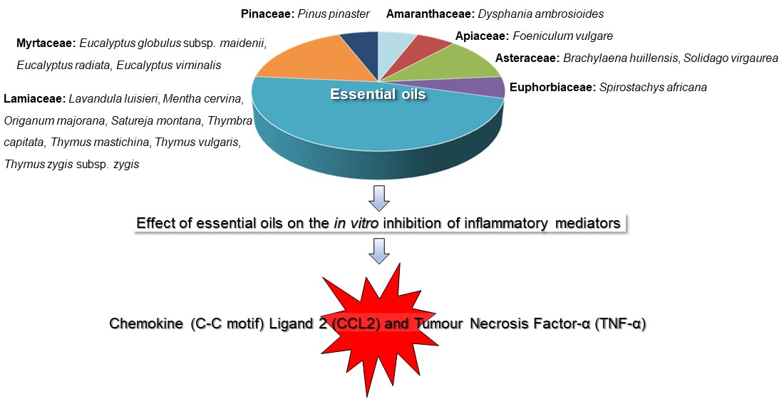
Effect of Essential Oils on the Release of TNF-α and CCL2 by LPS-Stimulated THP‑1 Cells

 , and
, and
Abstract
:
1. Introduction
2. Material and Methods
2.1. Plant Material
2.2. Extraction and Chemical Analysis of the Essential Oils
2.2.1. Gas Chromatography (GC)
2.2.2. Gas Chromatography-Mass Spectrometry (GC‑MS)
2.3. In Vitro Inhibition of TNF-α and CCL2
3. Results and Discussion
3.1. Chemical Composition of the Essential Oils
3.2. In Vitro Inhibition of TNF‑α Release by LPS-Stimulated THP-1 Cells
3.3. In Vitro Inhibition of CCL2 Release by LPS-Stimulated THP-1 Cells
4. Conclusions
Author Contributions
Funding
Institutional Review Board Statement
Informed Consent Statement
Data Availability Statement
Acknowledgments
Convention on Biodiversity
Conflicts of Interest
Abbreviations
| CCL2 | Chemokine (C-C motif) ligand 2 |
| COX-2 | Cyclooxygenase-2 |
| DF | Dry, flowering phase aerial parts |
| DL | Dry leaves |
| DV | Dry, vegetative phase aerial parts |
| EOs | Essential oils |
| FBS | Fetal bovine serum |
| FF | Fresh, flowering phase aerial parts |
| FL | Fresh leaves from fruiting phase |
| IC50 | Half-maximal inhibitory concentration |
| LDL | Low-density lipoprotein |
| LPS | Lipopolysaccharide |
| MCP-1 | Monocyte chemoattractant protein-1 |
| MEE | Mata Experimental do Escaroupim |
| MTT | 3-(4,5-Dimethylthiazol-2-yl)-2,5 diphenyltetrazolium bromide |
| THP-1 | Human acute monocytic leukemia cell line |
| TNF-α | Tumor necrosis factor-α |
References
- Vane, J.R.; Botting, R.M. The mechanism of action of aspirin. Thromb. Res. 2003, 110, 255–258. [Google Scholar] [CrossRef]
- Furman, D.; Campisi, J.; Verdin, E.; Carrera-Bastos, P.; Targ, S.; Franceschi, C.; Ferrucci, L.; Gilroy, D.W.; Fasano, A.; Miller, G.W.; et al. Chronic inflammation in the etiology of disease across the life span. Nat. Med. 2019, 25, 1822–1832. [Google Scholar] [CrossRef] [PubMed]
- Song, P.; Fang, Z.; Wang, H.; Cai, Y.; Rahimi, K.; Zhu, Y.; Fowkes, F.G.R.; Fowkes, F.J.I.; Rudan, I. Global and regional prevalence, burden, and risk factors for carotid atherosclerosis: A systematic review, meta-analysis, and modelling study. Lancet Glob. Health 2020, 8, e721–e729. [Google Scholar] [CrossRef]
- CCAP. Custo e Carga da Aterosclerose em-Portugal.Centro de Estudo de Medicina Baseada na Evidência da FML, Centro de Estudos Aplicados da Universidade Católica e Sociedade Portuguesa de Aterosclerose. 2019. Available online: https://spaterosclerose.org/highlights-de-2018/item/274-custo-e-carga-da-aterosclerose-em-portugal.html (accessed on 13 October 2020). (In Portuguese)
- Branco, J.C.; Faustino, A.; Carvalho, B.; Araújo, F.; Canhão, H.; Brito, I.; da Silva, J.A.P.; Costa, J.A.; Costa, L.; Maurício, L.; et al. Rede Nacional de especialidade hospitalar e de referenciação de reumatologia. 2015. Available online: https://www.sns.gov.pt/wp-content/uploads/2016/05/rede-referencia%C3%A7%C3%A3o-hospitalar-reumatologia.pdf (accessed on 13 October 2020). (In Portuguese)
- Usenbo, A.; Kramer, V.; Young, T.; Musekiwa, A. Prevalence of arthritis in Africa: A systematic review and meta-analysis. PLoS ONE 2015, 10, e0133858. [Google Scholar] [CrossRef]
- SESPA. Secretário de Estado para a Saúde Pública de Angola. 2018. Available online: http://jornaldeangola.sapo.ao/sociedade/angola_so_tem_oito_medicos_formados_em_reumatologia (accessed on 13 October 2020). (In Portuguese).
- Boudreault, J.; Desmeules, F.; Roy, J.-S.; Dionne, C.; Frémont, P.; MacDermid, J.C. The efficacy of oral non-steroidal anti-inflammatory drugs for rotator cuff tendinopathy: A systematic review and meta-analysis. J. Rehabil. Med. 2014, 46, 294–306. [Google Scholar] [CrossRef] [Green Version]
- Wallace, J.L. Eicosanoids in the gastrointestinal tract. Br. J. Pharmacol. 2019, 176, 1000–1008. [Google Scholar] [CrossRef] [Green Version]
- Miguel, M.G. Antioxidant and anti-inflammatory activities of essential oils: A short review. Molecules 2010, 15, 9252–9287. [Google Scholar] [CrossRef] [Green Version]
- Dandlen, S.; Lima, A.; Mendes, M.; Miguel, M.; Faleiro, M.L.; Sousa, M.; LG, P.; JG, B.; Figueiredo, A. Antimicrobial activity, cytotoxicity and intracellular growth inhibition of Portuguese Thymus essential oils. Rev. Bras. Farmacog. 2011, 21, 1012–1024. [Google Scholar] [CrossRef] [Green Version]
- De Lima, V.T.; Vieira, M.C.; Kassuya, C.A.L.; Cardoso, C.A.L.; Alves, J.M.; Foglio, M.A.; de Carvalho, J.E.; Formagio, A.S.N. Chemical composition and free radical-scavenging, anticancer and anti-inflammatory activities of the essential oil from Ocimum kilimandscharicum. Phytomedicine 2014, 21, 1298–1302. [Google Scholar] [CrossRef]
- Grassi, L.T.; Malheiros, A.; Meyre-Silva, C.; Buss, Z.S.; Monguilhott, E.D.; Fröde, T.S.; da Silva, K.A.B.S.; de Souza, M.M. From popular use to pharmacological validation: A study of the anti-inflammatory, anti-nocicpetive and healing effects of Chenopodium ambrosioides extract. J. Ethnopharmacol. 2013, 145, 127–138. [Google Scholar] [CrossRef] [Green Version]
- Alonso-Castro, A.J.; Domínguez, F.; Ruiz-Padilla, A.J.; Campos-Xolalpa, N.; Zapata-Morales, J.R.; Carranza-Alvarez, C.; Maldonado-Miranda, J.J. Medicinal plants from North and Central America and the Caribbean considered toxic for humans: The other side of the coin. Evid. Based Complement. Alternat. Med. 2017, 2017, 9439868. [Google Scholar] [CrossRef] [PubMed] [Green Version]
- Miguel, M.G.; Cruz, C.; Faleiro, L.; Simões, M.T.F.; Figueiredo, A.C.; Barroso, J.G.; Pedro, L.G. Foeniculum vulgare essential oils: Chemical composition, antioxidant and antimicrobial activities. Nat. Prod. Commun. 2010, 5, 319–328. [Google Scholar] [CrossRef] [PubMed] [Green Version]
- Maitai, C.K.; Talalaj, S.; Talalaj, D. Aromatic Plants of East Africa; Department of Pharmacy College of Health Sciences, University of Nairobi: Nairobi, Kenya, 1983. [Google Scholar]
- Oliva, M.M.; Demo, M.S.; Malele, R.S.; Mutayabarwa, C.K.; Mwangi, J.W.; Thoithi, G.N.; Kibwage, I.O.; Faillaci, S.M.; Scrivanti, R.L.; Lopez, A.G.; et al. Essential oil of Brachylaena hutchinsii Huthch from Tanzania: Antimicrobial activity and composition. East. Central Afr. J. Pharm. Sci. 2003, 6, 61–63. [Google Scholar]
- Mrema, J.P. Conservation of Brachylaena huillensis O.Hoffm (Asteraceae) in Dindili Forest Reserve, Morogoro, Tanzania. Ph.D. Thesis, Addis Ababa University, Addis Ababa, Ethiopia, 2006. [Google Scholar]
- Minciullo, P.L.; Calapai, G.; Miroddi, M.; Mannucci, C.; Chinou, I.; Gangemi, S.; Schmidt, R.J. Contact dermatitis as an adverse reaction to some topically used European herbal medicinal products—Part 4: Solidago virgaurea—Vitis vinifera. Contact Dermat. 2017, 77, 67–87. [Google Scholar] [CrossRef]
- Baarschers, W.H.; Horn, D.H.; Johnson, L.R.F. The structure of some diterpenes from tambooti wood, Spirostachys africana Sond. J. Chem. Soc. 1962, 10, 4046–4055. [Google Scholar] [CrossRef]
- Mathabe, M.C.; Hussein, A.A.; Nikolova, R.V.; Basson, A.E.; Meyer, J.J.M.; Lall, N. Antibacterial activities and cytotoxicity of terpenoids isolated from Spirostachys africana. J. Ethnopharmacol. 2008, 116, 194–197. [Google Scholar] [CrossRef]
- Mukandiwa, L.; McGaw, L.J.; Eloff, J.N.; Naidoo, V. Extracts of four plant species used traditionally to treat myiasis influence pupation rate, pupal mass and adult blow fly emergence of Lucilia cuprina and Chrysomya marginalis (Diptera: Calliphoridae). J. Ethnopharmacol. 2012, 143, 812–818. [Google Scholar] [CrossRef]
- Zuzarte, M.; Gonçalves, M.J.; Cruz, M.T.; Cavaleiro, C.; Canhoto, J.; Vaz, S.; Pinto, E.; Salgueiro, L. Lavandula luisieri essential oil as a source of antifungal drugs. Food Chem. 2012, 135, 1505–1510. [Google Scholar] [CrossRef]
- Figueiredo, A.C.; Pedro, L.G.; Barroso, J.G.; Trindade, H.; Sanches, J.; Oliveira, C.; Correia, M. Lavandula luisieri (Rozeira) Rivas-Martínez e Lavandula pedunculata (Mill.) Cav. Agrotec.
- Rodrigues, L.; Duarte, A.; Figueiredo, A.C.; Brito, L.; Teixeira, G.; Moldão, M.; Monteiro, A. Chemical composition and antibacterial activity of the essential oils from the medicinal plant Mentha cervina L. grown in Portugal. Med. Chem. Res. 2012, 21, 3485–3490. [Google Scholar] [CrossRef]
- Costa, D.C.; Costa, H.S.; Albuquerque, T.G.; Ramos, F.; Castilho, M.C.; Sanches-Silva, A. Advances in phenolic compounds analysis of aromatic plants and their potential applications. Trends Food Sci. Technol. 2015, 45, 336–354. [Google Scholar] [CrossRef]
- Mastelić, J.; Jerković, I. Gas chromatography-mass spectrometry analysis of free and glycoconjugated aroma compounds of seasonally collected Satureja montana L. Food Chem. 2003, 80, 135–140. [Google Scholar] [CrossRef]
- Figueiredo, A.C.; Barroso, J.G.; Pedro, L.G.; Salgueiro, L.; Miguel, M.G.; Faleiro, M.L. Portuguese Thymbra and Thymus species volatiles: Chemical composition and biological activities. Cur. Pharm. Des. 2008, 14, 3120–3140. [Google Scholar] [CrossRef] [PubMed]
- Fernandes, A.S.F.; Barros, L.; Carvalho, A.M.; Ferreira, I.C.F.R. Lipophilic and hydrophilic antioxidants, lipid peroxidation, inhibition and radical scavenging activity of two Lamiaceae food plants. Eur. J. Lip. Sci. Technol. 2010, 112, 1115–1121. [Google Scholar] [CrossRef]
- Figueiredo, A.C.; Pedro, G.; Barroso, J.G.; Sanches, J.; Correia, M. Óleos essenciais de espécies de Eucalyptus. Agrotec 2013, 8, 96–100. [Google Scholar]
- Mahumane, G.D.; van Vuuren, S.F.; Kamatou, G.; Sandasi, M.; Viljoen, A.M. Chemical composition and antimicrobial activity of Eucalyptus radiata leaf essential oil, sampled over a year. J. Essent. Oil Res. 2016, 28, 475–488. [Google Scholar] [CrossRef]
- Tolmacheva, A.A.; Rogozhin, E.A.; Deryabin, D.G. Antibacterial and quorum sensing regulatory activities of some traditional Eastern-European medicinal plants. Acta Pharm. 2014, 64, 173–186. [Google Scholar] [CrossRef] [Green Version]
- Figueiredo, A.C.; Pedro, L.G.; Barroso, J.G.; Trindade, H.; Sanches, J.; Oliveira, C.; Correia, M. Pinus pinaster Aiton e Pinus pinea L. Agrotec 2014, 12, 23–27. [Google Scholar]
- Takeya, M.; Yoshimura, T.; Leonard, E.J.; Takahashi, K. Detection of monocyte chemoattractant protein-1 in human atherosclerotic lesions by an anti-monocyte chemoattractant protein-1 monoclonal antibody. Hum. Pathol. 1993, 24, 534–539. [Google Scholar] [CrossRef]
- Bakheet, S.A.; Alrwashied, B.S.; Ansari, M.A.; Nadeem, A.; Attia, S.M.; Alanazi, M.M.; Aldossari, A.A.; Assiri, M.A.; Mahmood, H.M.; Al-Mazroua, H.A.; et al. CXC chemokine receptor 3 antagonist AMG487 shows potent anti-arthritic effects on collagen-induced arthritis by modifying B cell inflammatory profile. Immunol. Lett. 2020, 225, 74–81. [Google Scholar] [CrossRef]
- Tousoulis, D.; Oikonomou, E.; Economou, E.K.; Crea, F.; Kaski, J.C. Inflammatory cytokines in atherosclecrosis: Current therapeutic approaches. Eur. Heart J. 2016, 37, 1723–1735. [Google Scholar] [CrossRef] [Green Version]
- Aazza, S.; Lyoussi, B.; Megías, C.; Cortés-Giraldo, I.; Vioque, J.; Figueiredo, A.C.; Miguel, M.G. Anti-oxidant, anti-inflammatory and anti-proliferative activities of Moroccan commercial essential oils. Nat. Prod. Commun. 2014, 9, 587–594. [Google Scholar] [CrossRef] [PubMed] [Green Version]
- Rezayat, S.M.; Dehpour, A.-R.; Motamed, S.M.; Yazdanparast, M.; Chamanara, M.; Rashidian, A.; Sahebgharani, M. Foeniculum vulgare essential oil ameliorates acetic-induced colitis in rats through the inhibition of NF-kB pathway. Inflammopharmacology 2018, 26, 851–859. [Google Scholar] [CrossRef] [PubMed]
- Arantes, S.; Candeias, F.; Lopes, O.; Lima, M.; Pereira, M.; Tinoco, T.; Cruz-Morais, J.; Martins, M.R. Pharmacological and toxicological studies of essential oil of Lavandula stoechas subsp. luisieri. Planta Med. 2016, 82, 1266–1273. [Google Scholar] [CrossRef] [PubMed]
- Rufino, A.T.; Ribeiro, M.; Sousa, C.; Judas, F.; Salgueiro, L.; Cavaleiro, C.; Mendes, A.F. Evaluation of the anti-inflammatory, anti-catabolic and pro-anabolic effects of E-caryophyllene, myrcene and limonene in a cell model of osteoarthritis. Eur. J. Pharmacol. 2015, 750, 141–150. [Google Scholar] [CrossRef]
- Rufino, A.T.; Ferreira, I.; Judas, F.; Salgueiro, L.; Lopes, M.C.; Cavaleiro, C.; Mendes, A.F. Differential effects of the essential oils of Lavandula luisieri and Eryngium duriaei subsp. juresianum in cell models of two chronic inflammatory diseases. Pharm. Biol. 2015, 53, 1220–1230. [Google Scholar] [CrossRef] [Green Version]
- Arranz, E.; Jaime, L.; López de las Hazas, M.C.; Reglero, G.; Santoyo, S. Supercritical fluid extraction as an alternative process to obtain essential oils with anti-inflammatory properties from marjoram and sweet basil. Ind. Crops Prod. 2015, 67, 121–129. [Google Scholar] [CrossRef]
- Aazza, S.; El-Guendouz, S.; Miguel, M.G.; Antunes, M.D.; Faleiro, M.L.; Correia, A.I.; Figueiredo, A.C. Antioxidant, anti-inflammatory and anti-hyperglycamic activities of essential oils from Thymbra capitata, Thymus albicans, Thymus caespititius, Thymus carnosus, Thymus lotocephalus and Thymus mastichina from Portugal. Nat. Prod. Commun. 2016, 11, 1029–1038. [Google Scholar] [CrossRef] [Green Version]
- Carrasco, A.; Perez, E.; Cutillas, A.-B.; Martinez-Gutierrez, R.; Tomas, V.; Tudela, J. Origanum vulgare and Thymbra capitata essential oils from Spain: Determination of aromatic profile and bioactivities. Nat. Prod. Commun. 2016, 11, 113–120. [Google Scholar] [CrossRef]
- Wei, A.; Shibamoto, T. Antioxidant/lipoxygenase inhibitory activities and chemical compositions of selected essential oils. J. Agric. Food Chem. 2010, 58, 7218–7225. [Google Scholar] [CrossRef]
- Tsai, M.-L.; Lin, C.-C.; Lin, W.-C.; Yang, C.-H. Antimicrobial, antioxidant, and anti-inflammatory activities of essential oils from five selected herbs. Biosci. Biotech. Bioch. 2011, 75, 1977–1983. [Google Scholar] [CrossRef] [Green Version]
- Abdelli, W.; Bahri, F.; Romane, A.; Höferl, M.; Wanner, J.; Schmidt, E.; Jirovetz, L. Chemical composition and anti-inflammatory activity of Algerian Thymus vulgaris essential oil. Nat. Prod. Commun. 2017, 12, 611–614. [Google Scholar] [CrossRef] [PubMed] [Green Version]
- Fachini-Queiroz, F.C.; Kummer, R.; Estevão-Silva, C.F.; Carvalho, N.D.B.; Cunha, J.M.; Grespan, R.; Bersani-Amado, C.A.; Cuman, R.K.N. Effects of thymol and carvacrol, constituents of Thymus vulgaris L. essential oil, on the inflammatory response. Evid. Based Complement. Alternat. Med. 2012, 2012, 657026. [Google Scholar] [CrossRef] [PubMed] [Green Version]
- Hotta, M.; Nakata, R.; Katsukawa, M.; Hori, K.; Takahashi, S.; Inoue, H. Carvacrol, a component of thyme oil, activates PPARα and Gamma and suppresses COX-2 expression. J. Lip. Res. 2010, 51, 132–139. [Google Scholar] [CrossRef] [PubMed] [Green Version]
- Bukovská, A.; Čikoš, Š.; Juhás, Š.; Il’Ková, G.; Rehák, P.; Koppel, J. Effects of a combination of thyme and oregano essential oils on TNBS-induced colitis in mice. Mediat. Inflamm. 2007, 23296. [Google Scholar] [CrossRef] [PubMed] [Green Version]
- Ocaña, A.; Reglero, G. Effects of thyme extract oils (from Thymus vulgaris, Thymus zygis and Thymus hyemalis) on cytokine production and gene expression of oxLDL-stimulated THP-1 macrophages. J. Obes. 2012, 2012, 104706. [Google Scholar] [CrossRef] [PubMed] [Green Version]
- Juhás, Š.; Bujňáková, D.; Rehák, P.; Čikoš, Š.; Veselá, J.; Il’Ková, G.; Koppel, J. Anti-inflammatory effects of thyme essential oil in mice. Acta Vet. Brno 2008, 77, 327–334. [Google Scholar] [CrossRef] [Green Version]
- Cutillas, A.-B.; Carrasco, A.; Martinez-Gutierrez, R.; Tomas, V.; Tudela, J. Thyme essential oils from Spain: Aromatic profile ascertained by GC-MS, and their antioxidant, anti-lipoxygenase and antimicrobial activities. J. Food Drug Anal. 2018, 26, 529–544. [Google Scholar] [CrossRef] [Green Version]
- Tümen, I.; Guragac, F.T.; Keles, H.; Reunanen, M.; Kupeli-Akkol, E. Characterization and wound repair potential of essential oil Eucalyptus globulus Labill. Fresenius Environ. Bull. 2017, 26, 6390–6399. [Google Scholar]
- Tümen, I.; Akkol, E.K.; Taştan, H.; Süntar, I.; Kurtca, M. Research on the antioxidant, wound healing, and anti-inflammatory activities and the phytochemical composition of maritime pine (Pinus pinaster Ait). J. Ethnopharmacol. 2018, 211, 235–246. [Google Scholar] [CrossRef]
- Sá, R.C.S.; Andrade, L.N.; de Sousa, D.P. A review on anti-inflammatory activity of monoterpenes. Molecules 2013, 18, 1227–1254. [Google Scholar]
- Sá, R.C.S.; Andrade, L.N.; de Sousa, D.P. Sesquiterpenes from essential oils and anti-inflammatory activity. Nat. Prod. Commun. 2015, 10, 1767–1774. [Google Scholar]
- Faria, J.M.S.; Sena, I.; Ribeiro, B.; Rodrigues, A.M.; Maleita, C.M.N.; Abrantes, I.; Bennett, R.N.; Mota, M.; Figueiredo, A.C. First report on Meloidogyne chitwoodi hatching inhibition activity of essential oils and essential oils fractions. J. Pest. Sci. 2016, 89, 207–217. [Google Scholar] [CrossRef]
- Council of Europe (COE). European Directorate for the Quality of Medicines. European Pharmacopoeia, 7th ed.; COE: Strasbourg, France, 2007.
- Campana, P.R.V.; Mansur, D.S.; Gusman, G.S.; Ferreira, D.; Teixeira, M.M.; Braga, F.C. Anti-TNF-α activity of Brazilian medicinal plants and compounds from Ouratea semiserrata. Phytother. Res. 2015, 29, 1509–1515. [Google Scholar] [CrossRef] [PubMed]
- Mosmann, T. Rapid colorimetric assay for cellular growth and survival: Application to proliferation and cytotoxicity assays. J. Immunol. Methods 1983, 65, 55–63. [Google Scholar] [CrossRef]
- Matos, F.; Miguel, M.G.; Duarte, J.; Venâncio, F.; Moiteiro, C.; Correia, A.I.D.; Figueiredo, A.C.; Barroso, J.G.; Pedro, L.G. Antioxidant capacity of the essential oils from Lavandula luisieri, L. stoechas ssp. lusitanica, L. stoechas ssp. lusitanica x L. luisieri and L. viridis grown in Algarve (Portugal). J. Essent. Oil Res. 2009, 21, 327–336. [Google Scholar] [CrossRef]
- Lopes, V.R.; Barata, A.M.; Farias, R.; Mendes, M.D.; Lima, A.S.; Pedro, L.G.; Barroso, J.G.; Figueiredo, A.C. Morphological and essential oil variability from nine Portuguese fennel (Foeniculum vulgare Mill.) accessions. Acta Hort. 2010, 860, 33–50. [Google Scholar] [CrossRef]
- Faria, J.M.S.; Barbosa, P.; Bennett, R.N.; Mota, M.; Figueiredo, A.C. Bioactivity against Bursaphelenchus xylophilus: Nematotoxics from essential oils, essential oils fractions and decoction waters. Phytochemistry 2013, 94, 220–228. [Google Scholar] [CrossRef] [Green Version]
- Barbosa, P.; Faria, J.M.S.; Mendes, M.D.; Dias, L.S.; Tinoco, M.T.; Barroso, J.G.; Pedro, L.G.; Figueiredo, A.C.; Mota, M. Bioassays against pinewood nematode: Assessment of a suitable dilution agent and screening for bioactive essential oils. Molecules 2012, 17, 12312–12329. [Google Scholar] [CrossRef] [Green Version]
- Figueiredo, A.C.; Barroso, J.G.; Pedro, L.G. Volatiles from Thymbra and Thymus Species of the Western Mediterranean Basin, Portugal and Macaronesia. Nat. Prod. Commun. 2010, 5, 1465–1476. [Google Scholar] [CrossRef] [Green Version]
- Faria, J.M.S.; Lima, A.S.; Mendes, M.D.; Leiria, R.; Geraldes, D.A.; Figueiredo, A.C.; Trindade, H.; Pedro, L.G.; Barroso, J.G.; Sanches, J. Eucalyptus from Mata Experimental do Escaroupim (Portugal): Evaluation of the essential oil composition from sixteen species. Acta Hortic. 2011, 925, 61–66. [Google Scholar] [CrossRef]
- Miguel, M.G.; Gago, C.; Antunes, M.D.; Lagoas, S.; Faleiro, M.L.; Megías, C.; Cortés-Giraldo, I.; Vioque, J.; Figueiredo, A.C. Antibacterial, antioxidant and antiproliferative activities of Corymbia citriodora and eight Eucalyptus species essential oils. Medicines 2018, 5, 61. [Google Scholar] [CrossRef] [Green Version]
- Rodrigues, A.M.; Mendes, M.D.; Lima, A.S.; Barbosa, P.M.; Ascensão, L.; Barroso, J.G.; Pedro, L.G.; Mota, M.M.; Figueiredo, A.C. Pinus halepensis, Pinus pinaster, Pinus pinea and Pinus sylvestris essential oils chemotypes and monoterpene hydrocarbon enantiomers, before and after inoculation with the pinewood nematode Bursaphelenchus xylophilus. Chem. Biodivers. 2017, 14, e1600153. [Google Scholar] [CrossRef] [PubMed]
- Klein, E.; Schmidt, W. Structure of brachyl oxide. J. Agric. Food Chem. 1971, 19, 1115–1117. [Google Scholar] [CrossRef]
- Pereira, W.S.; da Silva, G.P.; Vigliano, M.V.; Leal, N.R.F.; Pinto, F.A.; Fernandes, D.C.; Santos, S.V.M.; Martino, T.; Nascimento, J.R.; de Azevedo, A.P.S.; et al. Anti-arthritic properties of crude extract from Chenopodium ambrosioides L. leaves. J. Pharm. Pharmacol. 2018, 70, 1078–1091. [Google Scholar] [CrossRef] [PubMed]
- Figueiredo, A.C. Biological properties of essentials oils and volatiles. Sources of variability. Nat. Volatiles Essent. Oils 2017, 4, 1–13. [Google Scholar]
- Ni, W.; Kitamoto, S.; Ishibashi, M.; Usui, M.; Inoue, S.; Huasa, K.-I.; Zhao, Q.; Nishida, K.-I.; Takeshita, A.; Eghashira, K. Monocyte chemoattractant protein-1 is an essential inflammatory mediator in angiotensin II-induced progression of established atherosclerosis in hypercholesterolemic mice. Arter. Thromb. Vasc. Biol. 2004, 24, 534–539. [Google Scholar] [CrossRef] [PubMed] [Green Version]
- Hirota, R.; Roger, N.N.; Nakamura, H.; Song, H.-S.; Sawamura, M.; Suganuma, N. Anti-inflammatory effects of limonene from Yuzu (Citrus junos Tanaka) essential oil on eosinophilis. J. Food Sci. 2010, 75, H87–H92. [Google Scholar] [CrossRef]
- Chen, L.-L.; Zhang, H.J.; Chao, J.; Liu, J.F. Essential oil of Artemisia argyi suppresses inflammatory responses by inhibiting JAK/STATs activation. J. Ethnopharmacol. 2017, 204, 107–117. [Google Scholar] [CrossRef]
- Luo, G.; Kong, J.; Cheng, B.C.-Y.; Zhao, H.; Fu, X.-Q.; Yan, L.-S.; Ding, Y.; Liu, Y.-L.; Pan, S.-Y.; Zhang, S.-F.; et al. Xiao Qing Long Tang essential oil exhibits inhibitory effects on the release of pro-inflammatory mediators by suppressing NF-alphaB, AP-1, and IRF3 signalling in the lipopolysaccharide-stimulated RAW264.7 cells. Rsc Adv. 2019, 9, 12977–12989. [Google Scholar] [CrossRef] [Green Version]
- Park, H.; Seol, G.H.; Ryu, S.; Choi, I.-Y. Neuroprotective effects of (-)-linalool against oxygen-glucose deprivation-induced neuronal injury. Arch. Pharmacal Res. 2016, 39, 555–564. [Google Scholar] [CrossRef]

| Family/Plant Species | Common Names (pt/en) | Medicinal Use | Other Uses | Reference |
|---|---|---|---|---|
| Amaranthaceae | ||||
| Dysphania ambrosioides (L.) Mosyakin & Clemants (= Chenopodium ambrosioides L.) | Quenopódio/Wormseed | Against respiratory, gastrointestinal and joint inflammatory disorders | Vermifuge, emetic | [13,14] |
| Apiaceae/Umbelliferae | ||||
| Foeniculum vulgare Mill. | Funcho/Fennel | Against respiratory and gastrointestinal inflammatory disorders | Culinary (seasoning) | [15] |
| Asteraceae/Compositae | ||||
| Brachylaena huillensis O. Hoffm. (= Brachylaena hutchinsii Hutch., Brachylaena mullensis O.Hoffm.) | Muhuhu */Silver oak | Against schistosomiasis and roots against diabetes | Firewood, charcoal, timber, poles, posts, tool handles, carving. Perfumery (essential oil distilled from wood) | [16,17,18] |
| Solidago virgaurea L. | Vara-de-ouro/European goldenrod or woundwort | External and internal against urinary inflammatory disorders | Cosmetic | [19] |
| Euphorbiaceae | ||||
| Spirostachys africana Sond. [= Excoecaria africana (Sond.) Müll.Arg., Excoecaria synandra Pax, Excoecariopsis synandra (Pax) Pa, Sapium africanum (Sond.) Kuntze, Spirostachys synandra (Pax) Pax, Stillingia africana (Sond.) Baill.] | Tambooti ** | External to treat myiasis, internal against gastrointestinal inflammatory disorders | Use of wood in furniture | [20,21,22] |
| Lamiaceae/Labiatae | ||||
| Lavandula luisieri (Rozeira) Rivas-Martínez | Rosmaninho/butterfly lavender | External and internal against respiratory, circulatory, gastrointestinal and joint inflammatory disorders | Ornamental, aromatic, cosmetic, culinary (seasoning) | [23,24] |
| Mentha cervina L. | Poejo fino/Hart’s pennyroyal | External and internal against respiratory and gastrointestinal inflammatory disorders | Aromatic, culinary (seasoning) | [25] |
| Origanum majorana L. | Oregão/Marjoram | External and internal against nervous, respiratory and gastrointestinal inflammatory disorders | Aromatic, culinary (seasoning) | [26] |
| Satureja montana L. | Segurelha/Winter savory | External and internal against nervous, respiratory and gastrointestinal inflammatory disorders | Culinary (seasoning) | [27] |
| Thymbra capitata (L.) Cav. [= Thymus capitatus Hoffms. et Link., Thymus creticus Brot., Corydothymus capitatus Rechenb. f., Satureja capitata L.] | Tomilho-de-Creta/Conehead thyme | External and internal against spasms and nervous, respiratory and gastrointestinal disorders | Aromatic, culinary (seasoning) | [28] |
| Thymus mastichina (L.) L. | Bela-luz/Spanish marjoram | External and internal against nervous, respiratory, gastrointestinal and joint inflammatory disorders | Aromatic, culinary (seasoning) | [28] |
| Thymus pulegioides L. | Serpão/Broad-leaved thyme, lemon thyme | External and internal against nervous, respiratory and gastrointestinal inflammatory disorders | Aromatic, culinary (seasoning) | [29] |
| Thymus vulgaris L. | Tomilho/thyme | External and internal against nervous, respiratory and gastrointestinal inflammatory disorders | Ornamental, aromatic, culinary (seasoning) | [19] |
| Thymus zygis Loefl. ex L. subsp. zygis | Erva-de-Santa-Maria/Spanish red thyme | External and internal against nervous, circulatory, respiratory and gastrointestinal inflammatory disorders | Aromatic, culinary (seasoning) | [28] |
| Myrtaceae | ||||
| Eucalyptus globulus subsp. maidenii (F.Muell.) J.B.Kirkp. | Eucalipto/Maiden’s gum | External and internal against circulatory, respiratory and gastrointestinal inflammatory disorders | Timber, fuel, paper pulp. Aromatic, culinary (seasoning) | [30] |
| Eucalyptus radiata A.Cunn. ex DC. | Eucalipto/Narrow-leaved peppermint eucalyptus | External and internal against mouth, respiratory and gastrointestinal inflammatory disorders | [31] | |
| Eucalyptus viminalis Labill. | Eucalipto/Manna gum | Internal against respiratory inflammatory disorders | Deodorant | [32] |
| Pinaceae | ||||
| Pinus pinaster Aiton | Pinheiro-bravo/Maritime pine | External for circulatory problems, and internal against respiratory, gastrointestinal and joint inflammatory disorders | Timber and oleoresin production | [33] |
| EO/EO Components’ Anti-Inflammatory Activity | Family/Species | Reference |
|---|---|---|
| Apiaceae | ||
| Foeniculum vulgare | EO inhibition of 5-lipoxygenase (IC50 = 0.04 mg/mL). Fenchone inhibition of 5-lipoxygenase (IC50 = 0.02 mg/mL). | [37] |
| EO (200 and 400 mg/kg) decreased the activity of mieloperoxidase (MPO) and the expression of TNF-α in the colon tissue previously submitted to acetic acid solution (acute colitis), and inhibited acetic acid-induced expression of p‑NF-kB p65 protein. | [38] | |
| Lamiaceae | ||
| Lavandula luisieri | EO (50–200 mg/kg) inhibition of paw edema (31–83%) induced by carrageenan administered in male Wistar rats. | [39] |
| EO (25 μg/mL) nitric oxide (NO) inhibition (75%) in IL-1β induced primary chondrocyte. | [40] | |
| EO reduction of iNOS in human chondrocytes and intestinal cell line C2BBe1 (54.9 and 81.0%, respectively) and phosphorylated IkB-α (87.4% and 62.3%, respectively). | [41] | |
| Origanum majorana | EO (10 μg/mL) diminished the TNF-α, IL‑1β, IL-6, IL-10 and COX-2 secretion and NFκB gene expression after activation of THP-1 cells by lipopolysaccharide or human ox – LDL. The activity was attributed to cis-sabinene hydrate and terpinen‑4‑ol. | [42] |
| Thymbra capitata | EO inhibition of 5-lipoxygenase (IC50 = 0.1 mg/mL). | [43] |
| EO inhibition of 5-lipoxygenase (IC50 = 0.2 mg/mL). | [44] | |
| Thymus mastichina | EO inhibition of 5-lipoxygenase (IC50 = 0.7 mg/mL). | [43] |
| Thymus vulgaris | EO inhibition of 5-lipoxygenase (IC50 = 0.19 μg/mL). | [37] |
| EO (0.5 μg/mL) inhibition (80%) of 5-lipoxygenase. | [45] | |
| EO inhibition of 5-lipoxygenase (IC50 = 0.005 μg/mL). EO reduced the TNF-α, IL-1β, IL-8 secretion levels of THP-1 cells. | [46] | |
| EO (400 mg/kg, after 6 h) reduced (50.4–58.4%) carrageenan-induced paw edema in mice. | [47] | |
| Carvacrol (10 mg/ear) reduced ear edema. Carvacrol (10 mg/ear) inhibited the activity of myeloperoxidase (MPO) (43.8%). Carvacrol (0.3–90 μg/mL) reduced (20.07–52.23%) neutrophil migration in response to fMLP stimulation. EO (750 mg/kg) and carvacrol (100–400 mg/kg) exerted inhibited leukocyte migration to the injury site. Carvacrol (0.3‑90μg/mL) reduced LTB4 stimulation (19.8–61.1%). | [48] | |
| EO and carvacrol suppressed lipopolysaccharide-induced COX-2 mRNA and protein expression in human macrophage-like U937 cells. | [49] | |
| EO (moderate concentration) decreased the mRNA levels of IL-1β, IL-6, granulocyte-macrophage colony stimulating factor (GM-CSF) and TNF-α, and lowered the amount of IL-1β and IL-6 proteins in animal models of colitis. | [50] | |
| EO reduced production and gene expression of the pro-inflammatory mediators TNF‑α, IL-1B and IL-6 and increased the parameters on the anti-inflammatory IL-10 cytokine. | [51] | |
| EO (5000 ppm) decreased paw edema and ear swelling, inhibited the total mRNA IL‑1β expression in the mouse colon. | [52] | |
| Thymus zygis subsp. zygis | EO thymol type inhibition of 5-lipoxygenase (IC50 = 54 – 73 μL/L).EO linalool type inhibition of 5-lipoxygenase (IC50 = 299 – 402 μL/L). | [53] |
| EO reduced production and gene expression of the pro-inflammatory mediators TNF‑α, IL-1B and IL-6 and increased the parameters on the anti-inflammatory IL-10 cytokine. | [51] | |
| Myrtaceae | ||
| Eucalyptus globulus | EO inhibition of 5-lipoxygenase (IC50 = 0.16 mg/mL). | [37] |
| subsp. maidenii | EO (0.5μg/mL) inhibition (50%) of lipoxygenase. | [45] |
| EO (200 mg/kg) inhibited by 28.8% the inflammatory phase of wound healing (Whittle method). | [54] | |
| Pinaceae | ||
| Pinus pinaster | EO (100 mg/kg dose) inhibition (30.3%) of paw edema in the Whittle method using carrageenan. | [55] |
| Family/Species | Code | Sampling Date | Plant Part | Collection Place/Source # | EO yield (%, v/w) | Main Components (≥10%) |
|---|---|---|---|---|---|---|
| Amaranthaceae | ||||||
| Dysphania ambrosioides | Da | 2013 | FF | Monsaraz | 0.56 | iso-Ascaridole 51, ascaridole 16 |
| Apiaceae | ||||||
| Foeniculum vulgare | Fv | 2013 | DV | Herbal shop | α-Pinene 27, trans-anethole 18, Limonene 11 | |
| Fv s | 2013 | Seeds | Herbal shop | 1.16 | Methyl chavicol 79, limonene 12 | |
| Asteraceae | ||||||
| Brachylaena huillensis | Bh | 2013 | EO | Angola | n.a. | Copaen-15-ol * 14 |
| Bh * | 2013 | EO* | Angola | n.a. | Copaen-15-ol * 12 | |
| Solidago virgaureaa | Sv | 2013 | FF | Pinheiro da Cruz | 0.72 | β-Pinene 22, α-pinene 21, germacrene D 15, limonene 12 |
| Euphorbiaceae | ||||||
| Spirostachys africana | 2013 | EO | Angola | n.a. | Stachenone * 28, Diosphenol (2) * 38 | |
| Lamiaceae | ||||||
| Lavandula luisieria,b | Ll | 2013 | DF | Herbal shop | 0.44 | 5-Methylene-2,3,4,4-tetramethylcyclopent-2-enone 18, 1,8-cineole 16 |
| Mentha cervina | Mc | 2013 | DV | Herbal shop | 1.54 | Pulegone 76 |
| Origanum majoranaa | Om | 2013 | DV | Herbal shop | 0.98 | Terpinen-4-ol 18, carvacrol 17, γ‑terpinene 13, carvacrol methyl ether 13 |
| Satureja montanaa | Sm | 2013 | DV | Herbal shop | 1.48 | Carvacrol 77 |
| Thymbra capitata | Tc | 2013 | FV | Algarve | 0.89 | Carvacrol 71 |
| Thymus mastichina | Thm | 2013 | FF | Bragança | 1.35 | 1,8-Cineole 69 |
| Thymus pulegioidesa | Thp | 2013 | DL | Herbal shop | 0.49 | Thymol 32, ρ-cymene 22 |
| Thymus vulgarisa | Thv | 2013 | DV | Herbal shop | 1.20 | Thymol 45, ρ-cymene 21, γ‑terpinene 16 |
| Thymus zygis subsp. zygis a | Thzz | 2013 | FF | Bragança | 0.71 | Carvacrol 45, ρ-cymeme 22, γ‑terpinene 17 |
| Myrtaceae | ||||||
| Eucalyptus globulus subsp. maidenii | Eg | 2013 | FL | MEE | 3.20 | α-Pinene 15, 1,8-Cineole 46, Limonene 23 |
| Eucalyptus radiata | Er | 2012 | FL | MEE | 7.20 | 1,8-Cineole 49 |
| Eucalyptus viminalis | Ev | 2012 | FL | MEE | 2.50 | α-Pinene 10, 1,8-Cineole 69 |
| Pinaceae | ||||||
| Pinus pinaster | Pp | 2013 | Oleoresin | Nazaré | 29.76 | α-Pinene 62, β-pinene 23 |
| Family/Species and Control | Concentrations (µg/mL) | TNF-α Inhibition (% ± S.D., n = 3) | CCL2 Inhibition (% ± S.D., n = 3) |
|---|---|---|---|
| Control | LPS (200 ng) | 2428.1 ± 587.8 a | 2382.3 ± 1480.8 a |
| DMSO (0.1%) | 96.5 ± 13.9 a | 24.1 ± 14.7 a | |
| Amaranthaceae | |||
| Dysphania ambrosioides | 90 | 48.6 ± 2.1 *** | 15.6 ± 0.7 *** |
| 30 | 30.9 ± 1.5 * | 9.4 ± 0.3 ** | |
| 10 | 13.6 ± 1.0 | 7.5 ± 0.3 * | |
| Apiaceae | |||
| Foeniculum vulgare | 90 | 22.3 ± 1.9 *** | NI |
| 30 | 0.5 ± 0.0 | NI | |
| 10 | NI | NI | |
| Asteraceae | |||
| Brachylaena huillensis | 30 | NI | 18.8 ± 2.3 ** |
| (re-distilled EO) | 10 | 4.4 ± 0.8 | 9.0 ± 0.5 |
| 3 | NI | 5.4 ± 0.2 | |
| Brachylaena huillensis | 30 | ND | ND |
| 10 | ND | ND | |
| 3 | ND | ND | |
| Solidago virgaurea | 90 | ND | ND |
| 30 | 5.0 ± 0.2 | ND | |
| 10 | NI | 4.9 ± 0.1 * | |
| 3 | ND | 8.0 ± 0.1 ** | |
| Euphorbiaceae | |||
| Spirostachys africanus | 30 | ND | ND |
| 10 | ND | ND | |
| 3 | ND | ND | |
| Lamiaceae | |||
| Lavandula luisieri | 90 | 82.9 ± 8.2 *** | 82.0 ± 12.4 *** |
| 30 | 23.2 ± 1.1 *** | 54.3 ± 3.0 *** | |
| 10 | 2.5 ± 0.1 | 22.7 ± 1.0 *** | |
| Mentha cervina | 90 | NI | NI |
| 30 | NI | NI | |
| 10 | NI | 2.5 ± 0.0 | |
| Origanum majonara | 90 | NI | NI |
| 30 | NI | NI | |
| 10 | NI | 4.8 ± 0.1 | |
| Satureja montana | 30 | ND | ND |
| 10 | ND | ND | |
| 3 | NI | 0.2 ± 0.0 | |
| Thymbra capitata | 30 | ND | NI |
| 20 | 51.1 ± 6.5 *** | ND | |
| 10 | 29.5 ± 1.7 *** | 0.4 ± 0.0 | |
| 5 | 9.1 ± 0.1 * | ND | |
| Thymus mastichina | 90 | ND | ND |
| 30 | ND | ND | |
| 10 | ND | ND | |
| Thymus pulegioides | 90 | ND | ND |
| 30 | NI | 0.9 ± 0.0 | |
| 10 | NI | 8.4 ± 0.5 | |
| Thymus vulgaris | 90 | ND | ND |
| 30 | ND | ND | |
| 10 | ND | ND | |
| Thymus zygis ssp. sygis | 30 | NI | 8.9 ± 1.0 |
| 10 | NI | 2.7 ± 0.2 | |
| 3 | NI | 0 | |
| Myrtaceae | |||
| Eucapyptus globulus subsp. | 90 | 6.5 ± 0.4 | 1.7 ± 0.0 |
| maidenii | 30 | 4.4 ± 0.3 | NI |
| 10 | 2.4 ± 0.2 | NI | |
| Eucalyptus radiata | 90 | 12.0 ± 0.1 * | NI |
| 30 | NI | NI | |
| 10 | NI | NI | |
| Eucalyptus viminalis | 90 | 3.3 ± 0.2 | NI |
| 30 | 0.2 ± 0.0 | NI | |
| 10 | NI | 1.4 ± 0.0 | |
| Pinaceae | |||
| Pinus pinaster (oleoresin) | 90 | ND | ND |
| 30 | 6.3 ± 0.1 | NI | |
| 10 | 6.8 ± 0.3 | NI |
Publisher’s Note: MDPI stays neutral with regard to jurisdictional claims in published maps and institutional affiliations. |
© 2020 by the authors. Licensee MDPI, Basel, Switzerland. This article is an open access article distributed under the terms and conditions of the Creative Commons Attribution (CC BY) license (http://creativecommons.org/licenses/by/4.0/).
Share and Cite
Miguel, M.G.; da Silva, C.I.; Farah, L.; Castro Braga, F.; Figueiredo, A.C. Effect of Essential Oils on the Release of TNF-α and CCL2 by LPS-Stimulated THP‑1 Cells. Plants 2021, 10, 50. https://doi.org/10.3390/plants10010050
Miguel MG, da Silva CI, Farah L, Castro Braga F, Figueiredo AC. Effect of Essential Oils on the Release of TNF-α and CCL2 by LPS-Stimulated THP‑1 Cells. Plants. 2021; 10(1):50. https://doi.org/10.3390/plants10010050
Chicago/Turabian StyleMiguel, Maria Graça, Carina Isabel da Silva, Luana Farah, Fernão Castro Braga, and Ana Cristina Figueiredo. 2021. "Effect of Essential Oils on the Release of TNF-α and CCL2 by LPS-Stimulated THP‑1 Cells" Plants 10, no. 1: 50. https://doi.org/10.3390/plants10010050