
Association of Serum Vaspin Concentration with Metabolic Disorders in Obese Individuals
 , ,
, ,
Abstract
:1. Introduction
2. Materials and Methods
2.1. Study Population
2.2. Study Design
2.2.1. Anthropometric Parameters
2.2.2. Blood Pressure Measurement
2.2.3. Biochemical Parameters
2.2.4. Statistical Analysis
3. Results
4. Discussion
5. Conclusions
Author Contributions
Funding
Institutional Review Board Statement
Informed Consent Statement
Data Availability Statement
Conflicts of Interest
References
- Barazzoni, R.; Gortan Cappellari, G.; Ragni, M.; Nisoli, E. Insulin resistance in obesity: An overview of fundamental alterations. Eat. Weight Disord. 2018, 23, 149–157. [Google Scholar] [CrossRef] [PubMed]
- Kojta, I.; Chacińska, M.; Błachnio-Zabielska, A. Obesity, bioactive lipids, and adipose tissue inflammation in insulin resistance. Nutrients 2020, 12, 1305. [Google Scholar] [CrossRef] [PubMed]
- Pasco, J.A.; Holloway, K.L.; Dobbins, A.G.; Kotowicz, M.A.; Williams, L.J.; Brennan, S.L. Body mass index and measures of body fat for defining obesity and underweight: A cross-sectional, population-based study. BMC Obes. 2014, 1, 9. [Google Scholar] [CrossRef] [PubMed] [Green Version]
- Singh, M.; Benencia, F. Inflammatory processes in obesity: Focus on endothelial dysfunction and the role of adipokines as inflammatory mediators: We reviewed obesity-induced metabolic and immunological changes at the level of vasculature and emphasize on the importance of adipokines. Int. Rev. Immunol. 2019, 38, 157–171. [Google Scholar] [CrossRef] [PubMed]
- Lin, X.; Li, H. Obesity: Epidemiology, Pathophysiology, and Therapeutics. Front. Endocrinol. 2021, 12, 706978. [Google Scholar] [CrossRef]
- Feng, R.; Li, Y.; Wang, C.; Luo, C.; Liu, L.; Chuo, F.; Li, Q.; Sun, C. Higher vaspin levels in subjects with obesity and type 2 diabetes mellitus: A meta-analysis. Diabetes Res. Clin. Pract. 2014, 106, 88–94. [Google Scholar] [CrossRef]
- Hida, K.; Wada, J.; Eguchi, J.; Zhang, H.; Baba, M.; Seida, A.; Hashimoto, I.; Okada, T.; Yasuhara, A.; Nakatsuka, A.; et al. Visceral adipose tissue-derived serine protease inhibitor: A unique insulin-sensitizing adipocytokine in obesity. Proc. Natl. Acad. Sci. USA 2005, 102, 10610–10615. [Google Scholar] [CrossRef] [Green Version]
- Teshigawara, S.; Wada, J.; Hida, K.; Nakatsuka, A.; Eguchi, J.; Murakami, K.; Kanzaki, M.; Inoue, K.; Terami, T.; Katayama, A.; et al. Serum Vaspin Concentrations Are Closely Related to Insulin Resistance, and rs77060950 at SERPINA12 Genetically Defines Distinct Group with Higher Serum Levels in Japanese Population. J. Clin. Endocrinol. Metabol. 2012, 97, E1202–E1207. [Google Scholar] [CrossRef] [PubMed] [Green Version]
- Youn, B.S.; Kloting, N.; Kratzsch, J.; Lee, N.; Park, J.W.; Song, E.S.; Ruschke, K.; Oberbach, A.; Fasshauer, M.; Stumvoll, M.; et al. Serum vaspin concentrations in human obesity and type 2 diabetes. Diabetes 2008, 57, 372–377. [Google Scholar] [CrossRef] [Green Version]
- Buyukinan, M.; Atar, M.; Can, U.; Pirgon, O.; Guzelant, A.; Deniz, I. The association between serum vaspin and omentin-1 levels in obese children with metabolic syndrome. Metab. Syndr. Relat. Disord. 2018, 16, 76–81. [Google Scholar] [CrossRef] [PubMed]
- Williams, B.; Mancia, G.; Spiering, W.; Agabiti Rosei, E.; Azizi, M.; Burnier, M.; Clement, D.L.; Coca, A.; de Simone, G.; Dominiczak, A.; et al. 2018 ESC/ESH Guidelines for the Management of Arterial Hypertension: The Task Force for the Management of Arterial Hypertension of the European Society of Cardiology and the European Society of Hypertension. J. Hypertens. 2018, 36, 1953–2041. [Google Scholar] [CrossRef] [PubMed] [Green Version]
- Tarabeih, N.; Kalinkovich, A.; Shalata, A.; Livshits, G. Circulating Levels of Visceral Adipose Tissue-Derived Serine Protease Inhibitor (Vaspin) Appear as a Marker of Musculoskeletal Pain Disability. Diagnostics 2020, 10, 797. [Google Scholar] [CrossRef]
- Taheri, E.; Hosseini, S.; Qorbani, M.; Mirmiran, P. Association of adipocytokines with lipid and glycemic profiles in women with normal weight obesity. BMC Endocr. Disord. 2020, 20, 171. [Google Scholar] [CrossRef]
- Yang, L.; Chen, S.J.; Yuan, G.Y.; Wang, D.; Chen, J.J. Changes and clinical significance of serum vaspin levels in patients with type 2 diabetes. Genet. Mol. Res. 2015, 14, 11356–11361. [Google Scholar] [CrossRef]
- Auguet, T.; Quintero, Y.; Riesco, D.; Morancho, B.; Terra, X.; Crescenti, A.; Broch, M.; Aguilar, C.; Olona, M.; Porras, J.A.; et al. New adipokines vaspin and omentin. Circulating levels and gene expression in adipose tissue from morbidly obese women. BMC Med. Genet. 2011, 12, 60. [Google Scholar] [CrossRef] [PubMed] [Green Version]
- Moradi, S.; Mirzaei, K.; Abdurahman, A.A.; Keshavarz, S.A.; Hossein-Nezhad, A. Mediatory effect of circulating vaspin on resting metabolic rate in obese individuals. Eur. J. Nutr. 2016, 55, 1297–1305. [Google Scholar] [CrossRef]
- Von Loeffelholz, C.; Möhlig, M.; Arafat, A.M.; Isken, F.; Spranger, J.; Mai, K.; Randeva, H.S.; Pfeiffer, A.F.; Weickert, M.O. Circulating vaspin is unrelated to insulin sensitivity in a cohort of nondiabetic humans. Eur. J. Endocrinol. 2010, 162, 507–513. [Google Scholar] [CrossRef] [Green Version]
- Tan, B.K.; Heutling, D.; Chen, J.; Farhatullah, S.; Adya, R.; Keay, S.D.; Kennedy, C.R.; Lehnert, H.; Randeva, H.S. Metformin decreases the adipokine vaspin in overweight women with polycystic ovary syndrome concomitant with improvement in insulin sensitivity and a decrease in insulin resistance. Diabetes 2008, 57, 1501–1507. [Google Scholar] [CrossRef] [PubMed] [Green Version]
- Chang, M.; Park, H.S.; Park, C.Y.; Song, Y.S.; Jang, Y.J. Association between serum vaspin concentrations and visceral adipose tissue in Korean subjects. Obesity 2010, 18, 2105–2110. [Google Scholar] [CrossRef] [PubMed]
- Suleymanoglu, S.; Tascilar, E.; Pirgon, O.; Tapan, S.; Meral, C.; Abaci, A. Vaspin and its correlation with insulin sensitivity indices in obese children. Diabetes Res. Clin. Pract. 2009, 84, 325–328. [Google Scholar] [CrossRef] [PubMed]
- Handisurya, A.; Riedl, M.; Vila, G.; Maier, C.; Clodi, M.; Prikoszovich, T.; Ludvik, B.; Prager, G.; Luger, A.; Kautzky-Willer, A. Serum vaspin concentrations in relation to insulin sensitivity following RYGB-induced weight loss. Obes. Surg. 2010, 20, 198–203. [Google Scholar] [CrossRef] [PubMed]
- Mlyczyńska, E.; Kieżun, M.; Kurowska, P.; Dawid, M.; Pich, K.; Respekta, N.; Daudon, M.; Rytelewska, E.; Dobrzyń, K.; Kamińska, B.; et al. New aspects of corpus luteum regulation in physiological and pathological conditions: Involvement of adipokines and neuropeptides. Cells 2022, 11, 957. [Google Scholar] [CrossRef]
- Klöting, N.; Berndt, J.; Kralisch, S.; Kovacs, P.; Fasshauer, M.; Schön, M.R.; Stumvoll, M.; Blüher, M. Vaspin gene expression in human adipose tissue: Association with obesity and type 2 diabetes. Biochem. Biophys. Res. Commun. 2006, 339, 430–436. [Google Scholar] [CrossRef] [PubMed]
- Kurowska, P.; Mlyczyńska, E.; Dawid, M.; Jurek, M.; Klimczyk, D.; Dupont, J.; Rak, A. Review: Vaspin (SERPINA12) expression and function in endocrine cells. Cells 2021, 10, 1710. [Google Scholar] [CrossRef] [PubMed]
- Gonzalez, C.R.; Caminos, J.E.; Vazquez, M.J.; Garces, M.F.; Cepeda, L.A.; Angel, A.; González, A.C.; García-Rendueles, M.E.; Sangiao-Alvarellos, S.; López, M.; et al. Regulation of visceral adipose tissue-derived serine protease inhibitor by nutritional status, metformin, gender and pituitary factors in rat white adipose tissue. J. Physiol. 2009, 587, 3741–3750. [Google Scholar] [CrossRef] [PubMed]
- Jung, H.N.; Jung, C.H. The role of anti-inflammatory adipokines in cardiometabolic disorders: Moving beyond adiponectin. Int. J. Mol. Sci. 2021, 22, 13529. [Google Scholar] [CrossRef]
- Klöting, N.; Kovacs, P.; Kern, M.; Heiker, J.T.; Fasshauer, M.; Schön, M.R.; Stumvoll, M.; Beck-Sickinger, A.G.; Blüher, M. Central vaspin administration acutely reduces food intake and has sustained blood glucose-lowering effects. Diabetologia 2011, 5, 1819–1823. [Google Scholar] [CrossRef] [Green Version]
- Heiker, J.T.; Klöting, N.; Kovacs, P.; Kuettner, E.B.; Sträter, N.; Schultz, S.; Kern, M.; Stumvoll, M.; Blüher, M.; Beck-Sickinger, A.G. Vaspin inhibits kallikrein 7 by serpin mechanism. Cell Mol. Life. Sci. 2013, 70, 2569–2583. [Google Scholar] [CrossRef] [PubMed] [Green Version]
- Nakatsuka, A.; Wada, J.; Iseda, I.; Teshigawara, S.; Higashio, K.; Murakami, K.; Kanzaki, M.; Inoue, K.; Terami, T.; Katayama, A.; et al. Vaspin is an adipokine ameliorating ER stress in obesity as a ligand for cell-surface GRP78/MTJ-1 complex. Diabetes 2012, 61, 2823–2832. [Google Scholar] [CrossRef] [Green Version]
- Recinella, L.; Orlando, G.; Ferrante, C.; Chiavaroli, A.; Brunetti, L.; Leone, S. Adipokines: New potential therapeutic target for obesity and metabolic, rheumatic, and cardiovascular diseases. Front. Physiol. 2020, 11, 578966. [Google Scholar] [CrossRef]
- Brunetti, L.; Di Nisio, C.; Recinella, L.; Chiavaroli, A.; Leone, S.; Ferrante, C.; Orlando, G.; Vacca, M. Effects of vaspin, chemerin and omentin-1 on feeding behavior and hypothalamic peptide gene expression in the rat. Peptides 2011, 32, 1866–1871. [Google Scholar] [CrossRef] [PubMed]
- Liu, S.; Li, X.; Wu, Y.; Duan, R.; Zhang, J.; Du, F.; Zhang, Q.; Li, Y.; Li, N. Effects of vaspin on pancreatic β cell secretion via PI3K/Akt and NF-κB signaling pathways. PLoS ONE 2017, 12, e0189722. [Google Scholar] [CrossRef] [Green Version]
- Pan, Z.; Zhuang, X.; Li, X.; Huang, S.; Zhang, L.; Lou, F.; Chen, S.; Ni, Y. Significance of vaspin in obstructive sleep apnea-hypopnea syndrome. Exp. Ther. Med. 2016, 11, 841–845. [Google Scholar] [CrossRef] [Green Version]
- Alizadeh, S.; Mirzaei, K.; Mohammadi, C.; Keshavarz, S.A.; Maghbooli, Z. Circulating omentin-1 might be associated with metabolic health status in different phenotypes of body size. Arch. Endocrinol. Metab. 2017, 61, 567–574. [Google Scholar] [CrossRef] [Green Version]
- Aliasghari, F.; Izadi, A.; Jabbari, M.; Imani, B.; Gargari, B.P.; Asjodi, F.; Ebrahimi, S. Are vaspin and omentin-1 related to insulin resistance, blood pressure and inflammation in NAFLD patients? J. Med. Biochem. 2018, 37, 470–475. [Google Scholar] [CrossRef] [PubMed]
- Bilir, B.E.; Güldiken, S.; Tunçbilek, N.; Demir, A.M.; Polat, A.; Bilir, B. The effects of fat distribution and some adipokines on insulin resistance. Endokrynol. Pol. 2016, 67, 277–2782. [Google Scholar] [CrossRef] [Green Version]
- Wada, J. Vaspin: A novel serpin with insulin-sensitizing effects. Expert Opin. Investig. Drugs 2008, 17, 327–333. [Google Scholar] [CrossRef]
- Shin, J.; Toyoda, S.; Nishitani, S.; Fukuhara, A.; Kita, S.; Otsuki, M.; Shimomura, I. Possible Involvement of Adipose Tissue in Patients With Older Age, Obesity, and Diabetes With SARS-CoV-2 Infection (COVID-19) via GRP78 (BIP/HSPA5): Significance of Hyperinsulinemia Management in COVID-19. Diabetes 2021, 70, 2745–2755. [Google Scholar] [CrossRef]
- Cakal, E.; Ustun, Y.; Engin-Ustun, Y.; Ozkaya, M.; Kilinç, M. Serum vaspin and C-reactive protein levels in women with polycystic ovaries and polycystic ovary syndrome. Gynecol. Endocrinol. 2011, 27, 491–495. [Google Scholar] [CrossRef] [PubMed]
- Phalitakul, S.; Okada, M.; Hara, Y.; Yamawaki, H. Vaspin prevents TNF-a-induced intracellular adhesion molecule-1 via inhibiting reactive oxygen species dependent NF-kB and PKCh activation in cultured rat vascular smooth muscle cells. Pharmacol. Res. 2011, 64, 493–500. [Google Scholar] [CrossRef]
- Jung, C.H.; Lee, M.J.; Kang, Y.M.; Lee, Y.L.; Yoon, H.K.; Kang, S.W.; Lee, W.J.; Park, J.Y. Vaspin inhibits cytokine-induced nuclear factor-kappa B activation and adhesion molecule expression via AMP-activated protein kinase activation in vascular endothelial cells. Cardiovasc. Diabetol. 2014, 13, 41. [Google Scholar] [CrossRef] [PubMed] [Green Version]
- Zieger, K.; Weiner, J.; Krause, K.; Schwarz, M.; Kohn, M.; Stumvoll, M.; Blüher, M.; Heiker, J.T. Vaspin suppresses cytokine induced inflammation in 3T3-L1 adipocytes via inhibition of NFκB pathway. Mol. Cell Endocrinol. 2018, 460, 181–188. [Google Scholar] [CrossRef] [PubMed] [Green Version]
- Qi, D.; Wang, D.; Zhang, C.; Tang, X.; He, J.; Zhao, Y.; Deng, W.; Deng, X. Vaspin protects against LPS-induced ARDS by inhibiting inflammation, apoptosis and reactive oxygen species generation in pulmonary endothelial cells via the Akt/GSK-3β pathway. Int. J. Mol. Med. 2017, 40, 1803–1817. [Google Scholar] [CrossRef]
- Zhu, X.; Zhang, L.; Chen, Y.; Chen, B.; Huang, H.; Lv, J.; Hu, S.; Shen, J. Vaspin protects mouse mesenchymal stem cells from oxidative stress-induced apoptosis through the MAPK/p38 pathway. Mol. Cell Biochem. 2019, 462, 107–114. [Google Scholar] [CrossRef] [PubMed]
- Jung, C.H.; Lee, W.J.; Hwang, J.Y.; Lee, M.J.; Seol, S.M.; Kim, Y.M.; Lee, Y.L.; Kim, H.S.; Kim, M.S.; Park, J.Y. Vaspin increases nitric oxide bioavailability through the reduction of asymmetric dimethylarginine in vascular endothelial cells. PLoS ONE 2012, 7, e52346. [Google Scholar] [CrossRef] [Green Version]
- Sato, K.; Shirai, R.; Yamaguchi, M.; Yamashita, T.; Shibata, K.; Okano, T.; Mori, Y.; Matsuyama, T.A.; Ishibashi-Ueda, H.; Hirano, T.; et al. Anti-Atherogenic Effects of Vaspin on Human Aortic Smooth Muscle Cell/Macrophage Responses and Hyper-lipidemic Mouse Plaque Phenotype. Int. J. Mol. Sci. 2018, 19, 1732. [Google Scholar] [CrossRef] [PubMed] [Green Version]
- Gao, J.H.; Zeng, M.Y.; Yu, X.H.; Zeng, G.F.; He, L.H.; Zheng, X.L.; Zhang, D.W.; Ouyang, X.P.; Tang, C.K. Visceral adipose tissue-derived serine protease inhibitor accelerates cholesterol efflux by up-regulating ABCA1 expression via the NF-κB/miR-33a pathway in THP-1 macropahge-derived foam cells. Biochem. Biophys. Res. Commun. 2018, 500, 318–324. [Google Scholar] [CrossRef]


| Parameter (Unit) | Obese Group (n = 40) | Control Group (n = 20) | p-Value | ||
|---|---|---|---|---|---|
| Mean | SD | Mean | SD | p | |
| Female/Male | 20/20 | - | 10/10 | - | NS |
| Age (years) | 43.3 | 13.4 | 38.9 | 14.7 | NS * |
| Body weight (kg) | 95.3 | 8.8 | 70.13 | 8.65 | <0.001 # |
| BMI (kg/m2) | 33.9 | 2.7 | 23.53 | 1.9 | <0.001 * |
| WHR | 1.02 | 0.14 | 0.87 | 0.14 | 0.001 * |
| Adipose tissue (%) | 35.4 | 6.3 | 24.7 | 5.7 | <0.001 # |
| Adipose tissue (kg) | 35.1 | 6.9 | 23.4 | 5.7 | <0.001 * |
| SBP (mmHg) | 132 | 12 | 132 | 6 | NS * |
| DBP (mmHg) | 79 | 7 | 78 | 7 | NS # |
| Total cholesterol (mmol/L) | 5.59 | 1.11 | 5.12 | 0.38 | NS # |
| LDL (mmol/L) | 3.59 | 1.14 | 3.17 | 0.53 | NS # |
| HDL (mmol/L) | 1.05 | 0.40 | 1.55 | 0.32 | <0.001 # |
| Triglycerides (mmol/L) | 2.07 | 0.87 | 0.97 | 0.45 | <0.001 * |
| Glucose (mmol/L) | 5.18 | 0.64 | 4.93 | 0.53 | NS * |
| Insulin (µU/mL) | 15.19 | 5.99 | 9.94 | 2.95 | 0.004 * |
| HOMA-IR | 3.65 | 1.55 | 2.05 | 0.63 | <0.001 * |
| hs-CRP (mg/L) | 2.80 | 0.79 | 1.66 | 0.66 | <0.001 # |
| IL-6 (pg/mL) | 4.31 | 0.62 | 2.81 | 0.68 | <0.001 * |
| Vaspin (ng/mL) | 0.82 | 0.62 | 0.43 | 0.59 | <0.001 * |
| Variables (Unit) | Correlation Coefficient (Rho) | p-Value * | Correlation Coefficient (Rho) Covariates-Age | p-Value * | Correlation Coefficient (Rho) Covariates–Age, BMI | p-Value * |
|---|---|---|---|---|---|---|
| Body weight (kg) | 0.452 | 0.003 | 0.198 | NS | −0.153 | NS |
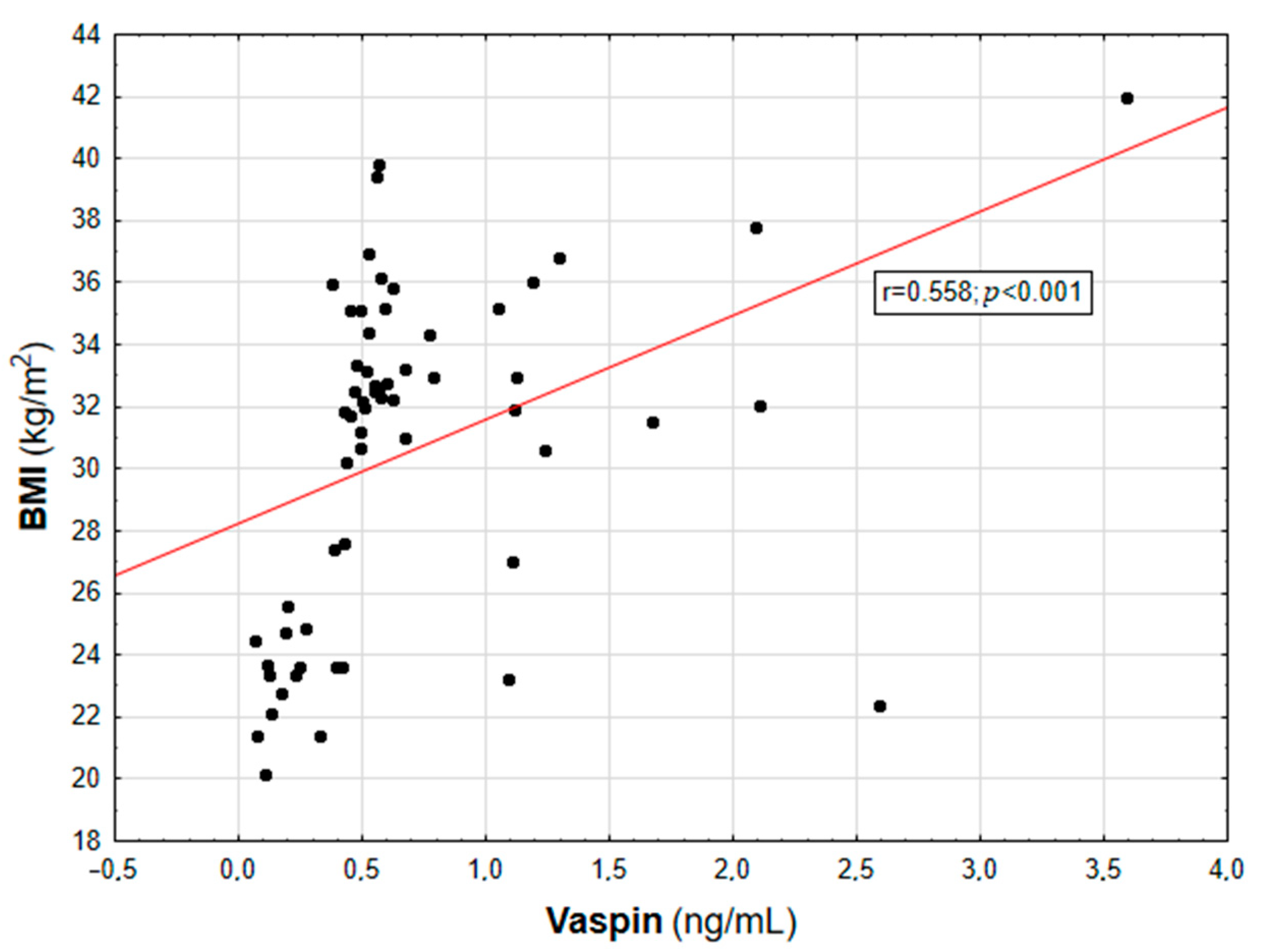
| BMI (kg/m2) | 0.558 | <0.001 | 0.393 | 0.004 | - | - |
| WHR | 0.447 | 0.003 | 0.398 | 0.004 | 0.247 | NS |
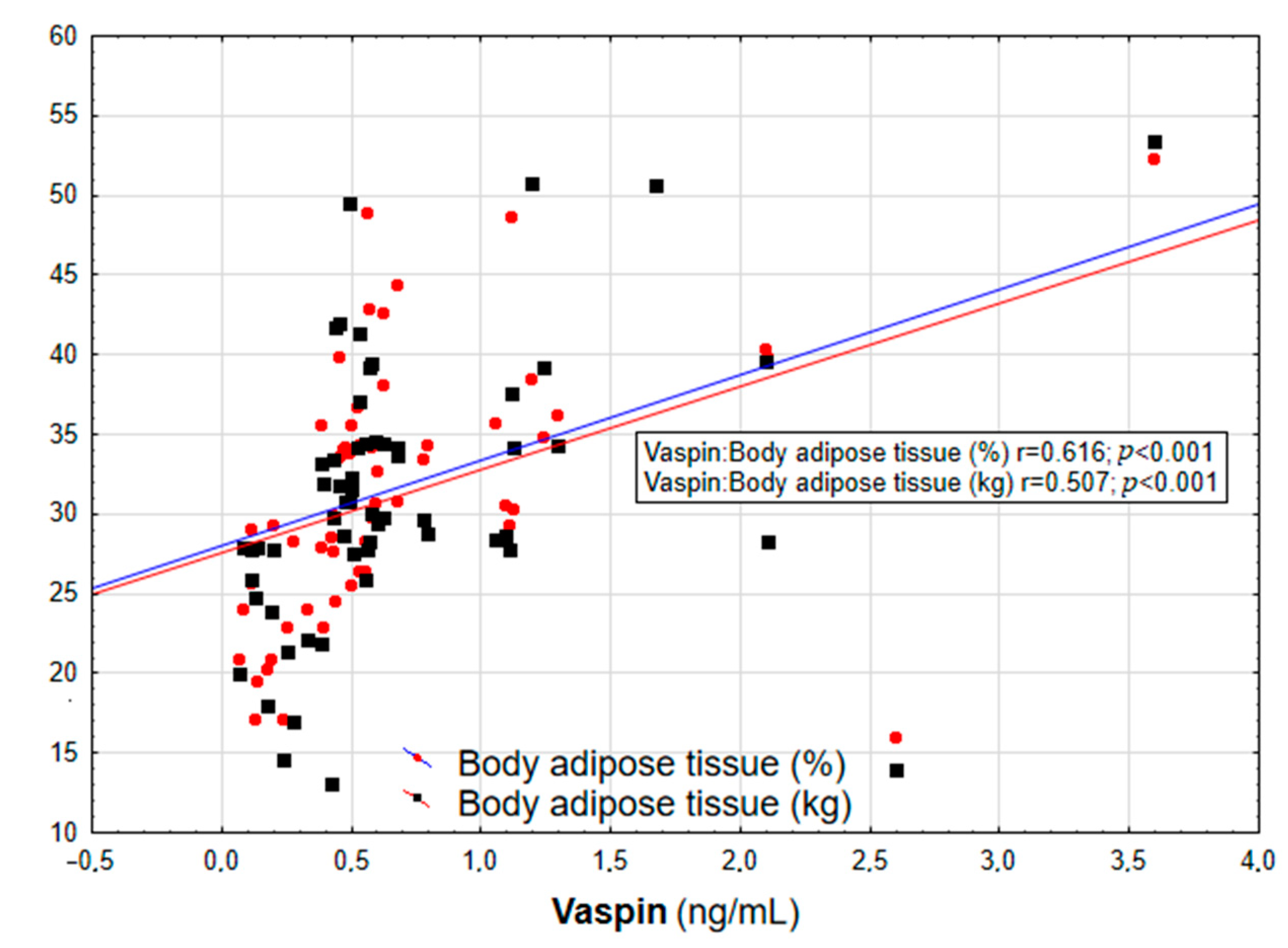
| Body adipose tissue (%) | 0.616 | <0.001 | 0.431 | <0.001 | 0.225 | NS |
| Body adipose tissue (kg) | 0.507 | <0.001 | 0.385 | 0.017 | 0.175 | NS |
| Total cholesterol (mmol/L) | 0.211 | NS | 0.105 | NS | 0.033 | NS |
| LDL cholesterol (mmol/L) | 0.184 | NS | 0.119 | NS | 0.065 | NS |
| HDL cholesterol (mmol/L) | −0.243 | NS | −0.134 | NS | 0.019 | NS |
| Triglycerides (mmol/L) | 0.337 | 0.013 | 0.089 | NS | −0.099 | NS |
| Glucose (mmol/L) | 0.141 | NS | 0.175 | NS | 0.084 | NS |
| Insulin (µU/mL) | 0.341 | 0.013 | 0.395 | 0.004 | 0.251 | NS |
| HOMA-IR | 0.382 | 0.022 | 0.374 | NS | 0.184 | NS |
| hs-CRP (mg/L) | 0.614 | <0.001 | 0.554 | <0.001 | 0.429 | <0.001 |
| IL-6 (pg/mL) | 0.457 | 0.003 | 0.229 | NS | −0.074 | NS |
| SBP (mmHg) | 0.001 | NS | 0.086 | NS | 0.037 | NS |
| DBP (mmHg) | 0.013 | NS | 0.086 | NS | 0.031 | NS |
| Variables (Unit) | Correlation Coefficient (Rho) | p-Value * |
|---|---|---|
| Body adipose tissue (%) | 0.382 | 0.030 |
| hs-CRP (mg/L) | 0.428 | 0.002 |
| OR | 95% CI | p | |
|---|---|---|---|
| raw | 8.5 | 1.18–61.35 | 0.0338 |
| adjusted * | 8.33 | 1.15–60.21 | 0.0338 |
Disclaimer/Publisher’s Note: The statements, opinions and data contained in all publications are solely those of the individual author(s) and contributor(s) and not of MDPI and/or the editor(s). MDPI and/or the editor(s) disclaim responsibility for any injury to people or property resulting from any ideas, methods, instructions or products referred to in the content. |
© 2023 by the authors. Licensee MDPI, Basel, Switzerland. This article is an open access article distributed under the terms and conditions of the Creative Commons Attribution (CC BY) license (https://creativecommons.org/licenses/by/4.0/).
Share and Cite
Pilarski, Ł.; Pelczyńska, M.; Koperska, A.; Seraszek-Jaros, A.; Szulińska, M.; Bogdański, P. Association of Serum Vaspin Concentration with Metabolic Disorders in Obese Individuals. Biomolecules 2023, 13, 508. https://doi.org/10.3390/biom13030508
Pilarski Ł, Pelczyńska M, Koperska A, Seraszek-Jaros A, Szulińska M, Bogdański P. Association of Serum Vaspin Concentration with Metabolic Disorders in Obese Individuals. Biomolecules. 2023; 13(3):508. https://doi.org/10.3390/biom13030508
Chicago/Turabian StylePilarski, Łukasz, Marta Pelczyńska, Anna Koperska, Agnieszka Seraszek-Jaros, Monika Szulińska, and Paweł Bogdański. 2023. "Association of Serum Vaspin Concentration with Metabolic Disorders in Obese Individuals" Biomolecules 13, no. 3: 508. https://doi.org/10.3390/biom13030508