Article

The Ribosome Is the Ultimate Receptor for Trypsin Modulating Oostatic Factor (TMOF)

by
Dov Borovsky
1,*,
Pierre Rougé
2 and
Robert G. Shatters, Jr.
3
1
Department of Biochemistry and Molecular Genetics, School of Medicine, University of Colorado Anschutz, Aurora, CO 80045, USA
2
UMR 152 Pharma-Dev, Faculté des Sciences Pharmaceutiques, Université Toulouse 3, CEDEX 09, F-31062 Toulouse, France
3
USDA ARS, U.S. Horticultural Research Laboratory, Subtropical Insects and Horticulture Research Unit, 2001 Rock Road, Fort Pierce, FL 34945, USA
*
Author to whom correspondence should be addressed.
Biomolecules 2022, 12(4), 577; https://doi.org/10.3390/biom12040577
Submission received: 16 March 2022 / Revised: 11 April 2022 / Accepted: 12 April 2022 / Published: 14 April 2022
(This article belongs to the Special Issue Insect Receptors: Biochemical, Physiological and Molecular Studies)

Abstract

:
Aedes aegypti Trypsin Modulating Oostatic Factor (AeaTMOF). a mosquito decapeptide that controls trypsin biosynthesis in female and larval mosquitoes. enters the gut epithelial cells of female mosquitoes using ABC-tmfA receptor/importer. To study the ultimate targeted receptor after AeaTMOF enters the cell, AeaTMOF was incubated in vitro with either Escherichia coli or Spodoptera frugiperda protein-expressing extracts containing 70S and 80S ribosomes, respectively. The effect of AeaTMOF on luciferase biosynthesis in vitro using 70S ribosomes was compared with that of oncocin112 (1–13), a ribosome-binding antibacterial peptide. The IC50 of 1 μM and 2 μM, respectively, for both peptides was determined. Incubation with a protein-expressing system and S. frugiperda 80S ribosomes determined an IC50 of 1.8 μM for Aedes aegypti larval late trypsin biosynthesis. Incubation of purified E. coli ribosome with increasing concentration of AeaTMOF shows that the binding of AeaTMOF to the bacterial ribosome exhibits a high affinity (KD = 23 ± 3.4 nM, Bmax = 0.553 ± 0.023 pmol/μg ribosome and Kassoc = 4.3 × 10⁷ M−1). Molecular modeling and docking experiments show that AeaTMOF binds bacterial and Drosophila ribosome (50S and 60S, respectively) at the entrance of the ribosome exit tunnel, blocking the tRNA entrance and preventing protein biosynthesis. Recombinant E. coli cells that express only ABC-tmfA importer are inhibited by AeaTMOF but not by oncocin112 (1–13). These results suggest that the ribosome is the ultimate targeted receptor of AeaTMOF.

Graphical Abstract

1. Introduction

Aedes aegypti Trypsin Modulating Oostatic Factor (AeaTMOF), a proline-rich peptide (YDPAPPPPPP), was originally isolated from the ovaries of female Ae. aegypti. AeaTMOF controls the biosynthesis of trypsin-like enzymes in the midgut epithelial cells of several species of mosquito including Aedes. aegypti, Culex quinquefasciatus, Culex. nigripalpus, and Anopheles albimanus [1]. AeaTMOF circulates in the hemolymph of female Ae. aegypti and Cx. quinquefasciatus after the blood meal and affects the translation of the late trypsin transcript in the gut epithelial cells [2,3]. AeaTMOF was also shown to inhibit the translation of the late trypsin transcript in Neobellieria [4] and in Heliothis virenscens. This was observed by expressing AeaTMOF on the coat protein of Tobacco Mosaic Virus and feeding it to the insects, where it stopped the translation of the trypsin transcript in the gut epithelial cells [5]. These earlier results indicated that AeaTMOF affects the translation of the trypsin transcript but does not affect the transcript abundance by inducing mRNA decay [6]. Therefore, it is possible to suggest that the effect is exerted on the ribosome where the translation of the trypsin message is blocked. Indeed, our earlier results show that AeaTMOF secreted by the mosquito ovaries binds to a AeaTMOF ABC gut receptor (ABCtmfA receptor/importer) that imports AeaTMOF into the gut epithelial cells [7]. AeaTMOF is a proline-rich peptide analogous to the antimicrobial proline-rich peptides (PR-AMP) that were first reported in honeybees [8] and subsequently identified in species including mammals, amphibians, crustaceans, and mollusks [9,10,11,12]. Initial efforts to find the bacterial target for PR-AMP identified a heat shock protein DnaK as the main candidate for inhibition; however, subsequent studies showed that dnaK deleted mutants were as efficiently inhibited by oncocin (Onc72 and Onc112) and apidaecin (Api88 and Api137) derivatives as the parental strain [13]. Further studies by these authors indicate that these PR-AMP target the bacterial cell ribosome. Several studies using X-ray crystallography showed that the first 13 amino acids of Onc112 bind within the ribosomal exit tunnel extending into the peptidyl transferase center overlapping with the binding site of the aminoacyl-tRNA, and thus preventing entry into the elongation phase. [14,15]. To find out the mechanism by which AeaTMOF stops the translation of trypsin in the female Ae. aegypti midgut epithelial cells, we used molecular modeling of Escherichia coli and Drosophila melanogaster ribosome with AeaTMOF to find out whether AeaTMOF-like oncocin binds the peptide exit tunnel as was shown by X-ray crystallography for oncocin binding to the 70S ribosomes of Thermus thermophilus [14,15]. We combined molecular modeling with binding studies of AeaTMOF to purified E. coli ribosomes in combination with in vitro studies using Spodoptera frugiperda and E. coli cell free translation of Ae. aegypti larval late trypsin and luciferase to find out how AeaTMOF stops trypsin and luciferase translation in vitro. We compared these results by using a short oncocin112 peptide with the first 13 amino acids, naming it oncocin112 (1–13) (VDKPPYLPRPRPP). These amino acids were shown by X-ray diffraction to occupy the peptide exit tunnel of the T. thermophilus ribosome [14,15]. The E. coli-purified ribosome was incubated with AeaTMOF-FITC, and the KD of AeaTMOF binding to the ribosome was determined showing that the binding affinity of AeaTMOF to the ribosome is high. This report shows for the first time the mechanism that AeaTMOF employs to modulate trypsin biosynthesis in the mosquito epithelial cells.

2. Materials and Methods

2.1. Bacterial Strains, and Chemicals

E. coli CGSC strain7636: F Δ(araD-araB)567,ΔlacZ4787(::rrnB-3),l,rph-1, Δ(rhaD-rhaB)568, hsdR514 and E. coli CGSC strain 8547: F Δ(araD-araB)567, ΔlacZ4787(::rrnB-3), ΔsbmA742::kan, λ, rph-1,Δ(rhaD-rhaB)568, hsdR514 (http://cgsc.biology.yale.edu/StrainRpt.php, accessed on 15 March 2022) were grown in Luria-Bertani (LB) at 37 °C under aerobic conditions with the addition, when required, of the appropriate antibiotics at the following concentrations—100 μg/mL for ampicillin and 50 μg/mL for kanamycin—which were purchased from Sigma-Aldrich (St. Louis, MO, USA) and diluted in LB medium.
E. coli CGSC strain 85447 lacking the SbmA importer expressing the ABCtmfA receptor/importer was prepared using plasmid pTAC-MAT-2 ABCtmfA importer/receptor as described earlier [7]. [3,4,5 3H]Leucine 250 μCi, Spc Act 100–150 Ci/mmol was purchased from Perkin Elmer (Waltham, MA, USA). AeaTMOF-FITC (H-YDPAPPPPPPK(FITC)K-OH) was synthesized at the University of Colorado Anschutz School of Medicine protein core. AeaTMOF-FITC was purified by HPLC showing a single peak, and mass spectrometry analysis of the peak identified the 3 expected ions at 455.7, 607.3, and 910.3, showing an expected Mr 1019.56 as was described earlier [7]. Synthetic AeaTMOF (H-YDPAPPPPPP-OH) and a short Oncocin112(1–13) (H-VDKPPYLPRPRPP-OH), a 13 amino acids oncocin112 peptide, 6 amino acids shorter than the original oncocin112 of 19 amino acids, were synthesized and purified by HPLC [16]. After HPLC purifications of AeaTMOF, oncocin112 (1–13), and AeaTMOF-FITC, the TFA ions were exchanged with phosphate ions.

2.2. Cloning of Late Larval Ae. aegypti Trypsin

A synthetic Ae. aegypti late larval trypsin gene (accession number AY198134.1) (857 bp) with SgfI and PmeI cloning sites at the 5′ and 3′ ends was synthesized by GeneScript (Piscataway, NJ, USA). The gene was cut with SgfI and PmeI restriction enzymes (Promega Madison, WI, USA) following the manufacturer’s guidelines and cloned into a plasmid pFA25A ICE T7 flexi that was opened with SgfI and PmeI following the manufacturer’s guidelines (Promega). The recombinant plasmid with the late larval trypsin gene was sequenced, confirming that a full-length Ae. aegypti larval late trypsin gene was cloned unidirectional at the 5′ to 3′ ends, replacing the Barnase gene on pFA25A ICE T7 flexi (Promega).

2.3. Inhibition of Luciferase Translation in S30 Bacterial Extract

To test the effect of AeaTMOF and Oncocin112 (1–13) on the translation of Luciferase by the bacterial 70 S ribosome, an E. coli S30 lysate extract system for circular DNA (Promega) containing a plasmid pBestluc DNA vector (4486 bp) (1 mg/mL) 0.3 μL, amino acid mixture without methionine (1 mM) 1.25 μL, amino acid mixture without Leucine (1 mM) 1.25 μL, S30 premix (including ribosomes) without amino acids 5 μL, S30 extract circular 3.5 μL, RNasin (8 U, 112 ng) 0.2 μL, different concentrations of AeaTMOF or Oncocin112 (1–13) (0.001 μM to 1000 μM), and nuclease-free water to a volume of 12.5 μL were incubated for 2 h at 37 °C. Control reactions did not contain pBestluc DNA. After incubation, 87.5 μL of GLO lysis buffer (Promega) containing beetle luciferin was added, mixed, and aliquots (5 μL) were removed, and their luminescence read in a GLOMAX multidetector system. Reactions were run in triplicates, and luminescence was corrected for control reactions that did not contain plasmid pBestluc DNA. Maximum luminescence (100%) was observed at 0.001 μM AeaTMOF or oncocin 112 (1–13) and was similar to incubations without the presence of the peptides. The decrease in luminescence (% ± SEM) in the presence of increasing log AeaTMOF or Oncocin112 (1–13) concentrations was then plotted using GrapPad Prism 3.

2.4. Inhibition of Larval Ae. aegypti Late Trypsin Translation in Insect Cell Extract

To test the effect of AeaTMOF on the translation of larval Ae. aegypti late trypsin, the TnT T7 insect cell extract protein expression system from S. frugiperda Sf21 cell line (Promega) was incubated with different concentrations of AeaTMOF in a reaction mixture containing TNT T7 ICE master mix of 10 μL, pFA25A ICE T7 flexi-late larval Trypsin (0.45 μg/mL) (see Section 2.2) 0.5 μL, [3,4,5 3H]Leucine 0.5 μL, AeaTMOF (0.01 to 1000 μM), and nuclease-free water to a total volume of 12.5 μL. The incubation mixtures were incubated at 30 °C for 4 h. At the end of the incubation period, aliquots (10 μL) were removed and spotted on filter paper squares (1 × 1 cm) and washed in cold 10% TCA for 10 min, and twice in cold 5% TCA for 5 min each followed by 2 min with ethanol (100%). The paper squares were dried at 70 °C in an oven and tritium labeled trypsin counted in a liquid scintillation counter PerkinElmer Tri-Carb 2910 TR. Controls were run in the presence of empty plasmid pFA25A ICE T7 flexi that was not cloned with Ae. aegypti larval late trypsin and subtracted from the experimental results. Incubations were run in triplicates, and results are expressed as means ± SEM. The decrease in [3H]trypsin biosynthesis was then plotted against the increase in AeaTMOF concentrations using GraphPad Prism 3.

2.5. Western Blotting and Mass Spectra Analyses

2.5.1. Western Blotting

To prove that the [3H]-labeled larval late trypsin synthesized by the TnT T7 insect cell lysate is indeed trypsin, the TnT T7 insect cell lysate was incubated at 30 °C for 4 h with Fluorotech Green tRNAlys (0.4 μL) in a reaction mixture (12.5 μL) containing TnT T7 ICE master mix of 10 μL, pFA25A ICE T7 flexi-larval late Trypsin (0.64 μg/mL) of 0.5 μL, and nuclease-free water of 1.6 μL. The incubation was repeated 3 times. An incubation without pFA25A ICE T7 flexi-Trypsin was used as a control. After incubation, aliquots (5 μL) were removed from each incubation mixture and mixed with SDS sample buffer (4 μL) containing Tris-HCl, 2% SDS, 5% ME, and 20% glycerol with tracking dye. The incubation mixtures containing SDS-ME and glycerol were heated at 70 °C for 5 min and were loaded on a 10% Bis-Tris (1.0 mm) mini gel (ThermoFisher, Waltham, MA, USA), and electrophoresis was run at 110 V for 90 min [17]. A separate lane was run with broad molecular weight-stained standards (10 μL) (ThermoFisher). After SDS-PAGE, the 10% gel was removed, and proteins were transferred to 100% methanol wet PVDF membrane in Tris/CAPS buffer using Western blotting and a semidry transfer apparatus (BioRad, Hercules, CA, USA) at 15 V for 3 min following the manufacturer’s guidelines. The PVDF membrane was then scanned to detect fluorescence-emitting proteins using Sapphire Biomolecular Imager (Azure Biosystems, Dublin, CA, USA) at a wavelength of 468 nm.

2.5.2. Mass Spectra Analysis

Four tubes containing four reactions mixtures (12.5 μL), TnT T7 ICE master mix (10 μL) and nuclease-free water (1.6 μL) were used. Tube 1 contained in addition BSA (40 μg; 0.5 μL), tube 2 contained only the master mix, tube 3 contained an empty plasmid pFA25A ICE T7 flexi (0.64 μg/mL; 0.5 μL), and tube 4 contained plasmid pFA25A ICE T7 flexi-late larval trypsin (0.64 μg/mL; 0.5 μL). The tubes were incubated at 30 °C for 4 h. After incubation, aliquots (5 μL) were removed from each incubation, put into sample buffer (4 μL) where the tubes were vortexed, heated at 70 °C for 5 min, and electrophoresed using SDS_PAGE as described above [17]. The gel was stained with Coomassie Brilliant Blue, and a protein band that ran at 28 kDa was cut from the destained gel, digested with trypsin, and analyzed by MS/MS [18].

2.6. Purification of E. coli Ribosomes

The purification of E. coli ribosomes is a modification of earlier published methods [19,20]. Briefly, E. coli (BL21) cells were grown in LB medium for 24 h to OD600 of 1.1, and the cells were centrifuged at 5000 rpm at 6 °C for 5 min. The supernatant was discarded and the pellet was washed with 40 mM HEPES-KOH pH7.4, 100 mM NaCl, and 2 mM Mg(OAc)2 buffer. The suspension was centrifuged at 6000 rpm for 5 min, the supernatant discarded, and the pellet was frozen at −80 °C and stored overnight. The frozen pellet was taken up in 10 mL of sonication buffer of 20 mM HEPES-KOH pH 7.5, 100 NH4OAc, 6 mM βME, 0.5 mM Na-EDTA, and 0.5% Tween 20 containing 100 units of RNase inhibitor, and the solution was sonicated at 40% power for 10 s, 50% power for 10 s, and 60% power for 20 s. During the sonication, the tube was immersed in an ice bath. The lysed cell solution (25 mL) in sonication buffer was centrifuged at 30,000 g in a Beckman ultracentrifuge using a 70Ti centrifuge rotor for 30 min at 4 °C. After centrifugation, the supernatant was collected, and the pellet discarded. High salt sucrose cushion (20 mM HEPES-KOH pH 7.5, 50 mM Mg(OAc)2, 100 mM NH4Cl, 6 mM βME, 0.5 mM Na-EDTA, and 1.1 M sucrose) solutions (25 mL each) containing 100 units of RNase inhibitor were added to the two centrifuge tubes. Each tube was carefully layered over the sucrose cushion, using a syringe with a needle, with the ribosomal solution (12.5 mL) obtained after the 30,000 g spin in 70Ti rotor. The tubes were centrifuged at 4 °C and 100,000 g (35,000 rpm) for 20 h. The clear pellets were each carefully layered with 0.5 mL of tight association buffer (20 mM HEPES-KOH pH 7.6, 6 mM Mg(OAc)2, 30 mM NH4OAc and 6 mM βME) to wash the top of the pellets, and the wash solutions were discarded. The pellets were then dissolved in tight association buffer (4.5 mL) and the tubes were shaken gently in bucket full of ice for 4 h. The solubilized pellets were centrifuged at 16,000 g in Eppendorf tubes for 10 min at 4 °C and aliquots (2 μL) were removed and diluted 1:100 in tight association buffer. The amount of the protein in the ribosome preparation and the ribosomal RNA was determined (2.28 mg/mL protein and 3.932 mg/mL ribosomal RNA). The ribosome preparation was then diluted with 10% glycerol and stored at −80 °C [19,20]. Aliquots (5 μL) of the purified ribosomes were then adsorbed unto carbon-coated 300 mesh of Cu/Rh grids and stained with 2% uranyl acetate. The grids were then observed by electron microscopy with 120 kV Talos L120C TEM at the CryoEM of the cancer center at the University of Colorado Anschutz Medical School.

2.7. Binding of AeaTMOF-FITC to E. coli Ribosome Assay and Kinetics

Purified E. coli ribosomes (1 μL, 9.4 μg) in tight association buffer (20 mM HEPES-KOH pH 7.6, 6 mM Mg(OAc)2, 30 mM NH4OAc, and 6 mM βME) were incubated in Eppendorf tubes containing 100 μL of tight association buffer and 5 μL of AeaTMOF-FITC (0–0.2 μM) (Specific activity 8333 Fluorescence Units/pmol) for 60 min at 30 °C with gentle shaking. After incubation, 12 μL of TCA (100%) was added, and the tubes were vortexed and incubated on ice for 10 min. The tubes were then centrifuged at 20,000 g for 5 min at 4 °C, and the supernatants were discarded. To the pellets, TCA (5%) in 100 μL of tight association buffer was added, the tubes were vortexed and centrifuged, and the supernatants discarded as described above. The washing by centrifugation with 5% TCA was repeated 3 times, and after the final centrifugation, the pellets were dissolved in 100 μL of PBS containing 0.4N NaOH, and the fluorescence of the dissolved pellets was read in a GloMax multidetector system using a blue filter (Excitation at 490 nm and Emission at 510–570 nm). All data were corrected for nonspecific fluorescence when incubations were run without AeaTMOF-FITC or with AeaTMOF-FITC and without ribosomes. The amount of AeaTMOF-FITC that bound the ribosome was determined from a linear calibration curve after plotting different concentrations of AeaTMOF-FITC (pmol) against fluorescence units. The results are expressed as AeaTMOF-FITC (pmol/μg ribosome). The kinetics of AeaTMOF-FITC binding to the purified bacterial ribosomes was followed at different concentrations of AeaTMOF-FITC using six independent experiments per concentration, the data was then fitted by nonlinear regression using GraphPad Prism 3 and the KD, Bmax and the affinity constant Kassoc determined. The binding experiments were repeated two independent times.

2.8. Molecular Modeling and Docking

To dock AeaTMOF to the Thermos thermophilus 70S ribosome, we first docked Oncocin112 to the T. thermophilus ribosome using the coordinates in PBD (ID: 4Z8C) [15] (results not shown). The proline-rich AeaTMOF structure [21] was manually positioned in the adenine-binding site of the ribosome and superimposed to oncocin112, refined, and energy minimized using the YASARA structure [22]. To dock AeaTMOF to the Drosophila melanogaster and E. coli ribosomes, the peptide was built up with Chimera and minimized with 1000 cycles of steepest descent and 100 additional cycles of conjugate gradient, using the Amber force field [23]. The atomic coordinates of the Drosophila melanogaster 80S ribosome were taken from the PDB (4V6W) [24]. The atomic coordinates of the E. coli 70S ribosome (PDB code 6O9J) associated with the proline-rich antimicrobial peptide oncocin112 [15] were used for the docking of AeaTMOF to the E. coli and Drosophila ribosomes. Docking trials were performed with YASARA [22]. Molecular cartoons were drawn with Chimera [23] and ChimeraX [25].

2.9. Effect of AeaTMOF and Oncocin112 (1–13) in Vivo on E. coli Growth

We aimed to find out if AeaTMOF can also stop bacterial growth as, was shown for the proline-rich Oncocin112 that binds to the bacterial ribosome, stopping protein translation, bacterial growth, and causing bacterial death [14]. E. coli CGSC strain7636 (2.7 × 10⁴ cells) SbmA+, expressing the proline-rich peptides importer SbmA, were incubated in 96-well plate containing 100 μL/well of LB medium, 0.4 mM of IPTG, 0.1 mM of ampicillin, and different concentrations of AeaTMOF and Oncocin112 (1–13) (3 mM and 10 mM) in different wells. The 96-well plate was incubated at 37 °C for 18 h and bacterial growth was followed at 630 nm using Biotech ElX808 absorbance microplate reader. Control wells contained bacterial cells growing without AeaTMOF or oncocin112 (1–13). In a second experiment, E. coli mutant CGSC strain 8547 (2.7 × 10⁴ cells) SbmA- that do not express the Oncocin112 importer SbmA but do express the ABC tmfA importer [7] were incubated in 100 μL of LB medium with AeaTMOF and oncocin112 (1–13) (3 mM and 10 mM) as described above for 18 h following bacterial growth. After the incubations in the presence of Oncocin112 (1–13) or AeaTMOF of 10 mM and 3 mM, aliquots (0.5 μL) were removed from each well and spread on LB agar plates containing 0.1 mM of ampicillin, and the plates were incubated overnight at 37 °C and viable colonies were counted. Incubations were done in triplicates and the experiments were repeated twice. Results are expressed as means of three determinations ± SEM.

2.10. Statistical Analysis

Statistical analyses were determined using GraphPad Prism using linear and nonlinear regression. Results were considered significant when p < 0.05. Kinetic parameters KD and Bmax were determined from a nonlinear regression (R2 > 0.84) using GraphPad Prism. Each experimental point is a mean of 3 or 6 determinations ± SEM.

3. Results

3.1. Inhibition of Luciferase Biosynthesis in E. coli S30 Extract System

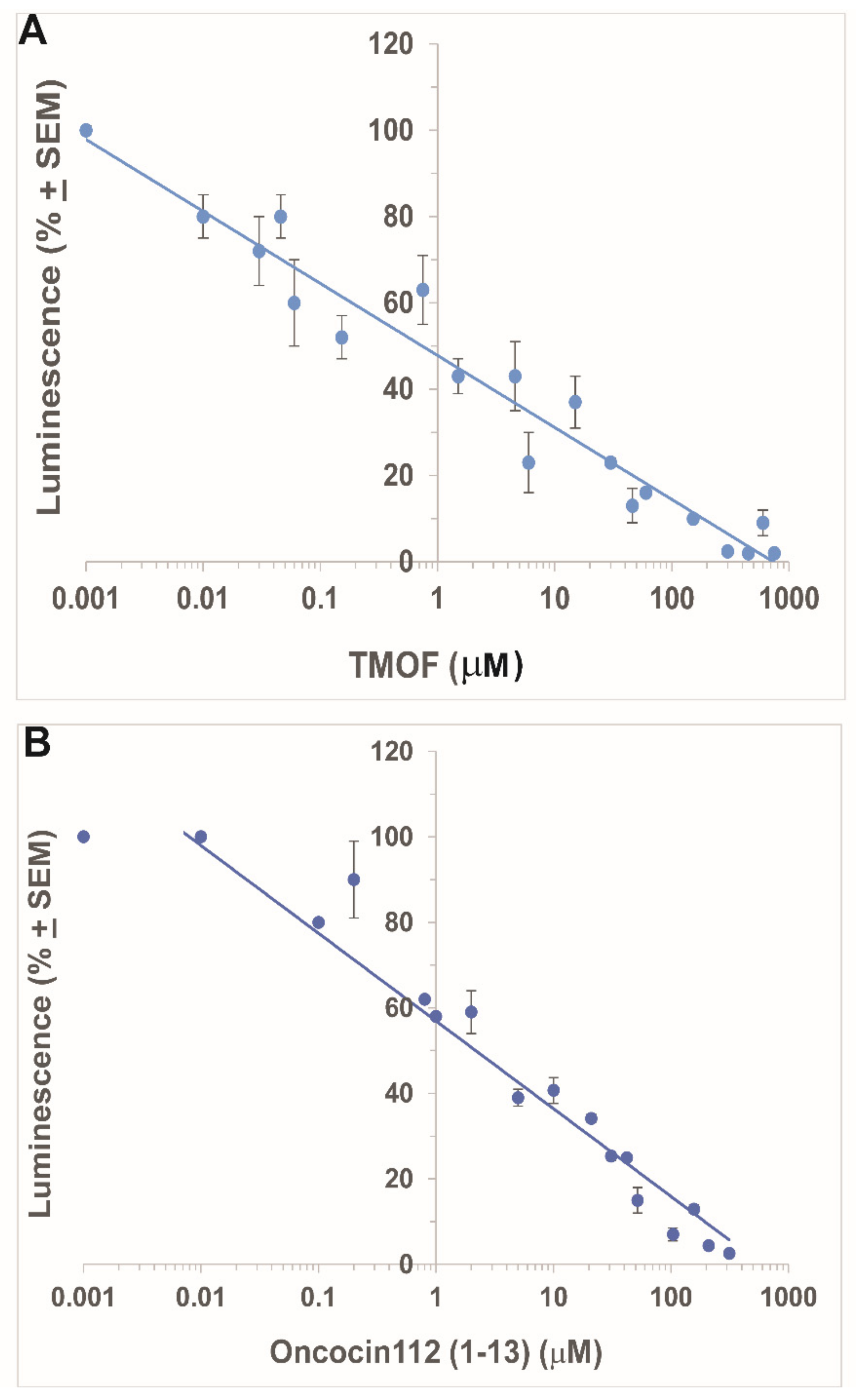
3.1.1. Inhibition by AeaTMOF

Incubations of E. coli S30 extract synthesizing Beetle luciferase in the presence of an increasing concentration of AeaTMOF (0.001 to 750 μM) stopped the biosynthesis of luciferase, diminishing its activity on Beetle luciferin as measured in luminescence units. The activity of the in vitro synthesized luciferase on Beetle luciferin at very low concentration of AeaTMOF (0.001 μM) was similar to luciferase activity without AeaTMOF, and this luminescence was used to calculate the decrease in luminescence (%) after incubation with increasing concentrations of AeaTMOF (Figure 1A). Higher concentrations of AeaTMOF (higher than 0.001 μM) caused a linear decrease in the synthesis of luciferase and a decline in luminescence (%) reaching IC50 (inhibition concentration of 50%) at 1 μM (Figure 1A). Complete inhibition of luciferase biosynthesis was reached at AeaTMOF concentrations of 250–750 μM. AeaTMOF is not an inhibitor of luciferase. Incubations of the E. coli S30 extract system (Promega) in the presence of plasmid pBESTluc (Promega) for 4 h without AeaTMOF and then adding AeaTMOF (1 to 100 μM) did not affect the activity of luciferase that was synthesized in vitro as compared with controls without the addition of AeaTMOF (results not shown).

3.1.2. Inhibition by Oncocin112 (1–13)

Incubations of E. coli S30 extract synthesizing Beetle luciferase in the presence of increasing concentrations of oncocin112 (1–13) (0.001 to 750 μM) also stopped the biosynthesis of luciferase and diminished its activity on Beetle luciferin measured in luminescence units as was shown in Section 3.1.1. The activity of the in vitro synthesized luciferase on Beetle luciferin at very low concentration of Oncocin112 (1–13) (0.001 and 0.01 μM) was the same and similar to luciferase activity without Oncocin112 (1–13), and this luminescence was used to calculate the decrease in luminescence (%) after incubation with increasing concentrations of Oncocin112 (1–13) (Figure 1B). Similar to the results obtained with AeaTMOF, Oncocin112 (1–13) also inhibits Beetle luciferase biosynthesis and not its activity (results not shown). The linear decline in the luminescence after incubation with Oncocin112 (1–13) reached an IC50 at 2 μM (Figure 1B), a twofold increase as compared with the AeaTMOF decapeptide. Oncocin112 (1–13) at 0.01 μM and AeaTMOF at 0.001 μM do not inhibit the biosynthesis of luciferase (Figure 1A,B).

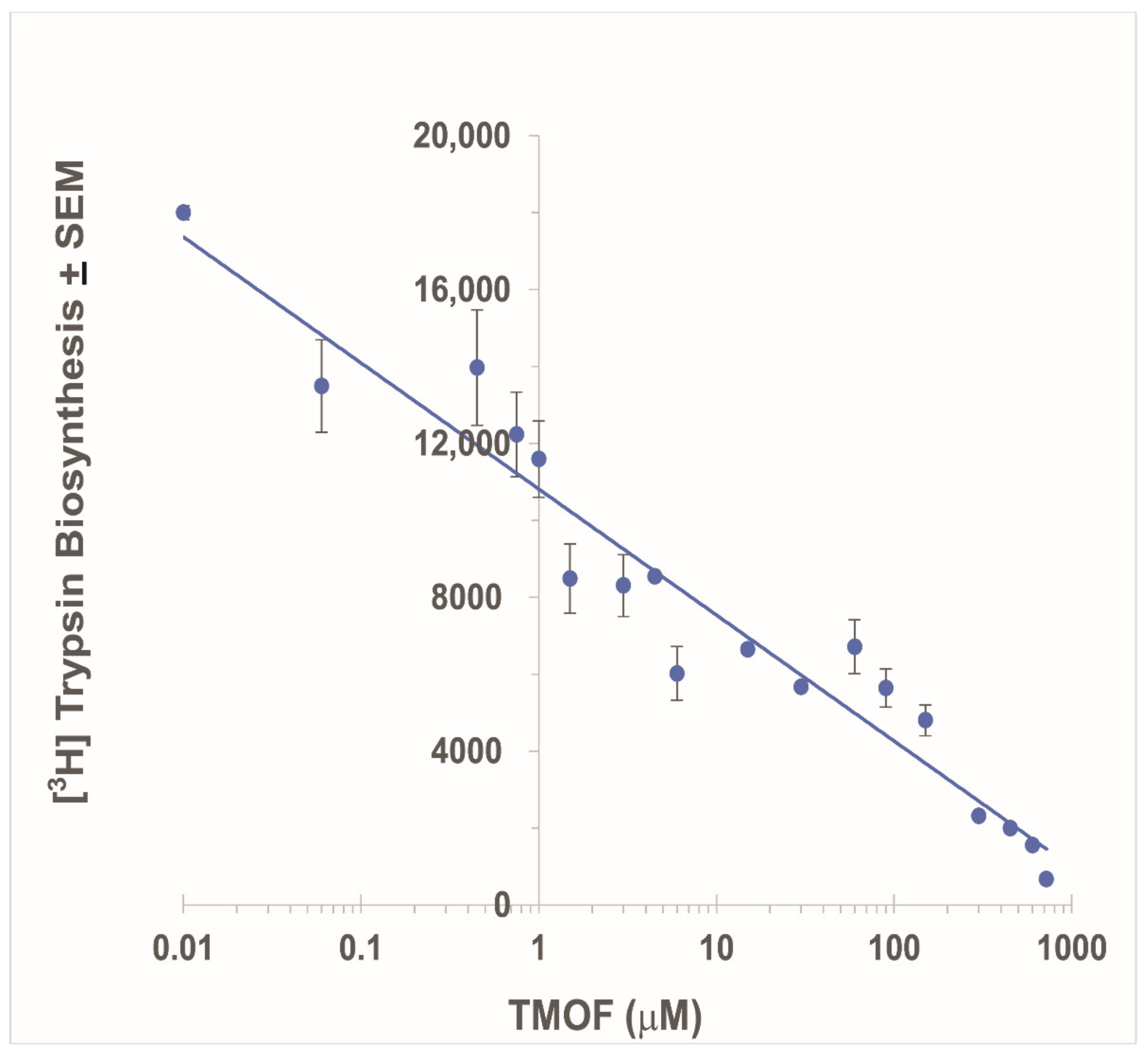
3.2. Inhibition of Ae. aegypti Late Larval Trypsin Biosynthesis with AeaTMOF

To find out how AeaTMOF blocks trypsin biosynthesis in mosquitoes and other insects [4,5], increasing concentrations of AeaTMOF (0.01 to 750 μM) were incubated in the TnT T7 insect cell extract protein expression system from S. frugiperda in the presence of recombinant plasmid pFA25A ICE T7 flexi carrying Ae. aegypti late larval trypsin in the presence of [3,4,5 3H]Leucine and [3H]trypsin biosynthesis. A control without AeaTMOF was also run showing that [3H]trypsin synthesized without AeaTMOF or in the presence of AeaTMOF (0.01 μM) was the same (results not shown). Increasing concentrations of AeaTMOF caused a rapid decrease in the synthesis of [3H]trypsin. A linear regression of the decrease in the synthesis was plotted against AeaTMOF (log μM) indicating that the IC50 of AeaTMOF is 1.8 μM (Figure 2). To prove that the [3 H]protein that was synthesized by the S. frugiperda 80S ribosomal lysate is trypsin, the protein expression system used above was first incubated with Fluorotech Green tRNAlys, and the synthesized fluorescent proteins were analyzed using Western blotting (Figure S1). A distinct fluorescence band (28.5 kDa) closer to the Ae. aegypti late larval trypsin 27.5 kDa, was detected in lanes b, c, and d after the insect protein expression system was incubated in the presence of Fluorotech Green tRNAlys and was absent when the tRNA was not added to the incubation mixture (Figure S1 lane a). Therefore, the fluorescence-emitting protein bands in lanes b, c, and d are probably the Ae. aegypti late larval trypsin (Figure S1). Incubating the insect cell extract protein expression system with (a) BSA, (b) only insect cell extract, or (c) pFA25A ICE T7 flexi (empty plasmid) and separating the proteins by SDS-PAGE (Figure S2A Supplementary Material, lanes 1, 2, and 3, respectively) produced faint bands at 28.5 kDa, but not as intense as the distinct protein band at 28.5 kDa that was observed when the insect cell extract protein expression system was incubated with pFA25A ICE T7 flexi carrying Ae. aegypti late larval trypsin (Figure S2, lane 4). MS/MS analysis of the 28.5 kDa band identified seven unique peptides with masses of 2633.31, 2060.1, 2373.13, 2072.94, 1187.61, 1209.57, and 775.358, and a 48% coverage of the protein (94 amino acids out of 250 amino acids) (Figure S2B). The protein was identified by mass spectrometry as Ae. aegypti larval late trypsin (accession number AAO43403.1). The very faint and diffused stained bands in lanes 2 and 3 that ran at 28.5 kDa (Figure S2A) were also analyzed by MS/MS but are not Ae. aegypti larval late trypsin.

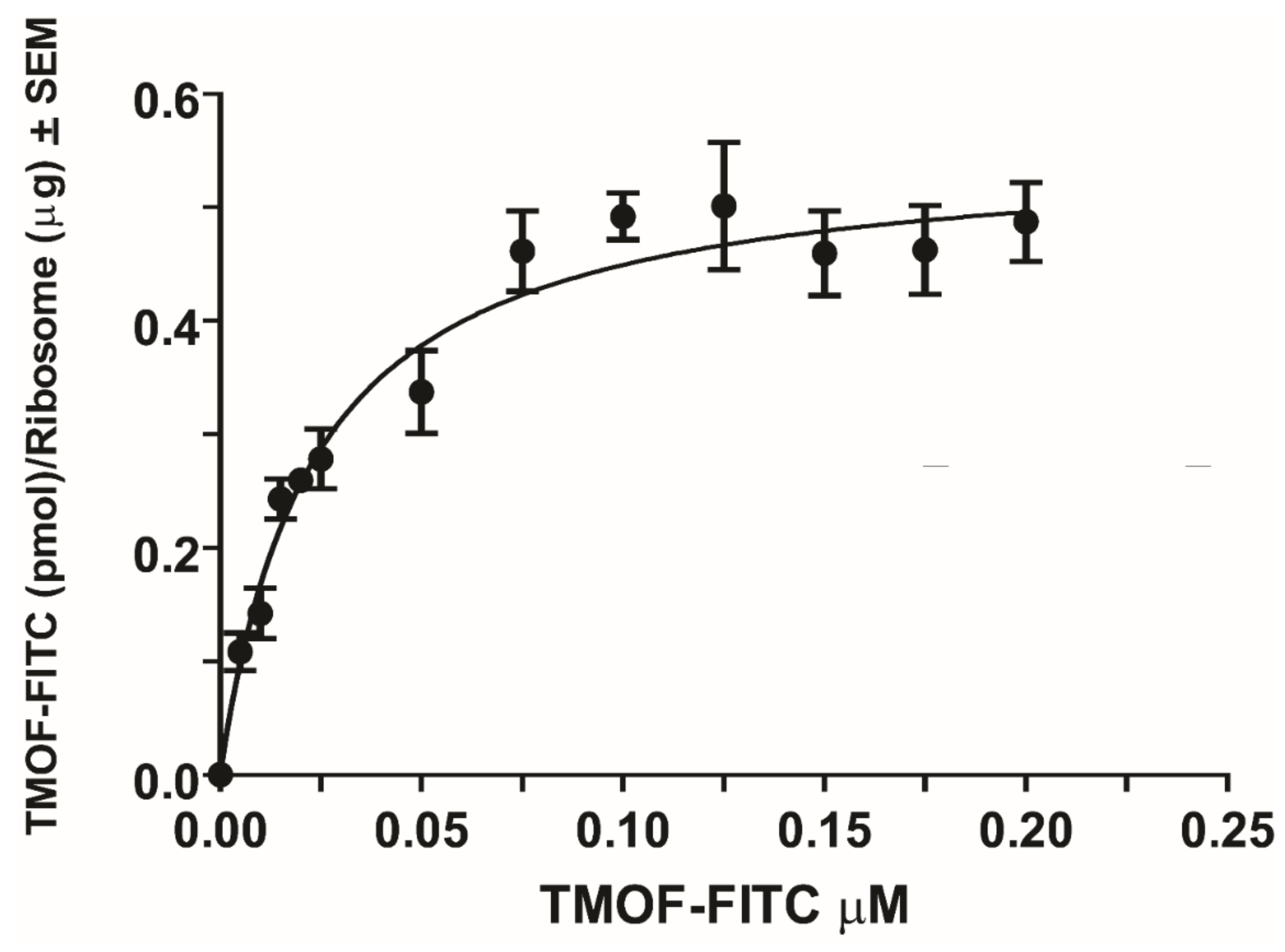
3.3. Binding Kinetics of AeaTMOF to E. coli Ribosomes

To find out whether the ribosome is the ultimate target of AeaTMOF as was shown for oncocin112 [14,15], a purified ribosome preparation (2.28 mg/mL protein and 3.932 mg/mL ribosomal RNA) that was negatively stained showing no damage or dissociation after the purification was used (Figure 3). AeaTMOF-FITC bound the purified E. coli ribosome preparation showing concentration dependence, specific, and high-affinity binding (KD = 23 ± 3.4 nM ± SEM and Bmax = 0.553 ± 0.023 pmol/μg ribosome ± SEM) (Figure 4, n = 6). Using the specific binding (KD), an affinity constant was calculated (Kassoc = 4.3 × 10⁷ M−1). Nonspecific binding was subtracted from each point (see Section 2.7).

3.4. Three-Dimensional Modeling of AeaTMOF Binding to Bacterial Ribosome

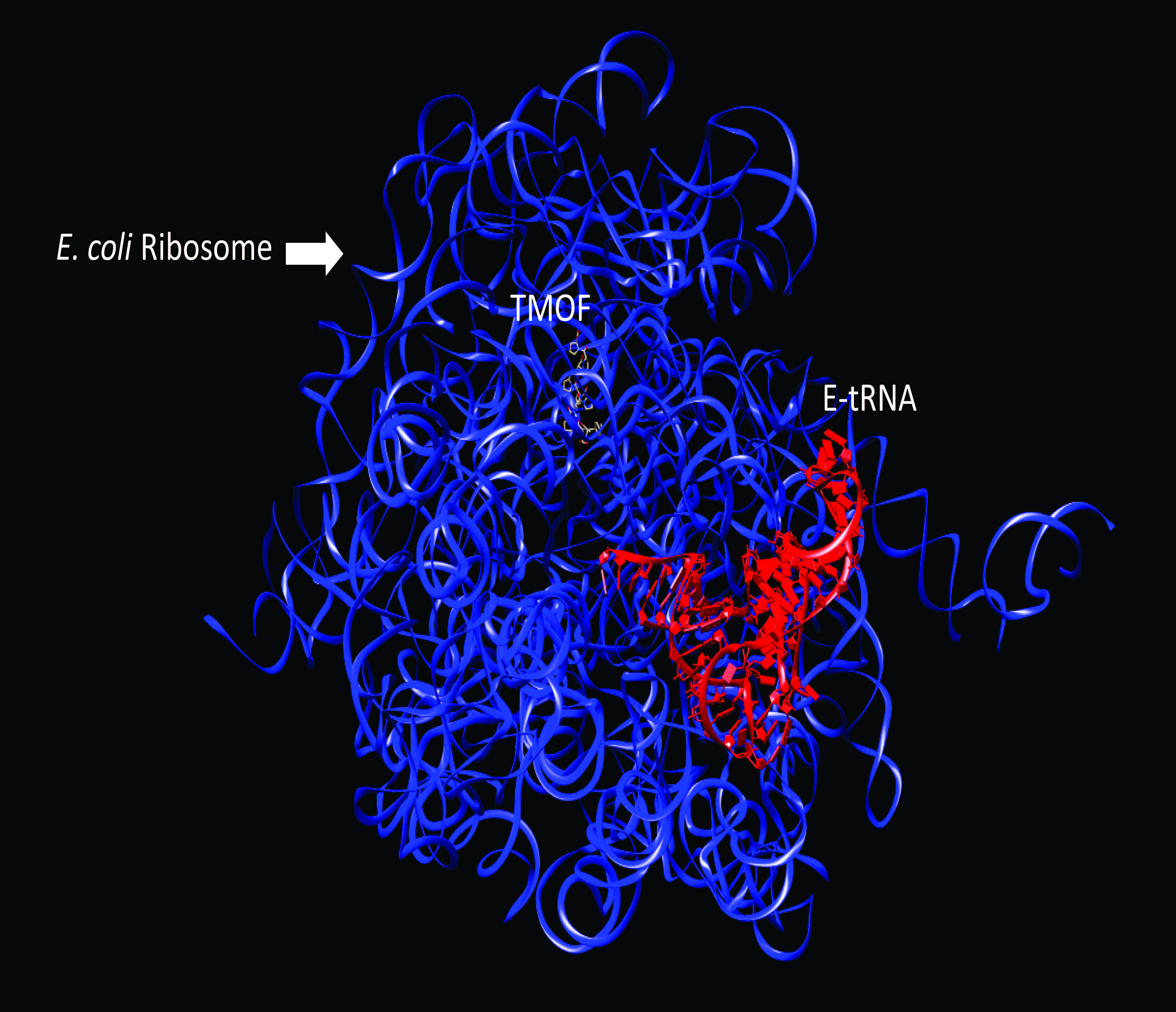
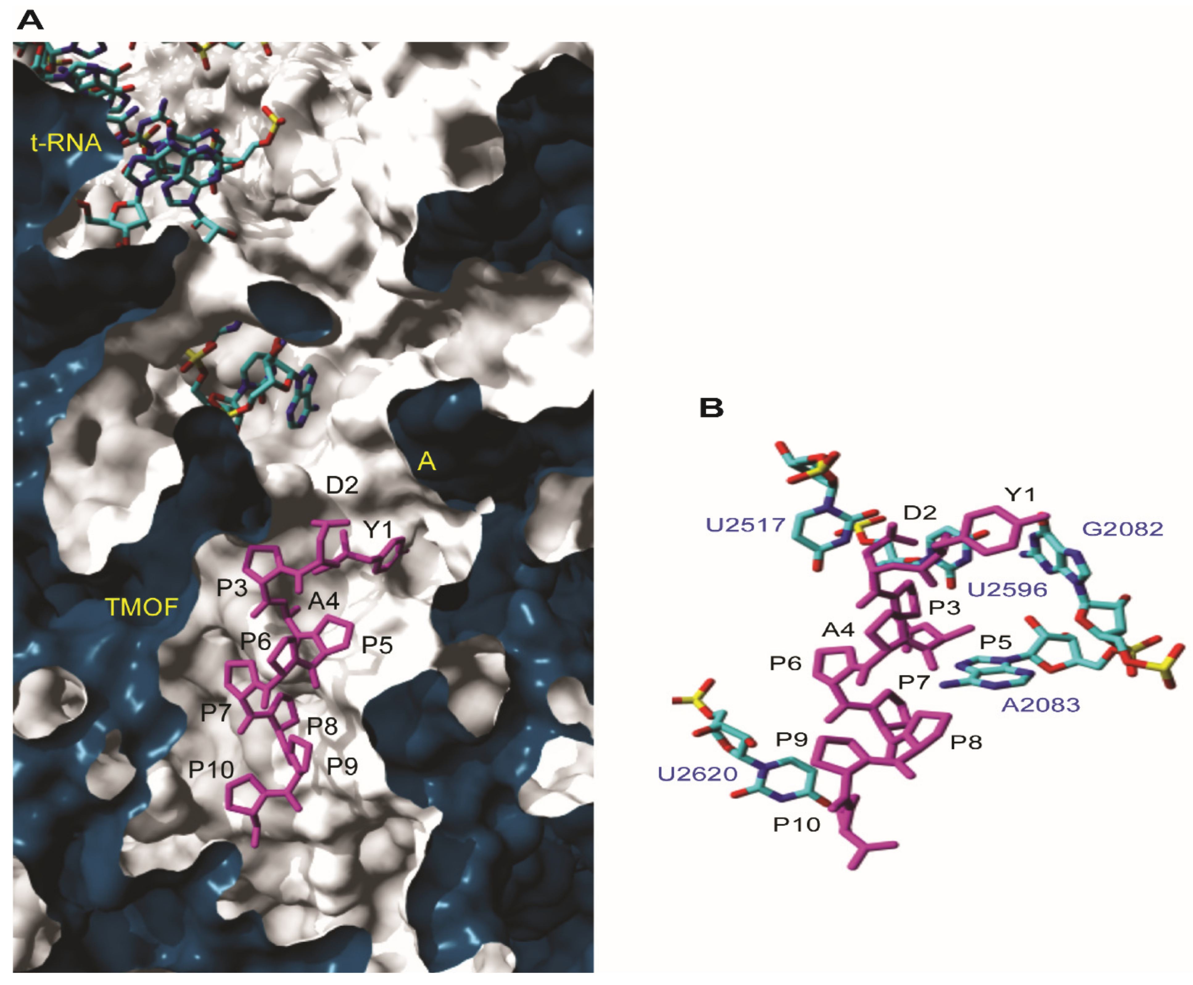
3.4.1. Docking to T. thermophilus Ribosome

For the initial molecular docking of AeaTMOF to the 70S ribosome of T. thermophilus, we used the X-ray crystallography of Onc112 binding to T. thermophilus 70S ribosome as a reference [14,15]. Docking of AeaTMOF to the 70S ribosome of T. thermophilus shows that the most favorable docking energy occurs when AeaTMOF interacted with the ribosome at the entrance to the peptide exit tunnel, just after the A-cleft of 50S RNA (Figure 5A). Several nucleotide bases (G2082, A2083, U2517, U2596, and U2620) create putative hydrogen bonds and hydrophobic stacking interactions with AeaTMOF (Figure 5B). Two of these nucleotide bases (in bold) participate in the binding of AeaTMOF to the ribosomal 50S RNA. Like Onc112 [14], the binding of AeaTMOF to the ribosomal 50S RNA probably interferes with the binding of the CCA end of the aminoacyl-tRNA, inhibiting the translation process by stopping the peptidyl transferase; however, more experiments will have to be done in the future to find out the exact mechanism.

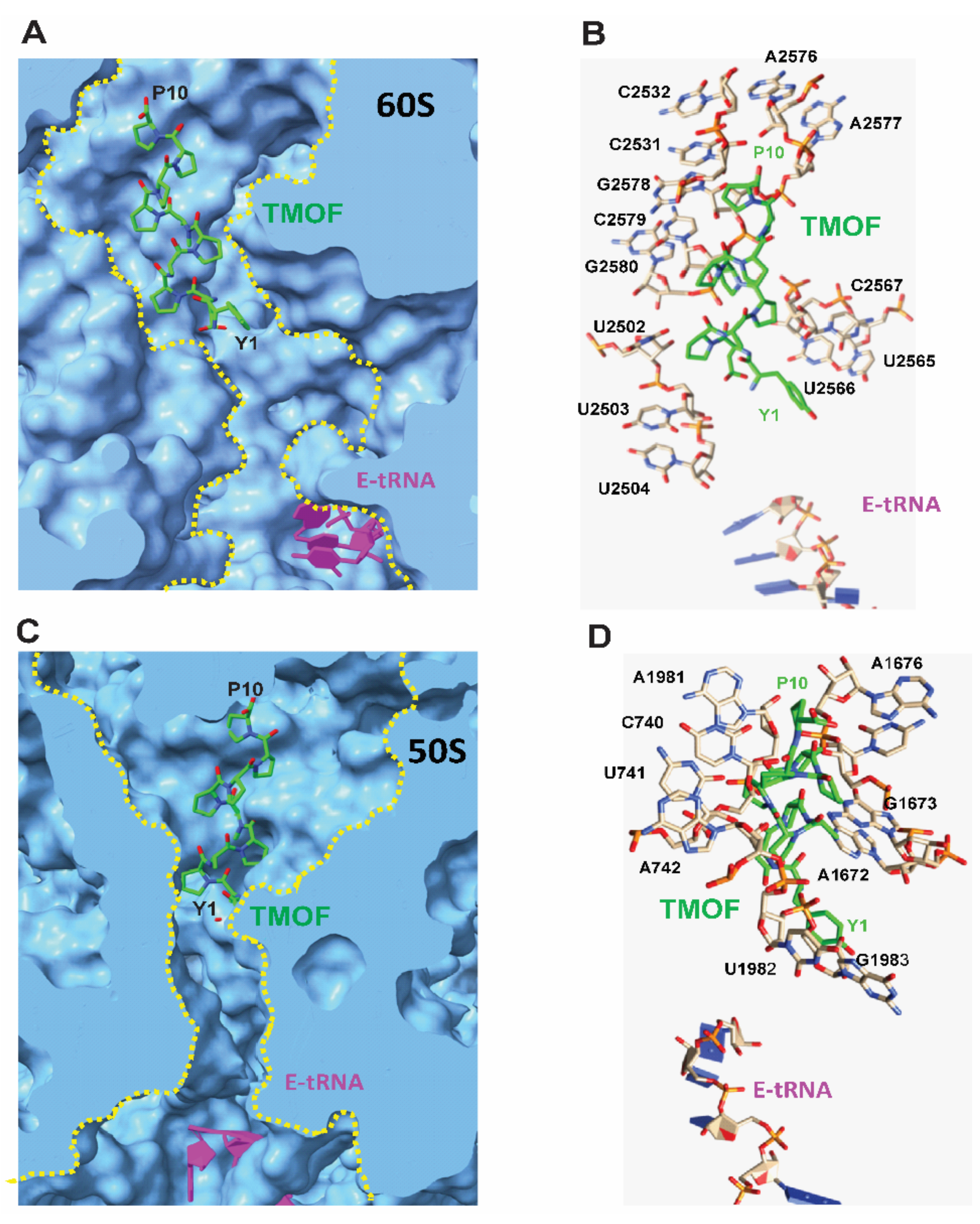
3.4.2. Docking to D. melanogaster and E. coli Ribosomes

Docking of AeaTMOF to D. melanogaster (60S) and E. coli (50S) ribosomes exhibited the most favorable energy when AeaTMOF occupied the peptide exit tunnel of both the Drosophila (60S) and E. coli (50S) ribosome large subunit (Figure 6A,C), blocking the entrance to the exit tunnel mainly by hydrophobic stacking interactions with the rings from purine and pyrimidine nucleotide bases of the Drosophila (28S) and E.coli (23S) r-RNA. A few hydrogen bonds between AeaTMOF and the nucleotide bases also participate in the interaction of AeaTMOF with the 28S and 23S r-RNAs (Figure 6B,D).

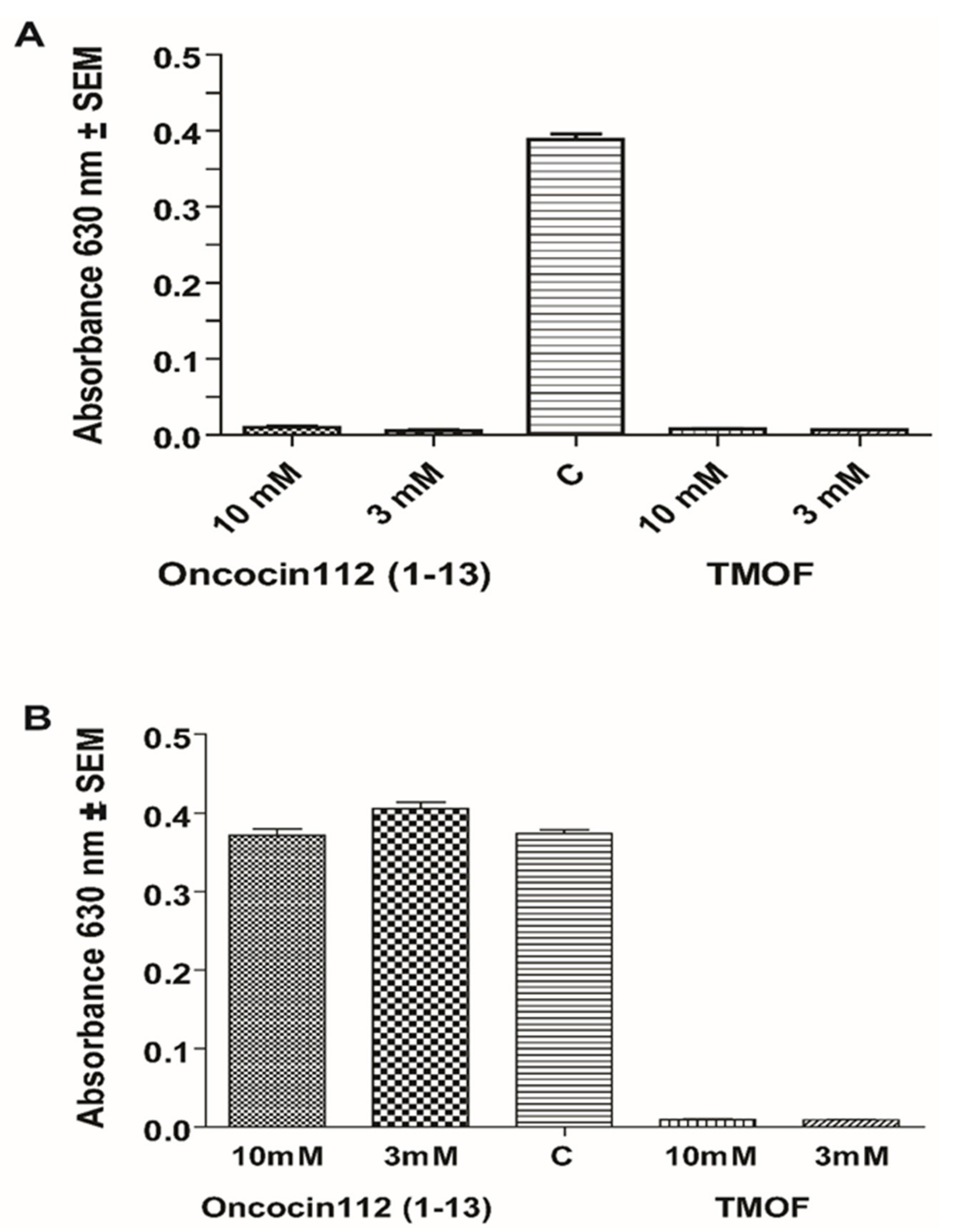
3.5. Inhibition of E.coli Growth with AeaTMOF and Oncocin112 (1–13)

Our results show that AeaTMOF binds to the E. coli-purified ribosome with high affinity (Figure 4), and that Onconcin112 (1–13) inhibits the biosynthesis of luciferase in vitro using the E. coli S30 lysate extract system for circular DNA (Promega) (Figure 1B). Oncocin112 was shown to kill E. coli by inhibiting bacterial protein translation at the 70S ribosome and using the SbmA transporter to get into the bacterial cytosol [13,26]. Our earlier results show that AeaTMOF enters bacterial cells that were engineered with the ABC-tmfA importer [7]. To find out if AeaTMOF-like oncocin 112 affects bacterial growth by entering the bacterial cell using SbmA importer, E. coli CGSC strain7636 cells expressing SbmA+ and E. coli mutant cells CGSC strain 8547 SbmA- expressing only ABC-tmfA importer were incubated with different concentrations of Oncocin112 (1–13) and AeaTMOF (3 and 10 mM). Both peptides significantly inhibited the growth of the E. coli cells, and only 4–10 colonies were still viable at the end of the incubation period from the initial 2.7 × 10⁴ cell, when compared with control cells that were not incubated with the peptides (p < 0.05) if the cells expressed a SbmA importer (Figure 7A). On the other hand, E. coli cells lacking a SbmA importer but expressing the ABC-tmfA importer were significantly (p < 0.05) inhibited by AeaTMOF (Figure 7B). Few colonies between 4 and 12 out of the starting 2.7 × 10⁴ colonies were still viable at the end of the incubation period. Control cells that were not incubated with the peptides grew at the same rate as cells that were incubated in the presence of Oncocin112 (1–13) (3 and 10 mM) (Figure 7B), indicating that AeaTMOF can use the mosquito ABCtmfA importer, as well as the bacterial SbmA importer. On the other hand, Oncocin112 (1–13) can only use the bacterial natural importer SbmA.

4. Discussion

AeaTMOF (YDPAPPPPPP) and NebTMOF (NPTNLH) were shown to stop the translation of trypsin mRNA in the gut epithelial cells of Ae. aegypti and Neobellieria bullata [4,5]. AeaTMOF binds a gut-specific receptor ABC-tmfA transporter (accession number MK895491) that imports AeaTMOF from the hemolymph after it is secreted by the mosquito ovaries into the gut epithelial cells, stopping the translation of the trypsin transcript in the gut epithelial cells [1,2,7]. Several of these ABC transporters with 80–100% similarity with the AeaTMOF receptor are also found in several Aedes, Anopheles, and Culex species, and their trypsin biosynthesis is controlled by AeaTMOF [7,27]. However, the mechanism by which AeaTMOF stops the translation of the trypsin transcript in the midgut epithelial cells of Ae. aegypti is unknown. We observed that AeaTMOF belongs to a family of proline-rich peptides that were shown to act as antimicrobial peptides by inhibiting the translation of proteins by bacterial ribosomes like Oncocin112 [13,14,15]. Therefore, we performed in vitro transcript/translation experiments using the E. coli 30S lysate extract system (Promega) with increasing concentrations of AeaTMOF or Oncocin112 (1–13). The latter was shown by X-ray crystallography to occupy the ribosomal exit tunnel at its entrance, blocking tRNA movement, mRNA translation, and protein biosynthesis [14,15]. Our results (Figure 1A,B) show that increasing concentrations of AeaTMOF or Oncocin112 (1–13) in vitro inhibit the biosynthesis of luciferase (IC50 1 μM and 2 μM, respectively). The peptides do not inhibit the activity of luciferase in the presence of AeaTMOF or Oncocin112 (1–13), indicating that the peptides act on the E. coli ribosome as was shown when Oncocin112 was incubated with E. coli ribosomes [14,15]. Although the full sequence of Oncocin112 (19 amino acids) was shown to inhibit in vitro protein biosynthesis [13], we decided to use a shorter Oncocin112 peptide, 13 amino acids long, because amino acids (1–13) occupy the entrance to the ribosomal exit tunnel [14,15] and a 13 amino acids peptide is closer in length to the decapeptide AeaTMOF [28]. AeaTMOF that was fed in vivo to Ae. aegypti larvae stopped larval trypsin biosynthesis and caused larval starvation and death when they were fed increasing amounts of AeaTMOF expressed by engineered Chlorella desiccate, Saccharomyces cerevisiae, and Pichia pastoris [29,30,31]. Our in vitro results also show that increasing concentrations of AeaTMOF incubated with S. frugiperda cell extract in the presence of plasmid pFA25A ICE T7 flexi engineered with Ae. aegypti late larval trypsin caused inhibition of the biosynthesis of [3H]trypsin (Figure 2). The IC50 (1.8 μM) that was determined is higher than the IC50 (1.0 μM) that was observed when AeaTMOF was incubated in vitro using E.coli S30 extract to synthesize luciferase (Figure 1A). Our results also show that AeaTMOF does not inhibit luciferase activity, and therefore does not interfere with the IC50 determinations. However, the two plasmids that were engineered to express luciferase and trypsin are different, and the transcription of luciferase and the late larval trypsin by the two plasmids is probably different, affecting the IC50. Indeed, different IC50 values from 0.150 μM to 0.250 μM were reported for oncocin112 when it was incubated in vivo with E. coli cells [13]. The synthesis of [3H]-labeled larval protein in vitro using a lysate extract of S. frugiperda ribosomes [32] in the presence of a plasmid carrying the larval trypsin gene does not prove that the late larval trypsin was synthesized. However, MS/MS analysis of the 28.5 kDa protein after the SDS-PAGE identified larval late trypsin only in incubations containing the plasmid pF25A ICE T7 flexi engineered with Ae. aegypti larval late trypsin. Several similar protein bands that were detected after staining of the SDS-PAGE gel in lanes (2–4) are from the S. frugiperda cell lysate that was used (Figure S2A). In lane 1, BSA was added, showing a heavily stained BSA band (66 kDa). A second fluorescent band that was observed at 25 kDA (Figure S1) might be due to an apparent internal translation start [33]. However, our SDS-PAGE did not detect this protein band (Figure S2A).
Our results suggest that AeaTMOF binds the E. coli ribosome similarly to the binding of the proline-rich peptide Oncocin112 that blocks the T. thermophilus ribosome exit tunnel, stopping protein biosynthesis [14,15]. For this reason, AeaTMOF ribosomal interaction studies were conducted using isolated intact E. coli ribosomes (Figure 3) that were incubated with increasing concentration of AeaTMOF-FITC. The binding of AeaTMOF to the ribosome shows high affinity (KD = 23 ± 3.4 nM ± SEM, Bmax = 0.553 ± 0.023 pmol/μg ribosome ± SEM and Kassoc = 4.3 × 10⁷ M−1) (Figure 4). A dissociation constant in the nanomolar range was also shown for other proline-rich peptides, oncocins, and apidaecins, which also bind the 70S bacterial ribosome [13]. Crystallization and X-ray diffraction of oncocin112 bound to the T. thermophilus 70S ribosome shows that the first 13 amino acids occupy the entrance to the exit tunnel of the 50S ribosome [14,15]. 3D modeling and docking of AeaTMOF to T. thermophilus shows that AeaTMOF-like Oncocin112 can also block the entrance to the exit tunnel of the 50S ribosome, interacting with several nucleotides by hydrogen bonding and hydrophobic stacking interactions, and thus can also stop protein synthesis and elongation by tRNA (Figure 5A,B). Molecular modeling and docking of AeaTMOF to the D. melanogaster (60S) and E. coli (50S) ribosomes shows that AeaTMOF also blocks the entrance to the exit tunnel of the 60S and 50S ribosomes by interacting with the D. melanogaster (28S) and E. coli (23S) r-RNA using hydrophobic and hydrogen bond interactions with the nucleotide bases (Figure 6A–D). The 3D models of AeaTMOF binding to bacterial and Drosophila ribosome (Figure 5 and Figure 6) suggest, for the first time, how AeaTMOF stops the translation of the trypsin transcript that was reported earlier [4,5].
The bacterial inner membrane protein SbmA is involved in the import of peptides, proline-rich peptides (including oncocin112), nucleic acids, antisense peptides, and several oligomers [34,35]. Incubation of E. coli CGSC strain7636 cells expressing SbmA+ and E.coli CGSC strain 8547 cells SbmA-,which was engineered with AeaTMOF receptor/importer (ABCtmfA) [7] with oncocin112 (1–13) or AeaTMOF (5 mM and 10 mM), inhibited bacterial growth in cells that expressed SbmA+ (Figure 7A), as was reported earlier for the proline-rich peptide oncocin112 [35]. However, when the peptides were incubated with E. coli cells that only expressed ABCtmfA receptor/importer [7], oncocin112 (1–13) did not affect the cell growth, even at high concentrations of 10 mM, whereas AeaTMOF entered the bacterial cell using the ABCtmfA importer inhibiting protein synthesis and cell growth (Figure 7B). In mosquitoes, AeaTMOF was shown by cytoimmunochemical analysis [36] to target specific gut epithelial cell that express the ABCtmfA (TMOF-specific receptor/importer) [7]. These cells express trypsin, which is secreted into the gut lumen and digests the blood meal to free amino acids that are used for egg development [1]. We hypothesize that AeaTMOF in these trypsin-synthesizing cells binds to the ribosomes that synthesize trypsin and does not inhibit the biosynthesis of other proteins that are synthesized in other midgut epithelial cells that lack the AeaTMOF-specific receptor/importer [7,36]. Indeed, injection of AeaTMOF into female mosquitoes that took a blood meal only inhibits trypsin biosynthesis, and as a result the blood meal is not digested and egg development is inhibited, making the female mosquitoes sterile but not killing them [1,16,27,28]. This would suggest that AeaTMOF blocks all protein synthesis of gut epithelial cells that express the ABCtmfA receptor. Alternatively, AeaTMOF may block translation of only specific transcripts within these receptor-expressing cells. Further work is needed to determine if AeaTMOF blocks all protein synthesis in receptor-bearing gut epithelial cells or can selectively block synthesis of specific transcripts.
In conclusion, this report shows for the first time that AeaTMOF, after entering the bacterial cytosol using a SbmA importer or engineered mosquito ABCtmfA-receptor/importer expressed in the inner membrane of E. coli cells, binds with high affinity (KD = 23 ± 3.4 nM ± SEM) to the bacterial 70S. 3D modeling indicates that AeaTMOF can also bind the Drosophila 80S ribosome and possibly the mosquito 80S ribosome in the gut epithelial cells. These observations explain, for the first time, how trypsin biosynthesis in blood-fed female mosquitoes is regulated. Our binding experiment results to bacterial and insect ribosomes do not show whether the translation of the trypsin transcript is immediately stopped, as was shown for Oncocin112, in which the binding to the bacterial ribosome at the A-site in the empty exit tunnel affects the elongation factor Tu and the ternary complexes blocking elongation [37]. More work is needed to study AeaTMOF binding to mosquito ribosomes, as was done using the E. coli ribosome as a model system.

Supplementary Materials

The following supporting information can be downloaded at: https://www.mdpi.com/article/10.3390/biom12040577/s1, Figure S1: Western blot analysis of Ae. aegypti late larval trypsin synthesized in TnT T7 insect cell lysate in the presence of Fluorotech Green tRNAlys and plasmid pFA25A ICE T7 flexi cloned with late larval trypsin lanes (b, c and d). Lane (a) is a control incubation without Fluorotech Green tRNAlys; Figure S2: A. SDS-PAGE of TnT 7T insect cell lysates that were incubated with BSA, B. Mass spectrometry analysis of the excised protein band marked with a black arrow.

Author Contributions

D.B. wrote the original manuscript. D.B., R.G.S.J. and P.R. edited the manuscript. D.B. and R.G.S.J. analyzed the results. P.R. contributed the 3D models and analysis. D.B. developed, purified, cloned and assayed the binding assays of TMOF to the E. coli ribosmes. D.B. genetically engineered and expressed the ABCtmfA receptor in the inner membrane of E. coli. All authors have read and agreed to the published version of the manuscript.

Funding

This research did not receive external funding.

Institutional Review Board Statement

Not applicable.

Informed Consent Statement

Not applicable.

Data Availability Statement

Not applicable.

Conflicts of Interest

The authors declare no conflict of interest.

References

  1. Borovsky, D. Oostatic hormone inhibits biosynthesis of midgut proteolytic enzymes and egg development in mosquitoes. Arch. Insect Biochem. Physiol. 1988, 7, 187–210. [Google Scholar] [CrossRef]
  2. Borovsky, D.; Song, Q.; Ma, M.; Carlson, D.A. Biosynthesis, secretion and cytoimmunochemistry of trypsin modulating oostatic factor of Aedes aegypti. Arch. Insect Biochem. Physiol. 1994, 27, 27–38. [Google Scholar] [CrossRef]
  3. Borovsky, D.; Verhaert, P.; Rouge, P.; Powell, C.A.; De Loof, A. Blood digestion in Culex quinquefasciatus by trypsin is translationally regulated by Trypsin Modulating Oostatic Factor (TMOF). Front. Physiol. 2021, 12, 764061. [Google Scholar] [CrossRef]
  4. Borovsky, D.; Janssen, I.; Vanden Broeck, J.; Huybrechts, R.; Verhaert, P.; DeBondt, H.L.; Bylemans, D.; DeLoof, A. Molecular sequencing and modeling of Neobellieria bullata trypsin: Evidence for translational control with Neb TMOF. Eur. J. Biochem. 1996, 237, 279–287. [Google Scholar] [CrossRef] [PubMed]
  5. Borovsky, D.; Rabindran, S.; Dawson, W.O.; Powell, C.R.; Iannotti, D.; Morris, T.; Shabanowitz, J.; Hunt, D.F.; De Bondt, H.L.; De Loof, A. Expression of Aedes TMOF on the virion of TMV: Potential larvicide. Proc. Natl. Acad. Sci. USA 2006, 103, 18963–18968. [Google Scholar] [CrossRef] [PubMed] [Green Version]
  6. Schoenberg, D.; Maquat, L.E. Regulation of cytoplasmic mRNA decay. Nat. Rev. Genet. 2012, 13, 246–259. [Google Scholar] [CrossRef] [PubMed]
  7. Borovsky, D.; Deckers, K.; Vanhove, A.C.; Verstraete, M.; Rouge, P.; Shatters, R.G., Jr.; Powel, C.A. Cloning and Characterization of Aedes aegypti Trypsin Modulating Oostatic Factor (TMOF) Gut-Receptor. Biomolecules 2021, 11, 934. [Google Scholar] [CrossRef]
  8. Casteels, P.; Ampe, C.; Jacobs, F.; Vaeck, M.; Tempst, P. Apidaecins: Antibacterial peptides from honeybees. EMBO J. 1989, 8, 2387–2391. [Google Scholar] [CrossRef]
  9. Gennaro, R.; Zanetti, M.; Benincasa, M.; Podda, E.; Miani, M. Pro-rich antimicrobial peptides from animals:structure, biological functions and mechanism of action. Curr. Pharm. Des. 2002, 8, 763–778. [Google Scholar] [CrossRef]
  10. Li, J.X.; Xu, X.Q.; Yu, H.N.; Yang, H.L.; Huang, Z.X.; Lai, R. Direct antimicrobial activities of PR-bombesin. Life Sci. 2006, 78, 1953–1956. [Google Scholar] [CrossRef]
  11. Destoumieux, D.; Bulet, P.; Strub, J.M.; Van Dorsselaer, A.; Bachère, E. Recombinant expression and range of activity of penaeidins, antimicrobial peptides from penaeid shrimp. Eur. J. Biochem. 1999, 266, 335–346. [Google Scholar] [CrossRef] [PubMed] [Green Version]
  12. Gueguen, Y.; Bernard, R.; Julie, F.; Paulina, S.; Delphine, D.G.; Franck, V.; Philippe, B.; Evelyne, B. Oyster hemocytes express a proline-rich peptide displaying synergistic antimicrobial activity with a defensin. Mol. Immunol. 2009, 46, 516–522. [Google Scholar] [CrossRef] [PubMed] [Green Version]
  13. Krizsan, A.; Volke, D.; Weinert, S.; Strater, N.; Knappe, D.; Hoffmann, R. Insect-derived proline-rich antimicrobial peptides kill bacteria by inhibiting bacterial protein translation at the 70 S ribosome. Angew. Chem. Int. Ed. Engl. 2014, 53, 12236–12239. [Google Scholar] [CrossRef] [PubMed]
  14. Seefeldt, A.C.; Nguyen, F.; Antunes, S.; Perebaskine, N.; Graf, M.; Arenz, S.; Inampudi, K.K.; Douat, C.; Guichard, G.; Wilson, D.N.; et al. The proline-rich antimicrobial peptide Onc112 inhibits translation by blocking and destabilizing the initiation complex. Nat. Struct. Mol. 2015, 22, 470–475. [Google Scholar] [CrossRef] [PubMed]
  15. Roy, R.N.; Lomakin, I.B.; Gagnon, M.G.; Steitz, T.A. The mechanism of inhibition of protein synthesis by the proline-rich peptide oncocin. Nat. Struct. Mol. Biol. 2015, 2, 466–469. [Google Scholar] [CrossRef] [PubMed] [Green Version]
  16. Borovsky, D.; Carlson, D.A.; Griffin, P.R.; Shabanowitz, J.; Hunt, D.F. Mass Spectrometry and characterization of Aedes aegypti trypsin modulating oostatic factor (TMOF) and its analogs. Insect Biochem. Mol. Biol. 1993, 23, 703–712. [Google Scholar] [CrossRef]
  17. Laemmli, U.K. Cleavage of structural proteins during the assembly of the head of bacteriophage T4. Nature 1970, 227, 680–685. [Google Scholar] [CrossRef]
  18. Van Ekert, E.; Heylen, K.; Rouge, P.; Powell, C.A.; Shatters, R.G., Jr.; Smagghe, G.; Borovsky, D. Aedes aegypti juvenile hormone acid methyltransferase, the ultimate enzyme in the biosynthetic pathway of juvenile hormone III, exhibits substrate control. J. Insect Physiol. 2014, 64, 62–73. [Google Scholar] [CrossRef]
  19. Mehta, P.; Woo, P.; Venkataraman, K.; Karzai, A.W. Ribosome purification approaches for studying interactions of regulatory proteins and RNAs with the ribosome. Methods Mol Biol. 2012, 905, 273–289. [Google Scholar] [CrossRef] [Green Version]
  20. Rivera, M.C.; Maguire, B.; Lake, J.A. Purification of 70S Ribosomes. Cold Spring Harb. Protoc. 2015, 2015, 300–302. [Google Scholar] [CrossRef]
  21. Curto, E.V.; Jarpe, M.A.; Blalock, J.B.; Borovsky, D.; Krishna, N.R. Solution structure of trypsin modulating oostatic factor is a left-handed helix. Biochem. Biophys. Res. Commun. 1993, 193, 688–693. [Google Scholar] [CrossRef]
  22. Krieger, E.; Koraimann, G.; Vriend, G. Increasing the precision of comparative models with YASARA NOVA—A self-parametrizing force field. Proteins 2002, 47, 393–402. [Google Scholar] [CrossRef] [PubMed]
  23. Pettersen, E.F.; Goddard, T.D.; Huang, C.C.; Couch, G.S.; Greenblatt, D.M.; Meng, E.C.; Ferrin, T.E. UCSF Chimera—A visualization system for exploratory research and analysis. J. Comput. Chem. 2004, 25, 1605–1612. [Google Scholar] [CrossRef] [Green Version]
  24. Anger, A.M.; Armache, J.-P.; Berninghausen, O.; Habeck, M.; Subklewe, M.; Wilson, D.N.; Beckmazn, R. Structure of the human and Drosophila 80S ribosome. Nature 2013, 497, 80–85. [Google Scholar] [CrossRef] [PubMed]
  25. Goddard, T.D.; Huang, C.C.; Meng, E.C.; Pettersen, E.F.; Couch, G.S.; Morris, J.H.; Ferrin, T.E. UCSF ChimeraX: Meeting modern challenges in visualization and analysis. Protein Sci. 2018, 27, 14–25. [Google Scholar] [CrossRef] [PubMed]
  26. Mattiuzzo, M.; Bandiera, A.; Gennaro, R.; Benincasa, M.; Pacor, S.; Antcheva, N.; Scocchi, M. Role of the Escherichia coli SbmA in the antimicrobialactivity of proline-rich peptides. Mol. Microbiol. 2007, 66, 151–163. [Google Scholar] [CrossRef]
  27. Borovsky, D. Trypsin modulating oostatic factor for developing resistant crops. In Insectidices Design Using Advanced Technologies; Ishaaya, I., Nauen, R., Horowitz, A.R., Eds.; Springer: Berlin, Germany, 2007. [Google Scholar]
  28. Borovsky, D.; Carlson, D.A.; Griffin, P.R.; Shabanowitz, J.; Hunt, D.F. Mosquito oostatic factor a novel decapeptide modulating trypsin-like enzyme biosynthesis in the midgut. FASEB J. 1990, 4, 3015–3020. [Google Scholar] [CrossRef]
  29. Borovsky, D.; Sterner, A.; Powell, C.A. Cloning and Expressing Trypsin Modulating Oostatic Factor (TMOF) in Chlorella desiccata to control Mosquito Larvae. Arch. Insect Biochem. Physiol. 2016, 91, 17–36. [Google Scholar] [CrossRef]
  30. Borovsky, D.; Nauwelaers, S.; Powell, C.A.; Shatters, R.G., Jr. Cloning, genetic engineering and characterization of TMOF expressed in Saccharomyces cerervisiae to control larval mosquitoes. J. Insect Physiol. 2018, 106, 134–146. [Google Scholar] [CrossRef]
  31. Borovsky, D.; Nauwelaers, S.; Shatters, R., Jr. Biochemical and molecular characterization of Pichia pastoris cells expressing multiple TMOF genes (tmfA) for mosquito larval control. Front. Physiol. 2020, 11, 527. [Google Scholar] [CrossRef]
  32. Ezure, T.; Suzuki, T.; Higashide, S.; Shintani, E.; Endo, K.; Kobayashi, S.-I.; Shikata, M.; Ito, M.; Tanimizu, K.; Nishimura, O. Cell-free protein synthesis system prepared from insect cells by freeze-thawing. Biotechnol. Prog. 2006, 22, 1570–1577. [Google Scholar] [CrossRef] [PubMed]
  33. Kearse, M.G.; Wilusz, J.E. Non-AUG translation: A new start for protein synthesis in eukaryotes. Genes Dev. 2017, 31, 1717–1731. [Google Scholar] [CrossRef] [PubMed] [Green Version]
  34. Runti, G.; del Carmen Lopez Ruitz, M.; Stoilova, T.; Hussain, R.; Jennions, M.; Choudhury, H.G.; Benincasa, M.; Gennaro, R.; Beis, K.; Scocchi, M. Functional characterization of SbmA, a bacterial inner membrane transporter required for importing the antimicrobial peptide Bac7 (1–35). J. Bacteriol. 2013, 195, 5343–5351. [Google Scholar] [CrossRef] [PubMed] [Green Version]
  35. Knappe, D.; Piantavigna, S.; Hansen, A.; Mechler, A.; Binas, A.; Nolte, O.; Martin, L.I. Oncocin (VDKPPYLPRPRPPPPIYNR-NH2): A novel antibacterial peptide optimized against gram-negative human pathogens. J. Med. Chem. 2010, 53, 5240–5247. [Google Scholar] [CrossRef] [PubMed]
  36. Borovsky, D.; Powell, C.A.; Nayar, J.K.; Blalock, J.E.; Hayes, T.K. Characterization and localization of mosquito-gut receptors for trypsin modulating oostatic factor (TMOF) using a complementary peptide and immunocytochemistry. FASEB J. 1994, 8, 350–355. [Google Scholar] [CrossRef]
  37. Zhu, Y.; Weisshaar, J.C.; Mustafi, M. Long-term effects of the proline-rich antimicrobial peptide Oncocin 112 on the Escherichia coli translation machinery. J. Biol. Chem. 2020, 295, 13314–13325. [Google Scholar] [CrossRef]
Figure 1. Inhibition of luciferase biosynthesis shown as reduction in luminescence (%) in E. coli S30 extract in the presence of increasing peptide concentrations (0.001-750 μM). (A) AeaTMOF showing IC50 of 1.0 μM, and (B) oncocin112 (1–13) showing an IC50 of 2.0 μM. Each point is the mean of three determinations + SEM (see Section 2.3 for details). Luminescence at 100% indicates no inhibition of luciferase biosynthesis.
Biomolecules 12 00577 g001
Figure 2. Inhibition of Ae. aegypti late larval [3H]trypsin biosynthesis by increasing concentrations (0.01–750 μM) of AeaTMOF in the S. frugiperda TnT T7 insect cell extract protein expression system, showing an IC50 of 1.8 μM. Each point is a mean of three determinations ± SEM (see Section 2.4 for details).
Biomolecules 12 00577 g002
Figure 3. Purified E. coli ribosomes negatively stained with uranyl acetate on a carbon-coated grid and observed by electron microscopy with 120 kV Talos L120C TEM. The white bar below shows the relative size in nanometer. The 50S and 30S subunits can be observed in many of the ribosomes.
Biomolecules 12 00577 g003
Figure 4. Specific binding of AeaTMOF to E.coli-purified ribosomes expressed as AeaTMOF-FITC (pmo/Ribosome (μg)) showing Michaelis Menten binding kinetics and using nonlinear regression (R2 = 0.84). Binding results were corrected for nonspecific binding by incubating ribosomes without AeaTMOF-FITC or with AeaTMOF-FITC and without ribosomes (see Section 2.7). Each point is an average of six determinations ± SEM. The data represent results of one experiment from two independent experiments with similar results. KD = 23 ± 3.4 nM ± SEM, Bmax = 0.553 ± 0.023 pmol/μg ribosome ± SEM, and Kassoc = 4.3 × 10⁷ M−1.
Biomolecules 12 00577 g004
Figure 5. (A) Close-up cut view showing the docking of AeaTMOF (magenta stick) within the peptide exit tunnel of 23S RNA of T. thermophilus ribosome. The 5tRNA and the A-site of the 23S RNA are indicated. (B) Docking of AeaTMOF (magenta stick) into the peptide exit tunnel of 23S RNA of T. thermophilus showing the nucleotide bases (blue sticks) involved in the interaction with the peptide through hydrogen bonds (not shown) and stacking interactions.
Biomolecules 12 00577 g005
Figure 6. (A) Docking of AeaTMOF (colored green) to the Drosophila ribosome peptide exit tunnel (yellow dashed line) of the 60S large subunit (colored blue). The E-tRNA is colored magenta. (B) Cartoon showing the nucleotide bases of the 28S r-RNA from the 60S subunit that interacts with AeaTMOF via stacking interactions. The E-tRNA is also shown. (C) Docking of AeaTMOF (colored green) to the E. coli ribosome showing AeaTMOF (colored green) in the peptide exit tunnel (yellow dashed line) of the 50S large subunit (colored blue). The E-tRNA is colored magenta. (D) Cartoon showing the nucleotide bases of the 23S r-RNA from the 50S subunit that interacts with AeaTMOF via stacking interactions. The E-tRNA is also shown.
Biomolecules 12 00577 g006
Figure 7. Incubation of E. coli cells with Oncocin112 (1–13) and AeaTMOF at concentrations of 3 and 10 mM (A) with E. coli cells expressing SbmA and (B) with E. coli mutant cells SbmA- expressing the ABC-tmfA importer. Control cells incubated without oncocin112 (1–13) or AeaTMOF (C).
Biomolecules 12 00577 g007
Publisher’s Note: MDPI stays neutral with regard to jurisdictional claims in published maps and institutional affiliations.

