Review

Advanced Strategies for Therapeutic Targeting of Wild-Type and Mutant p53 in Cancer

by
Shengliang Zhang
1,2,3,4,*,
Lindsey Carlsen
1,5,
Liz Hernandez Borrero
1,
Attila A. Seyhan
1,2,3,4,
Xiaobing Tian
1,2,3,4 and
Wafik S. El-Deiry
1,2,3,4,6,*
1
Laboratory of Translational Oncology and Experimental Cancer Therapeutics, Warren Alpert Medical School, Brown University, Providence, RI 02912, USA
2
Department of Pathology and Laboratory Medicine, Warren Alpert Medical School, Brown University, Providence, RI 02912, USA
3
Joint Program in Cancer Biology, Lifespan Health System and Brown University, Providence, RI 02912, USA
4
Legorreta Cancer Center, Brown University, Providence, RI 02912, USA
5
Pathobiology Graduate Program, Warren Alpert Medical School, Brown University, Providence, RI 02912, USA
6
Hematology-Oncology Division, Department of Medicine, Lifespan Health System and Brown University, Providence, RI 02912, USA
*
Authors to whom correspondence should be addressed.
Biomolecules 2022, 12(4), 548; https://doi.org/10.3390/biom12040548
Submission received: 1 January 2022 / Revised: 28 February 2022 / Accepted: 6 March 2022 / Published: 6 April 2022
(This article belongs to the Collection p53 Function and Dysfunction in Human Health and Diseases)

Abstract

:
TP53 is a tumor suppressor gene that encodes a sequence-specific DNA-binding transcription factor activated by stressful stimuli; it upregulates target genes involved in growth suppression, cell death, DNA repair, metabolism, among others. TP53 is the most frequently mutated gene in tumors, with mutations not only leading to loss-of-function (LOF), but also gain-of-function (GOF) that promotes tumor progression, and metastasis. The tumor-specific status of mutant p53 protein has suggested it is a promising target for cancer therapy. We summarize the current progress of targeting wild-type and mutant p53 for cancer therapy through biotherapeutic and biopharmaceutical methods for (1) boosting p53 activity in cancer, (2) p53-dependent and p53-independent strategies for targeting p53 pathway functional restoration in p53-mutated cancer, (3) targeting p53 in immunotherapy, and (4) combination therapies targeting p53, p53 checkpoints, or mutant p53 for cancer therapy.

1. Introduction

The p53 protein is a sequence-specific DNA-binding transcription factor regulating gene expression related to multiple cellular functions including, but not limited to, cell cycle arrest, cell apoptosis, cell growth, DNA repair, cell metabolism, and the immune response in response to stressful stimuli [1]. Clinical studies show that wild-type p53 sensitizes cancer cells to conventional chemoradiotherapy, for example, in the treatment of rectal cancer. Nearly half of tumors harbor mutant p53. Most TP53 mutations are single missense mutations in its functional domains, most commonly the central DNA-binding domain. Missense mutations cause mutant p53 protein to lose its wild-type functions (LOF) and acquire dominant-negative activities and endow mutant p53 with gain-of-function (GOF) capabilities that confer aggressive tumor behavior and drug resistance. Clinical studies correlate poor prognosis of cancer patients with mutant p53. p53 is considered as a biomarker for tumor progression and an excellent target for designing cancer therapeutic strategies. Even in cancers carrying wild-type p53, aberrant p53 pathway signaling often occurs due to abnormal regulation such as due to high MDM2 expression often from gene amplification. The different p53 status between cancer cells and normal cells has led to p53 becoming one of the most important and rational targets for cancer therapy.
Over the past two decades, studies have revealed p53 structure, function, and role in tumorigenesis and development [1,2,3]. These fundamental studies strongly support the design and development of new approaches for targeting p53 (mutant and wild-type) in cancer therapy [4,5,6]. Delivery of wild-type p53 by adenovirus infection kills cancer cells and suppresses tumor growth in pre-clinical and clinical studies [7]. Peptides targeting p53 can rescue p53 function and reduce tumor proliferation [8]. Reactivation of p53 by blocking p53-MDM2/MDMX(MDM4) interaction is one of the most promising approaches for cancer therapy, and MDM2 inhibitors (such as AMG232 or milademetan) and the dual inhibitors of MDM2/MDMX (such as ALRN6924) are currently undergoing clinical evaluation [9,10]. APR-246 and COTI-2 are small molecules targeting mutant p53 under clinical evaluation in different clinical trials [11,12,13,14]. Immunotherapy targeting mutant p53 to suppress tumor growth is becoming a promising approach in cancer therapy. These pioneering studies shed light on prospective cancer therapy that targets mutant and wild-type p53.

2. p53 Structure and Mutations Exploited for Drug Development

The frequency of TP53 mutations is approximately 50% across all cancers, however, this frequency varies greatly depending on the cancer type. TP53 mutations are most common in ovarian (47.8%), colorectal (43.2%), esophageal (43.1%), head & neck (40.6%), and laryngeal (40.4%) tumors, while cervix (5.8%), hematopoietic (12.7%), and endocrine gland (14.6%) tumors experience the lowest frequency of p53 mutations [15]. High-grade serous ovarian cancer has a p53 mutation rate approaching 95%.
TP53 mutations generally occur in the DNA-binding domain, with about 30% arising in specific hotspots including R175, G245, R248, R249, R273, and R282 (individual mutation rates range from ~3–7%) [16]. Clinical studies demonstrate that in many tumor types, the presence of mutated p53 correlates with a worse patient prognosis as compared to the presence of wild-type p53. Though this is still a topic of controversy in some contexts, it is most widely accepted that tumors with mutated p53 predict worse clinical outcomes in breast, head and neck, liver, hematopoietic, and lymphoid system cancers as compared to their wild-type counterparts [17]. One factor contributing to the complexity of this topic is that TP53 mutant or variant sequences can have unique effects on cellular function. For example, cancer-associated polymorphisms in codon 72 of TP53 can result in a non-conservative amino acid change to either proline (p53pro) or arginine (p53arg), which have different characteristics. p53pro is less efficient in inducing apoptosis compared to p53arg, while p53arg is more likely to be degraded by the human papillomavirus (HPV) E6 protein. Much work by Maureen Murphy has uncovered racial, ethnic, and geographic differences in the TP53 codon 72 polymorphism and others. The cancer-associated mutations in TP53 are most often missense mutations in the central DNA-binding domain. These mutations also vary greatly in terms of their impact on p53 function. Some mutations cause loss of p53 function, some provide p53 the ability to inactivate wild-type p53 that is expressed from the remaining wild-type allele (dominant-negative p53 mutations), and still others cause p53 to acquire oncogenic GOF properties [18]. These unique characteristics of different p53 mutants make mutant p53 a challenging drug target and also necessitate consideration of individual mutations when using mutant p53 as a prognostic biomarker [17].

2.1. p53 Structure Exploited for Drugs Reactivating p53 Signaling

Wild-type p53 consists of a p53 transactivation domain at its N-terminal region and a tetramerization domain at its C-terminal region (Figure 1). The N- and C-terminals flank the DNA binding domain, also called the core domain [3].
DNA-binding domain (amino acid residues 94–292) of the p53 protein is responsible for p53 binding in a DNA sequence-specific manner to gene regulatory regions to control gene expression (Figure 1). Functionally significant upregulated target genes include p21 (WAF1), which regulates the cell cycle; Bax, DR5, Puma, and Noxa, which regulate cellular apoptosis in response to cellular stresses; TIGAR, which regulates processes related to cellular metabolism; and IRF5/9, which regulates processes related to intracellular immune functioning [1]. p53 binds specifically to the consensus DNA sequence (5′-RRRCWWGYYY-(N = 0–13)-RRRCWWGYYY-3′) [19]. This discovery has been widely applied to engineer different vectors expressing reporter genes under the control of p53 response elements (p53RE), in addition to predicting endogenous p53 DNA-binding response elements. The use of p53 transcriptional activity reporters has provided a useful tool in the high-throughput screening (HTS) of p53-targeting drugs via functional cell-based assays. A tool was developed to identify a series of small molecules that reactivate the downstream p53 pathway in tumors with mutated p53 [20]. Multiple small molecular weight chemical compounds targeting p53 (wild-type and mutant) have been identified using this method such as NSC59984 [21], prodigiosin [22], and CB002 [23,24].
The p53 tetramerization domain is located at the C-terminus between residues 325–355 in the human p53 protein (Figure 1). The binding of p53 to specific DNA sequences is regulated by tetramer formation via this tetramerization domain; the C-terminus is also one of the major immunodominant epitopes of human p53 that can be recognized by anti-p53 antibodies in the sera of cancer patients [25,26]. The C-terminal regulatory domain is located at residues 356–393, just following the tetramerization domain (Figure 1). The C-terminal region of p53 is intrinsically disordered and regulated by different post-translational modifications such as phosphorylation, acetylation, methylation, and ubiquitination. The regulatory domain either suppresses or activates p53-dependent transcriptional activities based on the post-transcriptional modifications [27]. MDM2 ubiquitinates p53 at the C-terminal domain, resulting in p53 degradation through the proteasome. Small molecular compound Tenovin is an example of a drug which induces p53 acetylation in this region [28].
The p53 transactivation domains are located near the N-terminus of p53. This domain recognizes and binds to the transcriptional coactivators P300/CBP and components of transcriptional machinery (Figure 1). This N-terminal transactivation domain of p53 binds to MDM2, an E3 ligase [29]. MDM2 ubiquitinates p53 and this results in p53 degradation via the proteasome. MDMX(MDM4) is a homolog of MDM2 but lacks E3-ligase activity; MDMX(MDM4) also binds to p53 TAD and suppresses p53 transcriptional activity. By interacting with MDM2, MDMX(MDM4) stabilizes MDM2 and enhances MDM2 activity [30,31]. Regulation of p53 by MDM2/MDMX(MDM4) keeps basal p53 levels low or undetectable in cells under physiological conditions. The transcriptional domain region of p53 is considered an important domain to target in the development of effective drugs that reactivate p53, which led to the discovery of MDM2 inhibitors such as those in the nutlin family. Most MDM2 inhibitors are not able to significantly inhibit MDMX(MDM4) activity because of the structural differences between MDM2 and MDMX(MDM4) in their p53-binding pockets [31]. Therefore, MDM2 inhibition alone may ineffectively upregulate p53 activity in tumors, especially in the tumors with MDMX(MDM4) overexpression. Dual inhibition of MDM2/MDMX(MDM4) can efficiently release p53 transcriptional activity, which has led to the discoveries of dual inhibitors of MDM2/MDMX(MDM4), such as small-molecule RO-5963 and a stable peptide ALRN-6924 [32,33].
Small molecule MDM2/X inhibitors that activate p53 can do so without causing DNA damage, as occurs with cytotoxic DNA damaging chemotherapy or radiation. This domain is modulated by post-translational modifications after DNA damage, such as phosphorylation and acetylation, via cross-talk with many signaling pathways, such as ATM, ATR, and PKI [34]. Phosphorylation of residues clustered in the N-terminal regions regulates the MDM2-p53 interaction as well as p300/CBP. The phosphorylation of residues Ser15 and Ser20 inhibits the p53-MDM2 interaction [35]. There is a second transactivation domain (TAD II) in p53 that is more C-terminal to the N-terminal domain and includes serine 46 and Thr55. The phosphorylation of Serine 46 has been implicated in the activation of apoptosis [36], and the Thr55 phosphorylation inhibits DNA-binding by enhancing competitive interactions between the TAD-II and DNA-binding domain [37]. These studies provide new insight into the design of treatments targeting p53 in combination with other signals to improve the efficacy of cancer therapy.
There is an abundance of p53 isoforms which are truncated either at the N-terminus (such as Δ40-p53, Δ133-p53, and Δ160-p53) or the C-terminus (β-p53 and γ-p53) [38,39]. The p53 isoforms are generated by two p53 transcription promotors (P1 and P2) and alternative splicing of intron 9 or intron 2 in cells [40]. The p53 isoforms lacking the functional domains might contribute to fine-tuning the p53 pathway signaling. For example, Δ40-p53 and Δ133-p53 lack the TAD, and Δ160-p53 lacks the TAD and part of the DBD, but they retain the TD and RD at the C-terminus. Therefore, they can bind to p53 and act through dominant negative effects [41]. The remaining parts of the TAD of the isoforms might regulate different gene expression patterns compared to wild-type full-length p53, therefore possibly displaying GOF [42]. The exact mechanism of action of p53 isoforms in regulating p53 pathway signaling is not completely elucidated. Abnormal expression of p53 isoforms are correlated with aggressive tumor progression [43]. Current strategies targeting p53 are designed mostly based on full-length p53 (wild-type or mutant). It is unclear what is the effect of these approaches on the isoforms. Targeting the isoforms of p53 might be helpful for increasing the efficiency of the therapeutics targeting p53.

2.2. Conformational Changes of p53 Mutants Utilized for Drugs That Restore Wild-Type Function

Most missense mutations in p53 occur in the core domain. The wild-type p53 core domain is unstable with low thermodynamic and kinetic stability, allowing rapid cycling between folded and unfolded states. Mutations in such residues, such as Arg273 and Arg282, destabilize the core DNA-binding domain by enhancing the thermodynamic and kinetic instability of mutant p53. These effects are often exploited to design ligands that selectively bind to the native state of the p53 protein to reverse the thermodynamic and kinetic denaturation consequences of these mutations.
Mutation at Arg249 of p53 results in impaired DNA-binding due to enhanced flexibility of the L3 loop. Mutation at Y220 causes a cavity formation in the DNA binding core domain that causes loss of p53-DNA binding affinity; these mutations have been exploited in drug development—PhiKan083 (PK083) is a representative example. PK083 is the first small molecule identified to bind to mutant p53 (Y220C), which raises the melting temperature of the mutant and slows down its rate of denaturation [44]. Recently, many Y220C cavity binders have been developed to improve their binding affinities to the Y200C, such as PK9318 [45], MB710, and MB725 [46]. These small molecules increase Tm by 2–4 °C with affinities in the 2–4 μM. The treatment with these Y220C cavity binders restores p53 transcriptional activity and suppresses tumor growth.
Different from other mutations in the core domain, R175H has been described as a zinc-binding mutant. Zinc ions are important in coordinating with C176, H179, C238, and C242 in the core domain to ensure proper folding of the p53 protein [47]. The mutation R175H next to C176 reduces zinc ion binding to the coordinate sites, resulting in mutant p53 protein misfolding, which abolishes DNA binding. Small molecule NSC319726 (ZMC1) plays a role as a zinc metallochaperones in the stabilization of mutant p53-Zn2+ interaction, resulting in a wild-type conformational change [48].
Mutations at the C-terminus disturb p53 tetramer formation. For example, mutations at Arg337 and Asp352 stabilize the tetramer, and mutation at Arg337 (R337H) disrupts the inter monomer salt bridge, resulting in impaired tetramer formation [49]. However, it is known that not all p53 mutants are equal. The wide variety of mutations in p53 makes it challenging to design drugs that target mutant p53 because different mutations might lead to specific structural changes that may need unique small molecules to fit in.
Promising pharmaceutical strategies using small molecular compounds such as nutlins, ALRN-6924,PRIMA-1, COTI-2, and ZMC1 are being tested as monotherapy or as part of a combination in specific tumor types [50,51,52,53].

2.3. p53 Transgenic Mouse Models (p53 Knockout, Mutant p53 Knock-Ins, and Inducible Models) Provide a Powerful Tool to Investigate p53 Function

Cell culture in vitro models mostly used for cancer research provide insights into fundamental biological functions of p53 but do not faithfully reproduce events of tumorigeneses inside living tissues. Mouse p53 is highly homologous to human p53, therefore, p53 transgenic mouse models have been widely applied to explore wild-type and mutant p53 biological functions at physiological conditions and address the mechanism of p53 (wild-type or mutant) action on tumorigenesis and drug resistance in vivo. A broad range of mutant p53 knock-ins or p53 knockout mouse models has been generated. In addition to the constitutive germline-knock-in or knockout of p53, a Cre-LoxP system (Cre acts as a site-specific recombinase to catalyze recombination between specific LoxP sites, and thus depletes genes or gene fragments flanked by LoxP sites) has been applied to conditionally knock in or knock out p53 in mice and to address the role of p53 at different tumor developmental stages [54]. Tissue-specific transcription factor promoters have been exploited for generating tissue-specific expression of Cre, which leads to mutant p53 expression or knockdown of p53 only in the specific tissues [55], therefore, providing an opportunity to investigate the role of mutant p53 or p53 loss in sporadic tumor tissues during tumorigenesis and tumor development. These p53 knock-ins and knockout mouse models mimic initiating events in tumorigenesis and progression of human tumors such as tumor behaviors (tumor growth, metastasis, metabolism, and senescence) and the host immune response in the tumor microenvironment due to p53 loss or p53 mutations.
(1) Studies of p53 using p53-knockout mouse models demonstrate that p53 is a tumor suppressor. The early studies of p53 using p53 germline-knockout mice with different genetic backgrounds, such as 129/sv and C57BL/C, showed that p53−/− mice developed tumors earlier than p53+/− mice. T-cell lymphoma was mostly found in p53−/− mice, while osteosarcomas, soft tissue sarcomas, and lymphoma were the tumors that mostly developed in p53+/− mice [56]. p53 germline-knockout renders mice susceptible to carcinogen-induced tumors [57]. Studies of p53 function using p53 knockout mouse models have demonstrated that p53 loss of function (LOF) increases the incidence of carcinogen-induced tumorigenesis of different tumors [58,59,60,61,62,63]; for example, p53−/− knockout mice are more susceptible than p53+/− or p53+/+ mice to N-methyl-N-nitrosourea stomach carcinogenesis [64]. Conditional knockout of p53 in murine enterocytes is insufficient to initiate intestinal tumorigenesis but markedly enhances carcinogen-induced tumor incidence and leads to invasive cancer and lymph node metastasis [58].
It is well known that p53 is a stress sensor and functions as a guardian of the genome by regulating cell cycle arrest and DNA repair or inducing cell death if there is too much unrepaired damage. p53 deficiency leads to genome instability and facilitates the causation of malignant tumorigenesis in response to cellular stresses, such as hyperproliferation, DNA damage, hypoxia, and inflammation signals. Recently, p53-null mouse models showed that the tumor microenvironment could be shaped by p53. p53 loss changes the senescence-associated secretory phenotype (SASP), leading to suppression of the immune response by recruitment of immune inhibitory cells, which promotes tumor growth. For example, pharmacological activation of p53 in vivo unleashed the interferon program, promoted T-cell infiltration, and significantly enhanced the efficacy of checkpoint therapy in a xenograft tumor model [65]. Consistently, p53-null mice showed an accumulation of suppressive regulatory T (Treg) cells in the tumor microenvironment [66]. These fundamental discoveries based on P53-knockout mouse models demonstrate that p53 is a tumor suppressor via different mechanisms of p53 action. Conditional regulation of p53 expression in mouse tissues showed that the restoration of p53 expression suppressed lymphomas and sarcomas growth via cellular apoptosis in different mouse models [67,68]. These studies provide a rationale for the restoration of p53 in cancer therapy, and multiple pharmaceutical approaches to reactivate p53 are under clinical evaluation for targeting therapy and will be discussed in this review.
(2) Mutant p53 knock-in mouse models demonstrate mutant p53 gain-of-function (GOF). p53 missense mutations in the germline were found in patients with Li-Fraumeni syndrome and were associated with cancer development. Patients with Li-Fraumeni syndrome with mutated p53 show tumor onset much earlier than patients with p53 deficiency. p53R248Q/+ patients with Li-Fraumeni syndrome have higher tumor numbers and shorter tumor-free survival by 10.5 years than p53 null/+ patients [69]. This clinical observation was also found in the mice with knock-in of mutant p53R172H (mimic of human p53R175) and mouse p53 R270H (mimic of human mutant p53 R273). These mutant p53 knock-in mice showed greater tumor development and metastases as compared to the p53 null mice in the c57BL/6 and 129/Sv backgrounds, indicating a gain-of-function of mutant p53 [70,71]. p53 mutant knock-in mouse models further support the hypothesis of the unequal functions of p53 mutations. Mutant p53 Knock-in mice harboring the hot spot alleles R248Q and G245S showed their different impacts on tumor development. R248Q/− mice had accelerated onset of all tumor types and shorter survival compared with p53-null mice, but G245S/− mice were similar to null mice in tumor latency and survival, suggesting that mutant p53 GOF alleles are not biologically equal [69]. Conditional knockdown of mutant p53 in cancer cells showed extended mouse survival, and the tumors underwent apoptosis and tumor regression [72]. These studies suggest that tumors are addicted to the sustained high levels of mutant p53. These fundamental discoveries of mutant p53 GOF provide a rationale for depleting mutant p53 in tumors as a strategy for cancer therapy. Targeting mutant p53 degradation by HSP90 inhibitors significantly extended the survival of mutant p53 (R248Q allele2, and R172H allele3) mice as compared to p53−/− littermates [72]. Drug development targeting mutant p53 degradation is a promising strategy for cancer therapy.

3. Strategies for Boosting Wild-Type p53 Activity in Cancer: Gene Therapy, Cytotoxic Chemotherapy, MDM2/MDMX(MDM4) Inhibitors, p53-Binding Compounds, Targeting p53 PTMs

Chemotherapies used for the treatment of cancer include DNA damaging agents such as doxorubicin, 5-FU, irinotecan, actinomycin D, etoposide, mitomycin D, bleomycin, daunomycin, and cisplatin. These agents induce the DNA damage response and p53, ultimately resulting in apoptosis mediated by p53 target genes. A majority of clinical studies have demonstrated a correlation between adverse clinical outcomes after treatment with chemotherapy in patients with mutant p53-expressing tumors compared to patients with wild-type p53 tumors. Thus, it is thought that wild-type p53 function is at least partly responsible for the clinical efficacy of conventional chemotherapy [73]. Treatments such as conventional chemotherapy and γ-radiation activate p53, which mediates apoptosis through activation of a subset of p53 target genes such as Puma, Noxa, Bax, and death receptor 5 (DR5), among others [74]. Though effective, these conventional chemotherapies induce DNA damage that in some cases leads to secondary malignancies, therefore, novel strategies for specifically targeting p53 are needed [75]. Despite this, there are no FDA-approved therapies that target either wild-type or mutant p53, though some are in various stages of clinical trials [50,76,77]. Advanced strategies, including biotherapeutic and pharmaceutic approaches, have been developed for targeting p53 reactivation for cancer therapy (Figure 2). Biotherapeutic approaches mostly focus on the replacement of p53 with wild-type p53 by gene delivery. Wild-type p53 can be transferred into cancer cells to replace endogenous p53 function using a recombinant virus such as a recombinant adenovirus which fails to replicate efficiently with the E1B-55 Kd protein deletion in cells. Gene therapy based on p53 delivery is under clinical evaluation (Table 1). Pharmaceutical approaches using small molecules for reactivation of wild-type p53 function is a major effort for cancer therapy targeting wild-type p53 (Table 1) [6,30,31].

3.1. Small Molecules Can Directly Target Wild-Type p53 Protein

p53 is a DNA-binding transcription factor that has been characterized as an “undruggable” target, therefore, searching for drugs directly targeting p53 is challenging. Moreover, because the mutations in p53 cause loss of function, directly targeting the protein for functional restoration is challenging. The small molecular compound RITA provides a possibility to directly target p53. RITA was first discovered via cell-based screening in human colon carcinoma lines HCT116 and HCT116 TP53−/− [78]; it directly binds to p53 and interrupts p53 interaction with MDM2, therefore preventing p53 degradation. Further studies showed that RITA treatment leads to a p53-dependent global transcriptional response. RITA suppresses oncogene expression and increases tumor suppressor expression via p53 reactivation, leading to p53-mediated cell death [107]. However, RITA treatment also causes genotoxicity through the integrated stress response (ISR) in wild-type p53 cells as well as accumulated ROS in cancer cells [108,109,110]. ROS-dependent activation of c-Jun N-terminal kinase (JNK) plays a crucial role in RITA treatment-induced p53-mediated inhibition of oncogenes leading to cellular apoptosis [110]. Recent studies show that RITA treatment suppresses tumor growth in p53 defective cancer cells [110,111,112], suggesting non-specificity of RITA in reactivation of p53.

3.2. Small Molecules Can Activate p53 via Targeting MDM2/MDMX(MDM4)

Inhibition of MDM2 is one of the most promising strategies for targeting wild-type p53 activation. Structure-based screening has led to the identification of a series of small molecules which specifically bind to the p53 protein-binding site in MDM2. Several MDM2 or MDM2/MDMX(MDM4) inhibitors have been developed recently and most of them are under clinical evaluation in different clinical trials (Table 1). Nutlins are a representative example of a class of MDM2 inhibitors. Nutlins are cis-imidazoline analogs and were discovered by biochemical screening methods by the pharmaceutical company Roche in Nutley, New Jersey [113]. Nutlin-3 binds to MDM2 and disassociates MDM2 from p53, thereby rescuing p53 from degradation. In preclinical treatments, the induction of apoptosis by nutlin-3 seems to be specific to wild-type p53. Recently, additional compounds in the nutlin family have been developed which also inhibit MDM2, such as RG7112 [114] and RG7338 [115]. MDM2 inhibitors show specific and potent antitumor effects in various tumors carrying wild-type p53 in preclinical and clinical studies. For example, RG7112 demonstrated clinical activity against relapsed/refractory acute myeloid leukemia (AML) and chronic myelogenous leukemia (CLL). MDM2 inhibition stabilizes wild-type p53 and transcriptionally upregulates gene expression of p53 targets in different tumors such as AML [116,117,118]. In addition to nutlins, AMG232, a selective piperidinone inhibitor of the MDM2-p53 protein–protein interaction, also causes upregulation of p53 signaling and has a potent antitumor effect [119]. Another MDM2 inhibitor DS-3032b (Rain-32/Milademetan) has entered clinical trials for MDM2-amplified cancers, including Merkle cell cancers, among others.
Inhibition of MDM2 and MDMX(MDM4) is considered a potent antitumor approach via activation of p53. MDMX(MDM4) as a paralogue of MDM2 is involved in p53 degradation by augmenting MDM2 activity [120]. Combination treatment with MDM2 and MDMX(MDM4) inhibitors has been investigated since the first MDMX(MDM4) inhibitor SJ172550 was discovered. SJ-172550 binds the p53-binding pocket of MDMX(MDM4) and effectively kills retinoblastoma cells that have high expression of MDMX(MDM4) [121]. Treatment with SJ-172550 combined with MDM2 inhibitors additively suppresses tumor growth [121]. Similar results were also observed after combination treatment with nutlins and XI-011 (NSC146109), an MDMX(MDM4) inhibitor [88,104], suggesting that dual inhibition of MDM2/MDMX(MDM4) causes a more potent antitumor effect and activation of p53 compared to treatment with either alone. Compounds that dually inhibit MDM2 and MDMX(MDM4) have been developed, such as ALRN-6924 [33]. ALRN-6924 is a cell-penetrating stapled α-helical peptide that robustly activates p53-dependent transcription by preventing its interaction with MDM2/MDMX(MDM4) and induces cell cycle arrest and apoptosis in TP53 wild-type tumors [33]. Clinical evaluations at phase I show that ALRN-6924 is well tolerated and demonstrates anti-tumor activity [122].

3.3. Small Molecules Can Induce p53 Transcription via Post-Translational Modifications

Post-translational modification is one of the key steps for p53 activation as a transcription factor. The small molecule tenovins was identified as a p53 activator via acetylation of p53. Tenovins blocks deacetylation of SirT1 substrates, including p53, and enhances p53-dependent transcription in cells through SirT1 inhibition [28]. Phosphorylation of p53 at different sites in the C-terminus affects p53 functions. The small molecule curaxin phosphorylates p53 at Ser392 through casein kinase 2 [123]. TopIn treatment results in phosphorylation of p53 at Ser15 and stabilizes the p53 protein by dissociation of MDM2 and p53 [124]. These studies provide a rationale for therapeutic targeting of post-translational modification of p53 to activate its transcriptional function. It is recognized that there is potential for non-specific effects of these small molecular weight compounds since these kinases are not p53-specific. Even so, upregulation of p53 via post-translational modifications can be exploited in combination with other therapies which target other parts of the p53 regulatory network to increase the efficacy of p53-targeting therapy.

4. p53-Dependent and p53-Independent Strategies for Targeting p53 Pathway Restoration in p53-Mutated Cancers

The tumor-specific mutant p53 is an attractive target candidate for cancer therapy development. Small molecules have been used to restore p53 function and inhibit mutant p53 GOF (Figure 2). These pharmacological approaches have been investigated in different tumors carrying mutant p53. In addition, biotherapeutics appear to be potentially powerful approaches for targeting mutant p53 in cancer cells. CRISPR is a representative example of genome modification of mutant p53. Targeting mutant p53 genomic modifications by CRISPR is at its beginning and may soon be able to clinically target mutant p53 for cancer therapy. In this review, however, we focus on pharmacological approaches for targeting mutant p53 in cancer therapy.

4.1. Restoration of Wild-Type Function in Tumors Expressing Mutant p53

Restoration of wild-type function in mutant p53-expressing tumors is a promising strategy for cancer therapy; however, restoration of wild-type function of p53 is challenging because of the high variation of p53 mutations in cancer. PRIMA-1 (APR-246) is a representative example of a p53-restoring compound. PRIMA-1 (APR-246) is converted to the reactive electrophile methylene quinuclidinone (MQ), which covalently binds to Cys124 and 277 at the core domain of mutant p53. The binding of MQ causes refolding of mutant p53 and restores wild-type functions [86,125,126]. Similar thiol reactivity of mutant p53 was also found with other mutant p53-reactivating compounds, such as MIRA-1, CP-31398, STIMA-1, and 3-benzoylacrylic acid (Table 1). They bind to mutant p53 by Michael addition. Restoration of wild-type p53 by p53-reactivating small molecular weight compounds such as PRIMA-1 (ARP-246) induces p53 target gene expression (such as p21, Puma, and Noxa), triggers cell death, and suppresses tumor growth in vivo. APR-246 is the first p53-restoring compound under clinical evaluation either as monotherapy or in combination treatment. APR-246 in combination with azacytidine is under clinical evaluation at phase I and III in acute myeloid leukemia.
Phikan083 and SCH529074 are two small molecules identified via structure-based assays. Both compounds restore wild-type p53 function to mutant p53, but the mechanism is different. Phikan083 is a carbazole derivative identified based on a docking-based screen for a molecule that fits into this cavity. Phikan083 binds to the cavity that results from the Y220C mutation of p53 and restores wild-type function [44]. By contrast to Phikano83, SCH529074 acts as a chaperone and binds specifically to the p53 DNA-binding domain, restoring DNA-binding activity to mutant p53 [127]. A single amino acid change (N268R) abolishes the binding of SCH529074 [127]. Alkylating drugs 3-benzoylacrylic acid and its fluorinated derivative (E)-4-(4-fluorophenyl)-4-oxobut-2-enoic acid were identified to covalently bind to the p53 binding domain containing the mutations Y220C, R175H, G245S, R249S, or R282 by increasing the melting temperature of the core domain [90,92].
NSC319726 (ZMC1) is a small molecule zinc metallochaperone that specifically refolds mutant p53 (R175) to the wild-type conformation [48]. ZMC1 facilitates zinc ion binding to mutant p53 and leads to wild-type-like conformation changes, restoring wild-type-like functions. ZMC1 upregulates p53 target expression and induces cellular apoptosis in cancer cells harboring the R175 mutation. In addition, a novel thiosemicarbazone derivative COTI-2 was found to bind to different p53 mutants and wild-type p53. COTI-2 induces the conformational change of mutant p53 (including R245 and R175) to wild-type, therefore, restoring p53 transcriptional activities [14,93]. COTI-2 might likely work as a thiosemicarbazone metal ion chelator to induce a conformational change of mutant type p53 with the wild-type. However, unlike ZMC1, COTI-2 showed no effects on intracellular zinc levels in HNSCC-mutant TP53 cells, suggesting that COTI-2 is not a traditional zinc metallochaperone [14]. Current studies showed a potential antitumor efficacy of COTI-2 in different tumor types through both p53-dependent and p53-independent mechanisms [14,128]. COTI-2 treatment affects other targets that are independent of p53 activates tumor suppressor AMPK, and inactivates the oncogene mTOR in HNSCC [14].

4.2. Therapeutic Induction of Mutant p53 Degradation

Mutant p53 is stabilized in most cancer cells. Cancer cells are addicted to mutant p53 for their survival and proliferation. Preclinical studies showed that knockdown of mutant p53 extended mouse survival and repressed tumor growth [72], suggesting that depletion of mutant p53 is a promising therapeutic approach. However, genomic depletion of mutant p53 is not yet available for cancer therapy in the clinic. Most studies are focused on pharmacological approaches to induce mutant p53 degradation via the proteasome or lysosome.
One example of a group of compounds that exploit the proteasome to induce mutant p53 degradation are HSP90 inhibitors, such as geldanamycin [129] and 17AAG [130]. HSP90 inhibits MDM2, therefore, protecting mutant p53 from MDM2-mediated ubiquitination [131]. Inhibition of HSP90 activity releases MDM2 from HSP90 and allows MDM2 to interact with mutant p53. HDAC inhibitor, SAHA, inhibits HSP90 activity, thus resulting in mutant p53 degradation in cancer [132]. These studies suggest that MDM2 is a key factor in mutant p53 destabilization. The role of MDM2 in mutant p53 degradation was exploited by NSC59984 to induce mutant p53 degradation in human cancer cells. NSC59984 is a small molecule that induces mutant p53 degradation via activation of MDM2 [21]. In addition to MDM2-mediated mutant p53 degradation via the proteasome, HSP40-CHIP appears to be a new pathway involved in mutant p53 degradation. Small molecules statins (cholesterol-lowering drugs) were found to induce mutant p53 (R175) degradation via HSP40-CHIP [101]. These studies provide a potential strategy to deplete mutant p53 through a mevalonate pathway–HSP40 (DNAJA1) axis.
Spaurtin is a representative example for exploiting the lysosome to induce mutant p53 degradation through the chaperone-mediated autophagy (CMA) pathway [102]. These small molecules induce mutant p53 degradation via interruption of p53 stabilization. It is unclear whether they directly bind to mutant p53. The identification of CMA as a new degradative mechanism for mutant p53 provides the possibility of activating CMA as a new treatment for cancers with mutant TP53.
Recently, proteolysis-targeting chimeras (PROTACS) have been developed to modulate protein degradation. PROTACs are heterobifunctional molecules consisting of one ligand for binding to a protein of interest and another to an E3 ubiquitin (E3) ligase, connected via a linker [133]. This promising technique will provide a new approach for drug discovery by targeting mutant p53 degradation.

4.3. Activation of p73 to Bypassing Mutant p53 in Restoration of Wild-Type p53 Function

Mutant p53 forms an inhibitory complex with p73 to abolish the transactivation and proapoptotic activity of p73. p73 is a member of the p53 family and has a similar structure and function to p53. Active p73 can induce cellular apoptosis via the regulation of p53-targets. Our lab conducted an early HTS and found that a significant proportion of the selected candidates relied on p73 for p53-reporter activation in p53 mutant cancer cells, providing a rationale for activating p73, thereby bypassing mutant p53 loss-of-function [20].
The interruption of mutant p53 binding to p73 results in the release of p73 from the inhibitory complex. Small molecular compound RETRA is an example of a compound that interrupts mutant p53 GOF. RETRA binds to mutant p53, blocks interaction between mutant p53 and p73, and results in p73 activation [103]. Similar results were also found in treatment with prodigiosin. Prodigiosin promotes p53 pathway activity via releasing p73 from the inhibitory complex with mutant p53 in p53 mutant-cancer cells [22]. RETRA and prodigiosin produce tumor-suppressor effects through p73-like reactivation of p53.
NSC59984 also can activate the p73 pathway by degradation of mutant p53, which is induced via a ROS–ERK2–MDM2 axis [21,134]. Different from NSC59984, statins were not found to upregulate p73 signaling, though they can induce mutant p53 degradation [101]. These studies suggest that the release of p73 from the mutant p53 inhibitory complex is one of the steps for p73 activation. p73 activation may need further regulation through multiple signaling pathways. Targeting p73 activation in combination with depletion of mutant p53 GOF or small molecular weight compounds with the dual effect could be a promising approach for a potent antitumor effect.

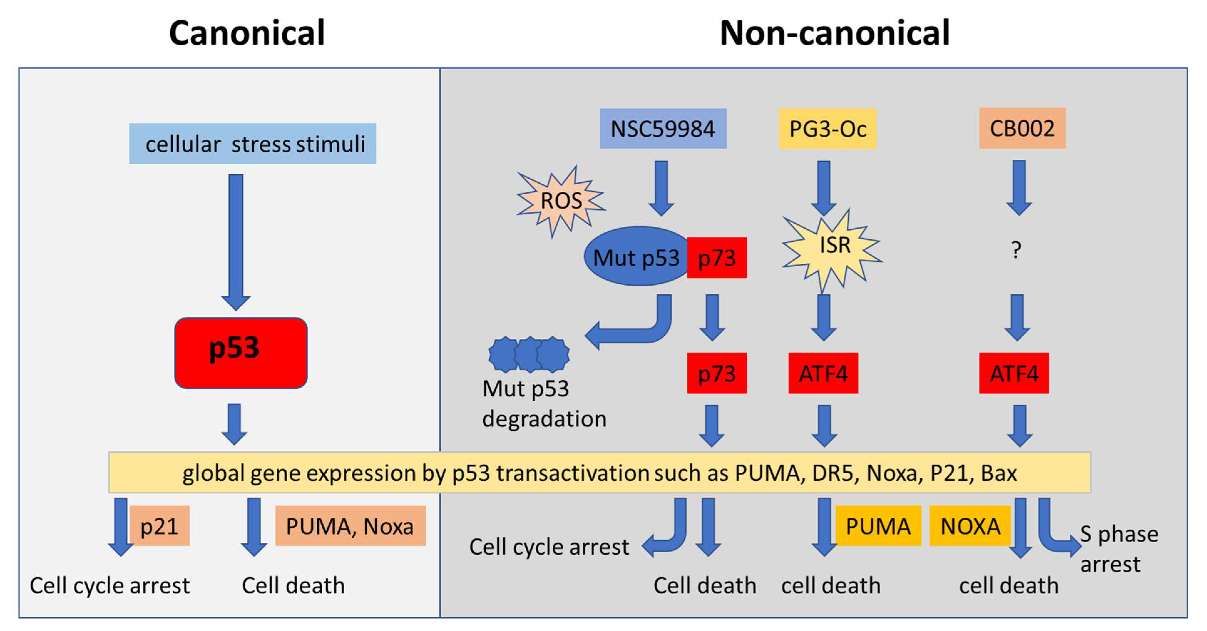
4.4. Restoration of p53 Pathway Signaling Independent of p53 through Non-Canonical Regulatory Pathways

Global gene expression panels show multiple genes transcriptionally regulated by p53 in a canonical way [135,136,137,138,139]. In addition, p53 pathway signaling was found to partially overlap with the other signaling pathways. It is known that the canonical p53 targets can be regulated via other factors via a p53 independent manner [140,141,142,143]. For example, p21 is a p53 transcriptional target and transcriptionally upregulated via p53 binding to its promoter. In addition, p21 also is regulated via other factors either at the transcriptional level or post-translational modifications independent of p53 [144]. Non-canonical pathways also regulate p53 pathway signaling in addition to the canonical p53 regulation. The proof concept of non-canonical regulation of p53 pathway signaling was raised in our recent investigations of p53 pathway restoration. The transcriptomic and proteomic analysis showed that the compounds PG3-Oc, CB002, and their analogues increased ATF3/4 transcriptomic and proteomic targets which overlapped with canonical p53 pathway signaling [145,146]. The integrated stress response ATF3 and ATF4 transcription factors play a role in the stimulation of pro-apoptotic p53 targets by the p53 pathway restoring compounds in tumor cells expressing mutant p53. We found that PG3-Oc partially upregulates the p53-transcriptome (13.7% of public p53 target-gene dataset; 15.2% of the in-house dataset) and the p53-proteome (18%, HT29; 16%, HCT116-p53−/−). ATF4 is a key regulator of PG3-Oc-induced p53 pathway restoration. ATF4 shares a subset of p53 target genes involved in cell cycle arrest and apoptosis, such as PUMA, DR5, p21, Noxa, and NAG-1 in cells in response to PG3-oc [145]. We detected 197 genes in the Fischer p53 dataset (343 genes) and out of those, 102 were differentially expressed by a CB002 analogue in p53 deficient cancer cells, indicating that nearly 50% of the established p53 target genes were altered [146]. Our investigation suggests that ATF4 is a non-canonical factor in restoring p53 pathway signaling independent of p53. ATF3/4 was found to induce NOXA expression by binding to NOXA promoter in head and neck squamous cell carcinoma (HNSCC) in response to cisplatin treatment [141]. These results suggest that p53 pathway signaling can be regulated via non-canonical regulatory pathways (Figure 3). These non-canonical regulation pathways compensate for p53 loss and play an important role in restoring p53 pathway signaling in tumor cells that express mutant p53. The p53 canonical targets Noxa and Puma are regulated by ATF3/4 via non-canonical regulatory pathways and are required to induce cell death by CB002 and PG3-Oc, respectively [145,146]. These results provide a therapeutic strategy for modulating components of non-canonical regulatory pathways to achieve partial restoration of the global p53 transcriptome and proteome that include critical p53-effectors of cell cycle arrest, apoptosis, and metastasis in p53-deficient cancer cells.

5. Targeting p53 Function in Immunotherapy: Bispecific Antibodies, Gene Therapy, Small Molecule Combinations

Immunotherapy is a revolutionary development for cancer therapy [147]. Mutant p53 and wild-type p53 both relate to the immune response. p53 function related to the immune response provides a possibility for combination treatments with immune checkpoint blockade (ICB) in different tumors. With high protein levels and tumor specificity, mutant p53 becomes one of most relevant tumor antigens to target with tumor vaccines and immunotherapy.

5.1. Activation of Wild-Type p53 for Immunotherapy Using Immune Checkpoint Inhibitors

Activation of p53 promotes T-cell infiltration and sensitizes tumor cells to checkpoint therapy. There are some successful examples of activation of p53 in combination with immune checkpoint blockade (ICB) in different tumors. rADp53 in situ gene therapy in combination with pembrolizumab has shown a potent antitumor effect and prolonged survival in different tumor models [148]. The P53MVA vaccine activates CD8+ cells [149] and the P53MVA vaccine in combination with pembrolizumab has appeared feasible, safe, and may offer clinical benefit in patients [150]. Similar to the overexpression of wild-type p53 such as rADp53 and P53MVA, MDM2/MDMX(MDM4) dual inhibitor ALRN-6924 shows that activation of p53 boosts the immune response against tumors by activating interferons in mouse models and human patients [151]. p53-restoring compound PRIMA-1 (APR-246) in combination with pembrolizumab is under clinical evaluation in phase I/II.

5.2. Targeting Mutant p53 in Immunotherapy

CD8+ and CD4+ T-lymphocytes recognize short peptides (epitopes as tumor antigens) derived from proteins presented on the cell surface in association with class I or II human leukocyte antigen (HLA) molecules. High expression of mutant p53 provides a possibility to generate vaccines to prevent tumor growth. However, mutant p53 is localized inside of cells, not on the cell surface.
Recent studies detected mutant p53 peptides presented on the cell surface with HLA complex. This mutant p53 peptide-HLA complex on the cancer cell surface provided a possibility for designing a new approach for immunotherapy by enhancing T-cell recognition of cancer cells based on these neoantigens. For this purpose, Vogelstein and his colleagues developed a new bispecific antibody H2-scDb, with one arm binding to a T-cell receptor and the other recognizing the mutant p53 (R175) peptide-HLA complex on the cancer cell surface [104]. This bispecific antibody function acts as a bridge between cancer cells and T-cells and stimulates T-cells to kill tumor cells. Treatment with this bispecific antibody specifically targets mutant p53-expressing tumor cells and significantly suppresses tumor growth. The bispecific antibody generated with different p53 mutations will bring T cell-based immunotherapy one step forward toward the development of precision cancer medicine.

6. Combination Therapies Targeting p53 Cell Cycle Checkpoints, MDM2-p53 Inhibition, or Mutant p53

Multiple oncogenic and signaling pathway abnormalities in individual cancers promote cancer cell resistance to conventional chemotherapy. Combinatorial treatment with two different drugs with individual targets can enhance antitumor efficacy compared to a monotherapy approach. This approach is widely used in cancer therapy in the clinic [152]. Clinical studies show the effect of AdP53 gene therapy combined with chemotherapy or radiotherapy. These studies provide a rationale for targeting p53 reactivation in combination treatments. p53 activation in combination treatment improves antitumor efficacy and minimizes side effects and newly developed resistance in tumors at different progressive stages.

6.1. Targeting p53 in Combination with Cell Stress Signaling

ROS stresses are common events in cancer cells. Administration of p53-restoring compounds often results in cell stress and increases cellular ROS levels. Cellular stress has been exploited in combination treatment with p53-targeting compounds. PRIMA-1 (APR-246) is one example of a p53-reactivating compound that induces cellular stress that can be exploited with combination treatments. PRIMA-1 (APR-246) was found to increase ROS by depleting GSH. SCL7A11 is a component of the xC(-) system in the regulation of GSH and is considered a major biomarker for APR-246 treatment in cancer [153,154]. PRIMA combined with an xC(-) system inhibitor, which increases ROS, promotes the antitumor effect of the combination treatment [155].
Signaling pathways related to cellular stresses can be targeted in combination with p53-reactivating compounds. JNK is a signaling pathway related to the stress response and has been found to be activated in the p53 pathway targeting treatments; the p53 restoring compound PRIMA was found to induce p53-dependent apoptosis through this pathway in colon cancer [156]. Treatment with the p53-reactivating compound RITA demonstrated a significant number of differentially expressed genes associated with the stress response, including the JNK signaling pathway. Combined treatment with RITA and a JNK activator synergizes in cytotoxic responses in MM cell lines and patient samples [157]. The mechanism by which small molecules induce p53-mediated apoptosis through the JNK signaling pathway provides a rationale for the combination of p53 activating drugs with JNK activators in the treatment of some types of cancer. These studies propose a new approach for targeting mutant p53 in combination with cellular stress signaling.

6.2. Targeting p53 in Combination with Inhibition of Raf/MEK/ERK Pathways

Abnormalities in p53 and the Raf/MEK/ERK axis are among the most common during tumorigenesis and tumor development in various cancers. Targeting p53 in combination with inhibition of the Raf/MEK/ERK pathway is effective in certain drug-resistant cancer cells. The simultaneous blockade of MEK and MDM2 signaling by nutlin-3a triggered synergistic, pro-apoptotic responses in AML cell lines [158]. Similar results were also observed with combination treatment of MDM2 antagonist (RG7388) and MEK inhibitors in dedifferentiated liposarcoma [159]. In melanoma cells, treatment with nutlins combined with inhibitors of ERK2 was found to synergistically induce cell apoptosis. Nutlin-3 combined with vemurafenib (inhibitor of mutant Raf) synergistically induced apoptosis and suppressed melanoma cell viability in vitro and tumor growth in vivo, suggesting that reactivation of p53 by nutlins overcomes vemurafenib resistance in melanoma [160]. Sorafenib, a Raf inhibitor, along with nutlin-3, synergistically induced apoptosis in RCC [161].
These results strongly indicate the therapeutic potential of combined MEK blockade and MDM2 inhibition to activate p53 in cancer cells. A p53-based combinatorial approach can reduce doses and side effects of agents in combinations and efficiently suppresses tumor growth.

6.3. Targeting p53 (Wild-Type and Mutant) in Combination with Conventional Chemotherapy

Clinical studies demonstrate improved outcomes after treatment with conventional chemotherapy in patients with tumors harboring wild-type p53 compared to mutant p53, suggesting a crucial role of wild-type p53 in the cellular response to chemotherapy. This idea is supported by the observation that lymphoid cells from p53 knockout mice experience complete chemoresistance [162]. Furthermore, others have observed that wild-type p53 is necessary for cytotoxicity of conventional chemotherapy in vitro [163,164,165]. These findings suggest that overexpression or reactivation of wild-type p53 may synergize with chemotherapy treatment to induce apoptosis in cancer cells, and this has indeed been observed in vitro [166,167].
p53-targeting small molecules have been investigated for an antitumor effect not only as a monotreatment, but also in combination with chemotherapy. Conventional chemotherapeutic agents 5-FU, doxorubicin, etoposide, and cisplatin are often used in combination treatment with p53-targeting agents. p53-based chemotherapy significantly reduces the genotoxic burden and induces high anti-tumor efficacy.
Nutlins have been widely used in treating cancer cells with wild-type p53. The effectiveness of nutlin-3 in combination with chemotherapy has been observed in preclinical studies. Nutlins and their analogs (such as MI-43, MI-63, and MI-319) synergize with genotoxic drugs such as 5-FU, doxorubicin, etoposide, and cisplatin in multiple tumors carrying wild-type p53 such as hematological malignancies, lymphoma, neuroblastoma, hepatocellular carcinoma, lung cancer, and pancreatic cancer. Nutlins in combination with chemotherapy show minimal toxic effects in normal cells. Dual inhibition of MDM2/MDMX(MDM4) by ALRN-6924 enhances antitumor efficacy of chemotherapy in TP53 wild-type hormone receptor-positive breast cancer models [51]. RITA, in combination with doxorubicin, enhanced the sensitivity of NALM-6 cells to doxorubicin and promoted doxorubicin-induced apoptosis. Small molecules targeting p53 activation decrease the necessary dose of conventional chemotherapy to reduce genotoxicity but enhance or magnify the effects of the chemotherapies.
Targeting mutant p53 in combination with conventional chemotherapy is another approach for increasing antitumor efficacy. Mutant P53 is a contributing factor for drug resistance. p53 mutations have been reported to correlate with resistance to platinum chemotherapy in different types of cancer cells. PRIMA-1 (APR-246) is one of the p53-restoring compounds that is administered in combination treatment with conventional chemotherapy. Combination treatment with PRIMA-1 (APR-246) overcomes cancer cell resistance to chemotherapy. PRIMA-1 was found to synergistically induce cell death in combination with multiple chemo-drugs such as in combination with adriamycin in NSCLC cell lines carrying different p53 mutations [168] or in combination with doxorubicin or cisplatin in thyroid cancer cells [169]. APR-246 has been reported to overcome chemoresistance to cisplatin and doxorubicin in p53-mutant ovarian cancer cells [170]. Combination treatment based on targeting the mutant P53 pathway prevents the development of drug resistance in cancer cells.
The synergistic effect of p53 overexpression or reactivation and chemotherapy provides a promising new direction for the development of combination therapies for patients with tumors containing mutant p53.

6.4. Combination Targeting p53 Cell Cycle Checkpoints in p53 Mutated Cancer Cells

p53 is involved in the G1-S cell cycle checkpoint via regulation of p21 and arrests cells at the G1 phase to allow enough time for DNA repair or otherwise induces cell death in response to DNA damage. Mutant p53-expressing cancer cells have defective G1 cell cycle checkpoints, and this causes such cancer cells to be more reliant on alternative mechanisms to maintain cell survival. The ataxia telangiectasia and Rad3-related kinase (ATR)- checkpoint kinase 1 (Chk1) axis and WEE1 are compensatory pathways independent of p53 involved in intra-S and G2/M checkpoints in response to DNA damage [171]. These cellular vulnerabilities enforced by mutant p53 can be exploited by further targeting of the checkpoints in response to DNA damage in p53-mutated cancer cells. Accumulating studies show that inhibition of ATR/Chk1/WEE1 sensitizes p53-deficient cancer cells to DNA-damaging agents.
Chk1 has a critical role in the DNA damage response (DDR) and cell cycle checkpoints at the S phase and at the G2/M transition [172]. Studies based on in vivo and in vitro experiments show that Chk1 inhibitors (such as UCN-01, AZD7762) sensitize p53-deficient cancer cells to DNA-damaging agents (such as irinotecan and cisplatin) in different tumor types, including TNBC, head and neck cancer, and pancreatic cancer [173,174,175]. The inhibition of Chk1 potentiates the cytotoxicity of DNA damaging agents in p53 deficient cells.
WEE1 is a crucial regulator of the G2/M checkpoint of the cell cycle independent of p53 [176]. Abrogation of the G2/M checkpoint by WEE1 inhibition can sensitize p53-deficient cells to DNA-damaging agents. This is supported by results from experiments with WEE inhibitors in combination with chemotherapy and radiation therapy in p53 mutant cancer cells. MK1775 (Adavosertib, AZD1775) is a small molecular weight WEE1 kinase inhibitor. Blockade of WEE1 by the inhibitor MK1775 synergistically induces cell death in p53 mutant colonic cancer when combined with irinotecan [177]. MK-1775 also synergizes with gemcitabine to suppress tumor growth selectively in p53-deficient pancreatic cancer and ovarian cancer [178,179]. MK1775 prevents WEE1-mediated phosphorylation CDK1.
WEE1 regulates CDK1 activity at the G2/M checkpoint and prevents entry into mitosis. Inhibition of WEE1 causes mitotic entry without completion of DNA repair and replication upon DNA damage, resulting in mitotic catastrophe and apoptosis. The combination results in significant DNA damage due to failure of DNA repair in the cells, which lose both the p53-checkpoint at G1 and the WEE1-checkpoint at G2, leading to synthetic lethality. Clinical studies at phase II show a promising antitumor efficiency of the combination of MK1775 with chemotherapy such as carboplatin and paclitaxel in p53-mutated ovarian cancer [180,181].
Many ATR, Chk1, and WEE1 inhibitors have been developed and evaluated in different tumors in clinical trials [182,183]. Mutant p53 can be a synthetic lethality factor in cancer cells to increase antitumor efficacy of ATR/Chk1/WEE1 inhibitors targeting checkpoints in combination when combined with chemotherapy for cancer therapy.

7. Summary and Prospects

The tumor-specific p53 status and the important role of p53 in the regulation of cell function indicate p53 as a promising target for cancer therapy. Although there are no FDA-approved drugs targeting p53 (wild-type or mutant), advanced strategies based on p53 structure and function have continued to be developed for targeting p53 in cancer therapy (Figure 2 and Figure 3), and some of them are under clinical evaluation (Table 1). Among the strategies, MDM2 is considered one of the key factors in drug targeting wild-type p53 or mutant p53. Interruption of p53 binding to MDM2 is one of the promising strategies to reactivate p53. This has led to the identification of MDM2 inhibitors. By contrast, inducing MDM2-mediated ubiquitination of mutant p53 is another promising strategy to induce mutant p53 degradation, and this has pointed to HSP90 inhibitors, HDAC inhibitors, and NSC59984 that activate MDM2 and destabilize mutant p53. Though clinical trials have demonstrated that MDM2 inhibitors have anti-tumor activity and acceptable safety profiles, some limitations exist, including the development of resistance and dose-limiting toxicity [184,185]. Common resistance mechanisms include the emergence of p53 mutations in tumors that were previously wild-type for p53, alteration of various oncogenic pathways, and MDMX(MDM4) overexpression [186]. Sequestration of wild-type p53 by MDMX(MDM4) after MDM2 inhibition has sparked interest in dual MDM2/MDMX(MDM4) inhibition [187]. Several clinical trials are investigating dual MDM2/MDMX(MDM4) inhibition, but none have been approved yet (Table 1). Further refinement of desirable effects of these small molecules will facilitate the translation of these promising compounds into clinical practice. Non-specific and off-target effects are a common weakness of small molecules when they are applied in cancer therapy. To improve the efficacy and reduce the off-target effects, targeting p53 in combination treatment has been widely investigated in preclinical and clinical research as a promising strategy for enhancing antitumor efficacy and reducing toxicity. Searching potential biomarkers will be essential for targeting p53 in combination therapy. Next genomic sequencing and proteomic assays provide global gene and gene expression patterns of tumors derived from patients. This information may be useful for rationally designing therapeutic approaches by targeting p53 and/or in combination with other altered signal pathways as additional targets in cancers. The patient-derived tumor organoids or patient-derived xenografts mouse models may become invaluable systems to further identify therapeutic approaches and contribute to personalized cancer medicine.
Besides the pharmacological approaches currently developed, biotherapeutic approaches targeting p53 (such as gene therapy and genomic editing) appear as promising strategies in cancer therapy. p53 based gene therapy has been considered a promising approach for cancer therapy as a monotherapy or in combination with chemo- and radiotherapy in a range of tumor types in clinical trials in China [83,188]. Selective delivery of wild-type p53 can increase the specificity of p53 gene therapy. Recently, cationic liposomes coated with anti-transferrin receptor single-chain Ab fragment (scL) has been used to help deliver wild-type p53 (scL-53). This scL53 nanocomplex can be specifically delivered to tumors based on the tumor transferrin receptor that is not expressed on normal cells [189]. The scL serves as an example and provides a new approach for p53-based gene therapy with high specificity and low toxicity. Genetic editing of mutant p53 appears to be an attractive strategy for p53-targeting therapy. The CRISPR/Cas9 system has been used to repair the TP53 414delC mutation to the wild-type TP53 genotype and inhibit the cell proliferation of PC-3 cells [190]. The CRISPR/Cas9 system could become a powerful technique that will bring genomic editing of p53 closer to cancer therapy [191,192].

Author Contributions

Conceptualization, S.Z. and W.S.E.-D.; writing—original draft preparation, S.Z., L.C., A.A.S. and W.S.E.-D.; writing—review and editing, S.Z., L.C., L.H.B., A.A.S., X.T. and W.S.E.-D.; supervision, S.Z. and W.S.E.-D.; project administration, W.S.E.-D.; funding acquisition, W.S.E.-D. All authors have read and agreed to the published version of the manuscript.

Funding

This work was supported in part by the Teymour Alireza P′98, P′00 Family Cancer Research Fund. grant number GFT640730, established by the Alireza Family.

Acknowledgments

W.S.E.-D. is an American Cancer Slociety Research Professor and is supported by the Mencoff Family University Professorship at Brown University.

Conflicts of Interest

W.S.E.-D. is the Founder of p53-Therapeutics. W.S.E-D. has disclosed this relationship and potential conflict of interest to his academic institution/employer and is fully compliant with NIH and institutional policy that is managing this potential conflict of interest. The remaining authors declare no conflict of interest.

References

  1. Kastenhuber, E.R.; Lowe, S.W. Putting p53 in Context. Cell 2017, 170, 1062–1078. [Google Scholar] [CrossRef] [Green Version]
  2. Gomes, A.S.; Ramos, H.; Inga, A.; Sousa, E.; Saraiva, L. Structural and Drug Targeting Insights on Mutant p53. Cancers 2021, 13, 3344. [Google Scholar] [CrossRef]
  3. Joerger, A.C.; Fersht, A.R. The tumor suppressor p53: From structures to drug discovery. Cold Spring Harb. Perspect. Biol. 2010, 2, a000919. [Google Scholar] [CrossRef]
  4. Blandino, G.; Di Agostino, S. New therapeutic strategies to treat human cancers expressing mutant p53 proteins. J. Exp. Clin. Cancer Res. 2018, 37, 30. [Google Scholar] [CrossRef] [Green Version]
  5. Lopes, E.A.; Gomes, S.; Saraiva, L.; Santos, M.M.M. Small Molecules Targeting Mutant P53: A Promising Approach for Cancer Treatment. Curr. Med. Chem. 2020, 26, 7323–7336. [Google Scholar] [CrossRef]
  6. Huang, J. Current developments of targeting the p53 signaling pathway for cancer treatment. Pharmacol. Ther. 2021, 220, 107720. [Google Scholar] [CrossRef]
  7. Valente, J.F.A.; Queiroz, J.A.; Sousa, F. p53 as the Focus of Gene Therapy: Past, Present and Future. Curr. Drug Targets 2018, 19, 1801–1817. [Google Scholar] [CrossRef]
  8. Soragni, A.; Janzen, D.M.; Johnson, L.M.; Lindgren, A.G.; Nguyen, T.Q.A.; Tiourin, E.; Soriaga, A.B.; Lu, J.; Jiang, L.; Faull, K.F.; et al. A Designed Inhibitor of p53 Aggregation Rescues p53 Tumor Suppression in Ovarian Carcinomas. Cancer Cell 2016, 29, 90–103. [Google Scholar] [CrossRef] [Green Version]
  9. Duffy, M.J.; Synnott, N.C.; O’Grady, S.; Crown, J. Targeting p53 for the treatment of cancer. Semin. Cancer Biol. 2020, 79, 58–67. [Google Scholar] [CrossRef]
  10. Sabapathy, K.; Lane, D. Therapeutic targeting of p53: All mutants are equal, but some mutants are more equal than others. Nat. Rev. Clin. Oncol. 2018, 15, 13–30. [Google Scholar] [CrossRef]
  11. Lane, D.P.; Cheok, C.F.; Lain, S. p53-based cancer therapy. Cold Spring Harb. Perspect. Biol. 2010, 2, a001222. [Google Scholar] [CrossRef] [Green Version]
  12. Menichini, P.; Monti, P.; Speciale, A.; Cutrona, G.; Matis, S.; Fais, F.; Taiana, E.; Neri, A.; Bomben, R.; Gentile, M.; et al. Antitumor Effects of PRIMA-1 and PRIMA-1(Met) (APR246) in Hematological Malignancies: Still a Mutant P53-Dependent Affair? Cells 2021, 10, 98. [Google Scholar] [CrossRef]
  13. Sallman, D.A.; DeZern, A.E.; Garcia-Manero, G.; Steensma, D.P.; Roboz, G.J.; Sekeres, M.A.; Cluzeau, T.; Sweet, K.L.; McLemore, A.; McGraw, K.L.; et al. Eprenetapopt (APR-246) and Azacitidine in TP53-Mutant Myelodysplastic Syndromes. J. Clin. Oncol. 2021, 39, 1584–1594. [Google Scholar] [CrossRef]
  14. Lindemann, A.; Patel, A.A.; Tang, L.; Liu, Z.; Wang, L.; Silver, N.L.; Tanaka, N.; Rao, X.; Takahashi, H.; Maduka, N.K.; et al. COTI-2, a novel thiosemicarbazone derivative, exhibits antitumor activity in HNSCC through p53-dependent and -independent mechanisms. Clin. Cancer Res. 2019, 25, 5650–5662. [Google Scholar]
  15. Olivier, M.; Hollstein, M.; Hainaut, P. TP53 Mutations in Human Cancers: Origins, Consequences, and Clinical Use. Cold Spring Harb. Perspect. Biol. 2010, 2, a001008. [Google Scholar] [CrossRef] [Green Version]
  16. Barta, J.A.; McMahon, S.B. Lung-Enriched Mutations in the p53 Tumor Suppressor: A Paradigm for Tissue-Specific Gain of Oncogenic Function. Mol. Cancer Res. 2019, 17, 3–9. [Google Scholar] [CrossRef] [Green Version]
  17. Robles, A.; Harris, C.C. Clinical Outcomes and Correlates of TP53 Mutations and Cancer. Cold Spring Harb. Perspect. Biol. 2009, 2, a001016. [Google Scholar] [CrossRef] [Green Version]
  18. Brosh, R.; Rotter, V. When mutants gain new powers: News from the mutant p53 field. Nat. Cancer 2009, 9, 701–713. [Google Scholar] [CrossRef]
  19. El-Deiry, W.S.; Kern, S.E.; Pietenpol, J.A.; Kinzler, K.W.; Vogelstein, B. Definition of a consensus binding site for p53. Nat. Genet. 1992, 1, 45–49. [Google Scholar] [CrossRef]
  20. Wang, W.; Kim, S.-H.; El-Deiry, W.S. Small-molecule modulators of p53 family signaling and antitumor effects in p53-deficient human colon tumor xenografts. Proc. Natl. Acad. Sci. USA 2006, 103, 11003–11008. [Google Scholar] [CrossRef] [Green Version]
  21. Zhang, S.; Zhou, L.; Hong, B.; van den Heuvel, A.P.J.; Prabhu, V.V.; Warfel, N.A.; Kline, C.L.B.; Dicker, D.T.; Kopelovich, L.; El-Deiry, W.S. Small-Molecule NSC59984 Restores p53 Pathway Signaling and Antitumor Effects against Colorectal Cancer via p73 Activation and Degradation of Mutant p53. Cancer Res. 2015, 75, 3842–3852. [Google Scholar] [CrossRef] [Green Version]
  22. Hong, B.; Prabhu, V.V.; Zhang, S.; van den Heuvel, A.P.J.; Dicker, D.T.; Kopelovich, L.; El-Deiry, W.S. Prodigiosin Rescues Deficient p53 Signaling and Antitumor Effects via Upregulating p73 and Disrupting Its Interaction with Mutant p53. Cancer Res. 2014, 74, 1153–1165. [Google Scholar] [CrossRef] [Green Version]
  23. Richardson, C.; Zhang, S.; Borrero, L.J.H.; El-Deiry, W.S. Small-molecule CB002 restores p53 pathway signaling and represses colorectal cancer cell growth. Cell Cycle 2017, 16, 1719–1725. [Google Scholar] [CrossRef] [Green Version]
  24. Hernandez-Borrero, L.J.; Zhang, S.; Lulla, A.; Dicker, D.T.; El-Deiry, W.S. CB002, a novel p53 tumor suppressor pathway-restoring small molecule induces tumor cell death through the pro-apoptotic protein NOXA. Cell Cycle 2018, 17, 557–567. [Google Scholar] [CrossRef] [Green Version]
  25. Schlichtholz, B.; Trédaniel, J.; Lubin, R.; Zalcman, G.; Hirsch, A.; Soussi, T. Analyses of p53 antibodies in sera of patients with lung carcinoma define immunodominant regions in the p53 protein. Br. J. Cancer 1994, 69, 809–816. [Google Scholar] [CrossRef] [Green Version]
  26. Schlichtholz, B.; Legros, Y.; Gillet, D.; Gaillard, C.; Marty, M.; Lane, D.; Calvo, F.; Soussi, T. The immune response to p53 in breast cancer patients is directed against immunodominant epitopes unrelated to the mutational hot spot. Cancer Res. 1992, 52, 6380–6384. [Google Scholar]
  27. Hafner, A.; Bulyk, M.L.; Jambhekar, A.; Lahav, G. The multiple mechanisms that regulate p53 activity and cell fate. Nat. Rev. Mol. Cell Biol. 2019, 20, 199–210. [Google Scholar] [CrossRef]
  28. Lain, S.; Hollick, J.J.; Campbell, J.; Staples, O.D.; Higgins, M.; Aoubala, M.; McCarthy, A.; Appleyard, V.; Murray, K.E.; Baker, L.; et al. Discovery, In Vivo Activity, and Mechanism of Action of a Small-Molecule p53 Activator. Cancer Cell 2008, 13, 454–463. [Google Scholar] [CrossRef] [Green Version]
  29. Haupt, Y.; Maya, R.; Kazaz, A.; Oren, M. Mdm2 promotes the rapid degradation of p53. Nature 1997, 387, 296–299. [Google Scholar] [CrossRef]
  30. Zhang, S.; Lou, J.; Li, Y.; Zhou, F.; Yan, Z.; Lyu, X.; Zhao, Y. Recent Progress and Clinical Development of Inhibitors that Block MDM4/p53 Protein-Protein Interactions. J. Med. Chem. 2021, 64, 10621–10640. [Google Scholar] [CrossRef]
  31. Espadinha, M.; Barcherini, V.; Lopes, E.A.; Santos, M.M.M. An Update on MDMX and Dual MDM2/X Inhibitors. Curr. Top Med. Chem. 2018, 18, 647–660. [Google Scholar] [CrossRef] [PubMed]
  32. Graves, B.; Thompson, T.; Xia, M.; Janson, C.; Lukacs, C.; Deo, D.; Di Lello, P.; Fry, D.; Garvie, C.; Huang, K.-S.; et al. Activation of the p53 pathway by small-molecule-induced MDM2 and MDMX dimerization. Proc. Natl. Acad. Sci. USA 2012, 109, 11788–11793. [Google Scholar] [CrossRef] [PubMed] [Green Version]
  33. Carvajal, L.A.; Ben Neriah, D.; Senecal, A.; Benard, L.; Thiruthuvanathan, V.; Yatsenko, T.; Narayanagari, S.-R.; Wheat, J.C.; Todorova, T.I.; Mitchell, K.; et al. Dual inhibition of MDMX and MDM2 as a therapeutic strategy in leukemia. Sci. Transl. Med. 2018, 10, eaao3003. [Google Scholar] [CrossRef] [PubMed] [Green Version]
  34. Jenkins, L.M.M.; Durell, S.R.; Mazur, S.J.; Appella, E. p53 N-terminal phosphorylation: A defining layer of complex regulation. Carcinogenesis 2012, 33, 1441–1449. [Google Scholar] [CrossRef] [Green Version]
  35. Carr, M.I.; Jones, S.N. Regulation of the Mdm2-p53 signaling axis in the DNA damage response and tumorigenesis. Transl. Cancer Res. 2016, 5, 707–724. [Google Scholar] [CrossRef] [Green Version]
  36. Liebl, M.C.; Hofmann, T.G. Cell fate regulation upon DNA damage: p53 Serine 46 kinases pave the cell death road. Bioessays 2019, 41, 1900127. [Google Scholar] [CrossRef] [Green Version]
  37. Sun, X.; Dyson, H.J.; Wright, P.E. A phosphorylation-dependent switch in the disordered p53 transactivation domain regulates DNA binding. Proc. Natl. Acad. Sci. USA 2021, 118, e2021456118. [Google Scholar] [CrossRef]
  38. Vieler, M.; Sanyal, S. p53 Isoforms and Their Implications in Cancer. Cancers 2018, 10, 288. [Google Scholar] [CrossRef] [Green Version]
  39. Khoury, M.P.; Bourdon, J.-C. The Isoforms of the p53 Protein. Cold Spring Harb. Perspect. Biol. 2009, 2, a000927. [Google Scholar] [CrossRef] [Green Version]
  40. Bourdon, J.-C.; Surget, S.; Khoury, M.P. Uncovering the role of p53 splice variants in human malignancy: A clinical perspective. Onco. Targets Ther. 2013, 7, 57–68. [Google Scholar] [CrossRef] [Green Version]
  41. Bourdon, J.-C.; Fernandes, K.; Murray-Zmijewski, F.; Liu, G.; Diot, A.; Xirodimas, D.P.; Saville, M.K.; Lane, D.P. p53 isoforms can regulate p53 transcriptional activity. Genes Dev. 2005, 19, 2122–2137. [Google Scholar] [CrossRef] [PubMed] [Green Version]
  42. Olivares-Illana, V.; Fåhraeus, R. p53 isoforms gain functions. Oncogene 2010, 29, 5113–5119. [Google Scholar] [CrossRef] [PubMed] [Green Version]
  43. Joruiz, S.M.; Bourdon, J.-C. p53 Isoforms: Key Regulators of the Cell Fate Decision. Cold Spring Harb. Perspect. Med. 2016, 6, a026039. [Google Scholar] [CrossRef] [PubMed] [Green Version]
  44. Boeckler, F.; Joerger, A.; Jaggi, G.; Rutherford, T.J.; Veprintsev, D.; Fersht, A.R. Targeted rescue of a destabilized mutant of p53 by an in silico screened drug. Proc. Natl. Acad. Sci. USA 2008, 105, 10360–10365. [Google Scholar] [CrossRef] [Green Version]
  45. Bauer, M.; Jones, R.N.; Tareque, R.K.; Springett, B.; Dingler, F.; Verduci, L.; Patel, K.J.; Fersht, A.R.; Joerger, A.C.; Spencer, J. A structure-guided molecular chaperone approach for restoring the transcriptional activity of the p53 cancer mutant Y220C. Future Med. Chem. 2019, 11, 2491–2504. [Google Scholar] [CrossRef] [Green Version]
  46. Baud, M.G.; Bauer, M.; Verduci, L.; Dingler, F.A.; Patel, K.J.; Roy, D.H.; Joerger, A.; Fersht, A.R. Aminobenzothiazole derivatives stabilize the thermolabile p53 cancer mutant Y220C and show anticancer activity in p53-Y220C cell lines. Eur. J. Med. Chem. 2018, 152, 101–114. [Google Scholar] [CrossRef]
  47. Butler, J.S.; Loh, S.N. Structure, Function, and Aggregation of the Zinc-Free Form of the p53 DNA Binding Domain. Biochemistry 2003, 42, 2396–2403. [Google Scholar] [CrossRef]
  48. Yu, X.; Vazquez, A.; Levine, A.J.; Carpizo, D.R. Allele-Specific p53 Mutant Reactivation. Cancer Cell 2012, 21, 614–625. [Google Scholar] [CrossRef] [Green Version]
  49. Galea, C.; Bowman, P.; Kriwacki, R.W. Disruption of an intermonomer salt bridge in the p53 tetramerization domain results in an increased propensity to form amyloid fibrils. Protein Sci. 2005, 14, 2993–3003. [Google Scholar] [CrossRef] [Green Version]
  50. Zawacka-Pankau, J.; Selivanova, G. Pharmacological reactivation of p53 as a strategy to treat cancer. J. Intern. Med. 2014, 277, 248–259. [Google Scholar] [CrossRef] [Green Version]
  51. Pairawan, S.; Zhao, M.; Yuca, E.; Annis, A.; Evans, K.; Sutton, D.; Carvajal, L.; Ren, J.; Santiago, S.; Guerlavais, V. First in class dual MDM2/MDMX inhibitor ALRN-6924 enhances antitumor efficacy of chemotherapy in TP53 wild-type hormone receptor-positive breast cancer models. Breast Cancer Res. 2021, 23, 29. [Google Scholar] [CrossRef] [PubMed]
  52. Perdrix, A.; Najem, A.; Saussez, S.; Awada, A.; Journe, F.; Ghanem, G.; Krayem, M. PRIMA-1 and PRIMA-1Met (APR-246): From Mutant/Wild Type p53 Reactivation to Unexpected Mechanisms Underlying Their Potent Anti-Tumor Effect in Combinatorial Therapies. Cancers 2017, 9, 172. [Google Scholar] [CrossRef] [PubMed] [Green Version]
  53. Vareki, S.M.; Salim, K.Y.; Danter, W.R.; Koropatnick, J. Novel anti-cancer drug COTI-2 synergizes with therapeutic agents and does not induce resistance or exhibit cross-resistance in human cancer cell lines. PLoS ONE 2018, 13, e0191766. [Google Scholar] [CrossRef] [PubMed] [Green Version]
  54. Lampreht Tratar, U.; Horvat, S.; Cemazar, M. Transgenic Mouse Models in Cancer Research. Front. Oncol. 2018, 8, 268. [Google Scholar] [CrossRef] [Green Version]
  55. Kersten, K.; De Visser, K.E.; Van Miltenburg, M.H.; Jonkers, J. Genetically engineered mouse models in oncology research and cancer medicine. EMBO Mol. Med. 2016, 9, 137–153. [Google Scholar] [CrossRef]
  56. Brož, D.K.; Attardi, L.D. In vivo analysis of p53 tumor suppressor function using genetically engineered mouse models. Carcinogenesis 2010, 31, 1311–1318. [Google Scholar] [CrossRef] [Green Version]
  57. Harvey, M.; McArthur, M.J.; Montgomery, C.A., Jr.; Butel, J.S.; Bradley, A.; Donehower, L.A. Spontaneous and carcinogen-induced tumorigenesis in p53-deficient mice. Nat. Genet. 1993, 5, 225–229. [Google Scholar] [CrossRef]
  58. Schwitalla, S.; Ziegler, P.K.; Horst, D.; Becker, V.; Kerle, I.; Begus-Nahrmann, Y.; Lechel, A.; Rudolph, K.L.; Langer, R.; Slotta-Huspenina, J.; et al. Loss of p53 in Enterocytes Generates an Inflammatory Microenvironment Enabling Invasion and Lymph Node Metastasis of Carcinogen-Induced Colorectal Tumors. Cancer Cell 2013, 23, 93–106. [Google Scholar] [CrossRef] [Green Version]
  59. French, J.E.; Lacks, D.; Trempus, C.; Dunnick, J.K.; Foley, J.; Mahler, J.; Tice, R.R.; Tennant, R.W. Loss of heterozygosity frequency at the Trp53 locus in p53-deficient (+/−) mouse tumors is carcinogen-and tissue-dependent. Carcinogenesis 2001, 22, 99–106. [Google Scholar] [CrossRef] [Green Version]
  60. Oda, H.; Zhang, S.; Tsurutani, N.; Shimizu, S.; Nakatsuru, Y.; Aizawa, S.; Ishikawa, T. Loss of p53 is an early event in induction of brain tumors in mice by transplacental carcinogen exposure. Cancer Res. 1997, 57, 646–650. [Google Scholar]
  61. Sakai, H.; Tsukamoto, T.; Yamamoto, M.; Shirai, N.; Iidaka, T.; Hirata, A.; Yanai, T.; Masegi, T.; Donehower, L.A.; Tatematsu, M. High susceptibility of nullizygous p53 knockout mice to colorectal tumor induction by 1,2-dimethylhydrazine. J. Cancer Res. Clin. Oncol. 2003, 129, 335–340. [Google Scholar] [CrossRef] [PubMed]
  62. Ozaki, K.; Sukata, T.; Yamamoto, S.; Uwagawa, S.; Seki, T.; Kawasaki, H.; Yoshitake, A.; Wanibuchi, H.; Koide, A.; Mori, Y. High susceptibility of p53(+/−) knockout mice in N-butyl-N-(4-hydroxybutyl)nitrosamine urinary bladder carcinogenesis and lack of frequent mutation in residual allele. Cancer Res. 1998, 58, 3806–3811. [Google Scholar] [PubMed]
  63. Kado, S.; Uchida, K.; Funabashi, H.; Iwata, S.; Nagata, Y.; Ando, M.; Onoue, M.; Matsuoka, Y.; Ohwaki, M.; Morotomi, M. Intestinal microflora are necessary for development of spontaneous adenocarcinoma of the large intestine in T-cell receptor beta chain and p53 double-knockout mice. Cancer Res. 2001, 61, 2395–2398. [Google Scholar] [PubMed]
  64. Yamamoto, M.; Tsukamoto, T.; Sakai, H.; Shirai, N.; Ohgaki, H.; Furihata, C.; Donehower, L.A.; Yoshida, K.; Tatematsu, M. p53 knockout mice (−/−) are more susceptible than (+/−) or (+/+) mice to N-methyl-N-nitrosourea stomach carcinogenesis. Carcinogenesis 2000, 21, 1891–1897. [Google Scholar] [CrossRef] [Green Version]
  65. Zhou, X.; Singh, M.; Santos, G.S.; Guerlavais, V.; Carvajal, L.A.; Aivado, M.; Zhan, Y.; Oliveira, M.M.S.; Westerberg, L.S.; Annis, D.A. Pharmacological activation of p53 triggers viral mimicry response thereby abolishing tumor immune evasion and promoting anti-tumor immunity. Cancer Discov. 2021. [Google Scholar] [CrossRef]
  66. Blagih, J.; Zani, F.; Chakravarty, P.; Hennequart, M.; Pilley, S.; Hobor, S.; Hock, A.K.; Walton, J.B.; Morton, J.P.; Gronroos, E.; et al. Cancer-Specific Loss of p53 Leads to a Modulation of Myeloid and T Cell Responses. Cell Rep. 2020, 30, 481–496.e6. [Google Scholar] [CrossRef]
  67. A Christophorou, M.; Martin-Zanca, D.; Soucek, L.; Lawlor, E.R.; Brown-Swigart, L.; Verschuren, E.; I Evan, G. Temporal dissection of p53 function in vitro and in vivo. Nat. Genet. 2005, 37, 718–726. [Google Scholar] [CrossRef]
  68. Martins, C.P.; Brown-Swigart, L.; Evan, G.I. Modeling the Therapeutic Efficacy of p53 Restoration in Tumors. Cell 2006, 127, 1323–1334. [Google Scholar] [CrossRef] [Green Version]
  69. Hanel, W.; Marchenko, N.D.; Xu, S.; Yu, S.X.; Weng, W.; Moll, U.M. Two hot spot mutant p53 mouse models display differential gain of function in tumorigenesis. Cell Death Differ. 2013, 20, 898–909. [Google Scholar] [CrossRef] [Green Version]
  70. Lang, G.A.; Iwakuma, T.; Suh, Y.-A.; Liu, G.; Rao, V.; Parant, J.M.; Valentin-Vega, Y.A.; Terzian, T.; Caldwell, L.C.; Strong, L.C.; et al. Gain of Function of a p53 Hot Spot Mutation in a Mouse Model of Li-Fraumeni Syndrome. Cell 2004, 119, 861–872. [Google Scholar] [CrossRef] [Green Version]
  71. Olive, K.; Tuveson, D.A.; Ruhe, Z.C.; Yin, B.; Willis, N.A.; Bronson, R.T.; Crowley, D.; Jacks, T. Mutant p53 Gain of Function in Two Mouse Models of Li-Fraumeni Syndrome. Cell 2004, 119, 847–860. [Google Scholar] [CrossRef] [PubMed] [Green Version]
  72. Alexandrova, E.M.; Yallowitz, A.R.; Li, D.; Xu, S.; Schulz, R.; A Proia, D.; Lozano, G.; Dobbelstein, M.; Moll, U.M. Improving survival by exploiting tumour dependence on stabilized mutant p53 for treatment. Nature 2015, 523, 352–356. [Google Scholar] [CrossRef] [PubMed] [Green Version]
  73. Tchelebi, L.; Ashamalla, H.; Graves, P.R. Mutant p53 and the Response to Chemotherapy and Radiation. Subcell. Biochem. 2014, 85, 133–159. [Google Scholar] [CrossRef] [PubMed]
  74. Chen, J. The Cell-Cycle Arrest and Apoptotic Functions of p53 in Tumor Initiation and Progression. Cold Spring Harb. Perspect. Med. 2016, 6, a026104. [Google Scholar] [CrossRef] [PubMed]
  75. Vega-Stromberg, T. Chemotherapy-induced Secondary Malignancies. J. Infus. Nurs. 2003, 26, 353–361. [Google Scholar] [CrossRef]
  76. Li, H.; Zhang, J.; Tong, J.H.M.; Chan, A.W.H.; Yu, J.; Kang, W.; To, K.F. Targeting the Oncogenic p53 Mutants in Colorectal Cancer and Other Solid Tumors. Int. J. Mol. Sci. 2019, 20, 5999. [Google Scholar] [CrossRef] [Green Version]
  77. Zhu, G.; Pan, C.; Bei, J.-X.; Li, B.; Liang, C.; Xu, Y.; Fu, X. Mutant p53 in Cancer Progression and Targeted Therapies. Front. Oncol. 2020, 10, 595187. [Google Scholar] [CrossRef]
  78. Issaeva, N.; Bozko, P.; Enge, M.; Protopopova, M.; Verhoef, L.G.G.C.; Masucci, M.; Pramanik, A.; Selivanova, G. Small molecule RITA binds to p53, blocks p53–HDM-2 interaction and activates p53 function in tumors. Nat. Med. 2004, 10, 1321–1328. [Google Scholar] [CrossRef]
  79. Sanz, G.; Singh, M.; Peuget, S.; Selivanova, G. Inhibition of p53 inhibitors: Progress, challenges and perspectives. J. Mol. Cell Biol. 2019, 11, 586–599. [Google Scholar] [CrossRef] [Green Version]
  80. Wurz, R.P.; Cee, V.J. Targeted Degradation of MDM2 as a New Approach to Improve the Efficacy of MDM2-p53 Inhibitors. J. Med. Chem. 2019, 62, 445–447. [Google Scholar] [CrossRef] [Green Version]
  81. Levine, A.J. Targeting Therapies for the p53 Protein in Cancer Treatments. Annu. Rev. Cancer Biol. 2019, 3, 21–34. [Google Scholar] [CrossRef]
  82. Heise, C.; Sampson-Johannes, A.; Williams, A.; Mccormick, F.; Von Hoff, D.D.; Kirn, D.H. ONYX-015, an E1B gene-attenuated adenovirus, causes tumor-specific cytolysis and antitumoral efficacy that can be augmented by standard chemotherapeutic agents. Nat. Med. 1997, 3, 639–645. [Google Scholar] [CrossRef] [PubMed]
  83. Zhang, W.-W.; Li, L.; Li, D.; Liu, J.; Li, X.; Li, W.; Xu, X.; Zhang, M.; Chandler, L.A.; Lin, H.; et al. The First Approved Gene Therapy Product for Cancer Ad-p53(Gendicine): 12 Years in the Clinic. Hum. Gene Ther. 2018, 29, 160–179. [Google Scholar] [CrossRef] [PubMed] [Green Version]
  84. Pearson, S.; Jia, H.; Kandachi, K. China approves first gene therapy. Nat. Biotechnol. 2004, 22, 3–4. [Google Scholar] [CrossRef] [PubMed]
  85. Foster, B.A. Pharmacological Rescue of Mutant p53 Conformation and Function. Science 1999, 286, 2507–2510. [Google Scholar] [CrossRef] [PubMed] [Green Version]
  86. Bykov, V.N.; Issaeva, N.; Shilov, A.; Hultcrantz, M.; Pugacheva, E.; Chumakov, P.; Bergman, J.; Wiman, K.; Selivanova, G. Restoration of the tumor suppressor function to mutant p53 by a low-molecular-weight compound. Nat. Med. 2002, 8, 282–288. [Google Scholar] [CrossRef]
  87. Bykov, V.N.; Zache, N.; Stridh, H.; Westman, J.; Bergman, J.; Selivanova, G.; Wiman, K.G. PRIMA-1MET synergizes with cisplatin to induce tumor cell apoptosis. Oncogene 2005, 24, 3484–3491. [Google Scholar] [CrossRef] [Green Version]
  88. Bykov, V.N.; Issaeva, N.; Zache, N.; Shilov, A.; Hultcrantz, M.; Bergman, J.; Selivanova, G.; Wiman, K. Reactivation of Mutant p53 and Induction of Apoptosis in Human Tumor Cells by Maleimide Analogs. J. Biol. Chem. 2005, 280, 30384–30391. [Google Scholar] [CrossRef] [Green Version]
  89. Zache, N.; Lambert, J.M.; Rökaeus, N.; Shen, J.; Hainaut, P.; Bergman, J.; Wiman, K.G.; Bykov, V.J. Mutant p53 targeting by the low molecular weight compound STIMA-1. Mol. Oncol. 2008, 2, 70–80. [Google Scholar] [CrossRef] [Green Version]
  90. Kaar, J.; Basse, N.; Joerger, A.; Stephens, E.; Rutherford, T.J.; Fersht, A.R. Stabilization of mutant p53 via alkylation of cysteines and effects on DNA binding. Protein Sci. 2010, 19, 2267–2278. [Google Scholar] [CrossRef] [Green Version]
  91. Punganuru, S.R.; Madala, H.R.; Venugopal, S.N.; Samala, R.; Mikelis, C.; Srivenugopal, K.S. Design and synthesis of a C7-aryl piperlongumine derivative with potent antimicrotubule and mutant p53-reactivating properties. Eur. J. Med. Chem. 2016, 107, 233–244. [Google Scholar] [CrossRef] [PubMed]
  92. Bauer, M.; Joerger, A.; Fersht, A.R. 2-Sulfonylpyrimidines: Mild alkylating agents with anticancer activity toward p53-compromised cells. Proc. Natl. Acad. Sci. USA 2016, 113, E5271–E5280. [Google Scholar] [CrossRef] [PubMed] [Green Version]
  93. Synnott, N.C.; O’Connell, D.; Crown, J.; Duffy, M.J. COTI-2 reactivates mutant p53 and inhibits growth of triple-negative breast cancer cells. Breast Cancer Res. Treat. 2019, 179, 47–56. [Google Scholar] [CrossRef]
  94. Gomes, A.S.; Ramos, H.; Gomes, S.; Loureiro, J.; Soares, J.; Barcherini, V.; Monti, P.; Fronza, G.; Oliveira, C.; Domingues, L.; et al. SLMP53-1 interacts with wild-type and mutant p53 DNA-binding domain and reactivates multiple hotspot mutations. Biochim. Biophys. Acta Gen. Subj. 2020, 1864, 129440. [Google Scholar] [CrossRef] [PubMed]
  95. Soares, J.; Raimundo, L.; Pereira, N.A.L.; Monteiro, Â.; Gomes, S.; Bessa, C.; Pereira, C.; Queiroz, G.; Bisio, A.; Fernandes, J.; et al. Reactivation of wild-type and mutant p53 by tryptophanolderived oxazoloisoindolinone SLMP53-1, a novel anticancer small-molecule. Oncotarget 2016, 7, 4326–4343. [Google Scholar] [CrossRef] [Green Version]
  96. Gomes, S.; Bosco, B.; Loureiro, J.B.; Ramos, H.; Raimundo, L.; Soares, J.; Nazareth, N.; Barcherini, V.; Domingues, L.; Oliveira, C.; et al. SLMP53-2 Restores Wild-Type-Like Function to Mutant p53 through Hsp70: Promising Activity in Hepatocellular Carcinoma. Cancers 2019, 11, 1151. [Google Scholar] [CrossRef] [Green Version]
  97. Tal, P.; Eizenberger, S.; Cohen, E.; Goldfinger, N.; Pietrokovski, S.; Oren, M.; Rotter, V. Cancer therapeutic approach based on conformational stabilization of mutant p53 protein by small peptides. Oncotarget 2016, 7, 11817–11837. [Google Scholar] [CrossRef] [Green Version]
  98. Proia, D.A.; Bates, R.C. Ganetespib and HSP90: Translating Preclinical Hypotheses into Clinical Promise. Cancer Res. 2014, 74, 1294–1300. [Google Scholar] [CrossRef] [Green Version]
  99. Li, D.; Marchenko, N.; Moll, U.M. SAHA shows preferential cytotoxicity in mutant p53 cancer cells by destabilizing mutant p53 through inhibition of the HDAC6-Hsp90 chaperone axis. Cell Death Differ. 2011, 18, 1904–1913. [Google Scholar] [CrossRef] [Green Version]
  100. Foggetti, G.; Ottaggio, L.; Russo, D.; Mazzitelli, C.; Monti, P.; Degan, P.; Miele, M.; Fronza, G.; Menichini, P. Autophagy induced by SAHA affects mutant P53 degradation and cancer cell survival. Biosci. Rep. 2019, 39, BSR20181345. [Google Scholar] [CrossRef] [Green Version]
  101. Parrales, A.; Ranjan, A.; Iyer, S.; Padhye, S.; Weir, S.J.; Roy, A.; Iwakuma, T. DNAJA1 controls the fate of misfolded mutant p53 through the mevalonate pathway. Nat. Cell Biol. 2016, 18, 1233–1243. [Google Scholar] [CrossRef] [PubMed]
  102. Vakifahmetoglu-Norberg, H.; Kim, M.; Xia, H.G.; Iwanicki, M.P.; Ofengeim, D.; Coloff, J.L.; Pan, L.; Ince, T.A.; Kroemer, G.; Brugge, J.S.; et al. Chaperone-mediated autophagy degrades mutant p53. Genes Dev. 2013, 27, 1718–1730. [Google Scholar] [CrossRef] [PubMed] [Green Version]
  103. Kravchenko, J.E.; Ilyinskaya, G.V.; Komarov, P.G.; Agapova, L.S.; Kochetkov, D.V.; Strom, E.; Frolova, E.I.; Kovriga, I.; Gudkov, A.; Feinstein, E.; et al. Small-molecule RETRA suppresses mutant p53-bearing cancer cells through a p73-dependent salvage pathway. Proc. Natl. Acad. Sci. USA 2008, 105, 6302–6307. [Google Scholar] [CrossRef] [PubMed] [Green Version]
  104. Hsiue, E.H.-C.; Wright, K.M.; Douglass, J.; Hwang, M.S.; Mog, B.J.; Pearlman, A.H.; Paul, S.; DiNapoli, S.R.; Konig, M.F.; Wang, Q.; et al. Targeting a neoantigen derived from a common TP53 mutation. Science 2021, 371, eabc8697. [Google Scholar] [CrossRef]
  105. Bykov, V.J.N.; Eriksson, S.E.; Bianchi, J.; Wiman, K.G. Targeting mutant p53 for efficient cancer therapy. Nat. Rev. Cancer 2018, 18, 89–102. [Google Scholar] [CrossRef]
  106. Duffy, M.J.; Synnott, N.C.; Crown, J. Mutant p53 as a target for cancer treatment. Eur. J. Cancer 2017, 83, 258–265. [Google Scholar] [CrossRef]
  107. Grinkevich, V.V.; Nikulenkov, F.; Shi, Y.; Enge, M.; Bao, W.; Maljukova, A.; Gluch, A.; Kel, A.; Sangfelt, O.; Selivanova, G. Ablation of key oncogenic pathways by RITA-reactivated p53 is required for efficient apoptosis. Cancer Cell 2009, 15, 441–453. [Google Scholar] [CrossRef] [Green Version]
  108. Ahmed, A.; Yang, J.; Maya-Mendoza, A.; Jackson, D.A.; Ashcroft, M. Pharmacological activation of a novel p53-dependent S-phase checkpoint involving CHK-1. Cell Death Dis. 2011, 2, e160. [Google Scholar] [CrossRef] [Green Version]
  109. Yang, J.; Ahmed, A.; Poon, E.; Perusinghe, N.; Brandon, A.D.H.; Box, G.; Valenti, M.; Eccles, S.; Rouschop, K.; Wouters, B.; et al. Small-Molecule Activation of p53 Blocks Hypoxia-Inducible Factor 1α and Vascular Endothelial Growth Factor Expression In Vivo and Leads to Tumor Cell Apoptosis in Normoxia and Hypoxia. Mol. Cell. Biol. 2009, 29, 2243–2253. [Google Scholar] [CrossRef] [Green Version]
  110. Shi, Y.; Nikulenkov, F.; Zawacka-Pankau, J.; Li, H.; Gabdoulline, R.; Xu, J.; Eriksson, S.; Hedström, E.; Issaeva, N.; Kel, A.; et al. ROS-dependent activation of JNK converts p53 into an efficient inhibitor of oncogenes leading to robust apoptosis. Cell Death Differ. 2014, 21, 612–623. [Google Scholar] [CrossRef] [Green Version]
  111. Wiegering, A.; Matthes, N.; Mühling, B.; Koospal, M.; Quenzer, A.; Peter, S.; Germer, C.-T.; Linnebacher, M.; Otto, C. Reactivating p53 and Inducing Tumor Apoptosis (RITA) Enhances the Response of RITA-Sensitive Colorectal Cancer Cells to Chemotherapeutic Agents 5-Fluorouracil and Oxaliplatin. Neoplasia 2017, 19, 301–309. [Google Scholar] [CrossRef] [PubMed]
  112. Weilbacher, A.; Gutekunst, M.; Oren, M.; E Aulitzky, W.; Van Der Kuip, H. RITA can induce cell death in p53-defective cells independently of p53 function via activation of JNK/SAPK and p38. Cell Death Dis. 2014, 5, e1318. [Google Scholar] [CrossRef] [PubMed] [Green Version]
  113. Hu, C.; Li, X.; Wang, W.; Zhang, L.; Tao, L.; Dong, X.; Sheng, R.; Yang, B.; Hu, Y. Design, synthesis, and biological evaluation of imidazoline derivatives as p53–MDM2 binding inhibitors. Bioorganic Med. Chem. 2011, 19, 5454–5461. [Google Scholar] [CrossRef] [PubMed]
  114. Ray-Coquard, I.; Blay, J.-Y.; Italiano, A.; Le Cesne, A.; Penel, N.; Zhi, J.; Heil, F.; Rueger, R.; Graves, B.; Ding, M.; et al. Effect of the MDM2 antagonist RG7112 on the P53 pathway in patients with MDM2-amplified, well-differentiated or dedifferentiated liposarcoma: An exploratory proof-of-mechanism study. Lancet Oncol. 2012, 13, 1133–1140. [Google Scholar] [CrossRef]
  115. Ding, Q.; Zhang, Z.; Liu, J.-J.; Jiang, N.; Zhang, J.; Ross, T.M.; Chu, X.-J.; Bartkovitz, D.; Podlaski, F.; Janson, C.; et al. Discovery of RG7388, a Potent and Selective p53–MDM2 Inhibitor in Clinical Development. J. Med. Chem. 2013, 56, 5979–5983. [Google Scholar] [CrossRef] [PubMed]
  116. Verreault, M.; Schmitt, C.; Goldwirt, L.; Pelton, K.; Haidar, S.; Levasseur, C.; Guehennec, J.; Knoff, D.; Labussière, M.; Marie, Y.; et al. Preclinical Efficacy of the MDM2 Inhibitor RG7112 in MDM2-Amplified and TP53 Wild-type Glioblastomas. Clin. Cancer Res. 2016, 22, 1185–1196. [Google Scholar] [CrossRef] [PubMed] [Green Version]
  117. Andreeff, M.; Kelly, K.R.; Yee, K.; Assouline, S.; Strair, R.; Popplewell, L.; Bowen, D.; Martinelli, G.; Drummond, M.W.; Vyas, P.; et al. Results of the Phase I Trial of RG7112, a Small-Molecule MDM2 Antagonist in Leukemia. Clin. Cancer Res. 2016, 22, 868–876. [Google Scholar] [CrossRef] [Green Version]
  118. Patnaik, A.; Tolcher, A.; Beeram, M.; Nemunaitis, J.; Weiss, G.J.; Bhalla, K.; Agrawal, M.; Nichols, G.; Middleton, S.; Beryozkina, A.; et al. Clinical pharmacology characterization of RG7112, an MDM2 antagonist, in patients with advanced solid tumors. Cancer Chemother Pharmacol. 2015, 76, 587–595. [Google Scholar] [CrossRef]
  119. Her, N.-G.; Oh, J.-W.; Oh, Y.J.; Han, S.; Cho, H.J.; Lee, Y.; Ryu, G.H.; Nam, D.-H. Potent effect of the MDM2 inhibitor AMG232 on suppression of glioblastoma stem cells. Cell Death Dis. 2018, 9, 1–12. [Google Scholar] [CrossRef]
  120. Shadfan, M.; Lopez-Pajares, V.; Yuan, Z.-M. MDM2 and MDMX: Alone and together in regulation of p53. Transl. Cancer Res. 2012, 1, 88–89. [Google Scholar]
  121. Reed, D.; Shen, Y.; Shelat, A.A.; Arnold, L.A.; Ferreira, A.M.; Zhu, F.; Mills, N.; Smithson, D.C.; Regni, C.A.; Bashford, D.; et al. Identification and Characterization of the First Small Molecule Inhibitor of MDMX. J. Biol. Chem. 2010, 285, 10786–10796. [Google Scholar] [CrossRef] [PubMed] [Green Version]
  122. Saleh, M.N.; Patel, M.R.; Bauer, T.M.; Goel, S.; Falchook, G.S.; Shapiro, G.I.; Chung, K.Y.; Infante, J.R.; Conry, R.M.; Rabinowits, G.; et al. Phase 1 Trial of ALRN-6924, a Dual Inhibitor of MDMX and MDM2, in Patients with Solid Tumors and Lymphomas Bearing Wild-type TP53. Clin. Cancer Res. 2021, 27, 5236–5247. [Google Scholar] [CrossRef] [PubMed]
  123. Gasparian, A.V.; Burkhart, C.A.; Purmal, A.A.; Brodsky, L.; Pal, M.; Saranadasa, M.; Bosykh, D.A.; Commane, M.; Guryanova, O.A.; Pal, S.; et al. Curaxins: Anticancer compounds that simultaneously suppress NF-kappaB and activate p53 by targeting FACT. Sci. Transl. Med. 2011, 3, 95ra74. [Google Scholar] [CrossRef] [PubMed]
  124. Bae, S.K.; Gwak, J.; Song, I.-S.; Park, H.-S.; Oh, S. Induction of apoptosis in colon cancer cells by a novel topoisomerase I inhibitor TopIn. Biochem. Biophys. Res. Commun. 2011, 409, 75–81. [Google Scholar] [CrossRef]
  125. Lambert, J.M.; Gorzov, P.; Veprintsev, D.; Söderqvist, M.; Segerbäck, D.; Bergman, J.; Fersht, A.R.; Hainaut, P.; Wiman, K.G.; Bykov, V.N. PRIMA-1 Reactivates Mutant p53 by Covalent Binding to the Core Domain. Cancer Cell 2009, 15, 376–388. [Google Scholar] [CrossRef] [Green Version]
  126. Zhang, Q.; Bykov, V.J.N.; Wiman, K.G.; Zawacka-Pankau, J. APR-246 reactivates mutant p53 by targeting cysteines 124 and 277. Cell Death Dis. 2018, 9, 439. [Google Scholar] [CrossRef] [Green Version]
  127. Demma, M.; Maxwell, E.; Ramos, R.; Liang, L.; Li, C.; Hesk, D.; Rossman, R.; Mallams, A.; Doll, R.; Liu, M.; et al. SCH529074, a Small Molecule Activator of Mutant p53, Which Binds p53 DNA Binding Domain (DBD), Restores Growth-suppressive Function to Mutant p53 and Interrupts HDM2-mediated Ubiquitination of Wild Type p53. J. Biol. Chem. 2010, 285, 10198–10212. [Google Scholar] [CrossRef] [Green Version]
  128. Salim, K.Y.; Vareki, A.M.; Danter, W.R.; Koropatnick, J. COTI-2, a novel small molecule that is active against multiple human cancer cell lines in vitro and in vivo. Oncotarget 2016, 7, 41363–41379. [Google Scholar] [CrossRef] [Green Version]
  129. Lin, K.; Rockliffe, N.; Johnson, G.G.; Sherrington, P.D.; Pettitt, A.R. Hsp90 inhibition has opposing effects on wild-type and mutant p53 and induces p21 expression and cytotoxicity irrespective of p53/ATM status in chronic lymphocytic leukaemia cells. Oncogene 2008, 27, 2445–2455. [Google Scholar] [CrossRef] [Green Version]
  130. Li, D.; Marchenko, N.D.; Schulz, R.; Fischer, V.; Velasco-Hernandez, T.; Talos, F.; Moll, U.M. Functional Inactivation of Endogenous MDM2 and CHIP by HSP90 Causes Aberrant Stabilization of Mutant p53 in Human Cancer Cells. Mol. Cancer Res. 2011, 9, 577–588. [Google Scholar] [CrossRef] [Green Version]
  131. Peng, Y.; Chen, L.; Li, C.; Lu, W.; Chen, J. Inhibition of MDM2 by hsp90 Contributes to Mutant p53 Stabilization. J. Biol. Chem. 2001, 276, 40583–40590. [Google Scholar] [CrossRef] [PubMed] [Green Version]
  132. Hu, Z.; Zhang, Y.; He, Y.; Cao, Q.; Zhang, T.; Lou, L.; Cai, Q. Full-Length Transcriptome Assembly of Italian Ryegrass Root Integrated with RNA-Seq to Identify Genes in Response to Plant Cadmium Stress. Int. J. Mol. Sci. 2020, 21, 1067. [Google Scholar] [CrossRef] [PubMed] [Green Version]
  133. Qi, Z.; Yang, G.; Deng, T.; Wang, J.; Zhou, H.; Popov, S.A.; Shults, E.E.; Wang, C. Design and linkage optimization of ursane-thalidomide-based PROTACs and identification of their targeted-degradation properties to MDM2 protein. Bioorganic Chem. 2021, 111, 104901. [Google Scholar] [CrossRef]
  134. Zhang, S.; Zhou, L.; El-Deiry, W.S. Small-Molecule NSC59984 Induces Mutant p53 Degradation through a ROS-ERK2-MDM2 Axis in Cancer Cells. Mol. Cancer Res. 2022. [Google Scholar] [CrossRef] [PubMed]
  135. Galbraith, M.D.; Andrysik, Z.; Sullivan, K.D.; Espinosa, J.M. Global Analyses to Identify Direct Transcriptional Targets of p53. Methods Pharmacol. Toxicol. 2021, 2267, 19–56. [Google Scholar] [CrossRef]
  136. Allen, M.A.; Andrysik, Z.; Dengler, V.L.; Mellert, H.S.; Guarnieri, A.; A Freeman, J.; Sullivan, K.D.; Galbraith, M.D.; Luo, X.; Kraus, W.L.; et al. Global analysis of p53-regulated transcription identifies its direct targets and unexpected regulatory mechanisms. eLife 2014, 3, e02200. [Google Scholar] [CrossRef] [PubMed]
  137. Broz, D.K.; Mello, S.S.; Bieging, K.T.; Jiang, D.; Dusek, R.L.; Brady, C.A.; Sidow, A.; Attardi, L.D. Global genomic profiling reveals an extensive p53-regulated autophagy program contributing to key p53 responses. Genes Dev. 2013, 27, 1016–1031. [Google Scholar] [CrossRef] [Green Version]
  138. Menendez, D.; Nguyen, T.-A.; Freudenberg, J.M.; Mathew, V.J.; Anderson, C.W.; Jothi, R.; Resnick, M.A. Diverse stresses dramatically alter genome-wide p53 binding and transactivation landscape in human cancer cells. Nucleic Acids Res. 2013, 41, 7286–7301. [Google Scholar] [CrossRef] [Green Version]
  139. Fischer, M. Census and evaluation of p53 target genes. Oncogene 2017, 36, 3943–3956. [Google Scholar] [CrossRef] [Green Version]
  140. Li, M. The role of P53 up-regulated modulator of apoptosis (PUMA) in ovarian development, cardiovascular and neurodegenerative diseases. Apoptosis 2021, 26, 235–247. [Google Scholar] [CrossRef]
  141. Sharma, K.; Vu, T.; Cook, W.; Naseri, M.; Zhan, K.; Nakajima, W.; Harada, H. p53-independent Noxa induction by cisplatin is regulated by ATF3/ATF4 in head and neck squamous cell carcinoma cells. Mol. Oncol. 2018, 12, 788–798. [Google Scholar] [CrossRef] [PubMed]
  142. Armstrong, J.L.; Veal, G.J.; Redfern, C.P.F.; Lovat, P. Role of Noxa in p53-independent fenretinide-induced apoptosis of neuroectodermal tumours. Apoptosis 2007, 12, 613–622. [Google Scholar] [CrossRef] [PubMed]
  143. Pérez-Galán, P.; Roué, G.; Villamor, N.; Montserrat, E.; Campo, E.; Colomer, D. The proteasome inhibitor bortezomib induces apoptosis in mantle-cell lymphoma through generation of ROS and Noxa activation independent of p53 status. Blood 2006, 107, 257–264. [Google Scholar] [CrossRef] [PubMed] [Green Version]
  144. Warfel, N.A.; El-Deiry, W.S. p21WAF1 and tumourigenesis: 20 years after. Curr. Opin. Oncol. 2013, 25, 52–58. [Google Scholar] [CrossRef] [PubMed]
  145. Tian, X.; Ahsan, N.; Lulla, A.; Lev, A.; Abbosh, P.; Dicker, D.T.; Zhang, S.; El-Deiry, W.S. P53-independent partial restoration of the p53 pathway in tumors with mutated p53 through ATF4 transcriptional modulation by ERK1/2 and CDK9. Neoplasia 2021, 23, 304–325. [Google Scholar] [CrossRef]
  146. Borrero, L.H.; Dicker, D.T.; Santiago, J.; Sanders, J.; Tian, X.; Ahsan, N.; Lev, A.; Zhou, L.; El-Deiry, W.S. A subset of CB002 xanthine analogs bypass p53-signaling to restore a p53 transcriptome and target an S-phase cell cycle checkpoint in tumors with mutated-p53. eLife 2021, 10, 10. [Google Scholar] [CrossRef]
  147. DeLeo, A.B.; Appella, E. The p53 Saga: Early Steps in the Development of Tumor Immunotherapy. J. Immunol. 2020, 204, 2321–2328. [Google Scholar] [CrossRef]
  148. Kunimura, N.; Kitagawa, K.; Sako, R.; Narikiyo, K.; Tominaga, S.; Bautista, D.S.; Wei Xu, M.F.; Shirakawa, T. Combination of rAd-p53 in situ gene therapy and anti-PD-1 antibody immunotherapy induced anti-tumor activity in mouse syngeneic urogenital cancer models. Sci. Rep. 2020, 10, 17464. [Google Scholar] [CrossRef]
  149. Hardwick, N.R.; Carroll, M.; Kaltcheva, T.; Qian, D.; Lim, D.; Leong, L.; Chu, P.; Kim, J.; Chao, J.; Fakih, M.; et al. p53MVA Therapy in Patients with Refractory Gastrointestinal Malignancies Elevates p53-Specific CD8+ T-cell Responses. Clin. Cancer Res. 2014, 20, 4459–4470. [Google Scholar] [CrossRef] [Green Version]
  150. Chung, V.; Kos, F.J.; Hardwick, N.; Yuan, Y.; Chao, J.; Li, D.; Waisman, J.; Li, M.; Zurcher, K.; Frankel, P.; et al. Evaluation of safety and efficacy of p53MVA vaccine combined with pembrolizumab in patients with advanced solid cancers. Clin. Transl. Oncol. 2018, 21, 363–372. [Google Scholar] [CrossRef]
  151. Hao, Q.; Lu, H.; Zhou, X. A potential synthetic lethal strategy with PARP inhibitors: Perspective on ‘Inactivation of the tumor suppressor p53 by long noncoding RNA RMRP’. J. Mol. Cell Biol. 2021, 13, 690–692. [Google Scholar] [CrossRef] [PubMed]
  152. Yap, T.A.; Omlin, A.; De Bono, J.S. Development of Therapeutic Combinations Targeting Major Cancer Signaling Pathways. J. Clin. Oncol. 2013, 31, 1592–1605. [Google Scholar] [CrossRef] [PubMed]
  153. Grellety, T.; Laroche-Clary, A.; Chaire, V.; Lagarde, P.; Chibon, F.; Neuville, A.; Italiano, A. PRIMA-1(MET) induces death in soft-tissue sarcomas cell independent of p53. BMC Cancer 2015, 15, 684. [Google Scholar] [CrossRef] [PubMed] [Green Version]
  154. Fujihara, K.M.; Benitez, M.C.; Cabalag, C.S.; Zhang, B.Z.; Ko, H.S.; Liu, D.S.; Simpson, K.J.; Haupt, Y.; Lipton, L.; Haupt, S.; et al. SLC7A11 Is a Superior Determinant of APR-246 (Eprenetapopt) Response than TP53 Mutation Status. Mol. Cancer Ther. 2021, 20, 1858–1867. [Google Scholar] [CrossRef]
  155. Liu, D.S.; Duong, C.P.; Haupt, S.; Montgomery, K.G.; House, C.M.; Azar, W.J.; Pearson, H.B.; Fisher, O.M.; Read, M.; Guerra, G.R.; et al. Inhibiting the system xC(-)/glutathione axis selectively targets cancers with mutant-p53 accumulation. Nat. Commun. 2017, 8, 14844. [Google Scholar] [CrossRef] [Green Version]
  156. Li, Y.; Mao, Y.; Brandt-Rauf, P.W.; Williams, A.C.; Fine, R.L. Selective induction of apoptosis in mutant p53 premalignant and malignant cancer cells by PRIMA-1 through the c-Jun-NH2-kinase pathway. Mol. Cancer Ther. 2005, 4, 901–909. [Google Scholar] [CrossRef] [Green Version]
  157. Saha, M.N.; Jiang, H.; Yang, Y.; Zhu, X.; Wang, X.; Schimmer, A.D.; Qiu, L.; Chang, H. Targeting p53 via JNK pathway: A novel role of RITA for apoptotic signaling in multiple myeloma. PLoS ONE 2012, 7, e30215. [Google Scholar] [CrossRef] [Green Version]
  158. Wang, X.; Yamamoto, Y.; Imanishi, M.; Zhang, X.; Sato, M.; Sugaya, A.; Hirose, M.; Endo, S.; Natori, Y.; Moriwaki, T.; et al. Enhanced G1 arrest and apoptosis via MDM4/MDM2 double knockdown and MEK inhibition in wild-type TP53 colon and gastric cancer cells with aberrant KRAS signaling. Oncol. Lett. 2021, 22, 558. [Google Scholar] [CrossRef]
  159. Roy, S.; Laroche-Clary, A.; Verbeke, S.; Derieppe, M.A.; Italiano, A. MDM2 Antagonists Induce a Paradoxical Activation of Erk1/2 through a P53-Dependent Mechanism in Dedifferentiated Liposarcomas: Implications for Combinatorial Strategies. Cancers 2020, 12, 2253. [Google Scholar] [CrossRef]
  160. Ji, Z.; Kumar, R.; Taylor, M.J.; Rajadurai, A.; Marzuka-Alcalá, A.; Chen, Y.E.; Njauw, C.-N.J.; Flaherty, K.T.; Jönsson, G.B.; Tsao, H. Vemurafenib Synergizes with Nutlin-3 to Deplete Survivin and Suppresses Melanoma Viability and Tumor Growth. Clin. Cancer Res. 2013, 19, 4383–4391. [Google Scholar] [CrossRef] [Green Version]
  161. Vatsyayan, R.; Singhal, J.; Nagaprashantha, L.P.D.; Awasthi, S.; Singhal, S.S. Nutlin-3 enhances sorafenib efficacy in renal cell carcinoma. Mol. Carcinog. 2013, 52, 39–48. [Google Scholar] [CrossRef] [PubMed] [Green Version]
  162. Aubrey, B.J.; Kelly, G.L.; Janic, A.; Herold, M.J.; Strasser, A. How does p53 induce apoptosis and how does this relate to p53-mediated tumour suppression? Cell Death Differ. 2018, 25, 104–113. [Google Scholar] [CrossRef] [PubMed] [Green Version]
  163. Bragado, P.; Armesilla, A.; Silva, A.; Porras, A. Apoptosis by cisplatin requires p53 mediated p38α MAPK activation through ROS generation. Apoptosis 2007, 12, 1733–1742. [Google Scholar] [CrossRef] [PubMed]
  164. Grandela, C.; Pera, M.F.; Grimmond, S.M.; Kolle, G.; Wolvetang, E.J. p53 is required for etoposide-induced apoptosis of human embryonic stem cells. Stem. Cell Res. 2007, 1, 116–128. [Google Scholar] [CrossRef] [Green Version]
  165. Sun, Y.; Xia, P.; Zhang, H.; Liu, B.; Shi, Y. P53 is required for Doxorubicin-induced apoptosis via the TGF-beta signaling pathway in osteosarcoma-derived cells. Am. J. Cancer Res. 2015, 6, 114–125. [Google Scholar]
  166. Saha, M.N.; Chen, Y.; Chen, M.-H.; Chen, G.; Chang, H. Small molecule MIRA-1 induces in vitro and in vivo anti-myeloma activity and synergizes with current anti-myeloma agents. Br. J. Cancer 2014, 110, 2224–2231. [Google Scholar] [CrossRef] [Green Version]
  167. Xie, Q.; Wu, M.Y.; Zhang, D.X.; Yang, Y.M.; Wang, B.S.; Zhang, J.; Xu, J.; Zhong, W.; Hu, J. Synergistic anticancer effect of exogenous wild-type p53 gene combined with 5-FU in human colon cancer resistant to 5-FU in vivo. World J. Gastroenterol. 2016, 22, 7342–7352. [Google Scholar] [CrossRef]
  168. Magrini, R.; Russo, D.; Ottaggio, L.; Fronza, G.; Inga, A.; Menichini, P. PRIMA-1 synergizes with adriamycin to induce cell death in non-small cell lung cancer cells. J. Cell. Biochem. 2008, 104, 2363–2373. [Google Scholar] [CrossRef]
  169. Messina, R.L.; Sanfilippo, M.; Vella, V.; Pandini, G.; Vigneri, P.; Nicolosi, M.L.; Gianì, F.; Vigneri, R.; Frasca, F. Reactivation of p53 mutants by prima-1 [corrected] in thyroid cancer cells. Int. J. Cancer 2012, 130, 2259–2270. [Google Scholar] [CrossRef]
  170. Mohell, N.; Alfredsson, J.; Fransson, A.; Uustalu, M.; Bystrom, S.; Gullbo, J.; Hallberg, A.; Bykov, V.J.N.; Bjorklund, U.; Wiman, K. APR-246 overcomes resistance to cisplatin and doxorubicin in ovarian cancer cells. Cell Death Dis. 2015, 6, e1794. [Google Scholar] [CrossRef] [Green Version]
  171. Lin, A.B.; McNeely, S.C.; Beckmann, R.P. Achieving Precision Death with Cell-Cycle Inhibitors that Target DNA Replication and Repair. Clin. Cancer Res. 2017, 23, 3232–3240. [Google Scholar] [CrossRef] [PubMed] [Green Version]
  172. Zhang, Y.; Hunter, T. Roles of Chk1 in cell biology and cancer therapy. Int. J. Cancer 2014, 134, 1013–1023. [Google Scholar] [CrossRef] [PubMed] [Green Version]
  173. Gadhikar, M.A.; Sciuto, M.R.; Alves, M.V.O.; Pickering, C.R.; Osman, A.A.; Neskey, D.M.; Mei Zhao, A.L.F.; Myers, J.N.; Frederick, M.J. Chk1/2 inhibition overcomes the cisplatin resistance of head and neck cancer cells secondary to the loss of functional p53. Mol. Cancer Ther. 2013, 12, 1860–1873. [Google Scholar] [CrossRef] [Green Version]
  174. Ma, C.X.; Cai, S.; Li, S.; Ryan, C.E.; Guo, Z.; Schaiff, W.T.; Lin, L.; Hoog, J.; Goiffon, R.J.; Prat, A.; et al. Targeting Chk1 in p53-deficient triple-negative breast cancer is therapeutically beneficial in human-in-mouse tumor models. J. Clin. Investig. 2012, 122, 1541–1552. [Google Scholar] [CrossRef] [PubMed] [Green Version]
  175. Vance, S.; Liu, E.; Zhao, L.; Parsels, J.D.; Parsels, L.A.; Brown, J.L.; Maybaum, J.; Lawrence, T.S.; Morgan, M.A. Selective radiosensitization of p53 mutant pancreatic cancer cells by combined inhibition of Chk1 and PARP1. Cell Cycle 2011, 10, 4321–4329. [Google Scholar] [CrossRef] [PubMed] [Green Version]
  176. Geenen, J.J.; Schellens, J.H. Molecular Pathways: Targeting the Protein Kinase Wee1 in Cancer. Clin. Cancer Res. 2017, 23, 4540–4544. [Google Scholar] [CrossRef] [Green Version]
  177. Yin, Y.; Shen, Q.; Tao, R.; Chang, W.; Li, R.; Xie, G.; Liu, W.; Zhang, P.; Tao, K. Wee1 inhibition can suppress tumor proliferation and sensitize p53 mutant colonic cancer cells to the anticancer effect of irinotecan. Mol. Med. Rep. 2017, 17, 3344–3349. [Google Scholar] [CrossRef]
  178. RajeshKumar, N.; De Oliveira, E.; Ottenhof, N.; Watters, J.; Brooks, D.; DeMuth, T.; Shumway, S.D.; Mizuarai, S.; Hirai, H.; Maitra, A.; et al. MK-1775, a Potent Wee1 Inhibitor, Synergizes with Gemcitabine to Achieve Tumor Regressions, Selectively in p53-Deficient Pancreatic Cancer Xenografts. Clin. Cancer Res. 2011, 17, 2799–2806. [Google Scholar] [CrossRef] [Green Version]
  179. Meng, X.; Bi, J.; Li, Y.; Yang, S.; Zhang, Y.; Li, M.; Liu, H.; Li, Y.; McDonald, M.E.; Thiel, K.W.; et al. AZD1775 Increases Sensitivity to Olaparib and Gemcitabine in Cancer Cells with p53 Mutations. Cancers 2018, 10, 149. [Google Scholar] [CrossRef] [Green Version]
  180. Oza, A.M.; Estevez-Diz, M.D.P.; Grischke, E.-M.; Hall, M.; Marmé, F.; Provencher, D.M.; Uyar, D.S.; Weberpals, J.I.; Wenham, R.M.; Laing, N.; et al. A Biomarker-enriched, Randomized Phase II Trial of Adavosertib (AZD1775) Plus Paclitaxel and Carboplatin for Women with Platinum-sensitive TP53-mutant Ovarian Cancer. Clin. Cancer Res. 2020, 26, 4767–4776. [Google Scholar] [CrossRef]
  181. Leijen, S.; Van Geel, R.M.J.M.; Sonke, G.; De Jong, D.; Rosenberg, E.; Marchetti, S.; Pluim, D.; van Werkhoven, E.; Rose, S.; Lee, M.A.; et al. Phase II Study of WEE1 Inhibitor AZD1775 Plus Carboplatin in Patients With TP53-Mutated Ovarian Cancer Refractory or Resistant to First-Line Therapy Within 3 Months. J. Clin. Oncol. 2016, 34, 4354–4361. [Google Scholar] [CrossRef] [PubMed] [Green Version]
  182. Gorecki, L.; Andrs, M.; Korabecny, J. Clinical Candidates Targeting the ATR–CHK1–WEE1 Axis in Cancer. Cancers 2021, 13, 795. [Google Scholar] [CrossRef] [PubMed]
  183. Kong, A.; Mehanna, H. WEE1 Inhibitor: Clinical Development. Curr. Oncol. Rep. 2021, 23, 1–8. [Google Scholar] [CrossRef] [PubMed]
  184. Haronikova, L.; Bonczek, O.; Zatloukalova, P.; Kokas-Zavadil, F.; Kucerikova, M.; Coates, P.J.; Fahraeus, R.; Vojtesek, B. Resistance mechanisms to inhibitors of p53-MDM2 interactions in cancer therapy: Can we overcome them? Cell Mol. Biol. Lett. 2021, 26, 53. [Google Scholar] [CrossRef] [PubMed]
  185. Wang, S.; Zhao, Y.; Aguilar, A.; Bernard, D.; Yang, C.-Y. Targeting the MDM2–p53 Protein–Protein Interaction for New Cancer Therapy: Progress and Challenges. Cold Spring Harb. Perspect. Med. 2017, 7, a026245. [Google Scholar] [CrossRef] [Green Version]
  186. Chapeau, E.A.; Gembarska, A.; Durand, E.Y.; Mandon, E.; Estadieu, C.; Romanet, V.; Wiesmann, M.; Tiedt, R.; Lehar, J.; de Weck, A.; et al. Resistance mechanisms to TP53-MDM2 inhibition identified by in vivo piggyBac transposon mutagenesis screen in an Arf−/− mouse model. Proc. Natl. Acad. Sci. USA 2017, 114, 3151–3156. [Google Scholar] [CrossRef] [Green Version]
  187. Jiang, L.; Zawacka-Pankau, J. The p53/MDM2/MDMX-targeted therapies-a clinical synopsis. Cell Death Dis. 2020, 11, 237. [Google Scholar] [CrossRef]
  188. Sobol, R.E.; Menander, K.B.; Chada, S.; Wiederhold, D.; Sellman, B.; Talbott, M.; Nemunaitis, J.J. Analysis of Adenoviral p53 Gene Therapy Clinical Trials in Recurrent Head and Neck Squamous Cell Carcinoma. Front. Oncol. 2021, 11, 645745. [Google Scholar] [CrossRef]
  189. Moore, E.C.; Sun, L.; Clavijo, P.E.; Friedman, J.; Harford, J.B.; Saleh, A.D.; Van Waes, C.; Chang, E.H.; Allen, C.T. Nanocomplex-based TP53 gene therapy promotes anti-tumor immunity through TP53- and STING-dependent mechanisms. OncoImmunology 2018, 7, e1404216. [Google Scholar] [CrossRef] [Green Version]
  190. Batır, M.B.; Şahin, E.; Çam, F.S. Evaluation of the CRISPR/Cas9 directed mutant TP53 gene repairing effect in human prostate cancer cell line PC-3. Mol. Biol. Rep. 2019, 46, 6471–6484. [Google Scholar] [CrossRef]
  191. Chira, S.; Gulei, D.; Hajitou, A.; Berindan-Neagoe, I. Restoring the p53 ‘Guardian’ Phenotype in p53-Deficient Tumor Cells with CRISPR/Cas9. Trends Biotechnol. 2018, 36, 653–660. [Google Scholar] [CrossRef] [PubMed]
  192. Mirgayazova, R.; Khadiullina, R.; Chasov, V.; Mingaleeva, R.; Miftakhova, R.; Rizvanov, A.; Bulatov, E. Therapeutic Editing of the TP53 Gene: Is CRISPR/Cas9 an Option? Genes 2020, 11, 704. [Google Scholar] [CrossRef] [PubMed]
Figure 1. p53 functional domains are utilized for drug development. Wild-type p53 protein contains several functional domains located at the N- and C terminal regions and the central core domain. The transactivation domain (TAD) is recognized and bound by transcriptional coactivators or MDM2/MDMX(MDM4). MDM2/MDMX(MDM4) binds to the TAD and sequentially ubiquitinates the C terminal region, resulting in p53 degradation. Blockade of p53- MDM2/MDMX(MDM4) interaction results in upregulation of p53 transcriptional activity through (1) post-translational modification (PTM) of the TAD such as phosphorylation at Ser15 and Ser20 in response to DNA damages, (2) small molecule occupies MDM2-binding sites at the TAD, such as RITA, or (3) inhibition of MDM2/MDMX(MDM4) activity with MDM2 inhibitors or MDM2/MDMX(MDM4) dual inhibitors. The DNA binding domain (DBD) binds to the consensus DNA sequences and regulates specific gene expression. Small molecules convert the mutated DBD to the wild-type, therefore restoring wild-type p53 transcriptional activity. The tetramerization domain (TD) is critical for p53 tetramer, which stabilizes p53DBD binding to the consensus DNA sequence. The C-terminal regulatory domain is involved in regulating p53 transcriptional activity by either suppressing or enhancing the DBD binding to the consensus DNA based on specific PTMs. Targeting specific post-translational modifications of the RD was exploited to identify tenovin-1 as a p53 activator via p53 acetylation at lysine 382. AC, acetylation; Me, methylation; P, phosphorylation; Ub, ubiquitination.
Biomolecules 12 00548 g001
Figure 2. Strategies for targeting mutant p53 and wild-type p53 in cancer cells. Pharmacological approaches for targeting wild-type and mutant p53 in cancer cells are focused on small molecules (upper panel). Small molecules targeting wild-type p53 activation via binding to p53 (such as RITA), inhibition of MDM2/X (such as an MDM2 inhibitor nutlin-3 and the dual inhibitor ALRN6924), post-translational modifications (such as tenovin). Small molecules target mutant p53 via restoration of p53 function (such as PRIMA-1), degradation of mutant p53 via activation of MDM2 (such as 17AAG and NSC59984) or interruption of mutant p53-p73 interaction (such as RETRA). Activation of p73 upregulates p53 target gene expression and induces cell death. Biotherapeutic approaches are based on gene transfection and genomic modifications (bottom panel). p53 is transfected into cancer cells with an adenovirus to replace mutant p53, and upregulates p53 signaling (such as rADp53). Genomic editing is used to restore wild-type p53 or delete mutant p53 in cancer cells by genome editing approaches (such as CRISPR). A bispecific antibody with mutant p53-specific peptide and ALH ligands promotes T cells to recognize and kill p53-mutant tumor cells in cancer immunotherapy.
Biomolecules 12 00548 g002
Figure 3. Restoration of p53 pathway signaling in canonical and non-canonical regulatory pathways in cancer cells. Cellular stresses activate p53 pathway signaling through p53 transactivation (canonical pathway, left panel). The p53 pathway signaling is regulated via p73, a p53 family member (such as by NSC59984), or via ATF4 (induction by small molecules such as PG3-Oc and CB002) in mutant p53-expressing cancer cells (non-canonical regulatory pathways, right panel). The gene expression via ATF4 or p73 partially overlaps with the canonical p53 targets, and some of the overlapping canonical p53 pathway signaling target genes (such as Noxa, PUMA, and p21) are crucial for the compounds to induce cell death and cell cycle arrest under different cellular conditions.
Biomolecules 12 00548 g003
Table 1. Overview of mutant-p53 targeting and wild-type p53 activating agents.
Compound/Peptide/AntibodyChemical Name and/or ClassTarget/MechanismClinical DevelopmentReferences
Targeting wild p53 activationp53 activatorRITA Binds to p53 and prevents WT p53 degradation by blocking interaction with MDM2Experimental and/or preclinical[78]
MDM2 inhbitorsNutlin-3aCis-imidazolineBlocks the interactive binding sites of p53 and MDM2, dramatically increasing the half-life of p53 and activating p53-mediated transcription.The listed inhibitors, except nutlin-3a, have undergone or are currently undergoing clinical trials[9,79]
RG7112Cis-imidazoline
RG7388Cis-imidazoline
RG7775Pegylated prodrug idasanutlin
MI-77301Spirooxindole
AMG232Piperidinone
SAR405838Piperidinone
MK-82422(1H)-Pyrimidinone
CGM097Dihydroisoquinolinone
DS-3032bUnknown
HDM201Imidazopyrrolidinone
MDM2/MDMX(MDM4) dual inhibitorsALRN-6924Stapled peptideBlocks the interactive binding sites of p53 and MDM2/MDMX(MDM4), dramatically increasing the half-life of p53 and activating p53-mediated transcription.currently undergoing clinical trials
RO-5963
RO-2443
indolyl hydantoinBinds to MDMX(MDM4)/MDM2 and blocks p53-MDM2/MDMX interaction [32]
MDM2 degradatorsPROTAC 8, A1874IMiD-based MDM2Targeted degradation of MDM2 using proteolysis targeting chimeras (PROTACs)Experimental and/or preclinical[9,80]
Gene therapy- based on oncolytic VirusesONYX-015Recombinant adenovirus with wild-type p53 (Ad-p53)A mutant adenovirus with a deleted E1B-55Kd gene commonly fails to replicate efficiently in cells with a wild-type p53 but replicates in many (but not all) cells with a mutant p53 gene.In clinical trials[9,81,82]
Gendicine (Ad-53)Recombinant adenovirus engineered to express wildtype-p53 (rAd-p53)Gene replacement (gene therapy)Approved in 2003 by the China Food and Drug Administration (CFDA) to treat head and neck cancer[9,83,84]
Targeting mutant p53Restoration of wild-type function to mutant p53CP-31398StyrylquinazolineCysteine-binding compounds, Michael acceptor binding to mutant p53Experimental and/or preclinical[85]
PRIMA-1QuinuclidinoneCysteine-binding compound is converted to MQ, which binds mutant p53 by Michael additionExperimental and/or preclinical[86]
APR-246QuinuclidinoneCysteine-binding compound is converted to MQ, which binds mutant p53 by Michael additionPhase Ib/II for ovarian cancer, MDS, and oesophageal cancer[87]
MIRA-1MaleimideMichael acceptor binding to mutant p53Experimental and/or preclinical[88]
STIMA-1StyrylquinazolineMichael acceptor binding to mutant p53Experimental and/or preclinical[89]
3-Benzoylacrylic acidBenzoylacrylateBinds to mutant p53 by Michael additionExperimental and/or preclinical[90]
KSS-9PiperlongumineMicrotubule poison; redox; Michael acceptor binding to mutant p53Experimental and/or preclinical[91]
PK11007SulfonylpyrimidineBinds to mutant p53 by nucleophilic aromatic substitutionExperimental and/or preclinical[92]
ZMC1
ZMC2
ZMC3
ZN-1
ThiosemicarbazoneZn2+ chelatorExperimental and/or preclinical[48]
COTI-2ThiosemicarbazoneZn2+ chelatorPhase I for gynecological tumors and head and neck cancer[93]
SLM P53-1Tryptophanol-derived oxazoloisoindolinonerestores wt-like DNA binding ability to mut p53R280K Bridges extra interaction between p53 and
DNA that rescues DNA binding
and transcription activity
Experimental and/or preclinical[94,95]
SLM p53-2Tryptopha-nol-derived oxa-zoloisoindolinoneRestores wild-type-like conformation and DNA-binding ability, possibly by enhancing interaction with Hsp70.Experimental and/or preclinical[96]
MB725
MB710
AminobenzothiazoleBinds to Y220C of p53 DBD Experimental and/or preclinical[46]
PK083
Pk9318
CarbazoleBinds to Y220C of p53 DBD [44,45]
pCAPsPeptidesBinds to mutant p53 and promotes refoldingExperimental and/or preclinical[97]
Mutant p53 degradationGanetespib
Onalespib Luminespib
TAS-116
Depletion of mutant p53 using HSP90 inhibitors or statinsIn clinical trials[9,98]
Vorinostat
Suberanilohydroxamic acid (SAHA)Histone deacetylase (HDAC) inhibitor, destabilizes mut p53 through inhibition
of the HDAC6-HSP90 chaperone axis, and at the same time, inhibit
the transcription of mutant p53 through HDAC8
In clinical trials[72,77,99,100]
Atorvastatin
Lovastatin
Statin drugs, Inhibition of mevalonate pathwayIn clinical trials[9,101]
NSC59984 Activation of MDM2 [21]
Spaurtin Chaperone-mediated autophagy (CMA) pathway [102]
Reacp53PeptideDisrupts mutant-p53 aggregatesExperimental and/or preclinical[8]
Interruption of mutant GOFRETRA2-(4,5-Dihydro-1,3-thiazol-2-ylthio)-1-(3,4-dihydroxyphenyl) ethanoneBinds to mutant p53 and disrupts mutant-p53–p73 complexesExperimental and/or preclinical[103]
Prodigiosin Disrupts mutant-p53–p73 complexesExperimental and/or preclinical[22]
ImmunotherapyH2-scDb
H2-Fab
Bispecific antibodyBispecific antibody links T cells to cancer cells with one arm binding to T cell receptor and the other arm binding to HLA-mutant p53 R175H peptide on cancer cell surface. Experimental and/or preclinical[104]
Abbreviations: HSP40, heat shock protein 40; MDS, myelodysplastic syndrome; MQ, methylene quinuclidinone; p73, tumor suppressor protein p73 (p53 family member). Data partially reviewed from references [9,10,77,105,106].
Publisher’s Note: MDPI stays neutral with regard to jurisdictional claims in published maps and institutional affiliations.

