
Toward Probing Surface Magnetism with Highly Charged Ions
 , , and
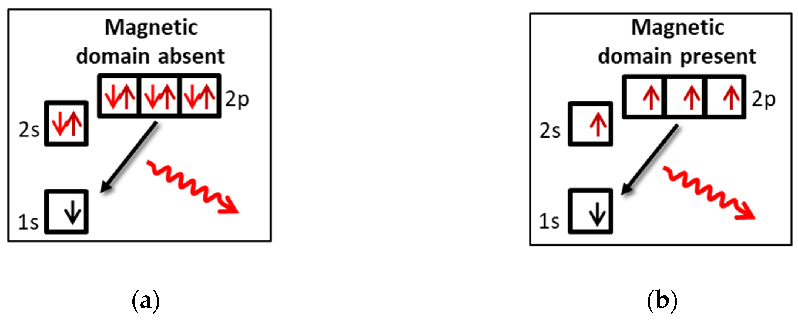
Abstract
:1. Introduction and Scientific Context
2. Setup and Installation
2.1. Ion Source and Beamline
2.2. Sample Preparation and Interaction Chamber
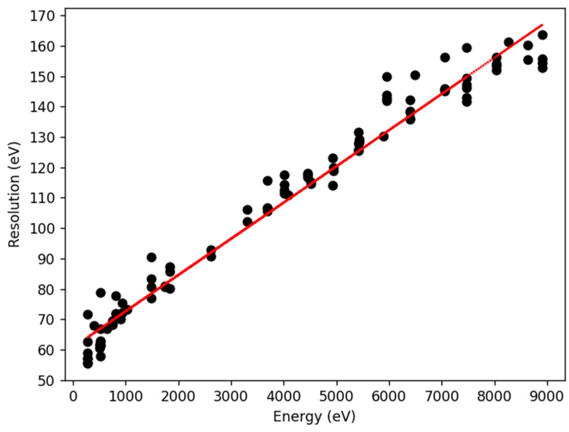
2.3. X-ray Detector Characterization
3. Preliminary Results with a Si Target
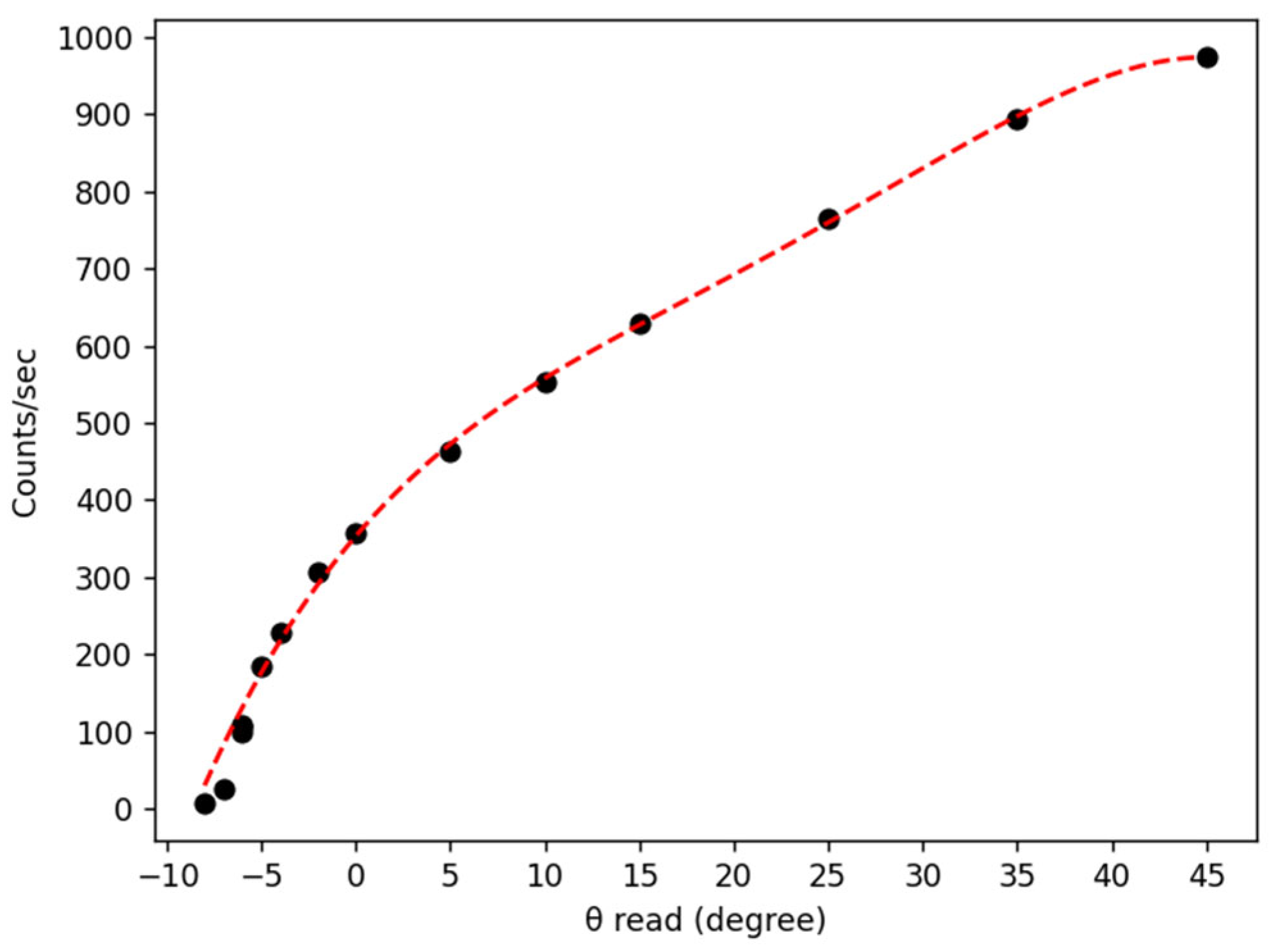
3.1. Determination of the Incident Angles Using X-ray Yield
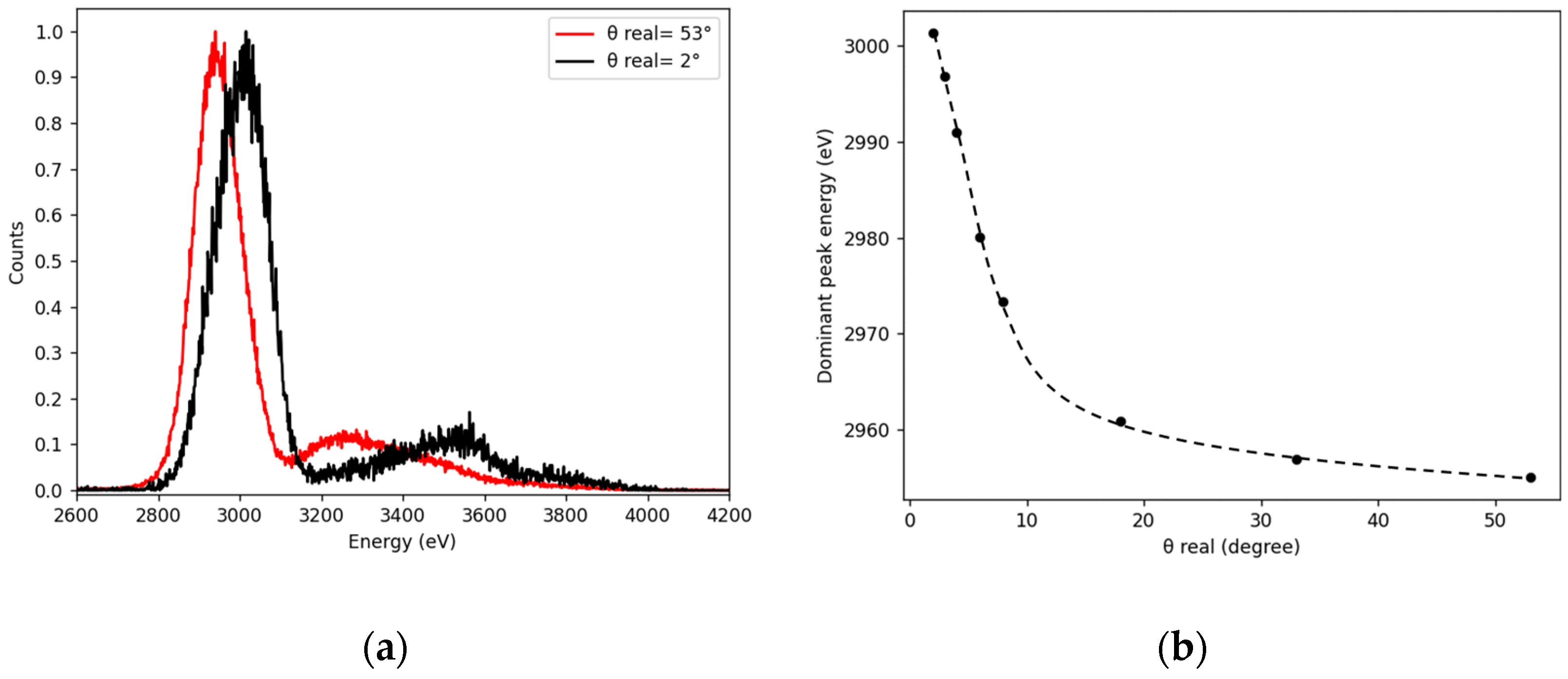
3.2. X-ray Spectra Dependency on the Incidence Angle
4. Conclusions
Author Contributions
Funding
Data Availability Statement
Conflicts of Interest
References
- Arnau, A.; Aumayr, F.; Echenique, P.; Grether, M.; Heiland, W.; Limburg, J.; Morgenstern, R.; Roncin, P.; Schippers, S.; Schuch, R.; et al. Interaction of slow multicharged ions with solid surfaces. Surf. Sci. Rep. 1997, 27, 113–239. [Google Scholar] [CrossRef]
- Schneider, D.H.G.; Briere, M.A. Investigations of the interactions of highest charge state ions with surfaces. Phys. Scr. 1996, 53, 228. [Google Scholar] [CrossRef]
- Winter, H.; Aumayr, F. Hollow atoms. J. Phys. B At. Mol. Opt. Phys. 1999, 32, R39. [Google Scholar] [CrossRef]
- Briand, J.P.; De Billy, L.; Charles, P.; Essabaa, S.; Briand, P.; Geller, R.; Desclaux, J.P.; Bliman, S.; Ristori, C. Production of hollow atoms by the excitation of highly charged ions in Interaction with a metallic surface. Phys. Rev. Lett. 1990, 65, 159. [Google Scholar] [CrossRef] [PubMed]
- Wilhelm, R.A.; Gruber, E.; Schwestka, J.; Kozubek, R.; Madeira, T.I.; Marques, J.P.; Kobus, J.; Krasheninnikov, A.V.; Schleberger, M.; Aumayr, F. Interatomic coulombic decay: The mechanism for rapid deexcitation of hollow atoms. Phys. Rev. Lett. 2017, 119, 103401. [Google Scholar] [CrossRef] [PubMed] [Green Version]
- Madesis, I.; Laoutaris, A.; Zouros, T.J.M.; Benis, E.P.; Gao, J.W.; Dubois, A. Pauli Shielding and Breakdown of Spin Statistics in Multielectron Multi-Open-Shell Dynamical Atomic Systems. Phys. Rev. Lett. 2020, 124, 113401. [Google Scholar] [CrossRef] [PubMed]
- Unipan, M.; Robin, A.; Winters, D.F.A.; Morgenstern, R.; Hoekstra, R. Probing local spin ordering at surfaces by He2+ ions. Phys. Rev. A 2006, 74, 062901. [Google Scholar] [CrossRef]
- Unipan, M.; Robin, A.; Morgenstern, R.; Hoekstra, R. Local spin polarization at surfaces probed by hollow atoms. Phys. Rev. Lett. 2006, 96, 177601. [Google Scholar] [CrossRef] [PubMed]
- Busch, M.; Wethekam, S.; Winter, H. Reexamination of local spin polarization at surfaces probed by hollow atoms. Phys. Rev. A 2008, 78, 010901. [Google Scholar] [CrossRef]
- Bhalla, C.P. K-shell auger rates, transition energies, and fluorescence yields of variously ionized states of argon. Phys. Rev. A 1973, 8, 2877. [Google Scholar] [CrossRef]
- Gumberidze, A.; Trassinelli, M.; Adrouche, N.; Szabo, C.I.; Indelicato, P.; Haranger, F.; Isac, J.M.; Lamour, E.; Le Bigot, E.O.; Mérot, J.; et al. Electronic temperatures, densities, and plasma X-ray emission of a 14.5 GHz electron-cyclotron resonance ion source. Rev. Sci. Instum. 2010, 81, 033303. [Google Scholar] [CrossRef] [PubMed] [Green Version]
- Dahman, Y. Nanotechnology and Functional Materials for Engineers; Elsevier: Amsterdam, The Netherlands, 2017; pp. 39–41. [Google Scholar]
- Winter, H. Collisions of atoms and ions with surfaces under grazing incidence. Phys. Rep. 2002, 367, 387–582. [Google Scholar] [CrossRef]
- Lamour, E.; Prigent, C.; Eberhardt, B.; Rozet, J.P.; Vernhet, D. 2E1 Ar17+ decay and conventional radioactive sources to determine efficiency of semiconductor detectors. Rev. Sci. Instrum. 2009, 80, 023103. [Google Scholar] [CrossRef] [PubMed] [Green Version]
- Winick, H. X-ray Data Booklet; Lawrence Berkeley National Laboratory: Oakland, CA, USA, 2001. [Google Scholar]
- d’Etat, B.; Briand, J.P.; Ban, G.; de Billy, L.; Desclaux, J.P.; Briand, P. Interaction of Ar17+ ions on metallic surfaces at grazing incidence. Phys. Rev. A 1993, 48, 1098. [Google Scholar] [CrossRef] [PubMed]


Publisher’s Note: MDPI stays neutral with regard to jurisdictional claims in published maps and institutional affiliations. |
© 2022 by the authors. Licensee MDPI, Basel, Switzerland. This article is an open access article distributed under the terms and conditions of the Creative Commons Attribution (CC BY) license (https://creativecommons.org/licenses/by/4.0/).
Share and Cite
Dergham, P.; Aumayr, F.; Lamour, E.; Macé, S.; Prigent, C.; Steydli, S.; Vernhet, D.; Werl, M.; Wilhelm, R.A.; Trassinelli, M. Toward Probing Surface Magnetism with Highly Charged Ions. Atoms 2022, 10, 151. https://doi.org/10.3390/atoms10040151
Dergham P, Aumayr F, Lamour E, Macé S, Prigent C, Steydli S, Vernhet D, Werl M, Wilhelm RA, Trassinelli M. Toward Probing Surface Magnetism with Highly Charged Ions. Atoms. 2022; 10(4):151. https://doi.org/10.3390/atoms10040151
Chicago/Turabian StyleDergham, Perla, Friedrich Aumayr, Emily Lamour, Stéphane Macé, Christophe Prigent, Sébastien Steydli, Dominique Vernhet, Matthias Werl, Richard Arthur Wilhelm, and Martino Trassinelli. 2022. "Toward Probing Surface Magnetism with Highly Charged Ions" Atoms 10, no. 4: 151. https://doi.org/10.3390/atoms10040151