
Inflammation and Oxidative Stress in Frailty and Metabolic Syndromes—Two Sides of the Same Coin


Abstract
:1. Introduction
2. Theories of Aging, Oxidative Stress and Inflammaging
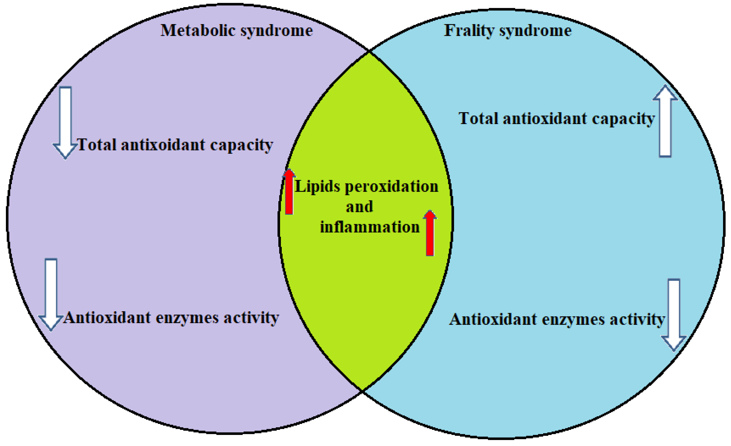
3. Oxidative Stress and Inflammation Markers in Elderly Individuals with Metabolic Syndrome
4. Oxidative Stress and Inflammation Markers in Elderly Individuals with Frailty Syndrome
5. Conclusions and Future Directions
Author Contributions
Funding
Conflicts of Interest
References
- Zsichla, L.; Müller, V. Risk Factors of Severe COVID-19: A Review of Host, Viral and Environmental Factors. Viruses 2023, 15, 175. [Google Scholar] [CrossRef]
- Popovic, D.; Papanas, N.; Koufakis, T.; Kotsa, K.; Al Mahmeed, W.; Al-Rasadi, K.; Al-Alawi, K.; Banach, M.; Banerjee, Y.; Ceriello, A.; et al. Glucometabolic perturbations in type 2 diabetes mellitus and coronavirus disease 2019: Causes, consequences, and how to counter them using novel antidiabetic drugs. Exp. Clin. Endocrinol. Diabetes 2023, 131, 182. [Google Scholar] [CrossRef]
- Gamarra-Morales, Y.; Molina-López, J.; Machado-Casas, J.F.; Herrera-Quintana, L.; Vázquez-Lorente, H.; Castaño-Pérez, J.; Perez-Villares, J.M.; Planells, E. Influence of Nutritional Parameters on the Evolution, Severity and Prognosis of Critically Ill Patients with COVID-19. Nutrients 2022, 14, 5363. [Google Scholar] [CrossRef]
- Ler, P.; Li, X.; Hassing, L.B.; Reynolds, C.A.; Finkel, D.; Karlsson, I.K.; Aslan, A.K.D. Independent and joint effects of body mass index and metabolic health in mid- and late-life on all-cause mortality: A cohort study from the Swedish Twin Registry with a mean follow-up of 13 Years. BMC Public Health 2022, 22, 718. [Google Scholar] [CrossRef] [PubMed]
- Bae, Y.S.; Choi, S.; Lee, K.; Son, J.S.; Lee, H.; Cho, M.H.; Koo, H.; Cho, I.Y.; Chang, J.; Kim, K.; et al. Association of Concurrent Changes in Metabolic Health and Weight on Cardiovascular Disease Risk: A Nationally Representative Cohort Study. J. Am. Hearth Assoc. 2019, 8, e011825. [Google Scholar] [CrossRef] [PubMed]
- Lee, Y.-B.; Kim, D.H.; Kim, S.M.; Kim, N.H.; Choi, K.M.; Baik, S.H.; Park, Y.G.; Han, K.; Yoo, H.J. Hospitalization for heart failure incidence according to the transition in metabolic health and obesity status: A nationwide population-based study. Cardiovasc. Diabetol. 2020, 19, 77. [Google Scholar] [CrossRef] [PubMed]
- Lorbergs, A.L.; Prorok, J.C.; Holroyd-Leduc, J.; Bouchard, D.R.; Giguere, A.; Gramlich, L.; Keller, H.; Tang, A.; Racey, M.; Ali, M.U.; et al. Nutrition and Physical Activity Clinical Practice Guidelines for Older Adults Living with Frailty. J. Frailty Aging 2022, 11, 3–11. [Google Scholar] [CrossRef]
- Angulo, J.; El Assar, M.; Álvarez-Bustos, A.; Rodríguez-Mañas, L. Physical activity and exercise: Strategies to manage frailty. Redox Biol. 2020, 35, 101513. [Google Scholar] [CrossRef]
- Englund, D.A.; Kirn, D.R.; Koochek, A.; Zhu, H.; Travison, T.G.; Reid, K.F.; von Berens, Å.; Melin, M.; Cederholm, T.; Gustafsson, T.; et al. Nutritional Supplementation with Physical Activity Improves Muscle Composition in Mobility-Limited Older Adults, The VIVE2 Study: A Randomized, Double-Blind, Placebo-Controlled Trial. J. Gerontol. Ser. A Biol. Sci. Med. Sci. 2017, 73, 95–101. [Google Scholar] [CrossRef] [PubMed]
- Vella, C.A.; Michos, E.D.; Sears, D.D.; Cushman, M.; Van Hollebeke, R.B.; Wiest, M.M.; Allison, M.A. Associations of Sedentary Behavior and Abdominal Muscle Density: The Multi-Ethnic Study of Atherosclerosis. J. Phys. Act. Health 2018, 15, 827–833. [Google Scholar] [CrossRef]
- Fahed, G.; Aoun, L.; Bou Zerdan, M.; Allam, S.; Bou Zerdan, M.; Bouferraa, Y.; Assi, H.I. Metabolic Syndrome: Updates on Pathophysiology and Management in 2021. Int. J. Mol. Sci. 2022, 23, 786. [Google Scholar] [CrossRef]
- Prasun, P. Mitochondrial dysfunction in metabolic syndrome. Biochim. Biophys. Acta –Mol. Basis Dis. 2020, 1866, 165838. [Google Scholar] [CrossRef]
- Saklayen, M.G. The Global Epidemic of the Metabolic Syndrome. Curr. Hypertens. Rep. 2018, 20, 12. [Google Scholar] [CrossRef] [PubMed] [Green Version]
- Alberti, K.G.M.M.; Eckel, R.H.; Grundy, S.M.; Zimmet, P.Z.; Cleeman, J.I.; Donato, K.A.; Fruchart, J.C.; James, W.P.T.; Loria, C.M.; Smith, S.C., Jr. Harmonizing the metabolic syndrome: A joint interim statement of the international diabetes federation task force on epidemiology and prevention; National heart, lung, and blood institute; American heart association; World heart federation; International Atherosclerosis Society; and International Association for the Study of Obesity. Circulation 2009, 120, 1640–1645. [Google Scholar] [CrossRef] [PubMed] [Green Version]
- Mardi, P.; Abdi, F.; Ehsani, A.; Seif, E.; Djalalinia, S.; Heshmati, J.; Shahrestanaki, E.; Gorabi, A.M.; Qorbani, M. Is non-high-density lipoprotein associated with metabolic syndrome? A systematic review and meta-analysis. Front. Endocrinol. 2022, 13, 957136. [Google Scholar] [CrossRef] [PubMed]
- Dent, E.; Martin, F.C.; Bergman, H.; Woo, J.; Romero-Ortuno, R.; Walston, J.D. Management of frailty: Opportunities, challenges, and future directions. Lancet 2019, 394, 1376–1386. [Google Scholar] [CrossRef]
- Hoogendijk, E.O.; Afilalo, J.; Ensrud, K.E.; Kowal, P.; Onder, G.; Fried, L.P. Frailty: Implications for clinical practice and public health. Lancet 2019, 394, 1365–1375. [Google Scholar] [CrossRef] [PubMed]
- Fried, L.P.; Tangen, C.M.; Walston, J.; Newman, A.B.; Hirsch, C.; Gottdiener, J.; Seeman, T.; Tracy, R.; Kop, W.J.; Burke, G.; et al. Frailty in Older adults: Evidence for a phenotype. J. Gerontol. Ser. A Biol. Sci. Med. Sci. 2001, 56, M146–M156. [Google Scholar] [CrossRef]
- Rockwood, K.; Hogan, D.; Macknight, C. Conceptualisation and Measurement of Frailty in Elderly People. Drugs Aging 2000, 17, 295–302. [Google Scholar] [CrossRef]
- Dzięgielewska-Gęsiak, S.; Wyszomirska, K.; Fatyga, E.; Wysocka, E.; Muc-Wierzgoń, M. The role of oxidant-antioxidant markers and resistin in metabolic syndrome elderly individuals. Sci. Prog. 2021, 104, 00368504211006510. [Google Scholar] [CrossRef]
- Brivio, P.; Paladini, M.S.; Racagni, G.; Riva, M.A.; Calabrese, F.; Molteni, R. From Healthy Aging to Frailty: In Search of the Underlying Mechanisms. Curr. Med. Chem. 2019, 26, 3685–3701. [Google Scholar] [CrossRef]
- Lauro, D.; Pastore, D.; Capuani, B.; Pacifici, F.; Palmirotta, R.; Abete, P.; Roselli, M.; Bellia, A.; Federici, M.; Di Daniele, N.; et al. Role of Serum and Glucocorticoid-Inducible Kinase (SGK)-1 in Senescence: A Novel Molecular Target Against Age-Related Diseases. Curr. Med. Chem. 2015, 22, 3765–3788. [Google Scholar] [CrossRef]
- Iannuzzi-Sucich, M.; Prestwood, K.M.; Kenny, A.M. Prevalence of Sarcopenia and Predictors of Skeletal Muscle Mass in Healthy, Older Men and Women. J. Gerontol. Ser. A Biol. Sci. Med. Sci. 2002, 57, M772–M777. [Google Scholar] [CrossRef] [Green Version]
- Carcaillon, L.; Garcia-Garcia, F.J.; Tresguerres, J.A.F.; Avila, G.G.; Kireev, R.; Rodríguez-Mañas, L. Higher Levels of Endogenous Estradiol are Associated with Frailty in Postmenopausal Women from the Toledo Study for Healthy Aging. J. Clin. Endocrinol. Metab. 2012, 97, 2898–2906. [Google Scholar] [CrossRef] [PubMed] [Green Version]
- Chew, J.; Tay, L.; Lim, J.P.; Leung, B.P.; Yeo, A.; Yew, S.; Ding, Y.Y.; Lim, W.S. Serum Myostatin and IGF-1 as Gender-Specific Biomarkers of Frailty and Low Muscle Mass in Community-Dwelling Older Adults. J. Nutr. Health Aging 2019, 23, 979–986. [Google Scholar] [CrossRef] [PubMed]
- Chen, L.-K.; Wu, Y.-H.; Liu, L.-K.; Lee, W.-J.; Hwang, A.-C.; Peng, L.-N.; Lin, M.-H. Association Among Serum Insulin-Like Growth Factor-1, Frailty, Muscle Mass, Bone Mineral Density, and Physical Performance Among Community-Dwelling Middle-Aged and Older Adults in Taiwan. Rejuvenation Res. 2018, 21, 270–277. [Google Scholar] [CrossRef] [PubMed]
- Pahlavani, M.; Ramalho, T.; Koboziev, I.; LeMieux, M.J.; Jayarathne, S.; Ramalingam, L.; Filgueiras, L.R.; Moustaid-Moussa, N. Adipose Tissue Inflammation in Insulin Resistance: Review of Mechanisms Mediating Anti-Inflammatory Effects of Omega-3 Polyunsaturated Fatty Acids. J. Investig. Med. 2017, 65, 1021–1027. [Google Scholar] [CrossRef] [Green Version]
- Hurrle, S.; Hsu, W.H. The etiology of oxidative stress in insulin resistance. Biomed J. 2017, 40, 257–262. [Google Scholar] [CrossRef]
- Bale, B.F.; Doneen, A.L.; Leimgruber, P.P.; Vigerust, D.J. The critical issue linking lipids and inflammation: Clinical utility of stopping oxidative stress. Front. Cardiovasc. Med. 2022, 9, 1042729. [Google Scholar] [CrossRef]
- Soriguer, F.; Colomo, N.; Valdés, S.; Goday, A.; Rubio-Martin, E.; Esteva, I.; Castaño, L.; De Adana, M.S.R.; Morcillo, S.; Calle, A.; et al. Modifications of the homeostasis model assessment of insulin resistance index with age. Acta Diabetol. 2014, 51, 917–925. [Google Scholar] [CrossRef]
- Shu, D.Y.; Chaudhary, S.; Cho, K.-S.; Lennikov, A.; Miller, W.P.; Thorn, D.C.; Yang, M.; McKay, T.B. Role of Oxidative Stress in Ocular Diseases: A Balancing Act. Metabolites 2023, 13, 187. [Google Scholar] [CrossRef] [PubMed]
- Kang, E.; Li, Y.; Kim, B.; Huh, K.Y.; Han, M.; Ahn, J.-H.; Sung, H.Y.; Park, Y.S.; Lee, S.E.; Lee, S.; et al. Identification of Serum Metabolites for Predicting Chronic Kidney Disease Progression according to Chronic Kidney Disease Cause. Metabolites 2022, 12, 1125. [Google Scholar] [CrossRef] [PubMed]
- Karamanakos, G.; Barmpagianni, A.; Kapelios, C.J.; Kountouri, A.; Bonou, M.; Makrilakis, K.; Lambadiari, V.; Barbetseas, J.; Liatis, S. The association of insulin resistance measured through the estimated glucose disposal rate with predictors of micro-and macrovascular complications in patients with type 1 diabetes. Prim. Care Diabetes 2022, 16, 837–843. [Google Scholar] [CrossRef]
- Dzięgielewska-Gęsiak, S.; Stołtny, D.; Brożek, A.; Muc-Wierzgoń, M.; Wysocka, E. Are insulin-resistance and oxidative stress cause or consequence of aging. Exp. Biol. Med. 2020, 245, 1260–1267. [Google Scholar] [CrossRef] [PubMed]
- Gysel, T.; Tonoli, C.; Pardaens, S.; Cambier, D.; Kaufman, J.-M.; Zmierczak, H.-G.; Goemaere, S.; Lapauw, B.; Calders, P. Lower insulin sensitivity is related to lower relative muscle cross-sectional area, lower muscle density and lower handgrip force in young and middle aged non-diabetic men. J. Musculoskelet. Neuronal Interact. 2016, 16, 302–309. [Google Scholar] [PubMed]
- Monti, D.; Ostan, R.; Borelli, V.; Castellani, G.; Franceschi, C. Inflammaging and human longevity in the omics era. Mech. Ageing Dev. 2016, 165, 129–138. [Google Scholar] [CrossRef]
- Trim, W.; Turner, J.E.; Thompson, D. Parallels in Immunometabolic Adipose Tissue Dysfunction with Ageing and Obesity. Front. Immunol. 2018, 9, 169. [Google Scholar] [CrossRef] [Green Version]
- Giorgi, C.; Marchi, S.; Simoes, I.C.; Ren, Z.; Morciano, G.; Perrone, M.; Patalas-Krawczyk, P.; Borchard, S.; Jędrak, P.; Pierzynowska, K.; et al. Mitochondria and Reactive Oxygen Species in Aging and Age-Related Diseases. Int. Rev. Cell Mol. Biol. 2018, 340, 209–344. [Google Scholar] [CrossRef] [Green Version]
- Derosa, G.; Catena, G.; Gaudio, G.; D’Angelo, A.; Maffioli, P. Adipose tissue dysfunction and metabolic disorders: Is it possible to predict who will develop type 2 diabetes mellitus? Role of markErs in the progreSsion of dIabeteS in obese paTIeNts (The RESISTIN trial). Cytokine 2020, 127, 154947. [Google Scholar] [CrossRef]
- Da Costa, J.P.; Vitorino, R.; Silva, G.M.; Vogel, C.; Duarte, A.C.; Rocha-Santos, T. A synopsis on aging—Theories, mechanisms and future prospects. Ageing Res. Rev. 2016, 29, 90–112. [Google Scholar] [CrossRef]
- McDonough, I.M.; Nolin, S.A.; Visscher, K.M. 25 years of neurocognitive aging theories: What have we learned? Front. Aging Neurosci. 2022, 14, 1002096. [Google Scholar] [CrossRef] [PubMed]
- Gavrilov, L.A.; Gavrilova, N.S. Evolutionary Theories of Aging and Longevity. Sci. World J. 2002, 2, 339–356. [Google Scholar] [CrossRef] [Green Version]
- Etienne, J.; Liu, C.; Skinner, C.M.; Conboy, M.J.; Conboy, I.M. Skeletal muscle as an experimental model of choice to study tissue aging and rejuvenation. Skelet. Muscle 2020, 10, 4. [Google Scholar] [CrossRef] [PubMed] [Green Version]
- Kowald, A.; Kirkwood, T.B.L. Evolution of the mitochondrial fusion–fission cycle and its role in aging. Proc. Natl. Acad. Sci. USA 2011, 108, 10237–10242. [Google Scholar] [CrossRef] [Green Version]
- Sacco, A.; Belloni, L.; Latella, L. From Development to Aging: The Path to Cellular Senescence. Antioxidants Redox Signal. 2021, 34, 294–307. [Google Scholar] [CrossRef]
- Harman, D. Aging: A Theory Based on Free Radical and Radiation Chemistry. J. Gerontol. 1956, 11, 298–300. [Google Scholar] [CrossRef] [Green Version]
- Forman, H.J. Redox signaling: An evolution from free radicals to aging. Free. Radic. Biol. Med. 2016, 97, 398–407. [Google Scholar] [CrossRef] [Green Version]
- Perridon, B.W.; Leuvenink, H.G.; Hillebrands, J.-L.; Van Goor, H.; Bos, E.M. The role of hydrogen sulfide in aging and age-related pathologies. Aging 2016, 8, 2264–2289. [Google Scholar] [CrossRef] [Green Version]
- Kaliszewska, A.; Allison, J.; Martini, M.; Arias, N. Improving Age-Related Cognitive Decline through Dietary Interventions Targeting Mitochondrial Dysfunction. Int. J. Mol. Sci. 2021, 22, 3574. [Google Scholar] [CrossRef]
- Bednarczyk, M.; Fatyga, E.; Muc-Wierzgoń, M.; Dzięgielewska-Gęsiak, S.; Wierzgoń, J. Age-related changes in expression of au-tophagy genes in human large intestine. J. Biol. Regul. Homeost. Agents 2021, 35, 1343–1347. [Google Scholar]
- Sadowska-Bartosz, I.; Bartosz, G. Effect of Antioxidants Supplementation on Aging and Longevity. BioMed Res. Int. 2014, 2014, 404680. [Google Scholar] [CrossRef] [PubMed]
- Kozakiewicz, M.; Kornatowski, M.; Krzywińska, O.; Kędziora-Kornatowska, K. Changes in the blood antioxidant defense of advanced age people. Clin. Interv. Aging 2019, 14, 763–771. [Google Scholar] [CrossRef] [Green Version]
- Vázquez-Lorente, H.; Herrera-Quintana, L.; Molina-López, J.; López-González, B.; Planells, E. Sociodemographic, Anthropometric, Body Composition, Nutritional, and Biochemical Factors Influenced by Age in a Postmenopausal Population: A Cross-Sectional Study. Metabolites 2023, 13, 78. [Google Scholar] [CrossRef]
- Vázquez-Lorente, H.; Herrera-Quintana, L.; Molina-López, J.; Gamarra-Morales, Y.; López-González, B.; Planells, E. Relationship between Body Composition and Biochemical Parameters with Antioxidant Status in a Healthy Cohort of Postmenopausal Women. Metabolites 2022, 12, 746. [Google Scholar] [CrossRef]
- Vitetta, L.; Anton, B. Lifestyle and nutrition, caloric restriction, mitochondrial health and hormones: Scientific interventions for anti-aging. Clin. Interv. Aging 2007, 2, 537–543. [Google Scholar] [CrossRef] [PubMed] [Green Version]
- Lippi, L.; Uberti, F.; Folli, A.; Turco, A.; Curci, C.; D’Abrosca, F.; de Sire, A.; Invernizzi, M. Impact of nutraceuticals and dietary supplements on mitochondria modifications in healthy aging: A systematic review of randomized controlled trials. Aging Clin. Exp. Res. 2022, 34, 2659–2674. [Google Scholar] [CrossRef]
- Santoro, A.; Bientinesi, E.; Monti, D. Immunosenescence and inflammaging in the aging process: Age-related diseases or longevity? Ageing Res. Rev. 2021, 71, 101422. [Google Scholar] [CrossRef]
- Cisneros, B.; García-Aguirre, I.; Unzueta, J.; Arrieta-Cruz, I.; González-Morales, O.; Domínguez-Larrieta, J.M.; Tamez-González, A.; Leyva-Gómez, G.; Magaña, J.J. Immune system modulation in aging: Molecular mechanisms and therapeutic targets. Front. Immunol. 2022, 13, 1059173. [Google Scholar] [CrossRef] [PubMed]
- Scheithauer, T.P.M.; Rampanelli, E.; Nieuwdorp, M.; Vallance, B.A.; Verchere, C.B.; van Raalte, D.H.; Herrema, H. Gut Microbiota as a Trigger for Metabolic Inflammation in Obesity and Type 2 Diabetes. Front. Immunol. 2020, 11, 571731. [Google Scholar] [CrossRef]
- Valacchi, G.; Virgili, F.; Cervellati, C.; Pecorelli, A. OxInflammation: From Subclinical Condition to Pathological Biomarker. Front. Physiol. 2018, 9, 858. [Google Scholar] [CrossRef] [Green Version]
- Karaman, A.; Aydın, H.; Geçkinli, B.; Çetinkaya, A.; Karaman, S. DNA damage is increased in lymphocytes of patients with metabolic syndrome. Mutat. Res. Genet. Toxicol. Environ. Mutagen. 2015, 782, 30–35. [Google Scholar] [CrossRef]
- Yan, M.; Li, X.; Sun, C.; Tan, J.; Liu, Y.; Li, M.; Qi, Z.; He, J.; Wang, D.; Wu, L. Sodium Butyrate Attenuates AGEs-Induced Oxidative Stress and Inflammation by Inhibiting Autophagy and Affecting Cellular Metabolism in THP-1 Cells. Molecules 2022, 27, 8715. [Google Scholar] [CrossRef]
- Vila, I.; Badin, P.-M.; Marques, M.-A.; Monbrun, L.; Lefort, C.; Mir, L.; Louche, K.; Bourlier, V.; Roussel, B.; Gui, P.; et al. Immune Cell Toll-like Receptor 4 Mediates the Development of Obesity- and Endotoxemia-Associated Adipose Tissue Fibrosis. Cell Rep. 2014, 7, 1116–1129. [Google Scholar] [CrossRef]
- Rius-Pérez, S.; Torres-Cuevas, I.; Millán, I.; Ortega, Á.L.; Pérez, S. PGC-1α, Inflammation, and Oxidative Stress: An Integrative View in Metabolism. Oxid. Med. Cell. Longev. 2020, 2020, 1452696. [Google Scholar] [CrossRef] [PubMed] [Green Version]
- Baez-Duarte, B.G.; Zamora-Ginez, I.; De Jésus, K.L.; Torres-Rasgado, E.; González-Mejía, M.E.; Porchia, L.; Ruiz-Vivanco, G.; Pérez-Fuentes, R. Association of the Metabolic Syndrome with Antioxidant Defense and Outstanding Superoxide Dismutase Activity in Mexican Subjects. Metab. Syndr. Relat. Disord. 2016, 14, 154–160. [Google Scholar] [CrossRef]
- Suriyaprom, K.; Kaewprasert, S.; Putpadungwipon, P.; Namjuntra, P.; Klongthalay, S. Association of antioxidant status and inflammatory markers with metabolic syndrome in Thais. J. Health Popul. Nutr. 2019, 38, 1. [Google Scholar] [CrossRef] [PubMed] [Green Version]
- Liu, Y.; Ma, C.; Lv, L.; Li, P.; Ma, C.; He, S.; Zeng, J.; Ping, F.; Zhang, H.; Li, W.; et al. Relationship between Decreased Serum Superoxide Dismutase Activity and Metabolic Syndrome: Synergistic Mediating Role of Insulin Resistance and β-Cell Dysfunction. Oxidative Med. Cell. Longev. 2020, 2020, 5384909. [Google Scholar] [CrossRef] [PubMed]
- Awadallah, S.; Hasan, H.; Attlee, A.; Raigangar, V.; Unnikannan, H.; Madkour, M.; Abraham, M.S.; Rashid, L.M. Waist circumference is a major determinant of oxidative stress in subjects with and without metabolic syndrome. Diabetes Metab. Syndr. Clin. Res. Rev. 2019, 13, 2541–2547. [Google Scholar] [CrossRef]
- Sakhaei, F.; Keshvari, M.; Asgary, S.; Salehizadeh, L.; Rastqar, A.; Samsam-Shariat, S.Z. Enzymatic antioxidant system and endothelial function in patients with metabolic syndrome. ARYA Atheroscler. 2020, 16, 94–101. [Google Scholar] [CrossRef]
- Gyawali, P.; Richards, R.S. Association of altered hemorheology with oxidative stress and inflammation in metabolic syndrome. Redox Rep. 2015, 20, 139–144. [Google Scholar] [CrossRef]
- Chung, H.-K.; Kim, J.H.; Choi, A.; Ahn, C.W.; Kim, Y.-S.; Nam, J.S. Antioxidant-Rich Dietary Intervention Improves Cardiometabolic Profiles and Arterial Stiffness in Elderly Koreans with Metabolic Syndrome. Yonsei Med. J. 2022, 63, 26–33. [Google Scholar] [CrossRef] [PubMed]
- Mendoza-Núñez, V.M.; Arista-Ugalde, T.L.; Rosado-Pérez, J.; Ruiz-Ramos, M.; Santiago-Osorio, E. Hypoglycemic and antioxidant effect of Tai chi exercise training in older adults with metabolic syndrome. Clin. Interv. Aging 2018, 13, 523–531. [Google Scholar] [CrossRef] [PubMed] [Green Version]
- Osali, A. Aerobic exercise and nano-curcumin supplementation improve inflammation in elderly females with metabolic syndrome. Diabetol. Metab. Syndr. 2020, 12, 26. [Google Scholar] [CrossRef] [PubMed] [Green Version]
- Smagula, S.F.; Stone, K.L.; Redline, S.; Ancoli-Israel, S.; Barrett-Connor, E.; Lane, N.E.; Orwoll, E.S.; Cauley, J.A.; Osteoporotic Fractures in Men (MrOS) Research Group. Actigraphy- and Polysomnography-Measured Sleep Disturbances, Inflammation, and Mortality Among Older Men. Psychosom. Med. 2016, 78, 686–696. [Google Scholar] [CrossRef] [Green Version]
- Tang, Z.; Wang, C.; Song, X.; Shi, J.; Mitnitski, A.; Fang, X.; Yu, P.; Rockwood, K. Co-occurrence of cardiometabolic diseases and frailty in older Chinese adults in the Beijing Longitudinal Study of Ageing. Age Ageing 2013, 42, 346–351. [Google Scholar] [CrossRef] [PubMed] [Green Version]
- Jayanama, K.; Theou, O.; Godin, J.; Mayo, A.; Cahill, L.; Rockwood, K. Relationship of body mass index with frailty and all-cause mortality among middle-aged and older adults. BMC Med. 2022, 20, 404. [Google Scholar] [CrossRef]
- Aguayo, G.A.; Vaillant, M.T.; Donneau, A.-F.; Schritz, A.; Stranges, S.; Malisoux, L.; Chioti, A.; Guillaume, M.; Muller, M.; Witte, D.R. Comparative analysis of the association between 35 frailty scores and cardiovascular events, cancer, and total mortality in an elderly general population in England: An observational study. PLoS Med. 2018, 15, e1002543. [Google Scholar] [CrossRef] [Green Version]
- Hoogendijk, E.O.; Huisman, M.; van Ballegooijen, A.J. The role of frailty in explaining the association between the metabolic syndrome and mortality in older adults. Exp. Gerontol. 2017, 91, 5–8. [Google Scholar] [CrossRef] [PubMed]
- Liu, X.; Tou, N.X.; Gao, Q.; Gwee, X.; Wee, S.L.; Ng, T.P. Frailty and risk of cardiovascular disease and mortality. PLoS ONE 2022, 17, e0272527. [Google Scholar] [CrossRef]
- Ligthart-Melis, G.C.; Luiking, Y.C.; Kakourou, A.; Cederholm, T.; Maier, A.B.; de van der Schueren, M.A.E. Frailty, Sarcopenia, and Malnutrition Frequently (Co-)occur in Hospitalized Older Adults: A Systematic Review and Meta-analysis. J. Am. Med. Dir. Assoc. 2020, 21, 1216–1228. [Google Scholar] [CrossRef]
- Zukeran, M.S.; Neto, J.V.; Romanini, C.V.; Mingardi, S.V.B.; Cipolli, G.C.; Aprahamian, I.; Ribeiro, S.M.L. The association between appetite loss, frailty, and psychosocial factors in community-dwelling older adults adults. Clin. Nutr. ESPEN 2021, 47, 194–198. [Google Scholar] [CrossRef] [PubMed]
- Sánchez-Garcí a, S.; García-Peña, C.; Salvà, A.; Sánchez-Arenas, R.; Granados-García, V.; Cuadros-Moreno, J.; Velázquez-Olmedo, L.B.; Cárdenas-Bahena, Á. Frailty in community-dwelling older adults: Association with adverse outcomes. Clin. Interv. Aging 2017, 12, 1003–1011. [Google Scholar] [CrossRef] [PubMed] [Green Version]
- Yuan, Y.; Lapane, K.L.; Tjia, J.; Baek, J.; Liu, S.-H.; Ulbricht, C.M. Trajectories of physical frailty and cognitive impairment in older adults in United States nursing homes. BMC Geriatr. 2022, 22, 339. [Google Scholar] [CrossRef] [PubMed]
- Rosas, C.; Oliveira, H.C.; Neri, A.L.; Ceolim, M.F. Stressful events, depressive symptoms, and frailty associated to older adults’ survival and mortality. Geriatr. Nurs. 2022, 46, 62–68. [Google Scholar] [CrossRef]
- Westbrook, R.; Zhang, C.; Yang, H.; Tian, J.; Guo, S.; Xue, Q.-L.; Walston, J.; Le, A.; Abadir, P.M. Metabolomics-Based Identification of Metabolic Dysfunction in Frailty. J. Gerontol. Ser. A Biol. Sci. Med. Sci. 2021, 77, 2367–2372. [Google Scholar] [CrossRef] [PubMed]
- Ramírez-Vélez, R.; Martínez-Velilla, N.; Correa-Rodríguez, M.; de Asteasu, M.L.S.; Zambom-Ferraresi, F.; Palomino-Echeverria, S.; García-Hermoso, A.; Izquierdo, M. Lipidomic signatures from physically frail and robust older adults at hospital admission. Geroscience 2022, 44, 1677–1688. [Google Scholar] [CrossRef]
- Liang, Y.-D.; Liu, Q.; Du, M.-H.; Liu, Z.; Yao, S.-M.; Zheng, P.-P.; Wan, Y.-H.; Sun, N.; Li, Y.-Y.; Liu, J.-P.; et al. Urinary 8-oxo-7,8-dihydroguanosine as a potential biomarker of frailty for elderly patients with cardiovascular disease. Free. Radic. Biol. Med. 2020, 152, 248–254. [Google Scholar] [CrossRef]
- Bernabeu-Wittel, M.; Gómez-Díaz, R.; González-Molina, Á.; Vidal-Serrano, S.; Díez-Manglano, J.; Salgado, F.; Soto-Martín, M.; Ollero-Baturone, M.; on Behalf of The Proteo Researchers. Oxidative Stress, Telomere Shortening, and Apoptosis Associated to Sarcopenia and Frailty in Patients with Multimorbidity. J. Clin. Med. 2020, 9, 2669. [Google Scholar] [CrossRef]
- Martínez-Ezquerro, J.D.; Rodríguez-Castañeda, A.; Ortiz-Ramírez, M.; Sánchez-García, S.; Rosas-Vargas, H.; Sánchez-Arenas, R.; la Torre, P.G.-D. Oxidative Stress, Telomere Length, and Frailty in an Old Age Population. Rev. Investig. Clin. 2020, 71, 393–401. [Google Scholar] [CrossRef] [Green Version]
- Serviddio, G.; Romano, A.; Greco, A.; Rollo, T.; Bellanti, F.; Altomare, E.; Vendemiale, G. Frailty Syndrome is Associated with Altered Circulating Redox Balance and Increased Markers of Oxidative Stress. Int. J. Immunopathol. Pharmacol. 2009, 22, 819–827. [Google Scholar] [CrossRef] [Green Version]
- Tembo, M.C.; Holloway-Kew, K.L.; Bortolasci, C.C.; Sui, S.X.; Brennan-Olsen, S.L.; Williams, L.J.; Kotowicz, M.A.; Pasco, J.A. Total Antioxidant Capacity and Frailty in Older Men. Am. J. Men’s Health 2020, 14, 1557988320946592. [Google Scholar] [CrossRef]
- Tembo, M.C.; Holloway-Kew, K.L.; Bortolasci, C.C.; Brennan-Olsen, S.L.; Williams, L.J.; Kotowicz, M.A.; Pasco, J.A. Association between serum interleukin-6 and frailty in older men: Cross-sectional data. Eur. Geriatr. Med. 2022, 12, 887–892. [Google Scholar] [CrossRef]
- Mu, L.; Jiang, L.; Chen, J.; Xiao, M.; Wang, W.; Liu, P.; Wu, J. Serum Inflammatory Factors and Oxidative Stress Factors Are Associated with Increased Risk of Frailty and Cognitive Frailty in Patients with Cerebral Small Vessel Disease. Front. Neurol. 2022, 12, 786277. [Google Scholar] [CrossRef]
- Puzianowska-Kuźnicka, M.; Owczarz, M.; Wieczorowska-Tobis, K.; Nadrowski, P.; Chudek, J.; Slusarczyk, P.; Skalska, A.; Jonas, M.; Franek, E.; Mossakowska, M. Interleukin-6 and C-reactive protein, successful aging, and mortality: The PolSenior study. Immun. Ageing 2016, 13, 21. [Google Scholar] [CrossRef] [Green Version]
- Hammami, S.; Ghzaiel, I.; Hammouda, S.; Sakly, N.; Hammami, M.; Zarrouk, A. Evaluation of pro-inflammatory cytokines in frail Tunisian older adults. PLoS ONE 2020, 15, e0242152. [Google Scholar] [CrossRef] [PubMed]
- Marcos-Pérez, D.; Sánchez-Flores, M.; Proietti, S.; Bonassi, S.; Costa, S.; Teixeira, J.P.; Fernández-Tajes, J.; Pásaro, E.; Laffon, B.; Valdiglesias, V. Association of inflammatory mediators with frailty status in older adults: Results from a systematic review and meta-analysis. Geroscience 2020, 42, 1451–1473. [Google Scholar] [CrossRef] [PubMed]
- Ribeiro, C.T.; Sangali, T.D.; Clausell, N.O.; Perry, I.S.; Souza, G.C. C-Reactive Protein and Frailty in Heart Failure. Am. J. Cardiol. 2022, 166, 65–71. [Google Scholar] [CrossRef] [PubMed]
- Mailliez, A.; Guilbaud, A.; Puisieux, F.; Dauchet, L.; Boulanger, É. Circulating biomarkers characterizing physical frailty: CRP, hemoglobin, albumin, 25OHD and free testosterone as best biomarkers. Results of a meta-analysis. Exp. Gerontol. 2020, 139, 111014. [Google Scholar] [CrossRef]
- Xu, Y.; Wang, M.; Chen, D.; Jiang, X.; Xiong, Z. Inflammatory biomarkers in older adults with frailty: A systematic review and meta-analysis of cross-sectional studies. Aging Clin. Exp. Res. 2022, 34, 971–987. [Google Scholar] [CrossRef]
- Reiner, A.P.; Aragaki, A.K.; Gray, S.L.; Wactawski-Wende, J.; Cauley, J.A.; Cochrane, B.B.; Kooperberg, C.L.; Woods, N.F.; LaCroix, A.Z. Inflammation and Thrombosis Biomarkers and Incident Frailty in Postmenopausal Women. Am. J. Med. 2009, 122, 947–954. [Google Scholar] [CrossRef] [Green Version]
- Landino, K.; Tanaka, T.; Fantoni, G.; Candia, J.; Bandinelli, S.; Ferrucci, L. Characterization of the plasma proteomic profile of frailty phenotype. Geroscience 2021, 43, 1029–1037. [Google Scholar] [CrossRef] [PubMed]
- Chen, S.-Y.; Wang, T.-Y.; Zhao, C.; Wang, H.-J. Oxidative stress bridges the gut microbiota and the occurrence of frailty syndrome. World J. Gastroenterol. 2022, 28, 5547–5556. [Google Scholar] [CrossRef] [PubMed]
- Margiotta, E.; Miragoli, F.; Callegari, M.L.; Vettoretti, S.; Caldiroli, L.; Meneghini, M.; Zanoni, F.; Messa, P. Gut microbiota composition and frailty in elderly patients with Chronic Kidney Disease. PLoS ONE 2020, 15, e0228530. [Google Scholar] [CrossRef] [PubMed] [Green Version]
- Kobayashi, S.; Suga, H.; Sasaki, S.; Three-generation Study of Women on Diets and Health Study Group. Diet with a combination of high protein and high total antioxidant capacity is strongly associated with low prevalence of frailty among old Japanese women: A multicenter cross-sectional study. Nutr. J. 2017, 16, 29. [Google Scholar] [CrossRef] [PubMed] [Green Version]
- Jayanama, K.; Theou, O.; Godin, J.; Cahill, L.; Rockwood, K. Association of fatty acid consumption with frailty and mortality among middle-aged and older adults. Nutrition 2020, 70, 110610. [Google Scholar] [CrossRef]
- Bo, Y.; Liu, C.; Ji, Z.; Yang, R.; An, Q.; Zhang, X.; You, J.; Duan, D.; Sun, Y.; Zhu, Y.; et al. A high whey protein, vitamin D and E supplement preserves muscle mass, strength, and quality of life in sarcopenic older adults: A double-blind randomized controlled trial. Clin. Nutr. 2019, 38, 159–164. [Google Scholar] [CrossRef]
- Lippi, L.; de Sire, A.; Mezian, K.; Curci, C.; Perrero, L.; Turco, A.; Andaloro, S.; Ammendolia, A.; Fusco, N.; Invernizzi, M. Impact of exercise training on muscle mitochondria modifications in older adults: A systematic review of randomized controlled trials. Aging Clin. Exp. Res. 2022, 34, 1495–1510. [Google Scholar] [CrossRef]
- Buchmann, N.; Spira, D.; König, M.; Demuth, I.; Steinhagen-Thiessen, E. Frailty and the metabolic syndrome—results of the berlin aging study ii (base-ii). J. Frailty Aging 2019, 8, 169–175. [Google Scholar] [CrossRef]
- Celli, A.; Barnouin, Y.; Jiang, B.; Blevins, D.; Colleluori, G.; Mediwala, S.; Armamento-Villareal, R.; Qualls, C.; Villareal, D.T. Lifestyle Intervention Strategy to Treat Diabetes in Older Adults: A Randomized Controlled Trial. Diabetes Care 2022, 45, 1943–1952. [Google Scholar] [CrossRef]
- Rodriguez-Mañas, L.; Laosa, O.; Vellas, B.; Paolisso, G.; Topinkova, E.; Oliva-Moreno, J.; Bourdel-Marchasson, I.; Izquierdo, M.; Hood, K.; Zeyfang, A.; et al. Effectiveness of a multimodal intervention in functionally impaired older people with type 2 diabetes mellitus. J. Cachex Sarcopenia Muscle 2019, 10, 721–733. [Google Scholar] [CrossRef] [Green Version]
- Bernabei, R.; Landi, F.; Calvani, R.; Cesari, M.; Del Signore, S.; Anker, S.D.; Bejuit, R.; Bordes, P.; Cherubini, A.; Cruz-Jentoft, A.J.; et al. Multicomponent intervention to prevent mobility disability in frail older adults: Randomised controlled trial (SPRINTT project). BMJ 2022, 377, e068788. [Google Scholar] [CrossRef] [PubMed]


Disclaimer/Publisher’s Note: The statements, opinions and data contained in all publications are solely those of the individual author(s) and contributor(s) and not of MDPI and/or the editor(s). MDPI and/or the editor(s) disclaim responsibility for any injury to people or property resulting from any ideas, methods, instructions or products referred to in the content. |
© 2023 by the authors. Licensee MDPI, Basel, Switzerland. This article is an open access article distributed under the terms and conditions of the Creative Commons Attribution (CC BY) license (https://creativecommons.org/licenses/by/4.0/).
Share and Cite
Dzięgielewska-Gęsiak, S.; Muc-Wierzgoń, M. Inflammation and Oxidative Stress in Frailty and Metabolic Syndromes—Two Sides of the Same Coin. Metabolites 2023, 13, 475. https://doi.org/10.3390/metabo13040475
Dzięgielewska-Gęsiak S, Muc-Wierzgoń M. Inflammation and Oxidative Stress in Frailty and Metabolic Syndromes—Two Sides of the Same Coin. Metabolites. 2023; 13(4):475. https://doi.org/10.3390/metabo13040475
Chicago/Turabian StyleDzięgielewska-Gęsiak, Sylwia, and Małgorzata Muc-Wierzgoń. 2023. "Inflammation and Oxidative Stress in Frailty and Metabolic Syndromes—Two Sides of the Same Coin" Metabolites 13, no. 4: 475. https://doi.org/10.3390/metabo13040475